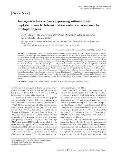
Transgenic tobacco plants expressing antimicrobial ... - Wdc-jp.biz
Transgenic tobacco plants expressing antimicrobial ... - Wdc-jp.biz
Transgenic tobacco plants expressing antimicrobial ... - Wdc-jp.biz

Create successful ePaper yourself
Turn your PDF publications into a flip-book with our unique Google optimized e-Paper software.
Plant Biotechnology 29, 383–389 (2012)<br />
DOI: 10.5511/plantbiotechnology.12.0619a<br />
Original Paper<br />
<strong>Transgenic</strong> <strong>tobacco</strong> <strong>plants</strong> <strong>expressing</strong> <strong>antimicrobial</strong><br />
peptide bovine lactoferricin show enhanced resistance to<br />
phytopathogens<br />
Shiro Fukuta 1, *, Kei-ichi Kawamoto 2,a , Yuko Mizukami 1 , Yukie Yoshimura 1 ,<br />
Jun-ichi Ueda 1 , Michio Kanbe 1<br />
1<br />
Aichi Agricultural Research Center, Nagakute, Aichi 480-1193, Japan; 2 Aichi College of Agriculture, Nagakute, Aichi<br />
480-1193, Japan<br />
* E-mail: shirou_fukuta@pref.aichi.lg.<strong>jp</strong>Tel: +81-561-62-0085Fax: +81-561-63-0815<br />
Received February 27, 2012; accepted June 19, 2012 (Edited by T. Anai)<br />
Abstract Lactoferricin B, the <strong>antimicrobial</strong>ly active N-terminal peptide derived from acid pepsin hydrolysis of bovine<br />
lactoferrin, has broad spectrum <strong>antimicrobial</strong> activities. We expressed the lactoferricin gene in <strong>tobacco</strong> <strong>plants</strong> to evaluate<br />
its <strong>antimicrobial</strong> activity. The coding region for the chimeric peptide gene of the signal peptide from <strong>tobacco</strong> pathogenrelated<br />
protein (PR-1) and bovine lactoferricin was synthesized using the recombinant polymerase chain reaction (PCR)<br />
method. <strong>Transgenic</strong> <strong>tobacco</strong> <strong>plants</strong> <strong>expressing</strong> the lactoferricin gene were developed using the Agrobacterium-mediated<br />
transformation method. The lactoferricin B gene was integrated into the <strong>tobacco</strong> genome and its transcription was detected<br />
by PCR, Southern blot analysis, and reverse transcription PCR (RT-PCR), respectively. The transgenic <strong>tobacco</strong> <strong>plants</strong><br />
were challenged with the pathogenic bacteria Pseudomonas syringae pv. tabaci and Botrytis cinerea. At 30 or 28 days postinoculation,<br />
the transgenic <strong>plants</strong> were still green and continued to grow, whereas the control <strong>plants</strong> were infected with<br />
bacterial or fungal pathogens from the roots to the tips, resulting in death of the <strong>plants</strong>. In conclusion, transgenic <strong>tobacco</strong><br />
<strong>plants</strong> that overexpressed the lactoferricin gene, linked to the signal peptide of <strong>tobacco</strong> PR-1 protein under the control of a<br />
high expression constitutive promoter, showed enhanced resistance to bacterial (P. syringae pv. tabaci) and fungal (B. cinerea)<br />
diseases.<br />
Key words: Lactoferricin, cationic peptide, transgenic <strong>tobacco</strong>, phytopathogens, disease resistance<br />
Lactoferrin is a glycoprotein found in bovine whey<br />
protein fractions. Lactoferrin has multiple biological<br />
functions, mostly related to host defense, including<br />
broad-spectrum <strong>antimicrobial</strong> properties.<br />
Lactoferricin B (Lfc) is a 25-amino acid peptide<br />
with a molecular weight of 3,124 that originates from<br />
the N-terminus of bovine lactoferrin (Bellamy et al.<br />
1992a). Lfc has stronger <strong>antimicrobial</strong> activity against<br />
various human pathogenic bacteria (Aguilera et al. 1999;<br />
Bellamy et al. 1992b; Sánchez-Gómez et al. 2008; Shin et<br />
al. 1998; Wakabayashi et al. 2002), fungi (Bellamy et al.<br />
1993), and viruses (Marr et al. 2009) compared to that<br />
of lactoferrin. The <strong>antimicrobial</strong> activity of Lfc against<br />
plant pathogenic bacteria (Zhang et al. 1998) and fungi<br />
(Muños and Marcos 2006) has also been studied. Lfc has<br />
characteristics of typical cationic <strong>antimicrobial</strong> peptides<br />
(Tomita et al. 2009), which are thought to be the result of<br />
actions on the cytoplasmic membrane of bacteria, such<br />
as the formation of pores, destabilization of the bilayer,<br />
thinning of the membrane, or depolarization of the<br />
membrane (Ulvatne et al. 2001).<br />
Many studies have shown the expression of<br />
<strong>antimicrobial</strong> cationic peptides in <strong>plants</strong>, e.g., cecropin<br />
in <strong>tobacco</strong> (Hightower et al. 1994; Jaynes et al. 1993)<br />
and tomato (Jan et al. 2010), sarcotoxin in <strong>tobacco</strong><br />
(Ohshima et al. 1999) and tomato (Radi et al. 2006),<br />
MsrA3 in potatoes (Osusky et al. 2004), and cecropinmelittin<br />
chimeric peptide in potato (Osusky et al. 2000).<br />
<strong>Transgenic</strong> <strong>plants</strong> showed considerably greater resistance<br />
to pathogens (Hightower et al. 1994; Jan et al. 2010;<br />
Jaynes et al. 1993; Ohshima et al. 1999; Osusky et al.<br />
2000; Osusky et al. 2004) and parasitic weeds (Radi et al.<br />
2006) than to the wild types.<br />
Zhang et al. (1998) reported that transgenic <strong>tobacco</strong><br />
<strong>expressing</strong> human lactoferrin protein demonstrated<br />
significant delays in developing bacterial wilt symptoms<br />
when inoculated with the bacterial pathogen Ralstonia<br />
solanacearum. They reported that lactoferricin, which<br />
has higher bactericidal activity and is resistant to pepsin<br />
digestion, could confer higher levels of bactericidal<br />
a<br />
Present address: AISAN Seed Co., Ltd., Mie 4908-0802, Japan.<br />
This article can be found at http://www.jspcmb.<strong>jp</strong>/<br />
Published online August 30, 2012<br />
Copyright © 2012 The Japanese Society for Plant Cell and Molecular Biology
384 Enhanced resistance of transgenic <strong>tobacco</strong> express Lfc<br />
Figure1. The sequences of the amino acids and the nucleotides of signal peptide of PR1 and lactoferricin. For improved plant expression, codon<br />
choice was made according to the codon usage in <strong>tobacco</strong>. Shadowed letters show the signal peptide of <strong>tobacco</strong> PR1 protein, and * shows the stop<br />
codon.<br />
disease resistance to transgenic <strong>plants</strong>. In this study, we<br />
developed transgenic <strong>tobacco</strong> <strong>plants</strong> that carried the Lfc<br />
gene fused to the signal peptide of <strong>tobacco</strong> pathogenrelated<br />
protein (PR-1) under the control of the enhanced<br />
cauliflower mosaic virus (CaMV) 35S promoter. The<br />
effects of Lfc gene expression in transgenic <strong>tobacco</strong><br />
<strong>plants</strong> against the phytopathogenic bacteria Pseudomonas<br />
syringae pv. tabaci and the fungi Botrytis cinerea were<br />
analyzed.<br />
Materials and methods<br />
Assay of Lfc antibacterial activity<br />
Synthesized bovine Lfc (N-FKC RRW QWR MKK LGA PSI<br />
TCV RRA F-C) was purchased from BioGate Co. (Gifu, Japan).<br />
The in vitro antibacterial activity of Lfc was determined by<br />
the method described by Zhang et al. (1998) with a slight<br />
modification. P. syringae pv. maculicola (PMC8301) and<br />
Xanthomonas campestris pv. vesicatoria (#613) were a gift from<br />
Dr. Takikawa, University of Shizuoka. R. solanacearum (OE1-<br />
1) was collected in Aichi Prefecture, Japan. The bacteria were<br />
grown to OD600 of 0.3 in NB liquid medium (Difco), and<br />
1 ml aliquots were transferred to culture tubes. Synthetic Lfc<br />
was added to the culture tubes to obtain final concentrations of<br />
100 µmol l −1 , 10 µmol l −1 , 1 µmol l −1 or 0 µmol l −1 , respectively.<br />
The bacterial suspensions were serially diluted 10-fold and<br />
plated on NB agar. The colonies were counted after an overnight<br />
culture.<br />
Plasmid construction<br />
The chimeric peptide gene coding region (Figure 1) of the<br />
signal peptide from <strong>tobacco</strong> PR-1 and Lfc (Sig-Lfc) was<br />
synthesized by the recombinant polymerase chain reaction<br />
(PCR) method (Fujimoto et al. 1993). The first and second<br />
recombinant PCR were performed in a total volume of 100 µl.<br />
The first reaction contained 1× AmpliTaq Gold Buffer (Applied<br />
Biosystems), 0.25 mmol l −1 dNTPs, 1 µmol l −1 of recombinant<br />
primer 1 (5′-TTG TCT CTA CAC TTC TCT TAT TCC TAG<br />
TAA TAT CCC ACT CTT GCC GTG CC-3′) and recombinant<br />
primer 2 (5′-CGA GCT TCT TCA TAC GCC ATT GCC AAC<br />
GAC GGC ATT TAA AGG CAC GGC AAG A-3′), and 2.5 units<br />
of AmpliTaq Gold (Applied Biosystems). The second reaction<br />
contained 1× AmpliTaq Gold Buffer, 0.25 mmol l −1 dNTPs,<br />
1 µmol l −1 of recombinant primer 3 (5′-ATG GGA TTT GTT<br />
Figure2. The expression constructs for sig-Lfc. The pEC-sigLfc<br />
plasmid expression vector for right and left border regions of the Ti<br />
plasmid; Nos-pro and Nos-ter, promoter and terminator, respectively,<br />
of the nopaline synthase gene; NPTII, neomycin phosphotransferase<br />
II; 2×35S, duplicated enhancer CaMV 35S promoter; KGMMV, 5′<br />
no coding region of Kyuri green mottle mosaic virus; sig-Lfc, peptide<br />
coding sequence of sig-Lfc.<br />
CTC TTT TCA CAA TTG CCT TCA TTT CTT CTT GTC TCT<br />
ACA C-3′) and recombinant primer 4 (5′-TTA AAA GGC ACG<br />
ACG AAC GCA TGT TAT TGA AGG GGC TCC GAG CTT CTT<br />
CA-3′), 2.5 units of AmpliTaq Gold, and 1 µl of the first PCR<br />
solution. The first and second PCR mixture were denatured<br />
for 9 min at 94°C and then amplified via five cycles of 94°C<br />
for 1 min, 45°C for 1 min, and 72°C for 2 min followed by 20<br />
cycles of 94°C for 1 min, 60°C for 1 min, and 72°C for 2 min.<br />
This was followed by a final extension period of 10 min at 72°C.<br />
The PCR products were cloned into the pGEM T-Easy Vector<br />
System (Promega) and sequenced with the CEQ8000 System<br />
(Beckman Coulter). The vector including the Sig-Lfc gene was<br />
digested with BamHI and SacI. The DNA fragment containing<br />
the chimeric peptide was inserted into the pEC vector, which<br />
had a high-expression promoter, including the double enhancer<br />
of cauliflower mosaic virus 35S promoter and the Kyuri green<br />
mottle mosaic virus (KGMMV) 5′ no coding region (Fukuta<br />
and Kanbe 2002). The binary vector pEC-sigLfc is shown in<br />
Figure 2.<br />
Plant material and transformation<br />
Tobacco (Nicotiana tabacum cv. Samsun) seeds were treated<br />
with 1% NaOCl and 0.1% (v/v) Tween20 for 10 min and then<br />
rinsed three times with sterile deionized water. The sterilized<br />
seeds were germinated and grown on Murashige and Skoog’s<br />
(MS) medium (Murashige and Skoog 1962) and maintained<br />
under a 16-h/8-h (light/dark) photoperiod at 25°C. Excised<br />
young leaves were co-cultivated with the overnight culture of<br />
Copyright © 2012 The Japanese Society for Plant Cell and Molecular Biology
S. Fukuta et al. 385<br />
Agrobacterium tumefaciens LBA4400 containing the pEC-sigLfc<br />
plasmid for 48 h at 25°C in the dark on co-culture medium<br />
[MS medium supplemented with 30 g l −1 sucrose, 0.2 mg l −1<br />
naphthalene acetic acid (NAA), 2 mg l −1 benzyladenine (BA),<br />
and 2.5 g l −1 gellan gum]. The ex<strong>plants</strong> were transferred<br />
to selection medium after two days (co-culture medium<br />
supplemented with 250 mg l −1 cefotaxim and 100 mg l −1<br />
kanamycin) at 25°C under a 16-h/8-h (light/dark) photoperiod.<br />
When shoots appeared from ex<strong>plants</strong>, they were separated<br />
and transferred into root formation medium (half strength<br />
MS medium, 15 g l −1 sucrose, 2.5 g l −1 gellan gum, 250 mg l −1<br />
cefotaxime, and 100 mg l −1 kanamycin).<br />
DNA isolation, PCR analysis, and Southern<br />
hybridization<br />
Genomic DNA was isolated from <strong>tobacco</strong> plant leaf tissue<br />
using the DNeasy Plant Mini Kit (Qiagen), according to the<br />
manufacturer’s instruction. Integration of the Sig-Lfc gene into<br />
the plant genome was confirmed by PCR amplification of the<br />
CaMV 35S promoter and the Sig-Lfc gene using the forward<br />
primer (5′-CGC CAA GCT TGC ATG CC-3′) and the reverse<br />
primer (5′-TTA AAA GGC ACG ACG AAC -3′). PCR products<br />
were analyzed on 1% agarose gels.<br />
Young leaves were collected to isolate DNA according to the<br />
CTAB method (Murray and Thompson 1980). About 5 µg of<br />
DNA samples were digested with BamHI and used for Southern<br />
analysis using Lfc gene as the probe. The Gene Images Random-<br />
Prime Labeling and Detection System (GE Healthcare) was<br />
used to detect the transgene.<br />
Analysis of Lfc gene expression<br />
Total RNA was extracted from 100 mg of young leaves<br />
with the RNeasy Plant Mini Kit (Qiagen) according to the<br />
manufacturer’s instructions. Total RNA was reverse-transcribed<br />
with an oligo-dT primer using Ready-To-Go You-Prime First-<br />
Strand Beads (Roche Applied Science). The resulting cDNA was<br />
used as a template for the PCR reaction to amplify the Lfc gene<br />
with the Lfc-F (5′-TTT AAA TGC CGT CGT TGG CAA -3′)<br />
and Lfc-B primers (5′-AAA GGC ACG ACG AAC GCA TGT<br />
-3′). PCR products were analyzed on 2% agarose gels.<br />
Evaluation of antibacterial and antifungal activity<br />
in transgenic <strong>tobacco</strong> <strong>plants</strong><br />
Antibacterial and antifungal assays using the plant pathogens<br />
P. syringae pv. tabaci and B. cinerea were performed in a plant<br />
culture box with two leaf-stage <strong>tobacco</strong> <strong>plants</strong> grown in MS<br />
medium. Bacterial solution (100 µl) containing 10 2 P. syringae<br />
cells and 100 µl of spore solution containing 10 2 B. cinerea<br />
spores were applied to the bottom of the culture box and<br />
incubated at 25°C under a 16-h/8-h (light/dark) photoperiod.<br />
The tests were performed using six <strong>plants</strong> infected with P.<br />
syringae and eight <strong>plants</strong> infected with B. cinerea. An infection<br />
rate was calculated according to the following formula:<br />
Infection rate<br />
= (the number of total infected <strong>plants</strong>/<br />
the number of total checked <strong>plants</strong>) × 100<br />
The mean and standard deviations were calculated based on<br />
three replications.<br />
Results<br />
Evaluation of biological activity of synthetic<br />
lactoferricin against plant pathogenic bacteria<br />
The biological activity of synthetic Lfc was evaluated<br />
against three strains of plant pathogenic bacteria, P.<br />
syringae pv. maculicola (PMC8301), X. campestris pv.<br />
vesicatoria (#613), and R. solanacearum (OE1-1). As<br />
a result of the growth inhibition experiment, even<br />
1 µmol l −1 Lfc showed bactericidal activity against the<br />
three bacterial species (Table 1). Furthermore, P. syringae<br />
and X. campestris were not detected at 100 µmol l −1 .<br />
Integration and expression of the Lfc gene<br />
It is imperative that Lfc is translocated into the<br />
intercellular spaces to make it effective for controlling<br />
bacterial and fungal diseases in <strong>tobacco</strong>. This was<br />
achieved by fusing Lfc to the signal peptide from <strong>tobacco</strong><br />
PR-1a, which directs secretion of the PR-1a protein into<br />
the intercellular spaces in <strong>tobacco</strong>. The Sig-Lfc gene was<br />
constructed using the recombinant PCR method.<br />
Tobacco (Nicotiana tabacum cv. Samsun) was<br />
transformed with pEC-sigLfc by A. tumefaciens-mediated<br />
transformation method. Integration of the Lfc gene into<br />
the genomic DNA of <strong>tobacco</strong> <strong>plants</strong> was confirmed by<br />
PCR amplification of the CaMV 35S promoter and Lfc<br />
gene sequences. A suitable 970 bp DNA fragment was<br />
detected in T 0 transgenic <strong>tobacco</strong> lines (No. 33 and 74),<br />
whereas no bands were detected in the untransformed<br />
control <strong>tobacco</strong> plant (data not shown).<br />
Table1.<br />
bacteria<br />
Antibacterial activity of Lfc against three phytopathogenic<br />
Bacterial species<br />
µM Lfc<br />
concentration<br />
Log10 CFU*/ml<br />
P. syringae 0 9.47±0.12<br />
1 5.51±0.45<br />
10 4.19±0.04<br />
100
386 Enhanced resistance of transgenic <strong>tobacco</strong> express Lfc<br />
Figure3. Southern blot analysis of transgenic <strong>tobacco</strong> <strong>plants</strong><br />
carrying the Lfc gene. DNA from transgenic <strong>tobacco</strong> <strong>plants</strong> No. 33<br />
(lane 1) and No. 74 (lane 2) was digested with BamHI. The Southern<br />
blot was probed with Lfc gene. The molecular weight marker was<br />
lambda/HindIII.<br />
Figure4. Analysis of the Lfc gene expression in the transgenic<br />
<strong>tobacco</strong> <strong>plants</strong> by RT-PCR. PCR products from RNA extracted from<br />
control (lane 1) and transgenic <strong>plants</strong> No. 33 (lane 2) and No. 74 (lane<br />
3) without reverse transcription, and RT-PCR products from RNA<br />
extracted from control (lane 4) and transgenic <strong>plants</strong> No. 33 (lane 5)<br />
and No. 74 (lane 6) were analyzed. Allow indicates Lfc gene amplified<br />
by RT-PCR. Lane M, 100 bp ladder.<br />
Furthermore, the transgenic <strong>tobacco</strong> lines were<br />
confirmed to have the transgene using Southern blot<br />
analysis. As a result, a signal was detected in No. 33<br />
and 74 (Figure 3), which revealed that the Lfc gene was<br />
transformed in these two lines.<br />
Lfc expression was tested at the RNA level using<br />
RT-PCR. The appropriate 78 bp DNA fragments were<br />
detected in two transgenic lines (Figure 4, lanes 5 and 6),<br />
whereas no bands appeared in the untransformed control<br />
<strong>tobacco</strong> <strong>plants</strong> (Figure 4, lane 4).<br />
Evaluation of <strong>antimicrobial</strong> activity in transgenic<br />
<strong>tobacco</strong><br />
To evaluate the resistance to bacterial disease, control<br />
<strong>tobacco</strong> <strong>plants</strong> and two kanamycin resistant T 1 transgenic<br />
lines (No. 33 and 74) were challenged with P. syringae pv.<br />
tabaci, an endemic pathogen of wildfire disease.<br />
Figure 5A shows the progression of P. syringae<br />
infection rate. The first symptoms were observed on the<br />
lower leaves in control and transgenic <strong>tobacco</strong> <strong>plants</strong><br />
nine days post-infection. The infection rate of control<br />
<strong>tobacco</strong> increased immediately and all control <strong>plants</strong><br />
were more or less damaged by bacterial infection, 16<br />
days post-infection, resulting in the yellowing of leaves<br />
and softening of stems. In contrast, symptoms in the<br />
transgenic <strong>tobacco</strong> <strong>plants</strong> were delayed. The average<br />
infection rates were much lower than those in the control<br />
<strong>plants</strong>. At 30 days post inoculation, the control <strong>plants</strong><br />
were infected with the disease from the roots to the tips,<br />
resulting in death of the <strong>plants</strong>. However, the transgenic<br />
<strong>plants</strong> were still green and continued to grow (Figure 5B).<br />
To investigate the ability of transgenic <strong>tobacco</strong> <strong>plants</strong><br />
to resist fungal infection, control <strong>tobacco</strong> <strong>plants</strong> and two<br />
kanamycin resistant T 1 transgenic lines (No. 33 and 74)<br />
were challenged with a pathogenic fungus (B. cinerea),<br />
and symptoms were recorded. The results are shown in<br />
Figure 6A. The fungus had grown all over the surface of<br />
the MS medium, and the roots and stems of the control<br />
<strong>plants</strong> were damaged 6 days post-inoculation. The<br />
control <strong>plants</strong> were infected from the roots to the tips 28<br />
days post-inoculation, and five control <strong>plants</strong> were dead.<br />
In contrast, symptoms in the transgenic <strong>tobacco</strong> <strong>plants</strong><br />
were delayed until 11 days post-inoculation. The average<br />
infection rates were much lower in the transgenic <strong>plants</strong><br />
than those in control <strong>plants</strong>. The control <strong>plants</strong> were<br />
damaged 28 days post-inoculation, resulting in yellowing<br />
of leaves. However, the transgenic <strong>plants</strong> were still green<br />
and continued to grow normally (Figure 6B).<br />
Discussion<br />
Lactoferrin is an iron-binding glycoprotein in the<br />
transferrin family. A high concentration of lactoferrin<br />
is found in human milk and has been suggested to have<br />
several biological activities, including protection from<br />
pathogens, regulation of iron absorption, modulation<br />
of the immune system, and cellular growth promoting<br />
activity (Lönnerdal and Iyer 1995). Recombinant<br />
lactoferrin has been produced in <strong>plants</strong> to protect against<br />
plant pathogenic bacteria (Mitra and Zhang 1994; Zhang<br />
et al. 1998) and to increase nutritive value (Nandi et al.<br />
2002; Nandi et al 2005). Lfc is the <strong>antimicrobial</strong> fragment<br />
derived from the full length lactoferrin protein upon<br />
pepsin cleavage. Lfc has characteristics of typical cationic<br />
<strong>antimicrobial</strong> peptides and higher <strong>antimicrobial</strong> activity<br />
than that of lactoferrin. Moreover, Lfc is not toxic to<br />
plant or animal cells (Muños and Marcos 2006). In the<br />
present study, transgenic <strong>tobacco</strong> <strong>plants</strong> <strong>expressing</strong> the<br />
Lfc gene were developed to enhance resistance against<br />
a broad spectrum of plant pathogens. As a result, Lfc<br />
exhibited bactericidal activity as high as bactericidal<br />
peptides, such as sarcotoxin IA derived from fresh fly<br />
Sarcophaga peregrina (Ohshima et al. 1999) and cecropin<br />
derived from giant silk moth Hylophora cecropiaie<br />
(Hightower et al. 1994; Jan et al. 2010; Jaynes et al. 1993).<br />
Trials for the overexpression of bactericidal peptides<br />
under the control of the CaMV35S promoter have been<br />
conducted without positive results, possibly due to the<br />
instability of the expressed peptides (Hightower et al.<br />
1994; Sharma et al. 2000; Zhang et al. 1998). Ohshima<br />
Copyright © 2012 The Japanese Society for Plant Cell and Molecular Biology
S. Fukuta et al. 387<br />
Figure5. <strong>Transgenic</strong> <strong>tobacco</strong> challenged with the bacterial pathogen<br />
P. syringae pv. tabaci. Progression of the infection rate in transgenic<br />
<strong>tobacco</strong> <strong>plants</strong> (No. 33 and No. 74) and control <strong>tobacco</strong> <strong>plants</strong> (A).<br />
Points represent the mean of three replicates inoculations using 6<br />
<strong>plants</strong>. Vertical bars represent standard deviations of three replications.<br />
Differences between values compared with control <strong>plants</strong> were tested<br />
for significance with Student’s t-test (* p
388 Enhanced resistance of transgenic <strong>tobacco</strong> express Lfc<br />
Lfc has antiviral (Andersen et al. 2001; Marr et al. 2009),<br />
antitumour (Iigo et al. 1999; Yoo et al. 1997), and antiinflammatory<br />
activities (Levay and Viljoen 1995).<br />
Introducing the Lfc gene into crops and fruit <strong>plants</strong><br />
is one method to develop plant resistance to bacterial<br />
and fungal diseases and to produce functional food for<br />
human health.<br />
Acknowledgments<br />
We would like to thank Dr. Takikawa for generous gift P. syringae<br />
pv. maculicola (PMC8301) and X. campestris pv. vesicatoria (#613),<br />
and Enago (http://www.enago.<strong>jp</strong>) for the English language review.<br />
References<br />
Aguilera O, Ostolaza H, Quirós LM, Fierro JF (1999)<br />
Permeabilizing action of an <strong>antimicrobial</strong> lactoferricin-derived<br />
peptide on bacterial and artificial membranes. FEBS Lett 462:<br />
273–277<br />
Andersen JH, Osbakk SA, Vorland LH, Traavik T, Gutteberg TJ<br />
(2001) Lactoferrin and cyclic lactoferricin inhibit the entry of<br />
human cytomegalovirus into human fibroblasts. Antiviral Res 51:<br />
141–149<br />
Bellamy W, Takase M, Yamauchi K, Wakabayashi H, Kawase K,<br />
Tomita M (1992a) Identification of the bactericidal domain of<br />
lactoferrin. Biochim Biophys Acta 1121: 130–136<br />
Bellamy W, Takase M, Wakabayashi H, Kawase K, Tomita M<br />
(1992b) Antibacterial spectrum of lactoferricin B, a potent<br />
bactericidal peptide derived from the N-terminal region of<br />
bovine lactoferrin. J Appl Bacteriol 73: 472–479<br />
Bellamy W, Wakabayashi H, Takase M, Kawase K, Shimamura S,<br />
Tomita M (1993) Killing of Candida albicans by lactoferricin B, a<br />
potent <strong>antimicrobial</strong> peptide derived from the N-terminal region<br />
of bovine lactoferrin. Med Microbiol Immunol 182: 97–105<br />
Fujimoto H, Itoh K, Yamamoto M, Kyozuka J, Shimamoto K (1993)<br />
Insect resistant rice generated by introduction of a modified<br />
δ-endotoxin gene of Bacillus thuringiensis. Biotechnology 11:<br />
1151–1155<br />
Fukuta S, Kanbe M (2002) Variants of the cauliflower mosaic<br />
virus 35S promoter for enhanced gene expression in transgenic<br />
<strong>tobacco</strong>. Res Bull Aichi Agric Res Ctr 34: 55–59<br />
Hightower R, Baden C, Penzes E, Dunsmuir P (1994) The<br />
expression of cecropin peptide in transgenic <strong>tobacco</strong> does not<br />
confer resistance to Pseudomonas syringae pv tabaci. Plant Cell<br />
Rep 13: 295–299<br />
Iigo M, Kuhara T, Ushida Y, Sekine K, Moore MA, Tsuda H (1999)<br />
Inhibitory effects of bovine lactoferrin on colon carcinoma 26<br />
lung metastasis in mice. Clin Exp Metastasis 17: 35–40<br />
Jan PS, Huang HY, Chen HM (2010) Expression of a synthesized<br />
gene encoding cationic peptide cecropin B in transgenic tomato<br />
<strong>plants</strong> protects against bacterial diseases. Appl Environ Microbiol<br />
76: 769–775<br />
Jaynes JM, Nagpala P, Destéfano-Beltrán L, Huang JH, Kim JH,<br />
Denny T, Cetiner S (1993) Expression of cecropin B lytic peptide<br />
analog in transgenic <strong>tobacco</strong> confers enhanced resistance to<br />
bacterial wilt caused by Pseudomonas solanacearum. Plant Sci 89:<br />
43–53<br />
Lee TJ, Coyne DP, Clemente TE, Mitra A (2002) Partial resistance<br />
to bacterial wilt in transgenic tomato <strong>plants</strong> <strong>expressing</strong><br />
antibacterial lactoferrin gene. J Am Soc Hortic Sci 127: 158–164<br />
Levay PF, Viljoen M (1995) Lactoferrin: a general review.<br />
Haematologica 80: 252–267<br />
Lönnerdal B, Iyer S (1995) Lactoferrin: molecular structure and<br />
biological function. Annu Rev Nutr 15: 93–110<br />
Marr AK, Jenssen H, Moniri MR, Hancock REW, Panté N (2009)<br />
Bovine lactoferrin and lactoferricin interfere with intracellular<br />
trafficking of Herpes simplex virus-1. Biochimie 91: 160–164<br />
Mitra A, Zhang Z (1994) Expression of a human lactoferrin cDNA<br />
in <strong>tobacco</strong> cells produces antibacterial protein(s). Plant Physiol<br />
106: 977–981<br />
Muñoz A, Marcos JF (2006) Activity and mode of action against<br />
fungal phytopathogens of bovine lactoferricin-derived peptides. J<br />
Appl Microbiol 101: 1199–1207<br />
Murashige T, Skoog F (1962) A revised medium for rapid growth<br />
and bioassays with <strong>tobacco</strong> tissue culture. Physiol Plant 15:<br />
473–479<br />
Murray MG, Thompson WF (1980) Rapid isolation of high<br />
molecular weight plant DNA. Nucleic Acids Res 8: 4321–4325<br />
Nandi S, Yalda D, Lu S, Nikolov Z, Misaki R, Fujiyama K, Huang<br />
N (2005) Process development and economic evaluation<br />
of recombinant human lactoferrin expressed in rice grain.<br />
<strong>Transgenic</strong> Res 14: 237–249<br />
Nandi S, Suzuki YA, Huang J, Yalda D, Pham P, Wu L, Bartley G,<br />
Huang N, Lönnerdal B (2002) Expression of human lactoferrin<br />
in transgenic rice grains for the application in infant formula.<br />
Plant Sci 163: 713–722<br />
Ohshima M, Mitsuhara I, Okamoto M, Sawano S, Nishiyama K,<br />
Kaku H, Natori S, Ohashi Y (1999) Enhanced resistance to<br />
bacterial diseases of transgenic <strong>tobacco</strong> <strong>plants</strong> over<strong>expressing</strong><br />
sarcotoxin IA, a bactericidal peptide of insect. J Biochem 125:<br />
431–435<br />
Osusky M, Osuska L, Hancock RE, Kay WW, Misra S (2004)<br />
<strong>Transgenic</strong> potatoes <strong>expressing</strong> a novel cationic peptide are<br />
resistant to late blight and pink rot. <strong>Transgenic</strong> Res 13: 181–190<br />
Osusky M, Zhou G, Osuska L, Hancock RE, Kay WW, Misra S<br />
(2000) <strong>Transgenic</strong> <strong>plants</strong> <strong>expressing</strong> cationic peptide chimeras<br />
exhibit broad-spectrum resistance to phytopathogens. Nat<br />
Biotechnol 18: 1162–1166<br />
Radi A, Dina P, Guy A (2006) Expression of sarcotoxin IA gene<br />
via a root-specific tob promoter enhanced host resistance against<br />
parasitic weeds in tomato <strong>plants</strong>. Plant Cell Rep 25: 297–303<br />
Sánchez-Gómez S, Lamata M, Leiva J, Blondelle SE, Jerala R,<br />
Andrä J, Brandenburg K, Lohner K, Moriyón I, Martínez-de-<br />
Tejada G (2008) Comparative analysis of selected methods for<br />
the assessment of <strong>antimicrobial</strong> and membrane-permeabilizing<br />
activity: a case study for lactoferricin derived peptides. BMC<br />
Microbiol 8: 196<br />
Sharma A, Sharma R, Imamura M, Yamakawa M, Machii H (2000)<br />
<strong>Transgenic</strong> expression of cecropin B, an antibacterial peptide<br />
from Bombyx mori, confers enhanced resistance to bacterial leaf<br />
blight in rice. FEBS Lett 484: 7–11<br />
Shin K, Yamauchi K, Teraguchi S, Hayasawa H, Tomita M, Otsuka<br />
Y, Yamazaki S (1998) Antibacterial activity of bovine lactoferrin<br />
and its peptides against enterohaemorrhagic Escherichia coli<br />
O157:H7. Lett Appl Microbiol 26: 407–411<br />
Tomita M, Wakabayashi H, Shin K, Yamauchi K, Yaeshima T,<br />
Iwatsuki K (2009) Twenty-five years of research on bovine<br />
lactoferrin applications. Biochimie 91: 52–57<br />
Ulvatne H, Haukland HH, Olsvik Ø, Vorland LH (2001)<br />
Lactoferricin B causes depolarization of the cytoplasmic<br />
membrane of Escherichia coli ATCC 25922 and fusion of<br />
negatively charged liposomes. FEBS Lett 492: 62–65<br />
Copyright © 2012 The Japanese Society for Plant Cell and Molecular Biology
S. Fukuta et al. 389<br />
van Esse HP, Thomma BPHJ, van’t Klooster JW, de Wit PJGM<br />
(2006) Affinity-tags are removed from Cladosporium fulvum<br />
effector proteins expressed in the tomato leaf apoplast. J Exp Bot<br />
57: 599–608<br />
Wakabayashi H, Teraguchi S, Tamura Y (2002) Increased<br />
Staphylococcus-killing activity of an <strong>antimicrobial</strong> peptide,<br />
lactoferricin B, with minocycline and monoacylglycerol. Biosci<br />
Biotechnol Biochem 66: 2161–2167<br />
Yoo Y-C, Watanabe S, Watanabe R, Hata K, Shimazaki K, Azuma<br />
I (1997) Bovine lactoferrin and lactoferricin, a peptide derived<br />
from bovine lactoferrin, inhibit tumor metastasis in mice. Jpn J<br />
Cancer Res 88: 184–190<br />
Zhang Z, Coyne DP, Vidaver AK, Mitra A (1998) Expression<br />
of human lactoferrin cDNA confers resistance to Ralstonia<br />
solanacearum in transgenic <strong>tobacco</strong> <strong>plants</strong>. Phytopathology 88:<br />
730–734<br />
Copyright © 2012 The Japanese Society for Plant Cell and Molecular Biology