Transgenic tobacco plants expressing antimicrobial ... - Wdc-jp.biz
Transgenic tobacco plants expressing antimicrobial ... - Wdc-jp.biz
Transgenic tobacco plants expressing antimicrobial ... - Wdc-jp.biz

Create successful ePaper yourself
Turn your PDF publications into a flip-book with our unique Google optimized e-Paper software.
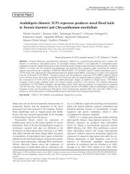
386 Enhanced resistance of transgenic <strong>tobacco</strong> express Lfc<br />
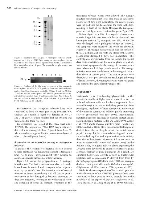
Figure3. Southern blot analysis of transgenic <strong>tobacco</strong> <strong>plants</strong><br />
carrying the Lfc gene. DNA from transgenic <strong>tobacco</strong> <strong>plants</strong> No. 33<br />
(lane 1) and No. 74 (lane 2) was digested with BamHI. The Southern<br />
blot was probed with Lfc gene. The molecular weight marker was<br />
lambda/HindIII.<br />
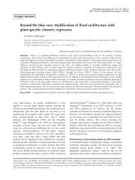
Figure4. Analysis of the Lfc gene expression in the transgenic<br />
<strong>tobacco</strong> <strong>plants</strong> by RT-PCR. PCR products from RNA extracted from<br />
control (lane 1) and transgenic <strong>plants</strong> No. 33 (lane 2) and No. 74 (lane<br />
3) without reverse transcription, and RT-PCR products from RNA<br />
extracted from control (lane 4) and transgenic <strong>plants</strong> No. 33 (lane 5)<br />
and No. 74 (lane 6) were analyzed. Allow indicates Lfc gene amplified<br />
by RT-PCR. Lane M, 100 bp ladder.<br />
Furthermore, the transgenic <strong>tobacco</strong> lines were<br />
confirmed to have the transgene using Southern blot<br />
analysis. As a result, a signal was detected in No. 33<br />
and 74 (Figure 3), which revealed that the Lfc gene was<br />
transformed in these two lines.<br />
Lfc expression was tested at the RNA level using<br />
RT-PCR. The appropriate 78 bp DNA fragments were<br />
detected in two transgenic lines (Figure 4, lanes 5 and 6),<br />
whereas no bands appeared in the untransformed control<br />
<strong>tobacco</strong> <strong>plants</strong> (Figure 4, lane 4).<br />
Evaluation of <strong>antimicrobial</strong> activity in transgenic<br />
<strong>tobacco</strong><br />
To evaluate the resistance to bacterial disease, control<br />
<strong>tobacco</strong> <strong>plants</strong> and two kanamycin resistant T 1 transgenic<br />
lines (No. 33 and 74) were challenged with P. syringae pv.<br />
tabaci, an endemic pathogen of wildfire disease.<br />
Figure 5A shows the progression of P. syringae<br />
infection rate. The first symptoms were observed on the<br />
lower leaves in control and transgenic <strong>tobacco</strong> <strong>plants</strong><br />
nine days post-infection. The infection rate of control<br />
<strong>tobacco</strong> increased immediately and all control <strong>plants</strong><br />
were more or less damaged by bacterial infection, 16<br />
days post-infection, resulting in the yellowing of leaves<br />
and softening of stems. In contrast, symptoms in the<br />
transgenic <strong>tobacco</strong> <strong>plants</strong> were delayed. The average<br />
infection rates were much lower than those in the control<br />
<strong>plants</strong>. At 30 days post inoculation, the control <strong>plants</strong><br />
were infected with the disease from the roots to the tips,<br />
resulting in death of the <strong>plants</strong>. However, the transgenic<br />
<strong>plants</strong> were still green and continued to grow (Figure 5B).<br />
To investigate the ability of transgenic <strong>tobacco</strong> <strong>plants</strong><br />
to resist fungal infection, control <strong>tobacco</strong> <strong>plants</strong> and two<br />
kanamycin resistant T 1 transgenic lines (No. 33 and 74)<br />
were challenged with a pathogenic fungus (B. cinerea),<br />
and symptoms were recorded. The results are shown in<br />
Figure 6A. The fungus had grown all over the surface of<br />
the MS medium, and the roots and stems of the control<br />
<strong>plants</strong> were damaged 6 days post-inoculation. The<br />
control <strong>plants</strong> were infected from the roots to the tips 28<br />
days post-inoculation, and five control <strong>plants</strong> were dead.<br />
In contrast, symptoms in the transgenic <strong>tobacco</strong> <strong>plants</strong><br />
were delayed until 11 days post-inoculation. The average<br />
infection rates were much lower in the transgenic <strong>plants</strong><br />
than those in control <strong>plants</strong>. The control <strong>plants</strong> were<br />
damaged 28 days post-inoculation, resulting in yellowing<br />
of leaves. However, the transgenic <strong>plants</strong> were still green<br />
and continued to grow normally (Figure 6B).<br />
Discussion<br />
Lactoferrin is an iron-binding glycoprotein in the<br />
transferrin family. A high concentration of lactoferrin<br />
is found in human milk and has been suggested to have<br />
several biological activities, including protection from<br />
pathogens, regulation of iron absorption, modulation<br />
of the immune system, and cellular growth promoting<br />
activity (Lönnerdal and Iyer 1995). Recombinant<br />
lactoferrin has been produced in <strong>plants</strong> to protect against<br />
plant pathogenic bacteria (Mitra and Zhang 1994; Zhang<br />
et al. 1998) and to increase nutritive value (Nandi et al.<br />
2002; Nandi et al 2005). Lfc is the <strong>antimicrobial</strong> fragment<br />
derived from the full length lactoferrin protein upon<br />
pepsin cleavage. Lfc has characteristics of typical cationic<br />
<strong>antimicrobial</strong> peptides and higher <strong>antimicrobial</strong> activity<br />
than that of lactoferrin. Moreover, Lfc is not toxic to<br />
plant or animal cells (Muños and Marcos 2006). In the<br />
present study, transgenic <strong>tobacco</strong> <strong>plants</strong> <strong>expressing</strong> the<br />
Lfc gene were developed to enhance resistance against<br />
a broad spectrum of plant pathogens. As a result, Lfc<br />
exhibited bactericidal activity as high as bactericidal<br />
peptides, such as sarcotoxin IA derived from fresh fly<br />
Sarcophaga peregrina (Ohshima et al. 1999) and cecropin<br />
derived from giant silk moth Hylophora cecropiaie<br />
(Hightower et al. 1994; Jan et al. 2010; Jaynes et al. 1993).<br />
Trials for the overexpression of bactericidal peptides<br />
under the control of the CaMV35S promoter have been<br />
conducted without positive results, possibly due to the<br />
instability of the expressed peptides (Hightower et al.<br />
1994; Sharma et al. 2000; Zhang et al. 1998). Ohshima<br />
Copyright © 2012 The Japanese Society for Plant Cell and Molecular Biology