Nanoassemblies of sulfonated polyaniline multilayers - ARPAL
Nanoassemblies of sulfonated polyaniline multilayers - ARPAL
Nanoassemblies of sulfonated polyaniline multilayers - ARPAL

Create successful ePaper yourself
Turn your PDF publications into a flip-book with our unique Google optimized e-Paper software.
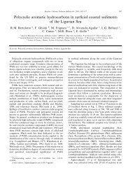
<strong>Nanoassemblies</strong> <strong>of</strong> <strong>sulfonated</strong> <strong>polyaniline</strong> <strong>multilayers</strong><br />
surface morphology <strong>of</strong> such bilayer films was investigated<br />
using atomic force microscopy. The electrochemical kinetics<br />
<strong>of</strong> such LBL films was also investigated by electrochemical<br />
surveying.<br />
2. Experimental details<br />
2.1. Solution preparation and deposition<br />
The emeraldine base form <strong>of</strong> PANI was synthesized as<br />
reported in [26]. The emeraldine base powder was dried, and<br />
<strong>sulfonated</strong> by dissolving in fuming sulfuric acid at 4–5 ◦ C<br />
with constant stirring for 2 h. Later, this solution was added<br />
drop-wise to methanol for the precipitation <strong>of</strong> the product<br />
and the temperature was maintained between 10 and 20 ◦ C.<br />
Precipitation was completed by the addition <strong>of</strong> acetone. The<br />
green powder was collected on a Buchner funnel, and washed<br />
repeatedly with methanol until the filtrate showed a value <strong>of</strong><br />
pH 7. The precipitate was dried under vacuum for 72 h [27].<br />
Microscopic glass, indium–tin-oxide (ITO) coated<br />
glass plates were used as substrate for the fabrication <strong>of</strong><br />
PDDA/SPANI multilayer films. Substrates were activated<br />
following a procedure reported previously [16]. The first<br />
layer <strong>of</strong> activated surface was deposited by <strong>sulfonated</strong><br />
polystyrene (PSS, M w = 70 000) solution for 15 min,<br />
prepared by using 2 mg ml −1 <strong>of</strong> PSS in water, which provided<br />
the charges necessary to adsorb the first layer <strong>of</strong> PDDA.<br />
SPANI (0.1 g) was dissolved in 10 ml <strong>of</strong> 0.1 N NaOH solution<br />
and diluted to 30 ml by distilled water. Then, this solution was<br />
filtered to remove any trace <strong>of</strong> undissolved SPANI particles.<br />
Later, this solution was adjusted to pH = 6 by the dropwise<br />
addition <strong>of</strong> 1 M HCl solution. The polyelectrolyte<br />
PDDA was purchased from Aldrich with M w = 200 000–<br />
350 000, and used in an aqueous solution (2 mg ml −1 ). The<br />
multilayer structure was fabricated by alternate dipping <strong>of</strong><br />
treated substrates in the PDDA and SPANI solution for 10 min<br />
each by rigorous washing in a solution <strong>of</strong> pH 2, and dried<br />
by nitrogen gas. The alternating layers <strong>of</strong> PDDA and SPANI<br />
were also deposited onto various substrates, prior to one layer<br />
deposition <strong>of</strong> PSS, polyanion. Such treated substrate was<br />
used to build <strong>multilayers</strong> structures <strong>of</strong> SPANI and PDDA.<br />
Later, SPANI solutions at pH = 5 as polycation, and at<br />
pH = 10 as polyanion were used for the deposition <strong>of</strong> SPANI<br />
LBL films on PSS-deposited glass or ITO-coated glass plates,<br />
respectively. SPANI (0.1 g) was dissolved in 10 ml <strong>of</strong> 0.1 N<br />
NaOH solution, and diluted to 30 ml by distilled water. Then,<br />
this solution was filtered to remove any trace <strong>of</strong> undissolved<br />
SPANI particles. This solution was divided into two parts,<br />
one was adjusted to pH = 5 and other to pH = 10 by slow<br />
addition <strong>of</strong> 1 M HCl. The pH 5 SPANI solution acted as<br />
polycation and the pH 10 solution acted as polyanion during<br />
deposition <strong>of</strong> multilayer films. Alternating layers <strong>of</strong> SPANI<br />
(pH = 5) and SPANI (pH = 10) were deposited onto the<br />
glass and ITO-coated glass plates, prior to the deposition <strong>of</strong><br />
one layer <strong>of</strong> PSS solution, by alternating submersions <strong>of</strong> the<br />
film samples in the electrolyte solutions to build a multibilayer<br />
structure. Between each deposition, the films were<br />
washed with a solution <strong>of</strong> pH 2 using HCl acid and dried by<br />
blowing nitrogen gas.<br />
2.2. Optical measurements<br />
The UV–visible spectra <strong>of</strong> LBL films deposited on the optical<br />
glass substrates were recorded by using the UV–visible<br />
spectrophotometer (Jasco model 7800).<br />
2.3. Electrical and electrochemical measurements<br />
The electrical characterization was performed using an<br />
electrometer (Keithley model 6517). Current–voltage (I–<br />
V ) characteristics were obtained by an applied potential<br />
(step <strong>of</strong> 0.05 V). Similar interdigitated electrodes were used<br />
for the electrical measurements [16]. The electrochemical<br />
measurements on LBL deposited SPANI films on ITO-coated<br />
glass plates were made by Potentiostat/Galvanostat (EG &<br />
G PARC, model 263A) with a supplied s<strong>of</strong>tware (M270).<br />
A standard three-electrode configuration was used, where<br />
PDDA/SPANI and SPANI (deposited at pH 5 and 10) films<br />
on PSS/ITO-coated glass plates acted as a working electrode,<br />
with platinum as a counter and Ag/AgCl as a reference<br />
electrode.<br />
2.4. Atomic force microscopy<br />
The surface morphology <strong>of</strong> the SPANI/PDDA LBL films was<br />
investigated by an atomic force microscope (AFM), which<br />
was a home-built instrument (Polo Nazionale Bioelettronica),<br />
working in contact mode in air at a constant contact force.<br />
Our AFM was operated in air, at constant deflection (i.e.<br />
vertical contact force) with triangular shaped gold-coated<br />
Si 3 N 4 . The tips <strong>of</strong> the microlevers had standard aspect ratio<br />
(about 1:1) and the levers had nominal force constant <strong>of</strong><br />
0.03 N m −1 . The constant force set point was about 0.1 nN,<br />
while the images acquired were 256×256 pixel maps. During<br />
the acquisition the row scanning frequency was set to 4 Hz,<br />
i.e. a physical tip–sample motion speed <strong>of</strong> 8, 4, 2 µm s −1 in<br />
the 2, 1, 0.5 µm scan size images, respectively. Some images<br />
presented features that were saturated in the post-processing<br />
redistribution <strong>of</strong> the available grey levels. Henceforth, it was<br />
possible to observe the finest structure <strong>of</strong> the samples. The<br />
images shown in this paper are representative <strong>of</strong> the samples,<br />
as similar looking images appeared in four different regions<br />
<strong>of</strong> the analysed samples, positioned at the vertices <strong>of</strong>a4mm<br />
side square, centred on the specimen [16, 28].<br />
3. Results and discussion<br />
3.1. UV–visible<br />
Figure 1(a) shows the optical absorption spectra <strong>of</strong><br />
PDDA/SPANI deposited on a PSS/glass slide as a function<br />
<strong>of</strong> the number <strong>of</strong> bilayers. As PDDA is not absorbing in<br />
the considered spectral region, the UV–visible absorption<br />
can only be emanating due to the SPANI layers. A typical<br />
UV–visible absorption spectrum <strong>of</strong> SPANI has three distinct<br />
absorption bands in the regions 340, 430–450 and 800–<br />
900 nm. These spectra depict the features characteristic<br />
<strong>of</strong> the SPANI forms, implying that the polymer is in the<br />
protonated form. The band at 340 is attributed to the<br />
π–π ∗ band-gap absorption and 430–450 and 800–900 are<br />
due to the protonation <strong>of</strong> SPANI. The constant absorbance<br />
31