Cardiac Involvement in Myasthenia Gravis - Romanian Journal of ...
Cardiac Involvement in Myasthenia Gravis - Romanian Journal of ...
Cardiac Involvement in Myasthenia Gravis - Romanian Journal of ...

You also want an ePaper? Increase the reach of your titles
YUMPU automatically turns print PDFs into web optimized ePapers that Google loves.
180<br />
C. Căl<strong>in</strong> et al. 2<br />
local hospital on an emergency basis for severe<br />
dysphagia, mastication problems and dysarthria. The<br />
diagnosis <strong>of</strong> myasthenia gravis was f<strong>in</strong>ally established<br />
after a positive neostigm<strong>in</strong>e test. The presence or<br />
absence <strong>of</strong> antibodies aga<strong>in</strong>st the acetylchol<strong>in</strong>e receptors<br />
could not be established at that po<strong>in</strong>t. Treatment with<br />
neostigm<strong>in</strong>e and theophyll<strong>in</strong>e was started, with a<br />
partial improvement <strong>of</strong> symptoms.<br />
After an uneventful period <strong>of</strong> time the patient<br />
was admitted to the local hospital for an episode <strong>of</strong><br />
loss <strong>of</strong> postural tone without loss <strong>of</strong> consciousness,<br />
right lid ptosis, diplopia, severe dyspnea and<br />
hypotension (BP 60/40mmHg). She was transferred<br />
to the Fundeni Cl<strong>in</strong>ic Institute, where a CT scan was<br />
performed, show<strong>in</strong>g a 5 cm thymic tumor. The patient<br />
underwent thymectomy <strong>in</strong>clud<strong>in</strong>g the resection <strong>of</strong><br />
both the thymic tumor and the <strong>in</strong>vaded pleura; the<br />
f<strong>in</strong>al diagnosis was: “Thymic tumor with <strong>in</strong>vasion<br />
<strong>of</strong> mediast<strong>in</strong>al and visceral pleura (T2N0M0).<br />
Generalized form <strong>of</strong> myasthenia gravis”. Ten sessions<br />
<strong>of</strong> radiotherapy consolidated the treatment. The<br />
outcome was favorable, with the complete remission<br />
<strong>of</strong> diplopia, dysathria, deglutition and mastication<br />
alter<strong>in</strong>g and the significant improvement <strong>of</strong> muscle<br />
weakness and dyspnea.<br />
Between 2002 and 2006 the patient had a<br />
regular follow-up, the evolution be<strong>in</strong>g stationary<br />
with slight <strong>in</strong>termittent relapses <strong>of</strong> the myasthenic<br />
weakness dur<strong>in</strong>g episodes <strong>of</strong> respiratory <strong>in</strong>fections.<br />
Start<strong>in</strong>g from July 2007 until January 2008<br />
the patient had seven episodes <strong>of</strong> syncope, both <strong>in</strong><br />
exertion and while sitt<strong>in</strong>g (<strong>in</strong>clud<strong>in</strong>g sup<strong>in</strong>e), the<br />
latest episode be<strong>in</strong>g two months previous to the<br />
admission <strong>in</strong> our cl<strong>in</strong>ic.<br />
At the admission <strong>in</strong> our cl<strong>in</strong>ic we were fac<strong>in</strong>g<br />
the situation <strong>of</strong> a patient previously diagnosed with<br />
generalized form <strong>of</strong> myasthenia gravis and a<br />
history <strong>of</strong> repeated episodes <strong>of</strong> syncope, without<br />
relationship to exertion, without prodroma, without<br />
ang<strong>in</strong>a or palpitations, with no postcritical motor<br />
deficits. She presented fatigue, dyspnea at less than<br />
ord<strong>in</strong>ary physical exertion and cough with mucopurulent<br />
sputum.<br />
The family history was unremarkable; as classical<br />
cardiovascular risk factors we can mention severe<br />
essential arterial hypertension (highest BP measured<br />
value <strong>of</strong> 180/90mmHg), dyslipidemia and obesity.<br />
Upon admission the physical exam<strong>in</strong>ation<br />
revealed a slightly altered general status, normal<br />
body temperature, plethora, ecchymoses on the legs,<br />
abdom<strong>in</strong>al type obesity. The muscle exam<strong>in</strong>ation<br />
revealed hypotonia and hypok<strong>in</strong>esia with preserved<br />
reflexes, right lid ptosis. The BP was 120/90mmHg,<br />
without postural hypotension, HR 83 bpm, regular<br />
heart sounds without murmurs, palpable pulses,<br />
without peripheral edema or jugular distension.<br />
The patient was <strong>in</strong> mild respiratory distress with a<br />
respiratory frequency <strong>of</strong> about 35/m<strong>in</strong>, course<br />
vesicular sounds and wheezes, cough with mucopurulent<br />
sputum.<br />
The laboratory evaluation revealed leucocytosis<br />
with neutrophilia, <strong>in</strong>terpreted as secondary to the<br />
respiratory <strong>in</strong>fection, dyslipidemia, slightly elevated<br />
blood urea nitrogen and uric acid.<br />
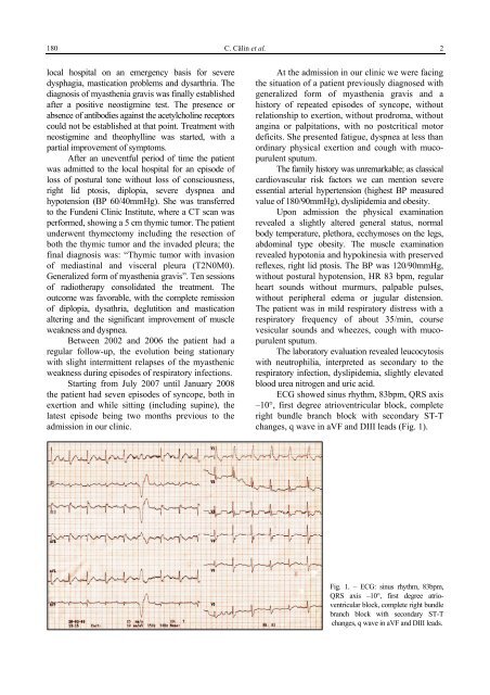
ECG showed s<strong>in</strong>us rhythm, 83bpm, QRS axis<br />
–10°, first degree atrioventricular block, complete<br />
right bundle branch block with secondary ST-T<br />
changes, q wave <strong>in</strong> aVF and DIII leads (Fig. 1).<br />
Fig. 1. – ECG: s<strong>in</strong>us rhythm, 83bpm,<br />
QRS axis –10°, first degree atrioventricular<br />
block, complete right bundle<br />
branch block with secondary ST-T<br />
changes, q wave <strong>in</strong> aVF and DIII leads.