Cardiac Involvement in Myasthenia Gravis - Romanian Journal of ...
Cardiac Involvement in Myasthenia Gravis - Romanian Journal of ...
Cardiac Involvement in Myasthenia Gravis - Romanian Journal of ...

You also want an ePaper? Increase the reach of your titles
YUMPU automatically turns print PDFs into web optimized ePapers that Google loves.
5 <strong>Cardiac</strong> <strong>in</strong>volvement <strong>in</strong> myasthenia gravis 183<br />
specific etiology, imposes several electrocardiographic<br />
differentials; this type <strong>of</strong> repolarisation<br />
changes may be seen <strong>in</strong> subendocardic ischaemianecrosis,<br />
central nervous system (CNS) disorders,<br />
pericarditis, myocarditis, obstructive hypertrophic<br />
cardiomyopathy, metabolic abnormalities, coca<strong>in</strong>e<br />
use, pheochromocytoma and hyperventilation.<br />
Chest X-ray showed a normal cardiothoracic<br />
ratio, without pulmonary or pleural abnormalities.<br />
Electrolytic disorders have been ruled out<br />
based on a normal biological panel. Due to the<br />
suspicion <strong>of</strong> myocardial ischaemia the serum<br />
tropon<strong>in</strong> was determ<strong>in</strong>ed (one measurement) –<br />
positive qualitative test.<br />
Based on the universal def<strong>in</strong>ition <strong>of</strong> myocardial<br />
<strong>in</strong>farction the positive diagnosis is made on the<br />
detection <strong>of</strong> the rise and/or fall <strong>of</strong> cardiac<br />
biomarkers together with at least one <strong>of</strong> the<br />
follow<strong>in</strong>g: ischemic symptoms, ECG changes<br />
<strong>in</strong>dicative <strong>of</strong> new ischaemia, new Q waves or<br />
imag<strong>in</strong>g evidence <strong>of</strong> new loss <strong>of</strong> viable myocardium<br />
or new wall motion abnormalities [7].<br />
In this case a second measurement <strong>of</strong> tropon<strong>in</strong><br />
was not available; although tropon<strong>in</strong> is specific for<br />
myocardial necrosis, there are false positive<br />
situations – critically ill patients, especially those with<br />
respiratory failure or sepsis, myocarditis, hypertrophic<br />
cardiomyopathy and so on.<br />
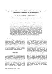
The first echocardiography showed the<br />
absence <strong>of</strong> regional wall motion abnormalities, but<br />
revealed moderate concentric left ventricular<br />
hypertrophy (<strong>in</strong>terventricular septum 13mm,<br />
posterior wall 13mm) (Fig. 5a and 5b) and normal<br />
left ventricular systolic function. The left ventricular<br />
hypertrophy was considered secondary to the<br />
arterial hypertension, a diagnosis <strong>of</strong> hypertrophic<br />
cardiomyopathy be<strong>in</strong>g considered unjustified <strong>in</strong><br />
this case. A small subendocardic necrosis could not<br />
be elim<strong>in</strong>ated, as <strong>in</strong> this case and especially <strong>in</strong> the<br />
presence <strong>of</strong> LV wall hypertrophy, echocardiography<br />
might not detect subtle wall motion<br />
abnormalities.<br />
Data on giant T wave <strong>in</strong>version shows that<br />
myocardial <strong>in</strong>farction and CNS disorders are major<br />
etiologies. In a series <strong>of</strong> 100 ECGs with T wave<br />
<strong>in</strong>version analyzed by Walder and Spodick [8]<br />
28 patients presented myocardial <strong>in</strong>farction and<br />
23 patients CNS disorders.<br />
In the reported case the cerebral CT scan<br />
was normal.<br />
Therefore, we were not able to determ<strong>in</strong>e a<br />
certa<strong>in</strong> cause <strong>of</strong> the ECG changes, but could not<br />
exclude myocardial necrosis <strong>in</strong> a patient with<br />
multiple cardiac risk factors. Coronary angiography<br />
on an emergency basis was not performed, <strong>in</strong> this<br />
phase the patient be<strong>in</strong>g treated for the acute<br />
respiratory failure; although coronary angiography<br />
is not contra<strong>in</strong>dicated <strong>in</strong> myasthenia gravis, iod<strong>in</strong>e<br />
contrast usage warrants precaution <strong>in</strong> these cases,<br />
as situations <strong>of</strong> muscle weakness exacerbation have<br />
been reported [9].<br />
Fig. 5a. – Transthoracic echocardiography – parasternal long<br />
axis. Interventricular septum 13mm.<br />
Fig. 5a. – Transthoracic echocardiography – parasternal long<br />
axis. Interventricular septum 13mm.<br />
Fig. 5b. – Transthoracic echocardiography – parasternal long<br />
axis; anatomic M-mode. Left ventricle dimensions.<br />
The myasthenic crisis had a favorable outcome<br />
with remission. Dur<strong>in</strong>g the follow<strong>in</strong>g week the ECG<br />
showed no further evolution. Further myocardial<br />
biomarkers determ<strong>in</strong>ations were <strong>in</strong> a normal range.<br />
The physical exam<strong>in</strong>ation <strong>of</strong> the patient after<br />
the myasthenic crisis showed normal BP measurements<br />
under treatment with ACEI and diuretics, without<br />
signs <strong>of</strong> pulmonary or systemic congestion; the<br />
follow<strong>in</strong>g echocardiographic exam<strong>in</strong>ations were similar<br />
to the first one.<br />
Two months after the myasthenic crisis the<br />
ECG shows dynamic changes <strong>in</strong> the anterior leads,<br />
with biphasic T waves <strong>in</strong> leads C2-C6, DI,aVL<br />
(Fig. 6), with no ischaemic or heart failure symptoms.