Use of Microcalcification Descriptors in BI-RADS 4th ... - Quantason
Use of Microcalcification Descriptors in BI-RADS 4th ... - Quantason
Use of Microcalcification Descriptors in BI-RADS 4th ... - Quantason

Create successful ePaper yourself
Turn your PDF publications into a flip-book with our unique Google optimized e-Paper software.
BREAST IMAGING: <strong>BI</strong>-<strong>RADS</strong> <strong>Microcalcification</strong> <strong>Descriptors</strong><br />
Burnside et al<br />
complished by us<strong>in</strong>g either imag<strong>in</strong>g<br />
and/or medical records to determ<strong>in</strong>e<br />
whether a patient had developed breast<br />
cancer at or adjacent to the biopsy site<br />
with<strong>in</strong> 1 year <strong>of</strong> a benign biopsy result.<br />
We used a 12-month follow-up as our<br />
reference standard because it has been<br />
recommended <strong>in</strong> mammographic practice<br />
audits as a sufficient <strong>in</strong>terval to<br />
identify false-negative f<strong>in</strong>d<strong>in</strong>gs (6). Because<br />
the lesions <strong>in</strong>cluded <strong>in</strong> this study<br />
may represent very early breast cancer,<br />
we performed additional follow-up<br />
when possible. Ten women who did not<br />
undergo mammographic follow-up underwent<br />
cl<strong>in</strong>ical follow-up rang<strong>in</strong>g from<br />
12 to 50 months (mean, 33.9 months <br />
13.2). Seven women who did not undergo<br />
cl<strong>in</strong>ical follow-up underwent<br />
mammographic follow-up rang<strong>in</strong>g from<br />
12 to 43 months (mean, 24.9 months <br />
13.7). Overall, mammographic (mean,<br />
30.6 months 13.7) and cl<strong>in</strong>ical follow-up<br />
(mean, 35.3 months 13.7) <strong>of</strong><br />
the 90 patients with benign lesions averaged<br />
far more than the 12-month <strong>in</strong>terval<br />
recommended to ensure benignity.<br />
Statistical Analysis<br />
We used s<strong>of</strong>tware (S-PLUS, version 6.2,<br />
Insightful, Seattle, Wash; R, version<br />
2.1.1, R Foundation for Statistical Comput<strong>in</strong>g,<br />
Vienna, Austria) for statistical<br />
analysis. The Fisher exact test was used<br />
to calculate the difference among the<br />
descriptor groups with respect to the<br />
risk <strong>of</strong> malignancy; the proportion <strong>of</strong><br />
malignant cases was the dependent<br />
variable. A P value <strong>of</strong> less than .05 was<br />
considered to <strong>in</strong>dicate a statistically significant<br />
difference. Post hoc analyses<br />
were used to expla<strong>in</strong> significant results<br />
from the Fisher exact test, <strong>in</strong>clud<strong>in</strong>g<br />
odds ratios and 95% confidence <strong>in</strong>tervals<br />
(CIs), for pairwise comparisons <strong>of</strong><br />
descriptor groups, which were derived<br />
by us<strong>in</strong>g either asymptotic methods (S-<br />
PLUS) or exact methods (R). Pairwise<br />
odds ratios were considered to <strong>in</strong>dicate<br />
a statistically significant difference if the<br />
95% CI for the odds ratio excluded 1.<br />
All analyses were based on a s<strong>in</strong>gle observation<br />
per patient so that the usual<br />
assumption <strong>of</strong> <strong>in</strong>dependent data was<br />
satisfied.<br />
Results<br />
<strong>Microcalcification</strong> Results<br />
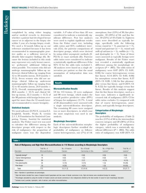
Of the 115 lesions, 25 were malignant<br />
and 90 were benign, which makes the<br />
overall positive predictive value (PPV)<br />
<strong>of</strong> biopsy for malignancy 21.7%. A total<br />
<strong>of</strong> 26 abnormalities were assessed with<br />
a s<strong>in</strong>gle microcalcification descriptor.<br />
The rema<strong>in</strong><strong>in</strong>g 89 cases were assigned<br />
two or more descriptors, <strong>in</strong> which case<br />
the most suspicious was used <strong>in</strong> our<br />
analysis.<br />
Morphologic <strong>Descriptors</strong><br />
Each <strong>of</strong> the microcalcification morphologic<br />
descriptors further stratified the<br />
probability <strong>of</strong> malignancy as follows:<br />
coarse heterogeneous, one (7%) <strong>of</strong> 14;<br />
amorphous, four (13%) <strong>of</strong> 30; f<strong>in</strong>e pleomorphic,<br />
10 (29%) <strong>of</strong> 34; and f<strong>in</strong>e l<strong>in</strong>ear,<br />
10 (53%) <strong>of</strong> 19 (Table 1). Eighteen<br />
cases were described as typically benign,<br />
specifically with the follow<strong>in</strong>g<br />
terms: round (n 7), punctate (n 7),<br />
round and punctate (n 1), round and<br />
milk <strong>of</strong> calcium (n 1), rodlike (n 1),<br />
and dystrophic (n 1). None <strong>of</strong> these<br />
cases assessed as typically benign were<br />
malignant. Results <strong>of</strong> the Fisher exact<br />
test revealed a statistically significant<br />
difference among the morphologic descriptors<br />
(P .005). The odds ratios <strong>of</strong><br />
malignancy were 0.07 (95% CI: 0.01,<br />
0.69) for coarse heterogeneous versus<br />
f<strong>in</strong>e l<strong>in</strong>ear, 0.14 (95% CI: 0.03, 0.66)<br />
for amorphous versus f<strong>in</strong>e l<strong>in</strong>ear, 0.37<br />
(95% CI: 0.09, 1.34) for f<strong>in</strong>e pleomorphic<br />
versus f<strong>in</strong>e l<strong>in</strong>ear, and 0 (95% CI:<br />
0, 0.32) for typically benign versus f<strong>in</strong>e<br />
l<strong>in</strong>ear. Results <strong>of</strong> this analysis suggest<br />
that the f<strong>in</strong>e l<strong>in</strong>ear descriptor, used as a<br />
base case, <strong>in</strong>dicates a significantly <strong>in</strong>creased<br />
risk <strong>of</strong> malignancy (95% CI for<br />
odds ratios excludes 1) compared with<br />
that <strong>of</strong> coarse heterogeneous, amorphous,<br />
and typically benign descriptors.<br />
Categorization <strong>of</strong> Morphologic<br />
<strong>Descriptors</strong><br />
The probability <strong>of</strong> malignancy (Table 2)<br />
was five (11%) <strong>of</strong> 44 <strong>in</strong> the <strong>in</strong>termediate<br />
concern category and 20 (38%) <strong>of</strong> 53 <strong>in</strong><br />
the higher probability <strong>of</strong> malignancy<br />
category, which was a statistically significant<br />
difference (P .002). The odds<br />
ratio <strong>of</strong> malignancy was 4.69 (95% CI:<br />
Table 1<br />
Rate <strong>of</strong> Malignancy and High-Risk <strong>Microcalcification</strong>s <strong>in</strong> 115 Women accord<strong>in</strong>g to Morphologic <strong>Descriptors</strong><br />
Descriptor<br />
Total No. <strong>of</strong><br />
<strong>Microcalcification</strong>s<br />
No. <strong>of</strong> Invasive<br />
Cancers<br />
No. <strong>of</strong> Ductal<br />
Carc<strong>in</strong>omas <strong>in</strong> Situ<br />
No. <strong>of</strong> High-Risk<br />
Lesions*<br />
Total No. <strong>of</strong><br />
Lesions †<br />
Typically benign ‡ 18 0 (0) 0 (0) 0 (0) 0 (0)<br />
Coarse heterogeneous 14 0 (0) 1 (7) 0 (0) 1 (7)<br />
Amorphous 30 2 (7) 2 (7) 4 (13) 4 (13)<br />
F<strong>in</strong>e pleomorphic 34 5 (15) 5 (15) 1 (3) 10 (29)<br />
F<strong>in</strong>e l<strong>in</strong>ear 19 6 (32) 4 (21) 1 (5) 10 (53)<br />
Total 115 13 (11.3) 12 (10.4) 6 (5.2) 25 (21.7)<br />
Note.—Numbers <strong>in</strong> parentheses are percentages.<br />
* High-risk lesions <strong>in</strong>cluded four cases <strong>of</strong> atypical ductal hyperplasia and two cases <strong>of</strong> lobular carc<strong>in</strong>oma <strong>in</strong> situ. High-risk lesions were considered benign for analysis.<br />
† Percentages calculated by us<strong>in</strong>g the number <strong>of</strong> <strong>in</strong>vasive cancers and ductal carc<strong>in</strong>omas <strong>in</strong> situ malignancies divided by the total number <strong>of</strong> cases for each morphologic descriptor.<br />
‡ Includes round, punctate, dystrophic, milk <strong>of</strong> calcium, and rodlike microcalcifications.<br />
Radiology: Volume 242: Number 2—February 2007 391