Use of Microcalcification Descriptors in BI-RADS 4th ... - Quantason
Use of Microcalcification Descriptors in BI-RADS 4th ... - Quantason
Use of Microcalcification Descriptors in BI-RADS 4th ... - Quantason

You also want an ePaper? Increase the reach of your titles
YUMPU automatically turns print PDFs into web optimized ePapers that Google loves.
ORIGINAL RESEARCH BREAST IMAGING<br />
Elizabeth S. Burnside, MD, MPH, MS<br />
Jennifer E. Ochsner, MD<br />
Kathryn J. Fowler, MD<br />
Jason P. F<strong>in</strong>e, PhD<br />
Lonie R. Salkowski, MD<br />
Daniel L. Rub<strong>in</strong>, MD, MS<br />
Gale A. Sisney, MD<br />
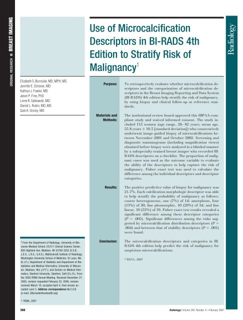
<strong>Use</strong> <strong>of</strong> <strong>Microcalcification</strong><br />
<strong>Descriptors</strong> <strong>in</strong> <strong>BI</strong>-<strong>RADS</strong> <strong>4th</strong><br />
Edition to Stratify Risk <strong>of</strong><br />
Malignancy 1<br />
Purpose:<br />
Materials and<br />
Methods:<br />
To retrospectively evaluate whether microcalcification descriptors<br />
and the categorization <strong>of</strong> microcalcification descriptors<br />
<strong>in</strong> the Breast Imag<strong>in</strong>g Report<strong>in</strong>g and Data System<br />
(<strong>BI</strong>-<strong>RADS</strong>) <strong>4th</strong> edition help stratify the risk <strong>of</strong> malignancy,<br />
by us<strong>in</strong>g biopsy and cl<strong>in</strong>ical follow-up as reference standards.<br />
The <strong>in</strong>stitutional review board approved this HIPAA-compliant<br />
study and waived <strong>in</strong>formed consent. The study <strong>in</strong>cluded<br />
115 women (age range, 26–82 years; mean age,<br />
55.8 years 10.5 [standard deviation]) who consecutively<br />
underwent image-guided biopsy <strong>of</strong> microcalcifications between<br />
November 2001 and October 2002. Screen<strong>in</strong>g and<br />
diagnostic mammograms (<strong>in</strong>clud<strong>in</strong>g magnification views)<br />
obta<strong>in</strong>ed before biopsy were analyzed <strong>in</strong> a bl<strong>in</strong>ded manner<br />
by a subspecialty-tra<strong>in</strong>ed breast imager who recorded <strong>BI</strong>-<br />
<strong>RADS</strong> descriptors on a checklist. The proportion <strong>of</strong> malignant<br />
cases was used as the outcome variable to evaluate<br />
the ability <strong>of</strong> the descriptors to help capture the risk <strong>of</strong><br />
malignancy. Fisher exact test was used to calculate the<br />
difference among the <strong>in</strong>dividual descriptors and descriptor<br />
categories.<br />
1 From the Department <strong>of</strong> Radiology, University <strong>of</strong> Wiscons<strong>in</strong><br />
Medical School, E3/311 Cl<strong>in</strong>ical Science Center,<br />
600 Highland Ave, Madison, WI 53792-3252 (E.S.B.,<br />
J.E.O., L.R.S., G.A.S.); Mall<strong>in</strong>ckrodt Institute <strong>of</strong> Radiology,<br />
Wash<strong>in</strong>gton University School <strong>of</strong> Medic<strong>in</strong>e, St Louis, Mo<br />
(K.J.F.); Department <strong>of</strong> Statistics and Department <strong>of</strong> Biostatistics<br />
and Medical Informatics, University <strong>of</strong> Wiscons<strong>in</strong>,<br />
Madison, Wis (J.P.F.); and Section on Medical Informatics,<br />
Stanford University, Stanford, Calif (D.L.R.). From<br />
the 2005 RSNA Annual Meet<strong>in</strong>g. Received December 27,<br />
2005; revision requested February 22, 2006; revision<br />
received March 10; accepted April 4; f<strong>in</strong>al version accepted<br />
June 5. Address correspondence to E.S.B.<br />
(e-mail: EBurnside@uwhealth.org).<br />
Results:<br />
Conclusion:<br />
The positive predictive value <strong>of</strong> biopsy for malignancy was<br />
21.7%. Each calcification morphologic descriptor was able<br />
to help stratify the probability <strong>of</strong> malignancy as follows:<br />
coarse heterogeneous, one (7%) <strong>of</strong> 14; amorphous, four<br />
(13%) <strong>of</strong> 30; f<strong>in</strong>e pleomorphic, 10 (29%) <strong>of</strong> 34; and f<strong>in</strong>e<br />
l<strong>in</strong>ear, 10 (53%) <strong>of</strong> 19. Fisher exact test results revealed a<br />
significant difference among these descriptor categories<br />
(P .005). Significant differences among the risks suggested<br />
by microcalcification distribution descriptors (P <br />
.004) and between that <strong>of</strong> stability descriptors (P .001)<br />
were found.<br />
The microcalcification descriptors and categories <strong>in</strong> <strong>BI</strong>-<br />
<strong>RADS</strong> <strong>4th</strong> edition help predict the risk <strong>of</strong> malignancy for<br />
suspicious microcalcifications.<br />
RSNA, 2007<br />
RSNA, 2007<br />
388 Radiology: Volume 242: Number 2—February 2007
BREAST IMAGING: <strong>BI</strong>-<strong>RADS</strong> <strong>Microcalcification</strong> <strong>Descriptors</strong><br />
Burnside et al<br />
The Breast Imag<strong>in</strong>g Report<strong>in</strong>g and<br />
Data System (<strong>BI</strong>-<strong>RADS</strong>) has standardized<br />
the description and management<br />
<strong>of</strong> f<strong>in</strong>d<strong>in</strong>gs identified on mammograms,<br />
thereby facilitat<strong>in</strong>g communication<br />
between radiologists and referr<strong>in</strong>g<br />
physicians. Each descriptor signifies important<br />
features <strong>of</strong> mammographic abnormalities<br />
that convey the radiologist’s<br />
level <strong>of</strong> suspicion. The standardized evaluation<br />
<strong>of</strong> f<strong>in</strong>d<strong>in</strong>gs with predictive terms<br />
enables stratification <strong>of</strong> patient risk to<br />
guide management decisions. For example,<br />
<strong>BI</strong>-<strong>RADS</strong> category 3 (“probably benign”)<br />
represents a standardized constellation<br />
<strong>of</strong> f<strong>in</strong>d<strong>in</strong>gs associated with an estimated<br />
low risk <strong>of</strong> malignancy ( 2%),<br />
which enables management that is less<br />
<strong>in</strong>vasive than biopsy but more aggressive<br />
than rout<strong>in</strong>e follow-up as the standard <strong>of</strong><br />
care (1–4). Authors <strong>of</strong> the <strong>BI</strong>-<strong>RADS</strong> lexicon<br />
also have divided microcalcification<br />
morphologic descriptors <strong>in</strong>to the follow<strong>in</strong>g<br />
designated categories that predict<br />
benignity or malignancy: (a) typically<br />
benign, (b) <strong>in</strong>termediate concern, and<br />
(c) higher probability <strong>of</strong> malignancy<br />
(5,6). Although the probably benign<br />
category is used <strong>in</strong> large prospective<br />
studies, the categorization <strong>of</strong> microcalcification<br />
descriptors <strong>in</strong> <strong>BI</strong>-<strong>RADS</strong> is<br />
based on a comb<strong>in</strong>ation <strong>of</strong> published<br />
research and the op<strong>in</strong>ion <strong>of</strong> the panel<br />
<strong>of</strong> specialists who developed the lexicon<br />
(7–11).<br />
In the past, authors <strong>of</strong> <strong>BI</strong>-<strong>RADS</strong><br />
have used cl<strong>in</strong>ical research to drive the<br />
evolution <strong>of</strong> the lexicon. For example,<br />
amorphous microcalcifications have a<br />
20% probability <strong>of</strong> malignancy, which<br />
supports the placement <strong>of</strong> amorphous<br />
microcalcifications <strong>in</strong> the category <strong>of</strong> <strong>in</strong>termediate<br />
concern (7). Investigators<br />
have identified weaknesses <strong>in</strong> the lexicon<br />
to prompt change. For example, <strong>in</strong><br />
a study <strong>of</strong> <strong>in</strong>terobserver variability <strong>of</strong><br />
<strong>BI</strong>-<strong>RADS</strong> usage (12), microcalcification<br />
descriptors were the most difficult to<br />
apply consistently between readers.<br />
Advance <strong>in</strong> Knowledge<br />
<strong>Microcalcification</strong> descriptors <strong>in</strong><br />
the Breast Imag<strong>in</strong>g Report<strong>in</strong>g and<br />
Data System <strong>4th</strong> edition help<br />
stratify the risk <strong>of</strong> malignancy.<br />
Furthermore, results <strong>of</strong> a large retrospective<br />
review (9) <strong>of</strong> biopsies <strong>in</strong>dicated<br />
that two-thirds <strong>of</strong> all microcalcifications<br />
sampled for biopsy were described as<br />
pleomorphic—a descriptor that encompasses<br />
“a broad spectrum <strong>of</strong> calcification<br />
lesions.”<br />
In response, the <strong>BI</strong>-<strong>RADS</strong> <strong>4th</strong> edition,<br />
which was published <strong>in</strong> 2003 (6),<br />
provided ref<strong>in</strong>ed microcalcification descriptors<br />
by divid<strong>in</strong>g the former pleomorphic<br />
descriptor <strong>in</strong>to two descriptors—coarse<br />
heterogeneous and f<strong>in</strong>e<br />
pleomorphic (Fig 1). Coarse heterogeneous<br />
microcalcifications (Fig 2) are “irregular,<br />
conspicuous calcifications that<br />
are generally larger than 0.5 mm” and<br />
are considered to be <strong>of</strong> <strong>in</strong>termediate<br />
concern, along with amorphous microcalcifications<br />
(6). F<strong>in</strong>e pleomorphic microcalcifications<br />
(Fig 3) “vary <strong>in</strong> sizes<br />
and shapes...usually less than 0.5 mm<br />
<strong>in</strong> diameter” and are considered to be <strong>of</strong><br />
higher probability <strong>of</strong> malignancy, along<br />
with f<strong>in</strong>e l<strong>in</strong>ear microcalcifications (6).<br />
The purpose <strong>of</strong> our study was to retrospectively<br />
evaluate whether microcalcification<br />
descriptors and the categorization<br />
<strong>of</strong> microcalcification descriptors <strong>in</strong><br />
the <strong>BI</strong>-<strong>RADS</strong> <strong>4th</strong> edition help stratify<br />
the risk <strong>of</strong> malignancy by us<strong>in</strong>g biopsy<br />
and cl<strong>in</strong>ical follow-up as reference standards.<br />
Materials and Methods<br />
Patients<br />
The <strong>in</strong>stitutional review board at the<br />
University <strong>of</strong> Wiscons<strong>in</strong> approved this<br />
study and waived the requirement for<br />
<strong>in</strong>formed consent. This study was also<br />
Health Insurance Portability and Accountability<br />
Act compliant. Our study<br />
<strong>in</strong>cluded results <strong>of</strong> 115 image-guided biopsies<br />
performed <strong>in</strong> 115 women between<br />
November 2001 and October<br />
2002 for microcalcifications deemed<br />
suspicious by radiologists. The women<br />
were 26–82 years <strong>of</strong> age (mean age,<br />
55.8 years 10.5 [standard deviation]).<br />
Results <strong>of</strong> 11-gauge stereotactic<br />
biopsies and needle localizations performed<br />
for diagnosis were <strong>in</strong>cluded.<br />
Our exclusion criteria were as follows:<br />
(a) patient’s images were not available<br />
for review, (b) calcifications were not<br />
identified <strong>in</strong> the histologic specimen,<br />
and (c) mammographic or cl<strong>in</strong>ical follow-up<br />
<strong>of</strong> at least 12 months was not<br />
performed after biopsy. The first two<br />
criteria ensured accurate characterization<br />
<strong>of</strong> the microcalcifications and appropriate<br />
correlation with the histologic<br />
f<strong>in</strong>d<strong>in</strong>gs for each abnormality. The third<br />
criterion allowed follow-up evaluation<br />
to recognize misclassification at biopsy.<br />
Of 131 consecutive patients (131 biopsies),<br />
16 were excluded (images were<br />
unavailable for 11 patients, calcifications<br />
were not identified <strong>in</strong> the histologic<br />
specimen for three patients, and<br />
follow-up was not performed <strong>in</strong> two patients),<br />
which resulted <strong>in</strong> the f<strong>in</strong>al group<br />
<strong>of</strong> 115.<br />
Imag<strong>in</strong>g and Evaluation<br />
All mammographic exam<strong>in</strong>ations were<br />
performed with dedicated mach<strong>in</strong>es. Analog<br />
mammographic exam<strong>in</strong>ations were<br />
performed with one <strong>of</strong> two units (DMR,<br />
GE Medical Systems, Milwaukee, Wis;<br />
M-IV, Lorad, Danbury, Conn) and with<br />
a screen-film technique (M<strong>in</strong>-R 2000;<br />
Kodak Health Imag<strong>in</strong>g, Rochester, NY).<br />
Digital mammograms were acquired by<br />
us<strong>in</strong>g a system with a cesium iodide–<br />
amorphous silicon detector (Senographe<br />
2000D; GE Medical Systems). Digital<br />
mammograms were pr<strong>in</strong>ted (DryView<br />
8610 pr<strong>in</strong>ter and DVB film; Kodak Health<br />
Imag<strong>in</strong>g) to be analyzed alongside analog<br />
Published onl<strong>in</strong>e<br />
10.1148/radiol.2422052130<br />
Radiology 2007; 242:388–395<br />
Abbreviations:<br />
<strong>BI</strong>-<strong>RADS</strong> Breast Imag<strong>in</strong>g Report<strong>in</strong>g and Data System<br />
CI confidence <strong>in</strong>terval<br />
PPV positive predictive value<br />
Author contributions:<br />
Guarantors <strong>of</strong> <strong>in</strong>tegrity <strong>of</strong> entire study, E.S.B., L.R.S.;<br />
study concepts/study design or data acquisition or data<br />
analysis/<strong>in</strong>terpretation, all authors; manuscript draft<strong>in</strong>g or<br />
manuscript revision for important <strong>in</strong>tellectual content, all<br />
authors; approval <strong>of</strong> f<strong>in</strong>al version <strong>of</strong> submitted manuscript,<br />
all authors; literature research, E.S.B., J.E.O.,<br />
K.J.F.; cl<strong>in</strong>ical studies, E.S.B., J.E.O., K.J.F.; statistical<br />
analysis, E.S.B., J.P.F., L.R.S.; and manuscript edit<strong>in</strong>g, all<br />
authors<br />
Authors stated no f<strong>in</strong>ancial relationship to disclose.<br />
Radiology: Volume 242: Number 2—February 2007 389
BREAST IMAGING: <strong>BI</strong>-<strong>RADS</strong> <strong>Microcalcification</strong> <strong>Descriptors</strong><br />
Burnside et al<br />
images at the bl<strong>in</strong>ded hard-copy read<strong>in</strong>g.<br />
Each mammographic exam<strong>in</strong>ation was<br />
monitored for optimal exposure, contrast,<br />
and position<strong>in</strong>g at process<strong>in</strong>g.<br />
Screen<strong>in</strong>g and diagnostic mammograms<br />
obta<strong>in</strong>ed prior to biopsy were<br />
retrospectively analyzed <strong>in</strong> a bl<strong>in</strong>ded<br />
manner by a subspecialty-tra<strong>in</strong>ed breast<br />
imager (E.S.B., 5 years <strong>of</strong> breast imag<strong>in</strong>g<br />
experience), who recorded <strong>BI</strong>-<br />
<strong>RADS</strong> descriptors on a checklist. Both<br />
screen<strong>in</strong>g views (craniocaudal and mediolateral<br />
oblique) were evaluated, and<br />
diagnostic magnification views and a<br />
true lateral view (either with or without<br />
magnification) were also evaluated.<br />
Only images obta<strong>in</strong>ed prior to biopsy<br />
were available to the <strong>in</strong>terpret<strong>in</strong>g radiologist.<br />
Biopsy specimen radiographs<br />
and follow-up images were not available.<br />
Data were entered <strong>in</strong>to a research<br />
database by us<strong>in</strong>g a Web-based <strong>in</strong>terface.<br />
For morphologic and distribution<br />
descriptors, the <strong>BI</strong>-<strong>RADS</strong> <strong>4th</strong> edition<br />
lexicon was used to guide the radiologist<br />
dur<strong>in</strong>g image evaluation. Because stability<br />
descriptors are not <strong>of</strong>ficially <strong>in</strong>cluded<br />
<strong>in</strong> the lexicon (but nonetheless are<br />
widely used), a list with the terms stable,<br />
<strong>in</strong>creas<strong>in</strong>g, decreas<strong>in</strong>g, and unknown<br />
was used by the radiologist. The<br />
descriptor unknown was used if the patient<br />
did not have prior images or the<br />
quality <strong>of</strong> or position<strong>in</strong>g at prior exam<strong>in</strong>ations<br />
precluded evaluation <strong>of</strong> stability.<br />
When available, the most recent<br />
prior study was reviewed. The majority<br />
<strong>of</strong> these microcalcifications were detected<br />
at screen<strong>in</strong>g mammography;<br />
therefore, prior studies used for comparison<br />
had usually been performed 1<br />
year or more prior to the study <strong>of</strong> <strong>in</strong>terest.<br />
The <strong>in</strong>terpret<strong>in</strong>g radiologist was allowed<br />
to use more than one descriptor<br />
from each descriptor class to describe<br />
the f<strong>in</strong>d<strong>in</strong>gs at mammography. Specifically,<br />
the read<strong>in</strong>g radiologist could select<br />
multiple terms from the morphologic descriptors<br />
(coarse heterogeneous, amorphous,<br />
f<strong>in</strong>e pleomorphic, and f<strong>in</strong>e l<strong>in</strong>ear),<br />
the distribution descriptors (scattered,<br />
regional, clustered, segmental, and l<strong>in</strong>ear<br />
ductal), and/or the stability descriptors<br />
(stable, <strong>in</strong>creas<strong>in</strong>g, decreas<strong>in</strong>g, and unknown).<br />
If more than one descriptor from<br />
a descriptor class was assigned to a s<strong>in</strong>gle<br />
f<strong>in</strong>d<strong>in</strong>g, we used the most suspicious descriptor<br />
<strong>in</strong> our analysis for two reasons.<br />
First, we wanted to avoid double-count<strong>in</strong>g<br />
lesions to preserve the <strong>in</strong>dependence<br />
<strong>of</strong> descriptor groups for statistical test<strong>in</strong>g.<br />
Second, accord<strong>in</strong>g to <strong>BI</strong>-<strong>RADS</strong>, management<br />
decisions should be based on the<br />
most suspicious descriptor assigned to<br />
the mammographic f<strong>in</strong>d<strong>in</strong>g <strong>of</strong> concern.<br />
Study End Po<strong>in</strong>ts<br />
Pathologic f<strong>in</strong>d<strong>in</strong>gs at the patient’s def<strong>in</strong>itive<br />
surgical <strong>in</strong>tervention served as<br />
the reference standard for malignant lesions.<br />
We considered lumpectomy with<br />
established negative marg<strong>in</strong>s or mastectomy<br />
to be the patient’s def<strong>in</strong>itive surgical<br />
<strong>in</strong>tervention to avoid the possibility<br />
<strong>of</strong> sampl<strong>in</strong>g error at percutaneous biopsy.<br />
Although it is possible that a malignancy<br />
may be removed completely at<br />
percutaneous biopsy and result <strong>in</strong> benign<br />
pathologic f<strong>in</strong>d<strong>in</strong>gs at lumpectomy,<br />
this did not occur <strong>in</strong> our study. For benign<br />
lesions, cl<strong>in</strong>ical follow-up was ac-<br />
Figure 2<br />
Figure 2: Magnification mammogram <strong>in</strong> 53-<br />
year-old woman with cluster <strong>of</strong> coarse heterogeneous<br />
microcalcifications <strong>in</strong> left breast. Pathologic<br />
f<strong>in</strong>d<strong>in</strong>gs at 11-gauge stereotactic biopsy revealed<br />
<strong>in</strong>traductal papilloma associated with benign<br />
calcifications.<br />
Figure 3<br />
Figure 1<br />
Figure 1: Illustration <strong>of</strong> revision <strong>of</strong> microcalcification morphologic descriptors from <strong>BI</strong>-<strong>RADS</strong> 3rd edition<br />
to <strong>4th</strong> edition. Pleomorphic microcalcifications were further divided <strong>in</strong>to coarse heterogeneous (<strong>in</strong> <strong>in</strong>termediate<br />
concern category) and f<strong>in</strong>e pleomorphic (<strong>in</strong> higher probability <strong>of</strong> malignancy category).<br />
Figure 3: Magnification mammogram <strong>in</strong> 57-<br />
year-old woman with cluster <strong>of</strong> f<strong>in</strong>e pleomorphic<br />
microcalcifications <strong>in</strong> right breast. Pathologic<br />
f<strong>in</strong>d<strong>in</strong>gs at 11-gauge stereotactic biopsy and subsequent<br />
lumpectomy revealed nuclear grade 2<br />
cribriform ductal carc<strong>in</strong>oma <strong>in</strong> situ with necrosis<br />
and <strong>in</strong>traductal calcifications and 1-mm tubular<br />
carc<strong>in</strong>oma.<br />
390 Radiology: Volume 242: Number 2—February 2007
BREAST IMAGING: <strong>BI</strong>-<strong>RADS</strong> <strong>Microcalcification</strong> <strong>Descriptors</strong><br />
Burnside et al<br />
complished by us<strong>in</strong>g either imag<strong>in</strong>g<br />
and/or medical records to determ<strong>in</strong>e<br />
whether a patient had developed breast<br />
cancer at or adjacent to the biopsy site<br />
with<strong>in</strong> 1 year <strong>of</strong> a benign biopsy result.<br />
We used a 12-month follow-up as our<br />
reference standard because it has been<br />
recommended <strong>in</strong> mammographic practice<br />
audits as a sufficient <strong>in</strong>terval to<br />
identify false-negative f<strong>in</strong>d<strong>in</strong>gs (6). Because<br />
the lesions <strong>in</strong>cluded <strong>in</strong> this study<br />
may represent very early breast cancer,<br />
we performed additional follow-up<br />
when possible. Ten women who did not<br />
undergo mammographic follow-up underwent<br />
cl<strong>in</strong>ical follow-up rang<strong>in</strong>g from<br />
12 to 50 months (mean, 33.9 months <br />
13.2). Seven women who did not undergo<br />
cl<strong>in</strong>ical follow-up underwent<br />
mammographic follow-up rang<strong>in</strong>g from<br />
12 to 43 months (mean, 24.9 months <br />
13.7). Overall, mammographic (mean,<br />
30.6 months 13.7) and cl<strong>in</strong>ical follow-up<br />
(mean, 35.3 months 13.7) <strong>of</strong><br />
the 90 patients with benign lesions averaged<br />
far more than the 12-month <strong>in</strong>terval<br />
recommended to ensure benignity.<br />
Statistical Analysis<br />
We used s<strong>of</strong>tware (S-PLUS, version 6.2,<br />
Insightful, Seattle, Wash; R, version<br />
2.1.1, R Foundation for Statistical Comput<strong>in</strong>g,<br />
Vienna, Austria) for statistical<br />
analysis. The Fisher exact test was used<br />
to calculate the difference among the<br />
descriptor groups with respect to the<br />
risk <strong>of</strong> malignancy; the proportion <strong>of</strong><br />
malignant cases was the dependent<br />
variable. A P value <strong>of</strong> less than .05 was<br />
considered to <strong>in</strong>dicate a statistically significant<br />
difference. Post hoc analyses<br />
were used to expla<strong>in</strong> significant results<br />
from the Fisher exact test, <strong>in</strong>clud<strong>in</strong>g<br />
odds ratios and 95% confidence <strong>in</strong>tervals<br />
(CIs), for pairwise comparisons <strong>of</strong><br />
descriptor groups, which were derived<br />
by us<strong>in</strong>g either asymptotic methods (S-<br />
PLUS) or exact methods (R). Pairwise<br />
odds ratios were considered to <strong>in</strong>dicate<br />
a statistically significant difference if the<br />
95% CI for the odds ratio excluded 1.<br />
All analyses were based on a s<strong>in</strong>gle observation<br />
per patient so that the usual<br />
assumption <strong>of</strong> <strong>in</strong>dependent data was<br />
satisfied.<br />
Results<br />
<strong>Microcalcification</strong> Results<br />
Of the 115 lesions, 25 were malignant<br />
and 90 were benign, which makes the<br />
overall positive predictive value (PPV)<br />
<strong>of</strong> biopsy for malignancy 21.7%. A total<br />
<strong>of</strong> 26 abnormalities were assessed with<br />
a s<strong>in</strong>gle microcalcification descriptor.<br />
The rema<strong>in</strong><strong>in</strong>g 89 cases were assigned<br />
two or more descriptors, <strong>in</strong> which case<br />
the most suspicious was used <strong>in</strong> our<br />
analysis.<br />
Morphologic <strong>Descriptors</strong><br />
Each <strong>of</strong> the microcalcification morphologic<br />
descriptors further stratified the<br />
probability <strong>of</strong> malignancy as follows:<br />
coarse heterogeneous, one (7%) <strong>of</strong> 14;<br />
amorphous, four (13%) <strong>of</strong> 30; f<strong>in</strong>e pleomorphic,<br />
10 (29%) <strong>of</strong> 34; and f<strong>in</strong>e l<strong>in</strong>ear,<br />
10 (53%) <strong>of</strong> 19 (Table 1). Eighteen<br />
cases were described as typically benign,<br />
specifically with the follow<strong>in</strong>g<br />
terms: round (n 7), punctate (n 7),<br />
round and punctate (n 1), round and<br />
milk <strong>of</strong> calcium (n 1), rodlike (n 1),<br />
and dystrophic (n 1). None <strong>of</strong> these<br />
cases assessed as typically benign were<br />
malignant. Results <strong>of</strong> the Fisher exact<br />
test revealed a statistically significant<br />
difference among the morphologic descriptors<br />
(P .005). The odds ratios <strong>of</strong><br />
malignancy were 0.07 (95% CI: 0.01,<br />
0.69) for coarse heterogeneous versus<br />
f<strong>in</strong>e l<strong>in</strong>ear, 0.14 (95% CI: 0.03, 0.66)<br />
for amorphous versus f<strong>in</strong>e l<strong>in</strong>ear, 0.37<br />
(95% CI: 0.09, 1.34) for f<strong>in</strong>e pleomorphic<br />
versus f<strong>in</strong>e l<strong>in</strong>ear, and 0 (95% CI:<br />
0, 0.32) for typically benign versus f<strong>in</strong>e<br />
l<strong>in</strong>ear. Results <strong>of</strong> this analysis suggest<br />
that the f<strong>in</strong>e l<strong>in</strong>ear descriptor, used as a<br />
base case, <strong>in</strong>dicates a significantly <strong>in</strong>creased<br />
risk <strong>of</strong> malignancy (95% CI for<br />
odds ratios excludes 1) compared with<br />
that <strong>of</strong> coarse heterogeneous, amorphous,<br />
and typically benign descriptors.<br />
Categorization <strong>of</strong> Morphologic<br />
<strong>Descriptors</strong><br />
The probability <strong>of</strong> malignancy (Table 2)<br />
was five (11%) <strong>of</strong> 44 <strong>in</strong> the <strong>in</strong>termediate<br />
concern category and 20 (38%) <strong>of</strong> 53 <strong>in</strong><br />
the higher probability <strong>of</strong> malignancy<br />
category, which was a statistically significant<br />
difference (P .002). The odds<br />
ratio <strong>of</strong> malignancy was 4.69 (95% CI:<br />
Table 1<br />
Rate <strong>of</strong> Malignancy and High-Risk <strong>Microcalcification</strong>s <strong>in</strong> 115 Women accord<strong>in</strong>g to Morphologic <strong>Descriptors</strong><br />
Descriptor<br />
Total No. <strong>of</strong><br />
<strong>Microcalcification</strong>s<br />
No. <strong>of</strong> Invasive<br />
Cancers<br />
No. <strong>of</strong> Ductal<br />
Carc<strong>in</strong>omas <strong>in</strong> Situ<br />
No. <strong>of</strong> High-Risk<br />
Lesions*<br />
Total No. <strong>of</strong><br />
Lesions †<br />
Typically benign ‡ 18 0 (0) 0 (0) 0 (0) 0 (0)<br />
Coarse heterogeneous 14 0 (0) 1 (7) 0 (0) 1 (7)<br />
Amorphous 30 2 (7) 2 (7) 4 (13) 4 (13)<br />
F<strong>in</strong>e pleomorphic 34 5 (15) 5 (15) 1 (3) 10 (29)<br />
F<strong>in</strong>e l<strong>in</strong>ear 19 6 (32) 4 (21) 1 (5) 10 (53)<br />
Total 115 13 (11.3) 12 (10.4) 6 (5.2) 25 (21.7)<br />
Note.—Numbers <strong>in</strong> parentheses are percentages.<br />
* High-risk lesions <strong>in</strong>cluded four cases <strong>of</strong> atypical ductal hyperplasia and two cases <strong>of</strong> lobular carc<strong>in</strong>oma <strong>in</strong> situ. High-risk lesions were considered benign for analysis.<br />
† Percentages calculated by us<strong>in</strong>g the number <strong>of</strong> <strong>in</strong>vasive cancers and ductal carc<strong>in</strong>omas <strong>in</strong> situ malignancies divided by the total number <strong>of</strong> cases for each morphologic descriptor.<br />
‡ Includes round, punctate, dystrophic, milk <strong>of</strong> calcium, and rodlike microcalcifications.<br />
Radiology: Volume 242: Number 2—February 2007 391
BREAST IMAGING: <strong>BI</strong>-<strong>RADS</strong> <strong>Microcalcification</strong> <strong>Descriptors</strong><br />
Burnside et al<br />
1.48, 17.65) for microcalcifications with<br />
a higher probability <strong>of</strong> malignancy; this<br />
suggests a significant <strong>in</strong>crease <strong>in</strong> breast<br />
cancer risk compared with that for microcalcifications<br />
<strong>of</strong> <strong>in</strong>termediate concern.<br />
Distribution <strong>Descriptors</strong><br />
The distribution descriptors were also<br />
highly predictive <strong>of</strong> malignancy (Table<br />
3). The risk <strong>of</strong> breast cancer <strong>in</strong>creased<br />
progressively from diffuse (scattered or<br />
regional) to focal (clustered) to ductal<br />
(segmental or l<strong>in</strong>ear ductal) <strong>in</strong> distribution.<br />
None <strong>of</strong> the more diffuse calcifications<br />
were malignant. The differences<br />
among these descriptors were also statistically<br />
significant (P .004). The<br />
odds ratios <strong>of</strong> malignancy were 3.26<br />
(95% CI: 0.45, 18.76) for segmental<br />
versus clustered microcalcifications, 0<br />
(95% CI: 0, 9.01) for regional versus<br />
clustered microcalcifications, 10.86<br />
(95% CI: 2.43, 54.37) for l<strong>in</strong>ear ductal<br />
versus clustered microcalcifications,<br />
and 0 (95% CI: 0, 213.79) for scattered<br />
versus clustered microcalcifications.<br />
The CI for l<strong>in</strong>ear ductal versus clustered<br />
microcalcifications excludes 1, and this<br />
<strong>in</strong>dicates a statistically significant difference.<br />
Stability <strong>Descriptors</strong><br />
The stability descriptors also stratified<br />
the risk <strong>of</strong> malignancy <strong>in</strong> these patients<br />
(Table 4). Increas<strong>in</strong>g microcalcifications<br />
were associated with the highest risk <strong>of</strong><br />
malignancy (32%), followed by microcalcifications<br />
for which stability was unknown<br />
(13%) (P .001). We did not<br />
see any cases <strong>of</strong> malignancy that had<br />
stable microcalcifications. No decreas<strong>in</strong>g<br />
microcalcifications were identified <strong>in</strong><br />
our study population. The odds ratio <strong>of</strong><br />
malignancy for <strong>in</strong>creas<strong>in</strong>g versus stable<br />
microcalcifications was <strong>in</strong>f<strong>in</strong>ity (95% CI:<br />
2.42, <strong>in</strong>f<strong>in</strong>ity), which suggests that <strong>in</strong>creas<strong>in</strong>g<br />
microcalcifications are strongly<br />
predictive <strong>of</strong> malignancy.<br />
Comb<strong>in</strong>ation <strong>of</strong> <strong>Descriptors</strong><br />
We determ<strong>in</strong>ed the rate <strong>of</strong> malignancy<br />
by us<strong>in</strong>g a comb<strong>in</strong>ation <strong>of</strong> descriptors<br />
(Figs 4, 5; Tables 3, 4). We did not<br />
perform statistical test<strong>in</strong>g on these comb<strong>in</strong>ed<br />
descriptors. Although these rates<br />
Table 2<br />
Rate <strong>of</strong> Malignancy accord<strong>in</strong>g to <strong>Microcalcification</strong> Categories for Suspicious<br />
F<strong>in</strong>d<strong>in</strong>gs<br />
<strong>Microcalcification</strong> Morphologic<br />
Category<br />
Total No. <strong>of</strong><br />
<strong>Microcalcification</strong>s<br />
are <strong>of</strong> <strong>in</strong>terest for future <strong>in</strong>vestigation,<br />
the numbers <strong>in</strong> our study are <strong>in</strong>sufficient<br />
to formally analyze these <strong>in</strong>teractions<br />
statistically.<br />
Discussion<br />
In this study, we found that the descriptors<br />
<strong>in</strong> the <strong>BI</strong>-<strong>RADS</strong> <strong>4th</strong> edition can help<br />
predict the risk <strong>of</strong> malignancy for microcalcifications<br />
detected at mammography.<br />
The microcalcification morphologic descriptors—coarse<br />
heterogeneous, amorphous,<br />
f<strong>in</strong>e pleomorphic, and f<strong>in</strong>e l<strong>in</strong>ear—<br />
have a progressively <strong>in</strong>creas<strong>in</strong>g risk <strong>of</strong><br />
malignancy. In addition, our data support<br />
the current categorization <strong>of</strong> microcalcification<br />
morphologic descriptors <strong>in</strong>to <strong>in</strong>termediate<br />
concern and higher probability <strong>of</strong><br />
malignancy categories.<br />
It is not surpris<strong>in</strong>g that <strong>BI</strong>-<strong>RADS</strong> descriptors<br />
help stratify risk, consider<strong>in</strong>g<br />
that <strong>in</strong>itial studies provided terms that<br />
were most predictive <strong>of</strong> malignancy<br />
(10,11). The <strong>BI</strong>-<strong>RADS</strong> lexicon was created<br />
and has evolved to help capture<br />
predictive mammographic descriptors<br />
<strong>in</strong> a standardized manner (5,6). Accord<strong>in</strong>g<br />
to results <strong>of</strong> prior research (9),<br />
the microcalcification descriptors <strong>in</strong> the<br />
<strong>BI</strong>-<strong>RADS</strong> 3rd edition stratified the risk<br />
<strong>of</strong> malignancy as follows: amorphous,<br />
26%; pleomorphic, 41%; and f<strong>in</strong>e l<strong>in</strong>ear,<br />
81%. However, a flaw <strong>in</strong> the lexicon became<br />
evident when these researchers realized<br />
that a large proportion <strong>of</strong> microcalcifications<br />
were described as pleomorphic.<br />
The probability <strong>of</strong> malignancy <strong>of</strong><br />
pleomorphic microcalcifications directly<br />
mirrored the PPV <strong>of</strong> biopsy rather than<br />
provid<strong>in</strong>g further stratification; the risk <strong>of</strong><br />
No. <strong>of</strong> Benign<br />
<strong>Microcalcification</strong>s<br />
Intermediate concern* 44 39 (89) 5 (11)<br />
Higher probability <strong>of</strong> malignancy † 53 33 (62) 20 (38)<br />
Total 97 72 (74) 25 (26)<br />
Note.—Numbers <strong>in</strong> parentheses are percentages.<br />
* Includes coarse heterogeneous and amorphous microcalcifications.<br />
† Includes f<strong>in</strong>e pleomorphic and f<strong>in</strong>e l<strong>in</strong>ear microcalcifications.<br />
No. <strong>of</strong> Malignant<br />
<strong>Microcalcification</strong>s<br />
malignancy for pleomorphic microcalcifications<br />
was 41%, and the overall PPV <strong>of</strong><br />
biopsy was 42% (9). In essence, pleomorphic<br />
microcalcifications, despite their categorization<br />
as <strong>in</strong>dicat<strong>in</strong>g a higher probability<br />
<strong>of</strong> malignancy, had a malignancy<br />
risk similar to that <strong>of</strong> the entire biopsy<br />
population. Similarly, <strong>in</strong> our study, if<br />
coarse heterogeneous and f<strong>in</strong>e pleomorphic<br />
microcalcifications were described<br />
as pleomorphic, the risk <strong>of</strong> malignancy for<br />
the pleomorphic cases would be 23% (11<br />
malignancies <strong>of</strong> 48 total cases), which<br />
closely parallels our overall PPV <strong>of</strong><br />
21.7%. The addition <strong>of</strong> coarse heterogeneous<br />
and f<strong>in</strong>e pleomorphic divided the<br />
previous pleomorphic descriptor <strong>in</strong>to two<br />
dist<strong>in</strong>ct groups that help better stratify<br />
risk. The <strong>BI</strong>-<strong>RADS</strong> <strong>4th</strong> edition provides<br />
descriptors that help stratify microcalcifications<br />
<strong>in</strong>to separate risk groups—each<br />
dist<strong>in</strong>ct from the basel<strong>in</strong>e malignancy risk<br />
for all microcalcifications recommended<br />
for biopsy—and appropriately designates<br />
these descriptors <strong>in</strong>to <strong>in</strong>termediate concern<br />
and higher probability <strong>of</strong> malignancy<br />
categories.<br />
Although microcalcification distribution<br />
descriptors are not divided <strong>in</strong>to<br />
specific risk categories <strong>in</strong> <strong>BI</strong>-<strong>RADS</strong>, we<br />
f<strong>in</strong>d that they do help stratify the risk <strong>of</strong><br />
malignancy. <strong>Microcalcification</strong>s <strong>in</strong> a<br />
ductal distribution (either segmental or<br />
l<strong>in</strong>ear) are much more likely to be located<br />
<strong>in</strong> contiguous term<strong>in</strong>al ductal lobular<br />
units and thus to represent ductal<br />
carc<strong>in</strong>oma <strong>in</strong> situ. Clustered microcalcifications<br />
represent an <strong>in</strong>termediate risk<br />
for malignancy. <strong>Descriptors</strong> related to<br />
diffuse microcalcifications (scattered or<br />
regional) show a low likelihood <strong>of</strong> malig-<br />
392 Radiology: Volume 242: Number 2—February 2007
BREAST IMAGING: <strong>BI</strong>-<strong>RADS</strong> <strong>Microcalcification</strong> <strong>Descriptors</strong><br />
Burnside et al<br />
nancy relat<strong>in</strong>g to the fact that these<br />
microcalcifications arise <strong>in</strong> scattered<br />
glands, lobules, or the stromal elements<br />
<strong>of</strong> the breast and thus are more likely<br />
benign. These results are similar to<br />
those <strong>of</strong> previous work (9).<br />
<strong>Descriptors</strong> assess<strong>in</strong>g the stability <strong>of</strong><br />
microcalcifications were also highly predictive<br />
<strong>of</strong> malignancy <strong>in</strong> our study; a<br />
large proportion <strong>of</strong> malignant cases exhibited<br />
<strong>in</strong>creas<strong>in</strong>g microcalcifications.<br />
Aga<strong>in</strong>, our results support a forego<strong>in</strong>g<br />
<strong>in</strong>vestigation (13) that analyzes the predictive<br />
value <strong>of</strong> stability for microcalcifications.<br />
Stability descriptors are not <strong>in</strong>cluded<br />
<strong>in</strong> the <strong>BI</strong>-<strong>RADS</strong> lexicon but are<br />
widely accepted to aid differentiation <strong>of</strong><br />
benign and malignant disease. Of note<br />
<strong>in</strong> our study, all stable microcalcifications<br />
were benign. However, this f<strong>in</strong>d<strong>in</strong>g<br />
must be viewed with caution because<br />
<strong>of</strong> our limited sample size. Results<br />
<strong>of</strong> previous research (13) document<br />
that stability <strong>of</strong> microcalcifications is not<br />
reliable for the exclusion <strong>of</strong> malignancy.<br />
In our study, we analyzed the predictive<br />
abilities <strong>of</strong> <strong>BI</strong>-<strong>RADS</strong> microcalcification<br />
descriptor classes (ie, morphologic,<br />
distribution, and stability) <strong>in</strong> isolation.<br />
In contrast to our study design,<br />
comb<strong>in</strong>ations <strong>of</strong> <strong>BI</strong>-<strong>RADS</strong> descriptors<br />
are rout<strong>in</strong>ely used <strong>in</strong> cl<strong>in</strong>ical practice to<br />
characterize mammographic abnormalities.<br />
In fact, the majority <strong>of</strong> abnormalities<br />
<strong>in</strong> our study were described with<br />
more than one descriptor. Although decid<strong>in</strong>g<br />
management on the basis <strong>of</strong> the<br />
most suspicious descriptor is accepted<br />
practice, the use <strong>of</strong> comb<strong>in</strong>ed descriptors<br />
may have a more powerful predictive<br />
ability than that <strong>of</strong> isolated descriptors.<br />
The <strong>BI</strong>-<strong>RADS</strong> <strong>4th</strong> edition gives ra-<br />
Table 3<br />
Rate <strong>of</strong> Malignancy <strong>of</strong> <strong>Microcalcification</strong>s as Function <strong>of</strong> Distribution and Morphologic <strong>Descriptors</strong><br />
Distribution<br />
Descriptor<br />
L<strong>in</strong>ear<br />
F<strong>in</strong>e<br />
Pleomorphic<br />
Morphologic Descriptor<br />
Coarse<br />
Amorphous<br />
Heterogeneous<br />
Typically<br />
Benign<br />
Total<br />
L<strong>in</strong>ear ductal 6/7 (86) 2/3 (67) 0/1 (0) NA 0/1 (0) 8/12 (67)<br />
Segmental NA 2/3 (67) 1/5 (20) NA NA 3/8 (38)<br />
Clustered 4/11 (36) 6/27 (22) 3/24 (13) 1/14 (7) 0/14 (0) 14/90 (16)<br />
Regional NA 0/1 (0) NA NA 0/3 (0) 0/4 (0)<br />
Scattered 0/1 (0) NA NA NA NA 0/1 (0)<br />
Total 10/19 (53) 10/34 (29) 4/30 (13) 1/14 (7) 0/18 (0) 25/115 (21.7)<br />
Note.—Numbers are raw data, with percentages <strong>in</strong> parentheses. NA not applicable.<br />
Figures 4, 5<br />
Figure 4: Graph demonstrates probability <strong>of</strong> malignancy <strong>of</strong> abnormalities accord<strong>in</strong>g<br />
to comb<strong>in</strong>ed morphologic and distribution descriptors. Pleo pleomorphic,<br />
Heter heterogeneous.<br />
Figure 5: Graph demonstrates probability <strong>of</strong> malignancy <strong>of</strong> abnormalities<br />
accord<strong>in</strong>g to comb<strong>in</strong>ed morphologic and stability descriptors. Pleo pleomorphic,<br />
Heter heterogeneous.<br />
Radiology: Volume 242: Number 2—February 2007 393
BREAST IMAGING: <strong>BI</strong>-<strong>RADS</strong> <strong>Microcalcification</strong> <strong>Descriptors</strong><br />
Burnside et al<br />
diologists the opportunity to categorize<br />
suspicious mammographic abnormalities<br />
<strong>in</strong>to dist<strong>in</strong>ct risk groups by us<strong>in</strong>g<br />
categories. <strong>BI</strong>-<strong>RADS</strong> categories 4A, 4B,<br />
4C, and 5 represent <strong>in</strong>creas<strong>in</strong>g estimates<br />
<strong>of</strong> the probability <strong>of</strong> malignancy<br />
<strong>of</strong> suspicious f<strong>in</strong>d<strong>in</strong>gs primarily based<br />
on the <strong>in</strong>terpret<strong>in</strong>g radiologist’s overall<br />
impression <strong>of</strong> the f<strong>in</strong>d<strong>in</strong>gs. <strong>BI</strong>-<strong>RADS</strong><br />
provides little guidance on how to perform<br />
this classification accurately. Further<br />
research with larger patient populations<br />
and formal statistical test<strong>in</strong>g may<br />
determ<strong>in</strong>e the ability <strong>of</strong> comb<strong>in</strong>ed <strong>BI</strong>-<br />
<strong>RADS</strong> <strong>4th</strong> edition descriptors to predict<br />
the risk <strong>of</strong> malignancy and determ<strong>in</strong>e<br />
the appropriate <strong>BI</strong>-<strong>RADS</strong> category assignments<br />
<strong>in</strong> a more quantitative manner.<br />
That the <strong>BI</strong>-<strong>RADS</strong> <strong>4th</strong> edition can<br />
help stratify the risk <strong>of</strong> malignancy has<br />
important cl<strong>in</strong>ical implications because<br />
this <strong>in</strong>formation can contribute to physician-patient<br />
communication and shared<br />
decision mak<strong>in</strong>g. There is <strong>in</strong>creas<strong>in</strong>g <strong>in</strong>terest<br />
<strong>in</strong> shared decision mak<strong>in</strong>g as it<br />
relates to screen<strong>in</strong>g tests (14,15). Each<br />
patient has a unique risk tolerance and<br />
comorbidities to weigh when contemplat<strong>in</strong>g<br />
the decision to undergo breast<br />
biopsy. A probability can be communicated<br />
to patients so that they are better<br />
<strong>in</strong>formed about their own <strong>in</strong>dividual<br />
risk. The availability <strong>of</strong> a probability <strong>of</strong><br />
malignancy would allow decisions to be<br />
based on personal preference <strong>in</strong> the<br />
context <strong>of</strong> discussions with radiologists,<br />
referr<strong>in</strong>g physicians, and others.<br />
There were limitations to our study.<br />
First, our study was a retrospective<br />
analysis with one radiologist and a limited<br />
number <strong>of</strong> patients, which potentially<br />
limits the generalizability <strong>of</strong> our<br />
results to results <strong>of</strong> studies <strong>of</strong> prospectively<br />
assessed <strong>BI</strong>-<strong>RADS</strong> descriptors<br />
throughout a diverse group <strong>of</strong> radiologists<br />
and patients. Second, our study<br />
did not test the extent <strong>of</strong> <strong>in</strong>terobserver<br />
variability. It would be valuable to prospectively<br />
test the predictive value <strong>of</strong><br />
<strong>BI</strong>-<strong>RADS</strong> <strong>4th</strong> edition descriptors by us<strong>in</strong>g<br />
multiple radiologists <strong>in</strong> a larger patient<br />
population to <strong>in</strong>form authors <strong>of</strong><br />
future editions <strong>of</strong> the lexicon.<br />
Third, because our study group consisted<br />
<strong>of</strong> patients selected to undergo<br />
Table 4<br />
Rate <strong>of</strong> Malignancy <strong>of</strong> <strong>Microcalcification</strong>s as Function <strong>of</strong> Stability and Morphologic<br />
<strong>Descriptors</strong><br />
Stability<br />
Descriptor<br />
L<strong>in</strong>ear<br />
F<strong>in</strong>e<br />
Pleomorphic<br />
Morphologic Descriptor<br />
Coarse<br />
Amorphous Heterogeneous<br />
Typically<br />
Benign<br />
Increas<strong>in</strong>g 8/16 (50) 9/22 (41) 4/14 (29) 1/8 (13) 0/8 (0) 22/68 (32)<br />
Unknown 2/2 (100) 1/8 (13) 0/3 (0) 0/4 (0) 0/7 (0) 3/24 (13)<br />
Stable 0/1 (0) 0/4 (0) 0/13 (0) 0/2 (0) 0/3 (0) 0/23 (0)<br />
Total 10/19 (53) 10/34 (29) 4/30 (13) 1/14 (7) 0/18 (0) 25/115 (21.7)<br />
Note.—Numbers are raw data, with percentages <strong>in</strong> parentheses.<br />
Total<br />
breast biopsy for suspicious microcalcifications,<br />
it is possible that some patients<br />
with microcalcifications with suspicious<br />
descriptors were omitted from<br />
our study because they were not referred<br />
to undergo biopsy. For example,<br />
some cases <strong>of</strong> coarse heterogeneous microcalcifications<br />
(particularly multifocal)<br />
may not have been recommended<br />
to undergo biopsy and, consequently,<br />
were omitted from this analysis. Although<br />
further research is needed to<br />
determ<strong>in</strong>e the global risk <strong>of</strong> microcalcification<br />
descriptors <strong>in</strong> a screen<strong>in</strong>g population,<br />
our research is valuable <strong>in</strong> that it<br />
effectively <strong>in</strong>forms us <strong>of</strong> the risk estimation<br />
for the specific population <strong>of</strong><br />
women selected for biopsy. F<strong>in</strong>ally, our<br />
PPV was lower than that <strong>of</strong> previous<br />
studies (7,9), a circumstance that may<br />
limit the generalizability <strong>of</strong> our results<br />
to results <strong>of</strong> other studies with a higher<br />
PPV. Despite our low PPV, stratification<br />
<strong>of</strong> malignancy risk based on descriptors<br />
still occurred. In fact, the level <strong>of</strong> our<br />
PPV appears to have uniformly lowered<br />
the risk for each type <strong>of</strong> microcalcification<br />
descriptor we <strong>in</strong>vestigated when<br />
compared with previous work (7,9). It<br />
is probable that further <strong>in</strong>vestigation<br />
with larger patient populations will confirm<br />
that microcalcification descriptors<br />
help stratify risk relative to the overall<br />
PPV <strong>of</strong> biopsy. We are encouraged by<br />
the prelim<strong>in</strong>ary results that demonstrate<br />
that the <strong>4th</strong> edition <strong>of</strong> the <strong>BI</strong>-<br />
<strong>RADS</strong> lexicon is a useful tool <strong>in</strong> risk<br />
stratification.<br />
In conclusion, our research provides<br />
cl<strong>in</strong>ical data regard<strong>in</strong>g the <strong>in</strong>termediate<br />
concern and higher probability <strong>of</strong> malignancy<br />
categories <strong>in</strong> the <strong>BI</strong>-<strong>RADS</strong> <strong>4th</strong><br />
edition for characteriz<strong>in</strong>g microcalcification<br />
morphology. Furthermore, results<br />
<strong>of</strong> our work demonstrate that each<br />
microcalcification descriptor <strong>in</strong> the new<br />
<strong>BI</strong>-<strong>RADS</strong> lexicon can help stratify the<br />
risk <strong>of</strong> malignancy <strong>in</strong> patients selected<br />
to undergo breast biopsy. We believe<br />
our results contribute additional cl<strong>in</strong>ical<br />
evidence show<strong>in</strong>g that the <strong>BI</strong>-<strong>RADS</strong> lexicon<br />
represents a powerful cl<strong>in</strong>ical tool<br />
for mammographic report<strong>in</strong>g.<br />
References<br />
1. Sickles EA. Periodic mammographic follow-up<br />
<strong>of</strong> probably benign lesions: results <strong>in</strong><br />
3184 consecutive cases. Radiology 1991;179:<br />
463–468.<br />
2. Varas X, Leborgne F, Leborgne JH. Nonpalpable,<br />
probably benign lesions: role <strong>of</strong> follow-up<br />
mammography. Radiology 1992;184:<br />
409–414.<br />
3. Varas X, Leborgne JH, Leborgne F, Mezzera<br />
J, Jaumandreu S, Leborgne F. Revisit<strong>in</strong>g the<br />
mammographic follow-up <strong>of</strong> <strong>BI</strong>-<strong>RADS</strong> category<br />
3 lesions. AJR Am J Roentgenol 2002;<br />
179:691–695.<br />
4. Vizca<strong>in</strong>o I, Gadea L, Andreo L, et al. Shortterm<br />
follow-up results <strong>in</strong> 795 nonpalpable<br />
probably benign lesions detected at screen<strong>in</strong>g<br />
mammography. Radiology 2001;219:<br />
475–483.<br />
5. American College <strong>of</strong> Radiology. Breast imag<strong>in</strong>g<br />
report<strong>in</strong>g and data system (<strong>BI</strong>-<strong>RADS</strong>).<br />
3rd ed. Reston, Va: American College <strong>of</strong> Radiology,<br />
1998.<br />
6. American College <strong>of</strong> Radiology. Breast imag<strong>in</strong>g<br />
report<strong>in</strong>g and data system (<strong>BI</strong>-<strong>RADS</strong>).<br />
<strong>4th</strong> ed. Reston, Va: American College <strong>of</strong> Radiology,<br />
2003.<br />
7. Berg WA, Arnoldus CL, Teferra E, Bhargavan<br />
M. Biopsy <strong>of</strong> amorphous breast<br />
394 Radiology: Volume 242: Number 2—February 2007
BREAST IMAGING: <strong>BI</strong>-<strong>RADS</strong> <strong>Microcalcification</strong> <strong>Descriptors</strong><br />
Burnside et al<br />
calcifications: pathologic outcome and yield<br />
at stereotactic biopsy. Radiology 2001;221:<br />
495–503.<br />
8. Berg WA, Campassi C, Langenberg P, Sexton<br />
MJ. Breast imag<strong>in</strong>g report<strong>in</strong>g and data<br />
system: <strong>in</strong>ter- and <strong>in</strong>traobserver variability<br />
<strong>in</strong> feature analysis and f<strong>in</strong>al assessment. AJR<br />
Am J Roentgenol 2000;174:1769–1777.<br />
9. Liberman L, Abramson AF, Squires FB,<br />
Glassman JR, Morris EA, Dershaw DD.<br />
The breast imag<strong>in</strong>g report<strong>in</strong>g and data<br />
system: positive predictive value <strong>of</strong> mammographic<br />
features and f<strong>in</strong>al assessment<br />
categories. AJR Am J Roentgenol 1998;<br />
171:35–40.<br />
10. Swets JA, Getty DJ, Pickett RM, D’Orsi CJ,<br />
Seltzer SE, McNeil BJ. Enhanc<strong>in</strong>g and evaluat<strong>in</strong>g<br />
diagnostic accuracy. Med Decis Mak<strong>in</strong>g<br />
1991;11:9–18.<br />
11. Moskowitz M. The predictive value <strong>of</strong> certa<strong>in</strong><br />
mammographic signs <strong>in</strong> screen<strong>in</strong>g for<br />
breast cancer. Cancer 1983;51:1007–1011.<br />
12. Baker JA, Kornguth PJ, Floyd CE Jr. Breast<br />
imag<strong>in</strong>g report<strong>in</strong>g and data system standardized<br />
mammography lexicon: observer variability<br />
<strong>in</strong> lesion description. AJR Am J<br />
Roentgenol 1996;166:773–778.<br />
13. Lev-Toaff AS, Feig SA, Saitas VL, F<strong>in</strong>kel<br />
GC, Schwartz GF. Stability <strong>of</strong> malignant<br />
breast microcalcifications. Radiology 1994;<br />
192:153–156.<br />
14. Chan EC. Promot<strong>in</strong>g an ethical approach to<br />
unproven imag<strong>in</strong>g tests. J Am Coll Radiol<br />
2005;2:311–320.<br />
15. Hillman BJ. Informed and shared decision<br />
mak<strong>in</strong>g: an alternative to the debate over<br />
unproven screen<strong>in</strong>g tests. J Am Coll Radiol<br />
2005;2:297–298.<br />
Radiology: Volume 242: Number 2—February 2007 395