1. SALT AND PEPPER STAINING PATTERNS - like
1. SALT AND PEPPER STAINING PATTERNS - like
1. SALT AND PEPPER STAINING PATTERNS - like

Create successful ePaper yourself
Turn your PDF publications into a flip-book with our unique Google optimized e-Paper software.
Original Articles / Prace Oryginalne<br />
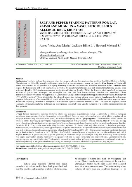
<strong>SALT</strong> <strong>AND</strong> <strong>PEPPER</strong> <strong>STAINING</strong> <strong>PATTERNS</strong> FOR LAT,<br />
ZAP-70 <strong>AND</strong> MUM-1 IN A VASCULITIC BULLOUS<br />
ALLERGIC DRUG ERUPTION<br />
WZÓR BARWIENIA SÓL I PIEPRZ DLA LAT, ZAP-70 I MUM-1 W<br />
NACZYNIOWYCH PĘCHERZACH REAKCJI ALERGICZNYCH<br />
NA LEK<br />
Abreu Velez Ana Maria 1 , Jackson Billie L. 2 , Howard Michael S. 1<br />
1 Georgia Dermatopathology Associates, Atlanta, Georgia, USA.<br />
abreuvelez@yahoo.com<br />
2 Billie L. Jackson, M.D., LLC, Macon, Georgia, USA.<br />
N Dermatol Online. 2011; 3(2): 104-107 Date of submission: 10.02.2011 / acceptance: 19.03.2011<br />
Conflicts of interest: None<br />
Abstract<br />
Background. The term bullous drug eruption refers to clinically adverse drug reactions that result in fluid-filled blisters or bullae.<br />
Blistering can be elicited by multiple medications, prescribed or over-the-counter, natural or synthetic. Case Report: A 78-year-old<br />
female was evaluated for the presence of a rapidly appearing, diffuse rash with vesicles, bullae and abdominal edema. Methods: Skin<br />
biopsies for hematoxylin and eosin examination, as well as for direct immunofluorescence and immunohistochemistry analysis were<br />
performed. Results: H&E staining demonstrated a subepidermal blistering disorder. Within the dermis, a mild, superficial, perivascular<br />
infiltrate of lymphocytes, histiocytes and eosinophils was seen. No frank leukocytoclastic vasculitis was appreciated. Direct<br />
immunofluorescence revealed a strong presence of Complement/C3, IgM and fibrinogen in the upper dermal blood vessels. Staining with<br />
LAT, MUM-1, and ZAP-70 was identified in the inflamed vessels, in a delicate salt and pepper pattern. Conclusions: In bullous drug<br />
eruptions, inflammation of the dermal blood vessels without frank leuckocytoclasis is often noted; vascular alterations subjacent to the<br />
blisters are frequently described as nonspecific. We document specific activation markers of the T cell immune response; further<br />
secondary cell signaling pathway molecules are overexpressed in dermal blood vessels, indicative of a complex immune response in<br />
these patients<br />
Streszczenie<br />
Wstęp: Termin pęcherzowa wysypka polekowa odnosi się klinicznie niepoŜądanych reakcji polekowych, w których występują<br />
wypełnione płynem większe (bullae) lub mniejsze pęcherze (blister). Pęcherze mogą być wywołane przez wiele leków, przepisanych na<br />
receptę albo bez recepty over-the-counter (OTC), naturalnych lub syntetycznych. Opis przypadku: 78-letnia kobieta została zbadana na<br />
obecność szybko pojawiającej się wysypki z rozproszonymi pęcherzami róŜnej wielkości i obrzęku brzucha. Metody: Wykonano biopsję<br />
skóry z barwieniem hematoksyliną i eozyną, jak równieŜ immunofluorescencję bezpośrednią i analizę immunohistochemiczną. Wyniki:<br />
Barwienie H & E wykazało zaburzenia- subepidermalne pęcherze. W skórze właściwej obserwowano, o łagodnym przebiegu,<br />
powierzchowną, okołonaczyniową infiltrację limfocytów, eozynofili i histiocytów. Nie prawdziwe leukocytoklastyczne zapalenie naczyń<br />
było mile widziane. Immunofluorescencja ujawniła silną obecność Complement/C3, IgM i fibrynogenu w górnych warstwach skórnych<br />
naczyń krwionośnych. Barwienie z LAT, MUM-1 i ZAP-70 było zidentyfikowane w zapaleniu naczyń, jako delikatny wzór soli i<br />
pieprzu. Wnioski: W pęcherzowych wysypkach polekowych, zapalenie naczyń krwionośnych skóry bez prawdziwej leuckocytoclazji<br />
było często zauwaŜalne; zmiany naczyniowe na spodzie pęcherza są często opisywane jako niespecyficzne. Udokumentowaliśmy<br />
swoiste markery aktywacji limfocytów T odpowiedzi immunologicznej; dalsze wtórne ogniwo cząsteczek sygnałowych szlaku są w<br />
nadmiernej ekspresji w naczyniach krwionośnych skóry, co wskazuje na złoŜoną odpowiedź odpornościową u tej pacjentki.<br />
Key words: vasculitides, bullous allergic drug reaction, LAT, ZAP-70, MUM 1, skin.<br />
Słowa klucze: vasculitides, reakcje alergiczne polekowe, LAT, ZAP-70, MUM 1, skóra<br />
Introduction<br />
Bullous drug reactions (BDRs) may occur<br />
secondary to various medications, both prescribed and<br />
over-the-counter, and natural or synthetic. Blistering may<br />
be clinically localized and mild, or widespread and<br />
severe. Blisters may be the major feature of the reaction;<br />
alternatively, blisters may be seen focally, or in localized<br />
areas of a more extensive rash. The reaction may show<br />
features of more than one condition (overlap) or be<br />
104<br />
© N Dermatol Online 3.2011
clinically unclassifiable. BDRs represent one of the most<br />
common blistering disorders encountered in<br />
dermatopathology, being more prevalent than the classic<br />
nosologic autoimmune cutaneous blistering diseases.<br />
Hematoxylin and eosin staining as well as direct<br />
immunofluorescence (DIF) of the skin, often<br />
demonstrate findings that can be shared by several<br />
diseases, thus being of limited help in establishing a<br />
definitive diagnosis. The typical histologic differential<br />
diagnosis includes 1) a bullous allergic drug reaction, 2)<br />
bullous pemphigoid, or 3) a bullous arthropod bite<br />
reaction. Indirect immunofluorescence (IIF)/salt split<br />
skin studies may be helpful in further distinguishing<br />
between these diagnostic possibilities, if clinically<br />
indicated. We obtained skin biopsies for hematoxylin<br />
and eosin (H&E) staining, for direct<br />
immunofluorescence (DIF), for indirect<br />
immunofluorescence (IIF) with salt split skin studies and<br />
for immunohistochemistry (IHC).<br />
Case report<br />
A 78-year-old female was evaluated for 2<br />
day duration of blisters on the abdomen that were mild to<br />
moderately painful. The patient was taking docusate,<br />
diltiazem, KCl, furosemide, omeprazole, ranitidine,<br />
pravachol and acetaminophen. On physical examination,<br />
the abdomen displayed one intact bulla that was tense,<br />
and showed mild erythema at the base. A second,<br />
adjacent site on the abdomen was consistent with a<br />
ruptured bulla with a denuded surface. There was no<br />
significant crusting to suggest pemphigus vulgaris. The<br />
face, chest and neck showed no involvement. The<br />
patient had an allergy to Codeine. A lesional skin biopsy<br />
was taken for hematoxylin and eosin (H&E) analysis. In<br />
addition, direct and indirect immunofluorescence (DIF,<br />
IIF) and immunohistochemistry (IHC) studies were<br />
performed.<br />
DIF and IIF/salt split skin were performed as<br />
previously described. Skin cryosections were prepared,<br />
and incubated with multiple fluorochromes, as<br />
previously reported [1-4].<br />
IHC: Performed as previously described, including<br />
antibodies against the linker of activated T cells (LAT<br />
protein), Zeta-chain-associated protein kinase 70 (ZAP-<br />
70) and MUM-1(multiple myeloma oncogene-1) protein<br />
[2-4].<br />
Results<br />
Microscopic description. H&E examination<br />
demonstrated a subepidermal blistering disorder. Partial<br />
blister re-epithelialization was noted. Within the blister<br />
lumen, eosinophils were present, with occasional<br />
neutrophils and lymphocytes. Mast cells were rare.<br />
Within the dermis, a mild, superficial, perivascular<br />
infiltrate of lymphocytes, histiocytes and eosinophils was<br />
seen. Direct immunofluorescence (DIF) studies were<br />
performed; results were classified as 4+ (very strong<br />
positvity) to (-) negative. Case results were as follows:<br />
IgG(+, focally positive BMZ); IgG3(-); IgG4(-); IgA(-);<br />
IgM (++, at superficial upper dermal vessels); IgD(-);<br />
IgE(-); Complement/C1q(-); Complement/C3 (++),<br />
positive around the upper dermal vessels; albumin(-) and<br />
fibrinogen (++), positive around the upper dermal blood<br />
vessels and inside the subepidermal blister. Antibodies to<br />
human plasminogen were negative (Fig. 1).<br />
Discussion<br />
Bullous drug eruptions are often diagnosed<br />
clinically, i.e., by careful history and physical<br />
examination. However, in many cases, these reactions<br />
can mimic other diseases [2,4]. The H&E skin biopsy<br />
may help to make the correct diagnosis, but does not<br />
usually help in establishing whether the reaction is druginduced.<br />
The presence of a vasculitis-<strong>like</strong> reaction is<br />
often noted, not fulfilling the diagnostic criteria of a<br />
leukocytoclastic vasculitis.[4-7]. Although we were not<br />
able to appreciate a true leukocytoclastic vasculitis, the<br />
case DIF clearly indicated an immune response against<br />
dermal blood vessels when utilizing FITC conjugated<br />
anti-human fibrinogen and complement/C3. Further, by<br />
IHC we were able to appreciate staining with antibodies<br />
against ZAP-70, LAT and MUM-1 proteins. The protein<br />
encoded by the LAT gene is phosphorylated by ZAP-<br />
70/SYK protein tyrosine kinases, following activation of<br />
the T-cell antigen receptor (TCR) signal transduction<br />
pathway [8]. The LAT transmembrane protein localizes<br />
to lipid rafts (also known as glycosphingolipid-enriched<br />
microdomains or GEMs), and acts as a docking site for<br />
SH2 domain-containing proteins [8]. Upon<br />
phosphorylation, this protein recruits multiple adaptor<br />
proteins and downstream signaling molecules into<br />
multimolecular signaling complexes located near the site<br />
of TCR engagement [8]. ZAP-70 is normally expressed<br />
in T cells and natural killer cells and has a critical role in<br />
the initiation of T-cell signaling [9]. ZAP-70 is a member<br />
in the protein-tyrosine kinase family. T lymphocytes are<br />
activated by engagement of the T cell receptor with<br />
processed antigen fragments presented by professional<br />
antigen presenting cells (e.g. macrophages, dendritic<br />
cells and B cells) [9]. Upon this activation, the tyrosine<br />
kinase Lck becomes activated, and phosphorylates the<br />
intracellular portions of the CD3 complex (called<br />
ITAMs). The most important member of the CD3 family<br />
is CD3-zeta, to which ZAP-70 binds [9].<br />
MUM-1 is a 50 kDa protein encoded by the MUM-1<br />
gene [10]. The MUM-1 molecule also has other<br />
monikers, including 1) interferon regulatory factor 4<br />
(IRF4) and 2) interferon consensus sequence binding<br />
protein for activated T cells (ICSAT). MUM-1 has been<br />
associated with melanocytic cells, and is involved in the<br />
DNA damage response pathway by contributing to the<br />
maintenance of chromatin architecture. Recruited to the<br />
vicinity of DNA breaks by TP53BP1, it plays an<br />
accessory role in facilitating damage-induced chromatin<br />
changes and promoting chromatin relaxation. MUM-1 is<br />
required for efficient DNA repair and cell survival<br />
following DNA damage [10].<br />
We conclude that in this case of a bullous<br />
allergic drug eruption, we observed some degree of<br />
inflammation of the dermal vessels without frank<br />
leukocytoclastic changes. We have described specific<br />
activation markers, especially of the T cell immune<br />
response; and further, overexpression of secondary cell<br />
signaling pathway molecules in dermal blood vessels,<br />
indicating a complex immune response in this patient.<br />
© N Dermatol Online 3.2011<br />
105
a b c<br />
d e f<br />
g h i<br />
Figure <strong>1.</strong> a-c. H&E shows a subepidermal blister (upper blue arrow) with a positive inflammatory infiltrate of the upper<br />
epidermal vessels (lower blue and maroon arrows). Positive staining of the blister containing FITC conjugated fibrinogen<br />
(white arrow). The blister lumen is shown by yellow stars. d, f. DIF. d. Positive staining of the upper dermal blood vessels<br />
with FITC conjugated anti-human C3(green staining; white arrows). The nuclei of the cells were counterstained with Topro<br />
III (red staining). Blister lumen is indicated by yellow star. f. FITC conjugated anti-human-IgM positive staining of the<br />
upper dermal vessels (green staining, white arrows). anti-human-IgM positive stain of the upper dermal vessels. The nuclei<br />
of the cells were counterstained with Topro III (red staining). Blister lumen is indicated by yellow star. e, g, h, i. IHC. e.<br />
Positive staining with MUM-1 in the small vessels under the blister (brown staining; red arrows). Blister lumen is indicated<br />
by yellow star. g. Positive staining with LAT in the same small vessels as MUM-1, under the blister (brown staining; red<br />
arrows). h. Positive staining with ZAP-70 in the same small vessels as MUM 1 and LAT, under the blister (brown staining;<br />
red arrows). Blister lumen is indicated by yellow star. i. Positive staining with HLA-DPDQDR in upper dermal inflamed<br />
vessels under the blister (brown staining; red arrows). Blister lumen is indicated by yellow star.<br />
106<br />
© N Dermatol Online 3.2011
REFERENCES / PIŚMIENNICTWO:<br />
<strong>1.</strong> Ghohestani RF, Nicolas JF, Rouselle P, Claudy AL:<br />
Diagnostic value of indirect immunofluorescence on sodium<br />
chloride-split skin in the differential diagnosis of<br />
subepidermal autoimmune blistering dermatoses. Arch<br />
Dermatol 1997; 133: 1102-1107.<br />
2. Abreu Velez AM, Jackson BL, Howard MS: Deposition<br />
of immunoreactants in a cutaneous allergic drug reaction.<br />
North Am J Med Sci . 2009; 1: 180-183.<br />
3. Abreu-Velez AM, Smith JG Jr., Howard MS: IgG/IgE<br />
bullous pemphigoid with CD45 lymphocytic reactivity to<br />
dermal blood vessels, nerves and eccrine sweat glands.<br />
North Am J Med Sci 2010; 2: 538-54<strong>1.</strong><br />
4. Abreu-Velez, AM, Klein AD III, Howard MS: Junctional<br />
adhesion molecule overexpression in Kaposi varicelliform<br />
eruption skin lesions -as a possible herpes virus entry site.<br />
North Am J Med Sci 2010; 2: 433-437.<br />
5. Cotliar J: Approach to the patient with a suspected drug<br />
eruption. Semin. Cutan. Med. Surg. 2007; 26: 147-154<br />
6. Carr DR, Houshmand E, Heffernan MP:. Approach to the<br />
acute, generalized, blistering patient. Semin. Cutan. Med.<br />
Surg. 2007; 26: 139-146.<br />
7. Roujeau JC: Clinical heterogeneity of drug<br />
hypersensitivity. Toxicology 2005; 209: 123–129.<br />
8. Zhang W, Sloan-Lancaster J, Kitchen J, Trible RP,<br />
Samelson LE: LAT: the ZAP-70 tyrosine kinase substrate<br />
that links T cell receptor to cellular activation. 1998. Cell.<br />
92; 1: 83–92.<br />
9. Chan AC, Iwashima M, Turck CW, Weiss A:. ZAP-70: a<br />
70 kd protein-tyrosine kinase that associates with the TCR<br />
zeta chain. Cell. 1992. 71: 649–662.<br />
10. Natkunam Y, Warnke RA, Montgomery K, Falini B,<br />
van de Rijn M: Analysis of MUM1/IRF4 protein expression<br />
using tissue microarrays and immunohistochemistry. Mod<br />
Pathol 2001; 14: 686-694.<br />
Funding source: Georgia Dermatopathology Associates, Atlanta, Georgia, USA<br />
© N Dermatol Online 3.2011<br />
107