Structural Basis for Recognition of the Intron Branch Site RNA by ...
Structural Basis for Recognition of the Intron Branch Site RNA by ...
Structural Basis for Recognition of the Intron Branch Site RNA by ...

Create successful ePaper yourself
Turn your PDF publications into a flip-book with our unique Google optimized e-Paper software.
R EPORTS<br />
<strong>Structural</strong> <strong>Basis</strong> <strong>for</strong> <strong>Recognition</strong><br />
<strong>of</strong> <strong>the</strong> <strong>Intron</strong> <strong>Branch</strong> <strong>Site</strong> <strong>RNA</strong><br />
<strong>by</strong> Splicing Factor 1<br />
Zhihong Liu, 1 * Ingrid Luyten, 1 * Mat<strong>the</strong>w J. Bottomley, 1<br />
Ana C. Messias, 1 Sophie Houngninou-Molango, 2<br />
Remco Sprangers, 1 Katia Zanier, 1 Angela Krämer, 2<br />
Michael Sattler 1 †<br />
During spliceosome assembly, splicing factor 1 (SF1) specifically recognizes <strong>the</strong><br />
intron branch point sequence (BPS) UACUAAC in <strong>the</strong> pre-m<strong>RNA</strong> transcripts. We<br />
show that <strong>the</strong> KH-QUA2 region <strong>of</strong> SF1 defines an enlarged KH (hn RNP K) fold<br />
which is necessary and sufficient <strong>for</strong> BPS binding. The 3 part <strong>of</strong> <strong>the</strong> BPS (UAAC),<br />
including <strong>the</strong> conserved branch point adenosine (underlined), is specifically<br />
recognized in a hydrophobic cleft <strong>for</strong>med <strong>by</strong> <strong>the</strong> Gly-Pro-Arg-Gly motif and<br />
<strong>the</strong> variable loop <strong>of</strong> <strong>the</strong> KH domain. The QUA2 region recognizes <strong>the</strong> 5<br />
nucleotides <strong>of</strong> <strong>the</strong> BPS (ACU). The branch point adenosine acting as <strong>the</strong><br />
nucleophile in <strong>the</strong> first biochemical step <strong>of</strong> splicing is deeply buried. BPS <strong>RNA</strong><br />
recognition suggests how SF1 may facilitate subsequent <strong>for</strong>mation <strong>of</strong> <strong>the</strong><br />
prespliceosomal complex A.<br />
The removal <strong>of</strong> introns from pre-m<strong>RNA</strong> transcripts<br />
is catalyzed <strong>by</strong> <strong>the</strong> spliceosome (1).<br />
Initial spliceosome assembly in higher eukaryotes<br />
involves cooperative binding <strong>of</strong> SF1<br />
[or branch point binding protein (BBP)] and U2<br />
auxiliary factor (U2AF) to intron sequences<br />
upstream <strong>of</strong> <strong>the</strong> 3 splice site (2, 3). SF1 specifically<br />
recognizes <strong>the</strong> BPS which is almost<br />
invariant in Saccharomyces cerevisiae (UAC-<br />
UAAC), but more divergent in mammals (YN-<br />
CURAY; Y, pyrimidine; R, purine; N, any<br />
nucleotide; <strong>the</strong> branch point adenosine is underlined)<br />
(4). SF1 belongs to <strong>the</strong> STAR (signal<br />
transduction and activation <strong>of</strong> <strong>RNA</strong>) (5) family<br />
<strong>of</strong> proteins, which share a conserved region,<br />
QUA2 (Quaking homology 2) located COOHterminal<br />
to a KH domain (6). We found that <strong>the</strong><br />
KH-QUA2 region <strong>of</strong> SF1 adopts an enlarged<br />
KH fold which is necessary and sufficient <strong>for</strong><br />
BPS binding. The BPS recognition involves<br />
numerous STAR-specific amino acids and is<br />
consistent with mutations that affect <strong>RNA</strong> binding<br />
in vitro and splicing (4, 7). The SF1/BPS<br />
structure suggests how SF1 may facilitate<br />
spliceosome assembly.<br />
The KH-QUA2 region and a zinc knuckle<br />
(Zn) located fur<strong>the</strong>r COOH-terminal have been<br />
implicated in branch site <strong>RNA</strong> binding (3, 7, 8).<br />
Here, we have defined <strong>the</strong> minimal region <strong>of</strong><br />
human SF1 required <strong>for</strong> <strong>RNA</strong> recognition <strong>by</strong><br />
nuclear magnetic resonance (NMR) titration<br />
1<br />
European Molecular Biology Laboratory (EMBL),<br />
Meyerh<strong>of</strong>strae 1, D-69117 Heidelberg, Germany.<br />
2<br />
University <strong>of</strong> Geneva, Department <strong>of</strong> Cell Biology,<br />
CH-1211 Geneva 4, Switzerland.<br />
*These authors contributed equally to this work.<br />
†To whom correspondence should be addressed. E-<br />
mail: sattler@EMBL-Heidelberg.de<br />
experiments with 5-UAUACUAACAA (BPS<br />
<strong>RNA</strong>). This <strong>RNA</strong> contains <strong>the</strong> S. cerevisiae<br />
BPS (underlined), which also represents <strong>the</strong><br />
preferred BPS in mammals (9). Addition <strong>of</strong><br />
BPS <strong>RNA</strong> to proteins encompassing <strong>the</strong> KH-<br />
QUA2 region <strong>of</strong> SF1 induces extensive changes<br />
in <strong>the</strong> NMR spectra as expected <strong>for</strong> <strong>the</strong><br />
<strong>for</strong>mation <strong>of</strong> a stable protein/<strong>RNA</strong> complex<br />
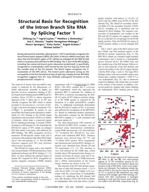
(Fig. 1). Additional experiments demonstrate<br />
that <strong>the</strong> KH-QUA2 region <strong>of</strong> SF1 is necessary<br />
and sufficient <strong>for</strong> recognition <strong>of</strong> <strong>the</strong> BPS <strong>RNA</strong><br />
(6). Based on iso<strong>the</strong>rmal titration calorimetry,<br />
<strong>the</strong> dissociation constant <strong>for</strong> <strong>the</strong> KH-QUA2/<br />
BPS <strong>RNA</strong> complex is 1 M, consistent with<br />
previous reports (3).<br />
The three-dimensional (3D) structure <strong>of</strong><br />
<strong>the</strong> KH-QUA2/BPS <strong>RNA</strong> complex was determined<br />
using heteronuclear multidimensional<br />
NMR spectroscopy (10). Recombinant<br />
13<br />
C, 15 N-labeled SF1 KH-QUA2 was complexed<br />
with unlabeled BPS <strong>RNA</strong>. Distance<br />
restraints were derived from 3D 15 N- and<br />
13<br />
C-edited NOE spectra. <strong>RNA</strong> assignments<br />
and intermolecular protein/<strong>RNA</strong> distance restraints<br />
were obtained from filtered 2D and<br />
3D experiments (11, 12). The structure is<br />
very well defined <strong>by</strong> <strong>the</strong> NMR data, which<br />
include about 300 intermolecular distance restraints<br />
involving 28 amino acids and 9 nucleotides.<br />
An ensemble <strong>of</strong> 15 NMR structures<br />
and a ribbon representation <strong>of</strong> <strong>the</strong> lowest<br />
energy structure are shown in Fig. 2A.<br />
The structure <strong>of</strong> <strong>the</strong> SF1 KH domain resembles<br />
that <strong>of</strong> o<strong>the</strong>r KH domains [rmsd 2.8 Å<br />
with Nova2 KH3 (13) and Vigilin (14)] consistent<br />
with <strong>the</strong> conservation <strong>of</strong> hydrophobic<br />
residues (6) stabilizing <strong>the</strong> KH domain fold<br />
(Fig. 2B). Residues 239 to 249 <strong>of</strong> QUA2 define<br />
an amphipathic helix (4) that <strong>for</strong>ms a hydrophobic<br />
interface with helices 1 (L155), 3<br />
(I225) and <strong>the</strong> GPRG loop (P159) <strong>of</strong> <strong>the</strong> KH<br />
domain (Fig. 2B). Based on secondary chemical<br />
shifts (15), <strong>the</strong> secondary structure <strong>of</strong> KH-<br />
QUA2, including 4, is pre<strong>for</strong>med and not<br />
induced <strong>by</strong> <strong>RNA</strong> binding. The sequence conservation<br />
<strong>of</strong> hydrophobic core residues in <strong>the</strong><br />
KH and QUA2 regions (6) suggests a similar<br />
1-1-2-2-3-3-4 topology <strong>for</strong> all STAR<br />
family proteins. Thus, <strong>the</strong> KH-QUA2 region <strong>of</strong><br />
STAR proteins defines a novel enlarged KH<br />
fold.<br />
The 5 and 3 ends <strong>of</strong> <strong>the</strong> <strong>RNA</strong> interact with<br />
<strong>the</strong> COOH- and NH 2<br />
-terminal regions <strong>of</strong> <strong>the</strong><br />
KH-QUA2 protein, respectively (Fig. 2). The<br />
BPS <strong>RNA</strong> adopts an extended single-stranded<br />
con<strong>for</strong>mation and is bound in a hydrophobic<br />
groove between QUA2, <strong>the</strong> GPRG loop, and<br />
<strong>the</strong> variable loop <strong>of</strong> <strong>the</strong> KH domain. Helices 1<br />
and 2 and strand 2 <strong>of</strong> <strong>the</strong> KH domain, and<br />
conserved residues in <strong>the</strong> QUA2 region interact<br />
with <strong>the</strong> <strong>RNA</strong> (Figs. 2 and 3). The large <strong>RNA</strong><br />
binding surface (solvent-accessible surface area<br />
buried upon complex <strong>for</strong>mation 2500 Å 2 )is<br />
very hydrophobic (Fig. 2C), due to numerous<br />
conserved aliphatic side chains that are located<br />
at <strong>the</strong> surface <strong>of</strong> <strong>the</strong> protein. In addition, conserved<br />
positively charged side chains flanking<br />
<strong>the</strong> hydrophobic <strong>RNA</strong> binding groove <strong>for</strong>m<br />
A<br />
B<br />
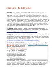
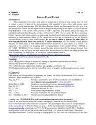
Fig. 1. The KH-QUA2 region <strong>of</strong> SF1 binds to <strong>the</strong><br />
BPS <strong>RNA</strong>. In (A) and (B), two well-resolved<br />
regions <strong>of</strong> 1 H, 15 N correlation spectra <strong>for</strong> SF1<br />
KH-QUA2 are shown in <strong>the</strong> absence (blue) and<br />
presence (red) <strong>of</strong> BPS <strong>RNA</strong>. Amino acid assignments<br />
are indicated. The backbone amide signals<br />
<strong>of</strong> R160 in <strong>the</strong> Gly-Pro-Arg-Gly motif and<br />
<strong>of</strong> I177 in strand 2 (B) experience very large<br />
chemical shift changes upon <strong>RNA</strong> binding consistent<br />
with <strong>the</strong>ir involvement in hydrogen<br />
bonding to Ura6 and <strong>the</strong> branch point adenosine<br />
Ade8 in <strong>the</strong> BPS <strong>RNA</strong>, respectively (Fig. 3).<br />
1098<br />
2 NOVEMBER 2001 VOL 294 SCIENCE www.sciencemag.org
compensating electrostatic interactions with <strong>the</strong><br />
solvent-exposed phosphate backbone <strong>of</strong> <strong>the</strong><br />
<strong>RNA</strong> (Figs. 2C and 3).<br />
R EPORTS<br />
The KH-QUA2/BPS <strong>RNA</strong> complex is stabilized<br />
<strong>by</strong> a combination <strong>of</strong> hydrophobic interactions,<br />
hydrogen bonding, and electrostatic<br />
contacts (Fig. 3A). Consistent with <strong>the</strong> conservation<br />
<strong>of</strong> <strong>the</strong> branch point adenosine in <strong>the</strong><br />
BPS, Ade8 is specifically recognized <strong>by</strong> <strong>the</strong> KH<br />
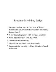
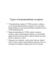
Fig. 2. Structure <strong>of</strong> <strong>the</strong> SF1/branch site <strong>RNA</strong><br />
complex. (A) Stereoview <strong>of</strong> <strong>the</strong> NMR ensemble<br />
<strong>of</strong> <strong>the</strong> SF1 KH-QUA2/BPS <strong>RNA</strong> complex. <strong>RNA</strong><br />
heavy atoms are shown in red; <strong>the</strong> N, C, C<br />
trace <strong>of</strong> <strong>the</strong> protein is shown in gray and colored<br />
blue <strong>for</strong> secondary structure elements. (B)<br />
Ribbon representation <strong>of</strong> <strong>the</strong> SF1 KH-QUA2<br />
domain bound to <strong>the</strong> BPS. Side chains <strong>of</strong> conserved<br />
hydrophobic core residues are shown in<br />
yellow; residues in <strong>the</strong> KH/QUA2 interface and<br />
<strong>the</strong> QUA2 helix 4 are colored magenta. The<br />
Gly-Pro-Arg-Gly motif and <strong>the</strong> variable loop <strong>of</strong><br />
<strong>the</strong> KH domain are colored green and red,<br />
respectively. (C) Ribbon and (D) surface representation<br />
<strong>of</strong> <strong>the</strong> KH-QUA2/BPS complex. Secondary<br />
structure elements <strong>of</strong> <strong>the</strong> KH-QUA2<br />
protein are labeled; <strong>RNA</strong> nucleotides are colored<br />
<strong>by</strong> atom type and annotated in magenta.<br />
The surface <strong>of</strong> <strong>the</strong> KH-QUA2 region is colored<br />
white, blue, and red <strong>for</strong> neutral, positive, and<br />
negative electrostatic potential, respectively.<br />
www.sciencemag.org SCIENCE VOL 294 2 NOVEMBER 2001 1099
R EPORTS<br />
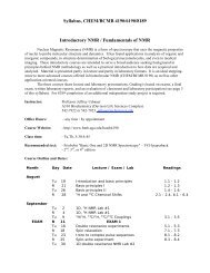
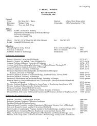
A<br />
Fig. 3. <strong>RNA</strong> recognition. (A) Schematic overview <strong>of</strong> protein/<strong>RNA</strong><br />
interactions. <strong>RNA</strong> nucleotides [single-letter<br />
code (42)] and protein side chains (three-letter code) are<br />
indicated. Hydrophobic interactions (red dotted lines),<br />
hydrogen bonds and electrostatic interactions (dashed<br />
blue lines) stabilizing <strong>the</strong> protein/<strong>RNA</strong> complex are<br />
shown. (B) <strong>Recognition</strong> <strong>of</strong> <strong>the</strong> branch point adenosine<br />
(Ade 8) and C9-A10-A11. Hydrogen bonds are indicated<br />
<strong>by</strong> black dashed lines. (C) <strong>Recognition</strong> <strong>of</strong> Ade7 <strong>by</strong> <strong>the</strong> KH<br />
domain. (D) <strong>Recognition</strong> <strong>of</strong> Ura6 and Cyt5 <strong>by</strong> <strong>the</strong><br />
158 GPRG 161 loop near <strong>the</strong> KH/QUA2 interface, and <strong>by</strong> <strong>the</strong><br />
QUA2 helix (4).<br />
domain. Most remarkably, <strong>the</strong> N6 and N1 <strong>of</strong><br />
Ade8 <strong>for</strong>m hydrogen bonds with <strong>the</strong> backbone<br />
amide and carbonyl oxygen <strong>of</strong> I177, which<br />
mimic <strong>the</strong> Watson-Crick functional groups <strong>of</strong> a<br />
uridine. Ano<strong>the</strong>r hydrogen bond is found between<br />
<strong>the</strong> 2 hydroxyl <strong>of</strong> Ade7 and <strong>the</strong> N7 <strong>of</strong><br />
Ade8. In combination, this pattern <strong>of</strong> hydrogen<br />
bonds uniquely specifies an adenosine base at<br />
this position. Consistently, <strong>the</strong> branch point<br />
adenosine is strictly conserved, and mutation to<br />
any o<strong>the</strong>r base abolishes SF1 binding (4). The<br />
sugar puckers and base stacking between Ade8,<br />
Cyt9, and Ade10 resemble an A-helical con<strong>for</strong>mation.<br />
Ade8 is surrounded <strong>by</strong> conserved aliphatic<br />
side chains (I157, L164, I175, I177,<br />
V183) <strong>of</strong> <strong>the</strong> KH domain (Fig. 3B). A L164N<br />
mutation in SF1 decreases <strong>the</strong> <strong>RNA</strong> binding<br />
affinity <strong>by</strong> 50% (7). The L164N mutation directly<br />
destabilizes <strong>the</strong> <strong>RNA</strong> interaction and<br />
does not affect <strong>the</strong> 3D fold <strong>of</strong> <strong>the</strong> KH domain<br />
[based on NMR spectra (15)]. In contrast, <strong>the</strong><br />
corresponding mutation in KH2 <strong>of</strong> FMR1,<br />
which is associated with <strong>the</strong> Fragile X syndrome,<br />
affects <strong>the</strong> KH fold in vitro (16, 17).<br />
A potential -cation interaction (18) between<br />
<strong>the</strong> Ade7 base and <strong>the</strong> side chain <strong>of</strong> K184<br />
(Fig. 3C) is consistent with <strong>the</strong> preference <strong>for</strong><br />
purine bases at this position in both <strong>the</strong> S.<br />
cerevisiae and mammalian branch sites. A<br />
K184A mutation abolishes <strong>RNA</strong> binding (Fig.<br />
4), demonstrating <strong>the</strong> importance <strong>of</strong> this interaction.<br />
The phosphates <strong>of</strong> Ade7 and Ade8<br />
closely approach <strong>the</strong> peptide backbone near <strong>the</strong><br />
158<br />
GPRG 161 loop. Consequently, a G161D mutation<br />
almost eliminates <strong>RNA</strong> binding (7), and<br />
mutation (R160E) <strong>of</strong> <strong>the</strong> R160 side chain,<br />
which contacts <strong>the</strong> 3 phosphate <strong>of</strong> <strong>the</strong> branch<br />
point adenosine (Ade8) (Fig. 3B), severely reduces<br />
<strong>RNA</strong> binding (Fig. 4). The N6 <strong>of</strong> Ade7<br />
interacts with <strong>the</strong> side chain <strong>of</strong> <strong>the</strong> conserved<br />
E149, which is positioned via a hydrogen bond<br />
with <strong>the</strong> backbone amide <strong>of</strong> I150 (Fig. 3C).<br />
Analysis <strong>of</strong> <strong>the</strong> structure also explains <strong>the</strong><br />
conservation <strong>of</strong> Ura6 in <strong>the</strong> BPS. Ura6 is located<br />
at <strong>the</strong> interface between <strong>the</strong> KH and QUA2<br />
domains (Fig. 3D). The Ura6 base interacts<br />
B<br />
C<br />
D<br />
with L244 and L247 in <strong>the</strong> QUA2 helix (4)<br />
and closely approaches <strong>the</strong> peptide backbone <strong>of</strong><br />
helix 1 consistent with <strong>the</strong> conservation <strong>of</strong><br />
G154. The imino proton <strong>of</strong> Ura6 is positioned<br />
to <strong>for</strong>m a hydrogen bond with <strong>the</strong> backbone<br />
amide <strong>of</strong> L155. Toge<strong>the</strong>r with steric constraints,<br />
<strong>the</strong>se interactions are specific <strong>for</strong> uridine, consistent<br />
with <strong>the</strong> conservation <strong>of</strong> this nucleotide<br />
and <strong>the</strong> loss <strong>of</strong> SF1 binding with a Ura6Ade<br />
mutant branch site (4). The sugar <strong>of</strong> Ura6<br />
stacks with P159 in <strong>the</strong> GPRG loop and is<br />
stabilized <strong>by</strong> a hydrogen bond between its 2-<br />
OH and <strong>the</strong> backbone amide <strong>of</strong> R160 (Fig. 3D).<br />
Both <strong>the</strong> sugar and base <strong>of</strong> Cyt5 exhibit numerous<br />
hydrophobic contacts with <strong>the</strong> exposed aliphatic<br />
side chains <strong>of</strong> L244 and L247. Consistent<br />
with this, <strong>the</strong> L244A or L247A mutations<br />
decrease <strong>the</strong> <strong>RNA</strong> interaction (Fig. 4). N151 is<br />
positioned to recognize <strong>the</strong> Watson-Crick face<br />
<strong>of</strong> Cyt5, consistent with reduced <strong>RNA</strong> binding<br />
<strong>of</strong> a N151A mutant protein (Fig. 4). Residues in<br />
<strong>the</strong> QUA2 region (L244, L247, T253, L254,<br />
R255) interact with Ade2, Ade4, and Cyt5.<br />
These interactions seem less important, because<br />
mutation <strong>of</strong> Ade4 or Cyt5 to guanosine in <strong>the</strong><br />
BPS (4), or <strong>the</strong> L254A and R255A mutations in<br />
KH-QUA2 (Fig. 4), have smaller effects on<br />
SF1 binding.<br />
The structure described herein provides <strong>the</strong><br />
first example <strong>for</strong> sequence-specific recognition<br />
<strong>of</strong> <strong>the</strong> conserved single-stranded BPS <strong>RNA</strong>.<br />
The complex <strong>of</strong> <strong>the</strong> Nova2-KH3 domain bound<br />
to a stem-loop <strong>RNA</strong> (13) is <strong>the</strong> only o<strong>the</strong>r<br />
high-resolution structure showing <strong>RNA</strong> recognition<br />
<strong>by</strong> a KH domain in <strong>the</strong> absence <strong>of</strong> a<br />
QUA2 domain. In both complexes <strong>the</strong> protein/<br />
<strong>RNA</strong> interfaces are very hydrophobic, but aromatic<br />
residues are not used <strong>for</strong> <strong>RNA</strong> recognition.<br />
These are likely to be general features <strong>of</strong><br />
KH domain/<strong>RNA</strong> interactions considering <strong>the</strong><br />
conservation <strong>of</strong> aliphatic amino acids and <strong>the</strong><br />
low abundance <strong>of</strong> aromatic residues in KH<br />
domains. In contrast, <strong>the</strong> recognition <strong>of</strong> singlestranded<br />
<strong>RNA</strong> <strong>by</strong> RNP domains always employs<br />
conserved aromatic residues <strong>for</strong> stacking<br />
interactions with <strong>RNA</strong> bases (19, 20). Interestingly,<br />
<strong>the</strong> recognition <strong>of</strong> Ura6-Ade7-Ade8-<br />
Cyt9 <strong>by</strong> SF1 resembles <strong>the</strong> interaction <strong>of</strong><br />
Ura12-Cyt13-Ade14-Cyt15 <strong>by</strong> <strong>the</strong> Nova2-KH3<br />
domain (13). Notably, <strong>the</strong> Watson-Crick recog-<br />
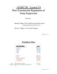
Fig. 4. Mutational analysis <strong>of</strong> KH-QUA2/BPS<br />
<strong>RNA</strong> interactions. For band shift experiments,<br />
<strong>the</strong> BPS <strong>RNA</strong> was incubated with buffer or<br />
wild-type and mutant SF1 proteins as indicated<br />
(10).<br />
1100<br />
2 NOVEMBER 2001 VOL 294 SCIENCE www.sciencemag.org
nition <strong>of</strong> adenosine <strong>by</strong> two hydrogen bonds<br />
with <strong>the</strong> peptide backbone is mediated <strong>by</strong><br />
equivalent positions in <strong>the</strong> 2 strand <strong>of</strong> <strong>the</strong> KH<br />
domains (21). However, BPS <strong>RNA</strong> binding <strong>by</strong><br />
SF1 requires <strong>the</strong> QUA2 region in addition to<br />
<strong>the</strong> KH domain. Thus, <strong>the</strong> enlarged KH fold<br />
defined <strong>by</strong> <strong>the</strong> KH-QUA2 region is crucial <strong>for</strong><br />
sequence-specific recognition <strong>of</strong> <strong>the</strong> singlestranded<br />
7-nucleotide BPS <strong>RNA</strong> (22).<br />
The structure <strong>of</strong> <strong>the</strong> SF1/<strong>RNA</strong> complex provides<br />
insight into early steps <strong>of</strong> spliceosome<br />
assembly. (i) It has been shown that binding <strong>of</strong><br />
U2AF65 to <strong>the</strong> polypyrimidine tract, located<br />
downstream <strong>of</strong> <strong>the</strong> BPS, directs its Arg-Ser–<br />
rich (RS) domain to contact <strong>the</strong> branch site (23).<br />
The structure presented herein is compatible<br />
with this and suggests that <strong>the</strong> positively<br />
charged RS domain could interact with <strong>the</strong><br />
solvent-exposed, negatively charged phosphate<br />
backbone <strong>of</strong> <strong>the</strong> BPS. (ii) Our NMR titrations<br />
show that <strong>the</strong> SF1 zinc knuckle does not contact<br />
<strong>the</strong> BPS. Thus, in contrast to a previously suggested<br />
model (8), we expect that <strong>the</strong> zinc<br />
knuckle may interact with nucleotides located<br />
upstream <strong>of</strong> <strong>the</strong> BPS in <strong>the</strong> pre-m<strong>RNA</strong>. (iii)<br />
The most striking feature <strong>of</strong> <strong>the</strong> SF1/BPS <strong>RNA</strong><br />
complex structure is that <strong>the</strong> branch point adenosine<br />
(Ade8) is deeply buried in a hydrophobic<br />
pocket <strong>of</strong> <strong>the</strong> KH domain. The base <strong>of</strong> Ade8 is<br />
oriented towards <strong>the</strong> protein and stabilized <strong>by</strong><br />
two hydrogen bonds with <strong>the</strong> peptide backbone,<br />
and thus shielded from interaction with o<strong>the</strong>r<br />
molecules. The next step <strong>of</strong> spliceosome assembly<br />
involves <strong>the</strong> <strong>for</strong>mation <strong>of</strong> a BPS/U2 sn<strong>RNA</strong><br />
duplex (complex A), in which <strong>the</strong> branch point<br />
adenosine is bulged out (24, 25). For duplex<br />
<strong>for</strong>mation, <strong>the</strong> complementary U2 sn<strong>RNA</strong> presumably<br />
approaches <strong>the</strong> SF1/BPS complex<br />
from its accessible, open face (Fig. 2C). The<br />
burial <strong>of</strong> <strong>the</strong> Ade8 base constrains it in an<br />
orientation that favors its exclusion from <strong>the</strong><br />
duplex with <strong>the</strong> U2 sn<strong>RNA</strong>. Even though <strong>for</strong>mation<br />
<strong>of</strong> <strong>the</strong> BPS/U2 sn<strong>RNA</strong> duplex requires<br />
disruption <strong>of</strong> <strong>the</strong> SF1/BPS complex, <strong>the</strong> recognition<br />
<strong>of</strong> Ade8 in a “prebulged” con<strong>for</strong>mation is<br />
likely to facilitate this transition and could explain<br />
<strong>the</strong> kinetic effects <strong>of</strong> SF1 during spliceosome<br />
assembly (26, 27). (iv) Protein/<strong>RNA</strong> recognition<br />
in <strong>the</strong> SF1/BPS complex may provide<br />
a structural basis <strong>for</strong> human inherited diseases<br />
that result from intronic mutations <strong>of</strong> Ura6 or<br />
Ade8 in <strong>the</strong> BPS (28, 29).<br />
References and Notes<br />
1. A. Krämer, Annu. Rev. Biochem. 65, 367 (1996).<br />
2. N. Abovich, M. Rosbash, Cell 89, 403 (1997).<br />
3. J. A. Berglund, N. Abovich, M. Rosbash, Genes Dev. 12,<br />
858 (1998).<br />
4. J. A. Berglund, K. Chua, N. Abovich, R. Reed, M.<br />
Rosbash, Cell 89, 781 (1997).<br />
5. C. Vernet, K. Artzt, Trends Genet. 13, 479 (1997).<br />
6. Supplementary materials showing a multiple sequence<br />
alignment <strong>of</strong> STAR family proteins and NMR<br />
titration experiments are available at Science Online<br />
at www.sciencemag.org/cgi/content/full/294/5544/<br />
1098/DC1.<br />
7. J. C. Rain, Z. Rafi, Z. Rhani, P. Legrain, A. Krämer, <strong>RNA</strong><br />
4, 551 (1998).<br />
R EPORTS<br />
8. J. A. Berglund, M. L. Fleming, M. Rosbash, <strong>RNA</strong> 4, 998<br />
(1998).<br />
9. Y. A. Zhuang, A. M. Goldstein, A. M. Weiner, Proc.<br />
Natl. Acad. Sci. U.S.A. 86, 2752 (1989).<br />
10. The KH-QUA2 region (residues 134 to 260) <strong>of</strong> human<br />
SF1 was cloned into a modified pET24d expression<br />
vector (Novagen) containing an NH 2<br />
-terminal histidine-tag<br />
followed <strong>by</strong> a TEV protease cleavage site. SF1<br />
mutants were prepared using <strong>the</strong> QuickChange sitedirected<br />
mutagenesis kit (Stratagene). Uni<strong>for</strong>mly 15 N-<br />
labeled and 15 N- and 13 C-labeled proteins were prepared<br />
<strong>by</strong> growing <strong>the</strong> Escherichia coli strain BL21(DE3)<br />
overexpressing KH-QUA2 in a minimal medium containing<br />
15 NH 4<br />
Cl with or without uni<strong>for</strong>mly 13 C-labeled<br />
glucose. Recombinant proteins were purified on a Nichelating<br />
affinity column. The histidine-tag was removed<br />
<strong>by</strong> TEV protease cleavage and additional purification<br />
was achieved with chromatography on a Q-<br />
Sepharose column. The BPS <strong>RNA</strong> 5-UAUACUAACAA<br />
was syn<strong>the</strong>sized in-house or purchased from Biospring,<br />
Frankfurt, Germany. Samples <strong>for</strong> NMR contained a 1:1<br />
complex <strong>of</strong> KH-QUA2 and <strong>the</strong> <strong>RNA</strong> at a concentration<br />
<strong>of</strong> 1 mM in 20 mM sodium phosphate buffer (pH 6.5),<br />
50 mM NaCl, 2 mM dithiothreitol. For NMR titrations,<br />
15<br />
N-labeled samples comprising <strong>the</strong> KH (residues 134<br />
to 229) and KH-QUA2-Zn regions (residues 134 to 296)<br />
<strong>of</strong> human SF1 were prepared as described above. To<br />
determine <strong>RNA</strong> binding, band-shift experiments were<br />
per<strong>for</strong>med in 10-l reactions containing 20 mM tris-<br />
HCl (pH 8.0), 100 mM NaCl, 5 mM imidazole, 2.5 mM<br />
-mercaptoethanol, 2.5 g t<strong>RNA</strong>, 2 g <strong>of</strong> KH-QUA2 or<br />
mutant proteins, and 0.01 pmol 5 [ 32 P]ATP-labeled BPS<br />
<strong>RNA</strong>. After 15 min at room temperature, reaction products<br />
were separated in a 6% polyacrylamide gel (acrylamide:bisacrylamide,<br />
80:1) in 25 mM tris, 25 mM boric<br />
acid, and 1 mM EDTA at 4°C. Reaction products were<br />
visualized <strong>by</strong> autoradiography. NMR spectra were recorded<br />
at 22°C on Bruker DRX 600 or DRX 800 NMR<br />
spectrometers. Spectra were processed with NMRPIPE<br />
(30) and analyzed using XEASY (31). Backbone and side<br />
chain 1 H, 15 N and 13 C resonances were assigned using<br />
standard triple resonance experiments (12). Distance<br />
restraints were derived from 13 C- and 15 N-edited 3D<br />
NOESY experiments. Assignments <strong>for</strong> <strong>the</strong> <strong>RNA</strong> were<br />
obtained from 2D isotope-filtered experiments. Intermolecular<br />
distance restraints were measured in 3D 13 C-<br />
edited/filtered experiments (11, 12). Dihedral angle restraints<br />
<strong>for</strong> <strong>the</strong> backbone angle were derived from<br />
3<br />
J(H N ,H ) coupling constants measured in an HNHA-J<br />
experiment (32); additional / restraints were derived<br />
from TALOS (33). Hydrogen bond restraints <strong>for</strong> secondary<br />
structure elements in <strong>the</strong> protein were defined from<br />
slowly exchanging amide protons, identified after exchange<br />
<strong>of</strong> <strong>the</strong> H 2<br />
O buffer to D 2<br />
O. For NMR titrations,<br />
chemical shifts were recorded with 1 H, 15 N-HSQC (heteronuclear<br />
single-quantum correlation) experiments on<br />
a 600-MHz spectrometer using 1 mM 15 N-labeled NMR<br />
samples <strong>of</strong> SF1 proteins comprising <strong>the</strong> KH, KH-QUA2,<br />
and KH-QUA2-Zn regions. The experimentally determined<br />
distance restraints (2855 NOEs, including 175<br />
<strong>RNA</strong>/<strong>RNA</strong>, 298 protein/<strong>RNA</strong>, and 76 hydrogen bond<br />
restraints) and dihedral angle (169) restraints were applied<br />
in a mixed torsion angle/Cartesian dynamics simulated<br />
annealing protocol using CNS (34) and ARIA (35).<br />
The simulated annealing protocol was greatly extended<br />
compared to standard protein structure calculations.<br />
NOEs involving <strong>the</strong> <strong>RNA</strong> were manually assigned and<br />
calibrated. A floating chirality approach (36) was used<br />
to define stereospecific assignments <strong>of</strong> isopropyl<br />
groups. <strong>Structural</strong> quality was evaluated using PRO-<br />
CHECK (37). In <strong>the</strong> final ensemble <strong>of</strong> 10 NMR structures,<br />
<strong>the</strong> atomic root-mean-square deviation (rmsd)<br />
about <strong>the</strong> mean coordinate positions is 0.60 0.17 <strong>for</strong><br />
<strong>the</strong> backbone and 1.37 0.28 <strong>for</strong> all heavy atoms<br />
excluding loop regions. No distance restraint is violated<br />
<strong>by</strong> more than 0.4 Å. Deviations from experimental<br />
distance, dihedral and residual dipolar coupling restraints<br />
are 0.0027 0.0009 Å and 0.49 0.08°,<br />
respectively. Deviations from idealized covalent geometry<br />
<strong>for</strong> bond lengths, bond angles, and improper dihedral<br />
angles are 0.0032 0.00006 Å, 0.458 0.007°,<br />
and 0.345 0.001°, respectively. 75.3 2.3% and<br />
19.5 2.8% <strong>of</strong> <strong>the</strong> / angles lie in <strong>the</strong> most favored<br />
and additionally allowed regions <strong>of</strong> <strong>the</strong> Ramachandran<br />
plot, respectively. Hydrogen bonds and electrostatic<br />
interactions are identified if <strong>the</strong> corresponding geometrical<br />
constraints are observed in more than 50% <strong>of</strong> <strong>the</strong><br />
final NMR structures. Figures showing 3D structures and<br />
molecular surfaces were prepared using MOLMOL (38)<br />
and GRASP (39), respectively.<br />
11. G. Varani, A. Fareed, F. H.-T. Allain, Prog. NMR Spectrosc.<br />
29, 51 (1996).<br />
12. M. Sattler, J. Schleucher, C. Griesinger, Prog. NMR<br />
Spectrosc. 34, 93 (1999).<br />
13. H. A. Lewis et al., Cell 100, 323 (2000).<br />
14. G. Musco et al., Cell 85, 237 (1996).<br />
15. Z. Liu, M. J. Bottomley, A. C. Messias, R. Sprangers, M.<br />
Sattler, data not shown.<br />
16. G. Musco et al., Nature Struct. Biol. 4, 712 (1997).<br />
17. H. A. Lewis et al., Structure 7, 191 (1999).<br />
18. J. P. Gallivan, D. A. Dougherty, Proc. Natl. Acad. Sci.<br />
U.S.A. 96, 9459 (1999).<br />
19. N. Handa et al., Nature 398, 579 (1999).<br />
20. C. Oubridge, N. Ito, P. R. Evans, C. H. Teo, K. Nagai,<br />
Nature 372, 432 (1994).<br />
21. O<strong>the</strong>r KH domains also bind small (three to four<br />
nucleotides) single-stranded <strong>RNA</strong> elements, most <strong>of</strong><br />
which contain adenosine residues (17). <strong>RNA</strong> binding<br />
<strong>of</strong> <strong>the</strong> STAR protein Sam68 is abolished upon mutation<br />
<strong>of</strong> <strong>the</strong> third adenosine in <strong>the</strong> tetranucleotide<br />
sequence Ura-Ade-Ade-Ade (40). These KH domains<br />
could thus employ similar modes <strong>of</strong> adenosine<br />
recognition.<br />
22. <strong>Recognition</strong> <strong>of</strong> <strong>the</strong> BPS <strong>RNA</strong> <strong>by</strong> SF1 involves residues<br />
which are characteristic <strong>for</strong> STAR family proteins.<br />
For example, K184, which contacts Ade7, is a<br />
conserved residue at <strong>the</strong> beginning <strong>of</strong> a STAR<br />
protein-specific extension <strong>of</strong> <strong>the</strong> variable loop (6).<br />
The K184A mutation abolishes BPS binding (Fig. 4),<br />
and <strong>the</strong> corresponding R185C mutation in <strong>the</strong><br />
STAR protein How/Who induces an embryonic lethal<br />
phenotype in Drosophila (41) demonstrating<br />
<strong>the</strong> importance <strong>of</strong> this residue. In contrast, in <strong>the</strong><br />
Nova2-KH3/<strong>RNA</strong> complex Cyt13 (which corresponds<br />
to Ade7 in <strong>the</strong> BPS) is recognized <strong>by</strong> a<br />
residue (R54) in strand 3 (13). Given <strong>the</strong> conservation<br />
<strong>of</strong> <strong>the</strong> SF1 KH-QUA2 region among members<br />
<strong>of</strong> <strong>the</strong> STAR family and <strong>the</strong> importance <strong>of</strong><br />
STAR-specific residues <strong>for</strong> <strong>RNA</strong> binding, it is likely<br />
that o<strong>the</strong>r STAR family proteins recognize singlestranded<br />
<strong>RNA</strong> in a similar way.<br />
23. J. Valcárcel, R. K. Gaur, R. Singh, M. R. Green, Science<br />
273, 1706 (1996).<br />
24. C. C. Query, M. J. Moore, P. A. Sharp, Genes Dev. 8,<br />
587 (1994).<br />
25. J. A. Berglund, M. Rosbash, S. C. Schultz, <strong>RNA</strong> 7, 682<br />
(2001).<br />
26. B. Rutz, B. Seraphin, <strong>RNA</strong> 5, 819 (1999).<br />
27. S. Guth, J. Valcárcel, J. Biol. Chem. 275, 38059<br />
(2000).<br />
28. A. De Klein et al., Hum. Mol. Genet. 7, 393 (1998).<br />
29. J. A. Kuivenhoven et al., J. Clin. Invest. 98, 358 (1996).<br />
30. F. Delaglio et al., J. Biomol. NMR 6, 277 (1995).<br />
31. C. Bartels, T.-H. Xia, M. Billeter, P. Güntert, K. Wüthrich,<br />
J. Biomol. NMR 5, 1 (1995).<br />
32. H. Kuboniwa, S. Grzesiek, F. Delaglio, A. Bax, J. Biomol.<br />
NMR 4, 871 (1994).<br />
33. G. Cornilescu, F. Delaglio, A. Bax, J. Biomol. NMR 13,<br />
289 (1999).<br />
34. A. T. Brünger et al., Acta Crystallogr. D 54, 905<br />
(1998).<br />
35. M. Nilges, S. I. O’Donoghue, Prog. NMR Spectrosc. 32,<br />
107 (1998).<br />
36. R. H. Folmer, C. W. Hilbers, R. N. Konings, M. Nilges,<br />
J. Biomol. NMR 9, 245 (1997).<br />
37. R. A. Laskowski, J. A. Rullmannn, M. W. MacArthur, R.<br />
Kaptein, J. M. Thornton, J. Biomol. NMR 8, 477<br />
(1996).<br />
38. R. Koradi, M. Billeter, K. Wüthrich, J. Mol. Graph. 14,<br />
51 (1996).<br />
39. A. Nicholls, K. A. Sharp, B. Honig, Proteins Struct.<br />
Function Genet. 11, 282 (1991).<br />
40. Q. Lin, S. J. Taylor, D. Shalloway, J. Biol. Chem. 272,<br />
27274 (1997).<br />
41. E. H. Baehrecke, Development 124, 1323 (1997).<br />
42. Single-letter abbreviations <strong>for</strong> <strong>the</strong> amino acid residues<br />
are as follows: A, Ala; C, Cys; D, Asp; E, Glu; F,<br />
Phe; G, Gly; H, His; I, Ile; K, Lys; L, Leu; M, Met; N, Asn;<br />
P, Pro; Q, Gln; R, Arg; S, Ser; T, Thr; V, Val; W, Trp; and<br />
Y, Tyr.<br />
www.sciencemag.org SCIENCE VOL 294 2 NOVEMBER 2001 1101
43. We thank J. Valcarcel and I. Mattaj <strong>for</strong> suggestions<br />
and critical reading <strong>of</strong> <strong>the</strong> manuscript; <strong>the</strong> European<br />
Large Scale facility in Frankfurt, Germany, and<br />
W. Bermel (Bruker, Karlsruhe, Germany) <strong>for</strong> time<br />
on <strong>the</strong> 800-MHz spectrometer; and M. Nilges and J.<br />
Linge <strong>for</strong> ARIA s<strong>of</strong>tware. Support from EMBL and<br />
R EPORTS<br />
<strong>the</strong> Deutsche Forschungsgemeinschaft (M.S.);<br />
Fonds voor Wetenschappelijk Onderzoek (FWO),<br />
Vlaanderen, Ne<strong>the</strong>rlands, and European Molecular<br />
Biology Organization (I.L.); Fundação para a Ciência<br />
e Tecnologia (A.C.M.); and <strong>the</strong> Swiss National Science<br />
Foundation (SNF) and <strong>the</strong> Canton <strong>of</strong> Geneva<br />
(A.K.) is acknowledged. Coordinates <strong>for</strong> <strong>the</strong> NMR<br />
structure <strong>of</strong> <strong>the</strong> SF1/<strong>RNA</strong> complex have been deposited<br />
in <strong>the</strong> Protein Data Bank under accession<br />
code 1K1G.<br />
25 July 2001; accepted 13 September 2001<br />
Mammalian TOR: A<br />
Homeostatic ATP Sensor<br />
Patrick B. Dennis, 1 Anja Jaeschke, 1 Masao Saitoh, 1 Brian Fowler, 2<br />
Sara C. Kozma, 1 George Thomas 1 *<br />
The bacterial macrolide rapamycin is an efficacious anticancer agent against<br />
solid tumors. In a hypoxic environment, <strong>the</strong> increase in mass <strong>of</strong> solid tumors<br />
is dependent on <strong>the</strong> recruitment <strong>of</strong> mitogens and nutrients. When nutrient<br />
concentrations change, particularly those <strong>of</strong> essential amino acids, <strong>the</strong> mammalian<br />
Target <strong>of</strong> Rapamycin (mTOR) functions in regulatory pathways that<br />
control ribosome biogenesis and cell growth. In bacteria, ribosome biogenesis<br />
is independently regulated <strong>by</strong> amino acids and adenosine triphosphate (ATP).<br />
Here we demonstrate that <strong>the</strong> mTOR pathway is influenced <strong>by</strong> <strong>the</strong> intracellular<br />
concentration <strong>of</strong> ATP, independent <strong>of</strong> <strong>the</strong> abundance <strong>of</strong> amino acids, and that<br />
mTOR itself is an ATP sensor.<br />
The survival <strong>of</strong> organisms is dependent on<br />
<strong>the</strong>ir ability to maintain cellular homeostasis.<br />
Environmental cues are deciphered <strong>by</strong> cellular<br />
regulatory elements, which adjust an organism’s<br />
metabolic state to reflect external<br />
conditions. The phosphatidylinositide kinase–<br />
related family <strong>of</strong> protein kinases contains a<br />
number <strong>of</strong> critical effectors that sense environmental<br />
factors that control <strong>the</strong> ability <strong>of</strong> an<br />
organism to survive. One member <strong>of</strong> <strong>the</strong> family<br />
is mTOR, which resides at <strong>the</strong> interface<br />
between nutrient sensing and <strong>the</strong> regulation<br />
<strong>of</strong> major metabolic responses (1–3). Depending<br />
on mitogen and amino acid availability,<br />
mTOR positively regulates translation and<br />
ribosome biogenesis while negatively controlling<br />
autophagy (1), suggesting that mTOR<br />
sets protein syn<strong>the</strong>tic rates as a function <strong>of</strong><br />
<strong>the</strong> availability <strong>of</strong> translational precursors (4,<br />
5). In response to mitogens and amino acids,<br />
mTOR phosphorylates and controls <strong>the</strong> activities<br />
<strong>of</strong> two key translational regulators, S6<br />
kinase 1 (S6K1) and initiation factor 4E binding<br />
protein (4E-BP1) (2). However, changes<br />
in mTOR activity in vitro, after ei<strong>the</strong>r mitogen<br />
or amino acid treatment, have been small<br />
and controversial (2, 3). The importance <strong>of</strong><br />
understanding <strong>the</strong> molecular mechanisms that<br />
control mTOR function is underscored <strong>by</strong><br />
recent phase 1 clinical trials showing that<br />
rapamycin is efficacious in <strong>the</strong> treatment <strong>of</strong><br />
solid tumors in patients with metastatic renal<br />
1<br />
The Friedrich Miescher Institute <strong>for</strong> Biomedical Research,<br />
Maulbeerstrasse 66, CH–4058, Basel, Switzerland.<br />
2 University Childrens Hospital CH–4005, Basel,<br />
Switzerland.<br />
*To whom correspondence should be addressed. E-<br />
mail: gthomas@fmi.ch<br />
cell carcinoma and non–small cell lung, prostate,<br />
and breast cancer (6).<br />
Protein syn<strong>the</strong>sis is <strong>the</strong> major energy-consuming<br />
process in <strong>the</strong> cell (7). In bacteria,<br />
protein syn<strong>the</strong>sis and cell growth are linked<br />
<strong>by</strong> ribosome biogenesis, which is independently<br />
controlled <strong>by</strong> amino acid and adenosine<br />
triphosphate (ATP) availability (8). Because<br />
mTOR is sensitive to amino acids and<br />
regulates ribosome biogenesis (1–3), we tested<br />
its activity <strong>for</strong> sensitivity to alterations in<br />
intracellular ATP concentrations. We used<br />
insulin-induced S6K1 activation and 4E-BP1<br />
phosphorylation as reporters <strong>for</strong> mTOR function<br />
in <strong>the</strong> presence <strong>of</strong> glycolytic or mitochondrial<br />
inhibitors (9). The glycolytic inhibitor<br />
2-deoxyglucose (2-DG) was more effective<br />
in inhibiting S6K1 Thr 389 phosphorylation<br />
and S6K1 activation than was <strong>the</strong><br />
mitochondrial inhibitor rotenone (Fig. 1A).<br />
Similar results were obtained when an<br />
mTOR-mediated phosphorylation site in 4E-<br />
BP1 (Ser 65 ) was measured (Fig. 1B) and<br />
when iodoacetic acid and dinitrophenol were<br />
used as glycolytic and mitochondrial inhibitors,<br />
respectively (10). The effectiveness <strong>of</strong><br />
each agent in inhibiting S6K1 activation par-<br />
Fig. 1. The effects <strong>of</strong><br />
metabolic inhibitors.<br />
(A) Serum-starved<br />
HEK293 cells were extracted<br />
directly or<br />
stimulated with 200<br />
nM insulin in <strong>the</strong> presence<br />
<strong>of</strong> 100 mM 2-DG<br />
or 20 M rotenone <strong>for</strong><br />
30 min. S6K1 levels and<br />
Thr 389 phosphorylation<br />
were measured <strong>by</strong><br />
Western blot analysis<br />
(9). S6K1 activity was<br />
assayed as described<br />
previously (22). (B) The<br />
expression and phosphorylation<br />
<strong>of</strong> transiently<br />
transfected HA-<br />
4E-BP1 was measured<br />
as in (A), with a polyclonal<br />
antibody to HA<br />
and phosphospecific antibody against Ser 65 , respectively<br />
(9). (C) ATP levels were measured from mock-transfected<br />
HEK293 cells treated as in (A), with a luciferase-based<br />
assay (11), with <strong>the</strong> results expressed as a percentage <strong>of</strong><br />
<strong>the</strong> insulin-stimulated control. (D) Transiently transfected,<br />
HA-tagged PKB activation was measured in vitro with H2B<br />
as substrate (22) after immunoprecipitation from serumstarved<br />
cells extracted directly or after insulin stimulation<br />
with or without <strong>the</strong> addition <strong>of</strong> 100 mM 2-DG, 20 nM<br />
rapamycin, or 100 nM wortmannin (9). HA-PKB expression<br />
and Ser 473 phosphorylation were measured as in (B) and<br />
(9), respectively. (E) HA-MAPK kinase activity toward myelin<br />
basic protein was measured from serum-starved cells<br />
extracted directly or after 100 nM 12-o-tetradecanoylphorbol-13-acetate<br />
(TPA) stimulation in <strong>the</strong> presence or<br />
absence <strong>of</strong> 20 nM rapamycin or 100 mM 2-DG <strong>for</strong> 30 min (22). All results and <strong>the</strong> SE in (C) are<br />
representative <strong>of</strong> at least three independent experiments.<br />
1102<br />
2 NOVEMBER 2001 VOL 294 SCIENCE www.sciencemag.org