Acidic Electrolysed Water in the Disinfection of the Ocular Surface
Acidic Electrolysed Water in the Disinfection of the Ocular Surface
Acidic Electrolysed Water in the Disinfection of the Ocular Surface

Create successful ePaper yourself
Turn your PDF publications into a flip-book with our unique Google optimized e-Paper software.
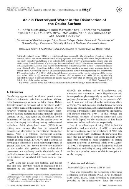
Exp. Eye Res. (2000) 70, 1–6<br />
Article No. exer.1999.0776, available onl<strong>in</strong>e at http:www.idealibrary.com on<br />
<strong>Acidic</strong> <strong>Electrolysed</strong> <strong>Water</strong> <strong>in</strong> <strong>the</strong> Dis<strong>in</strong>fection <strong>of</strong><br />
<strong>the</strong> <strong>Ocular</strong> <strong>Surface</strong><br />
SHIGETO SHIMMURA a *†, KOKI MATSUMOTO b †, HIROMOTO YAGUCHI a ,<br />
TOSHIYA OKUDA b , SEIYA MIYAJIMA b , AKIRA NEGI b , JUN SHIMAZAKI a<br />
AND KAZUO TSUBOTA a<br />
a<br />
Department <strong>of</strong> Ophthalmology, Tokyo Dental College, Chiba, Japan and b Department <strong>of</strong><br />
Ophthalmology, Kumamoto University School <strong>of</strong> Medic<strong>in</strong>e, Kumamoto, Japan<br />
(Received Lund 14 September 1998 and accepted <strong>in</strong> revised form 25 March 1999)<br />
<strong>Acidic</strong> electrolysed water (AEW) is a colorless solution prepared by <strong>the</strong> electrolysis <strong>of</strong> sodium chloride<br />
solution, which has potent anti-microbial activity aga<strong>in</strong>st a wide variety <strong>of</strong> bacteria, fungi and virus. In<br />
this study, <strong>the</strong> safety and efficacy <strong>of</strong> an isotonic AEW solution (iAEW) was <strong>in</strong>vestigated both <strong>in</strong> vitro and<br />
<strong>in</strong> vivo us<strong>in</strong>g denuded corneas <strong>of</strong> gu<strong>in</strong>ea pigs. Povidone iod<strong>in</strong>e (001–10%) was used as control. Exposure<br />
<strong>of</strong> P. aerug<strong>in</strong>osa to iAEW for 5 sec caused total <strong>in</strong>hibition <strong>of</strong> bacterial growth <strong>in</strong> vitro. Comparable results<br />
were obta<strong>in</strong>ed with 1% povidone iod<strong>in</strong>e, while more dilute solutions required longer exposure times for<br />
similar effects. Cytotoxicity <strong>of</strong> iAEW on corneal epi<strong>the</strong>lial cells <strong>in</strong> vitro was significantly less compared to<br />
1% povidone iod<strong>in</strong>e (P 005), while m<strong>in</strong>imal damage was observed <strong>in</strong> vivo by irrigation <strong>of</strong> <strong>the</strong> cornea<br />
with ei<strong>the</strong>r iAEW or 1% povidone iod<strong>in</strong>e. Treatment <strong>of</strong> P. aerug<strong>in</strong>osa with iAEW (15 sec) significantly<br />
<strong>in</strong>hibited <strong>the</strong> onset <strong>of</strong> corneal <strong>in</strong>fection <strong>in</strong> gu<strong>in</strong>ea pigs. iAEW may <strong>of</strong>fer an effective and safe method for<br />
dis<strong>in</strong>fection <strong>of</strong> <strong>the</strong> ocular surface.<br />
2000 Academic Press<br />
Key words: Corneal ulcer; free radicals; dis<strong>in</strong>fection; oxidative damage; Pseudomonas keratitis.<br />
1. Introduction<br />
Dis<strong>in</strong>fect<strong>in</strong>g agents used <strong>in</strong> cl<strong>in</strong>ical practice must<br />
effectively elim<strong>in</strong>ate <strong>in</strong>fectious organisms without<br />
be<strong>in</strong>g biohazardous or toxic to liv<strong>in</strong>g tissue. Halide<br />
derivatives such as povidone iod<strong>in</strong>e have been widely<br />
used for this purpose (Shelanski and Shelanski, 1956),<br />
however, problems such as hypersensitivity, coloration<br />
<strong>of</strong> sk<strong>in</strong> and bacterial resistance cannot be ignored<br />
(Zamora, 1986). These agents are <strong>of</strong>ten diluted for <strong>the</strong><br />
dis<strong>in</strong>fection <strong>of</strong> <strong>the</strong> sk<strong>in</strong> and ocular surface prior to<br />
<strong>in</strong>traocular surgery to avoid any toxicity to <strong>the</strong> corneal<br />
epi<strong>the</strong>lium. <strong>Acidic</strong> electrolysed water (AEW) is an<br />
entirely new concept that has <strong>the</strong> potential <strong>of</strong><br />
becom<strong>in</strong>g an alternative to conventional dis<strong>in</strong>fect<strong>in</strong>g<br />
agents. AEW is a colorless, transparent solution<br />
prepared by <strong>the</strong> electrolysis <strong>of</strong> tap water conta<strong>in</strong><strong>in</strong>g<br />
sodium chloride (NaCl, 20% wv), and is def<strong>in</strong>ed as<br />
hav<strong>in</strong>g a pH <strong>of</strong> less than 3 and a reduction potential <strong>of</strong><br />
greater than 1100 mV. Several devices are available<br />
on <strong>the</strong> market that produce AEW with<strong>in</strong> <strong>the</strong>se<br />
parameters, and are be<strong>in</strong>g used <strong>in</strong> <strong>the</strong> dis<strong>in</strong>fection <strong>of</strong><br />
medical <strong>in</strong>struments and food products as well as <strong>in</strong><br />
<strong>the</strong> treatment <strong>of</strong> superficial <strong>in</strong>fections such as g<strong>in</strong>givitis.<br />
AEW alone has potent anti-bacterial, anti-fungal<br />
and anti-viral activities, which is speculated to be<br />
mediated by <strong>the</strong> actions <strong>of</strong> sodium hypochlorite<br />
* Address correspondences to: Shigeto Shimmura, Department <strong>of</strong><br />
Ophthalmology, Tokyo Dental College, 5-11-13 Sugano, Ichikawa,<br />
Chiba 272-8513, Japan. E-mail: shigeeyebank.or.jp<br />
† Both authors contributed equally to this work.<br />
0014–48350001000106 $35.000<br />
(NaOCl), <strong>the</strong> sodium salt <strong>of</strong> hypochlorous acid<br />
(Iwasawa and Nakamura, 1996). Hypochlorous acid<br />
is also produced physiologically by myeloperoxidase <strong>in</strong><br />
polymorphonucleocytes (PMN) <strong>in</strong> <strong>the</strong> presence <strong>of</strong> Cl−<br />
and I− ions, and is <strong>in</strong>volved <strong>in</strong> <strong>the</strong> bactericidal effects<br />
<strong>of</strong> PMNs. The anti-microbial mechanisms <strong>of</strong> povidone<br />
iod<strong>in</strong>e are also not clear, although it is related with <strong>the</strong><br />
presence <strong>of</strong> free iod<strong>in</strong>e (I2) <strong>in</strong> proximity to <strong>the</strong> bacterial<br />
cell surface (Zamora, 1986). In this respect, <strong>the</strong><br />
bactericidal activities <strong>of</strong> povidone iod<strong>in</strong>e and AEW<br />
may both depend on <strong>the</strong> availability <strong>of</strong> free halide<br />
molecules, iod<strong>in</strong>e and chlor<strong>in</strong>e, respectively.<br />
One <strong>of</strong> <strong>the</strong> advantages <strong>of</strong> us<strong>in</strong>g AEW is that <strong>the</strong><br />
production and degradation <strong>of</strong> AEW is m<strong>in</strong>imally<br />
<strong>in</strong>vasive to tissue, s<strong>in</strong>ce <strong>the</strong> breakdown <strong>of</strong> AEW only<br />
produces sal<strong>in</strong>e (NaCl) and traces <strong>of</strong> chloride gas. This<br />
may <strong>of</strong>fer an advantage over iod<strong>in</strong>e products which<br />
have been reported to cause alterations <strong>in</strong> thyroid<br />
function as a result <strong>of</strong> <strong>in</strong>creased iodide levels (Lyen et<br />
al., 1982). The present study was designed to evaluate<br />
<strong>the</strong> feasibility and safety <strong>of</strong> us<strong>in</strong>g AEW on <strong>the</strong> ocular<br />
surface as a possible means <strong>of</strong> dis<strong>in</strong>fection prior to<br />
ocular surgery.<br />
2. Materials and Methods<br />
Bactericidal Effects <strong>of</strong> Isotonic AEW <strong>in</strong> vitro<br />
AEW was prepared by <strong>the</strong> electrolysis <strong>of</strong> tap water<br />
conta<strong>in</strong><strong>in</strong>g sodium chloride (NaCl, 20%) (Acitron-S,<br />
MAC Japan, Tokyo, Japan) accord<strong>in</strong>g to <strong>in</strong>structions<br />
provided by <strong>the</strong> manufacturer. S<strong>in</strong>ce <strong>the</strong> osmotic<br />
2000 Academic Press
2 S. SHIMMURA ET AL.<br />
pressure <strong>of</strong> AEW thus produced is hypotonic, isotonicity<br />
was ma<strong>in</strong>ta<strong>in</strong>ed by <strong>the</strong> addition <strong>of</strong> extra NaCl<br />
(40 gl−), which was confirmed by an osmometer.<br />
The addition <strong>of</strong> extra NaCl did not affect <strong>the</strong> pH (24)<br />
or redox potential (1100 mV) <strong>of</strong> <strong>the</strong> solution (iAEW).<br />
iAEW was prepared and stored at 4C <strong>in</strong> opaque<br />
conta<strong>in</strong>ers, and used with<strong>in</strong> 1 week <strong>of</strong> preparation.<br />
Pseudomonas aerug<strong>in</strong>osa (ATGC stra<strong>in</strong>) were harvested<br />
<strong>in</strong> SCDLP medium (Soybean-Case<strong>in</strong> Digest<br />
Broth with Lecith<strong>in</strong> and Polysorbate 80, Nihon<br />
Seiyaku, Tokyo) at a concentration <strong>of</strong> 29<br />
10 CFU ml− and mixed 1:1 with iAEW for reaction<br />
times <strong>of</strong> 5, 15, 30, 60, 180, 300 and 900 sec.<br />
Follow<strong>in</strong>g <strong>the</strong> specified reaction time, 10 µl <strong>of</strong> <strong>the</strong><br />
bacteriaiAEW solution was suspended <strong>in</strong> 1 ml <strong>of</strong><br />
SCDLP medium and cultured for 48 hr at 35C.<br />
Povidone iod<strong>in</strong>e solutions (10, 01 and 001%) served<br />
as control. After <strong>the</strong> 48 hr <strong>in</strong>cubation period, <strong>the</strong><br />
bactericidal effects <strong>of</strong> each solution was determ<strong>in</strong>ed by<br />
observ<strong>in</strong>g for any bacterial growth <strong>in</strong> culture. The<br />
experiment was repeated twice.<br />
Prophylactic Effects <strong>of</strong> Isotonic iAEW <strong>in</strong> vivo<br />
Alb<strong>in</strong>o Hartley gu<strong>in</strong>ea pigs <strong>of</strong> both sexes (400–<br />
450 g) were used and treated accord<strong>in</strong>g to <strong>the</strong> ARVO<br />
Statement for <strong>the</strong> Use <strong>of</strong> Animals <strong>in</strong> Ophthalmology<br />
and Vision Research. The cl<strong>in</strong>ical stra<strong>in</strong> <strong>of</strong> P. aerug<strong>in</strong>osa<br />
used <strong>in</strong> <strong>the</strong> <strong>in</strong> vivo study [serotype I, elastase (),<br />
alkal<strong>in</strong>e protease ()] was isolated from a patient with<br />
a severe corneal ulcer (Matsumoto et al., 1998), and<br />
stored <strong>in</strong> 10% skimmed milk medium at 70C until<br />
use. A 01 ml sample <strong>of</strong> medium was cultivated <strong>in</strong> 5 ml<br />
<strong>of</strong> tryptosoy broth (Eiken Chemical, Tokyo, Japan)<br />
with reciprocal shak<strong>in</strong>g a 1 Hz at 32C for 12 hr, and<br />
P. aerug<strong>in</strong>osa were subsequently collected by centrifugation<br />
at 3500 rpm for 20 m<strong>in</strong>. One ml samples <strong>of</strong> <strong>the</strong><br />
bacterial suspension (810 CFU ml−) were centrifuged<br />
at 9000 rpm for 3 m<strong>in</strong>, and treated with 095 ml<br />
<strong>of</strong> ei<strong>the</strong>r iAEW or sal<strong>in</strong>e control for 15 sec. Bacteria<br />
were <strong>the</strong>n collected by centrifugation for 2 m<strong>in</strong>,<br />
washed once and suspended <strong>in</strong> sterile sal<strong>in</strong>e. P.<br />
aerug<strong>in</strong>osa were <strong>the</strong>refore exposed to iAEW for a total<br />
<strong>of</strong> 135 sec prior to use <strong>in</strong> <strong>the</strong> animal experiment.<br />
After gu<strong>in</strong>ea pigs were anes<strong>the</strong>tized topically with<br />
04% oxybuproca<strong>in</strong>e hydrochloride and systemically<br />
with pentobarbital sodium (25 mg kg−), a 40 mm<br />
paper disc (Whatman International Ltd. Maidstone,<br />
U.K.) immersed <strong>in</strong> 1-heptanol (Nacalai Tesqu, Kyoto,<br />
Japan) was applied to <strong>the</strong> right cornea <strong>of</strong> <strong>the</strong> gu<strong>in</strong>ea<br />
pigs for 1 m<strong>in</strong>. The paper discs were <strong>the</strong>n removed,<br />
creat<strong>in</strong>g a reproducible circular defect <strong>in</strong> <strong>the</strong> corneal<br />
epi<strong>the</strong>lium. Epi<strong>the</strong>lial defects were made <strong>in</strong> all animals<br />
s<strong>in</strong>ce P. aerug<strong>in</strong>osa cannot efficiently <strong>in</strong>fect corneas<br />
with <strong>in</strong>tact epi<strong>the</strong>lium. After <strong>the</strong> corneal surface was<br />
washed with 40 ml <strong>of</strong> sterile sal<strong>in</strong>e, 30 µl <strong>of</strong> bacterial<br />
suspension treated with ei<strong>the</strong>r isotonic iAEW or sal<strong>in</strong>e<br />
control (n 6 each) was applied to <strong>the</strong> denuded area<br />
<strong>of</strong> each cornea. Signs <strong>of</strong> <strong>in</strong>flammation were observed<br />
TABLE I<br />
Criteria and scor<strong>in</strong>g <strong>of</strong> corneal lesions after <strong>the</strong><br />
<strong>in</strong>oculation <strong>of</strong> organisms<br />
Macroscopic f<strong>in</strong>d<strong>in</strong>g Severity Score<br />
Epi<strong>the</strong>lial damage Irregular epi<strong>the</strong>lium 1<br />
Epi<strong>the</strong>lial erosion 2<br />
Mild ulcer 3<br />
Severe ulcer 4<br />
Corneal opacity Slightly opaque 1<br />
Iris visible 2<br />
Iris <strong>in</strong>visible 3<br />
Dense 4<br />
Area<br />
Subscore<br />
(ratio <strong>of</strong> lesion to whole<br />
cornea)<br />
110<br />
01<br />
15<br />
02<br />
25<br />
04<br />
35<br />
06<br />
45<br />
08<br />
55<br />
10<br />
Corneal damage <strong>in</strong>dex (CDI). The grade <strong>of</strong> corneal damage was<br />
calculated as follows: CDI (epi<strong>the</strong>lial damage scorearea subscore)(opacity<br />
scorearea subscore).<br />
for 5 days, and <strong>the</strong>n eyes were enucleated for<br />
histological evaluation after animals were killed with<br />
an overdose <strong>of</strong> sodium pentobarbital. Sections were<br />
fixed with 10% formal<strong>in</strong>, embedded <strong>in</strong> paraff<strong>in</strong> and<br />
sta<strong>in</strong>ed with hematoxyl<strong>in</strong> and eos<strong>in</strong> (HE).<br />
The grade <strong>of</strong> corneal <strong>in</strong>flammation was semiquantified<br />
by <strong>the</strong> corneal damage <strong>in</strong>dex (CDI) as<br />
described <strong>in</strong> our previous publication (Matsumoto et<br />
al., 1998), with some modifications as shown <strong>in</strong> Table<br />
I. The modifications were to allow better evaluation <strong>of</strong><br />
<strong>the</strong> milder changes observed <strong>in</strong> this study, compared<br />
to <strong>the</strong> previous study that directly <strong>in</strong>oculated <strong>the</strong><br />
stroma with P. aerug<strong>in</strong>osa. Statistical analysis was<br />
carried out us<strong>in</strong>g <strong>the</strong> ANOVA test.<br />
Cytotoxic Effects <strong>of</strong> iAEW<br />
The cytotoxicity <strong>of</strong> iAEW was observed <strong>in</strong> vitro<br />
us<strong>in</strong>g an immortalized human corneal epi<strong>the</strong>lial cell<br />
l<strong>in</strong>e (T-HCEC) cultured <strong>in</strong> SHEM medium <strong>in</strong> 250 ml<br />
flasks (Iwaki Glass Inc., Tokyo) as previously described<br />
(Araki-Sasaki et al., 1995; Shimmura and Tsubota,<br />
1997). Prior to experiments, cells were passaged to 96<br />
well plates (Iwaki) at a seed<strong>in</strong>g density <strong>of</strong> 10 cells per<br />
well and <strong>in</strong>cubated for 48 hr until confluence. T-HCEC<br />
were <strong>the</strong>n exposed to ei<strong>the</strong>r isotonic iAEW, PBS() or<br />
povidone iod<strong>in</strong>e sal<strong>in</strong>e solution (01, 01or10%)(n <br />
8) for a total <strong>of</strong> 15 sec. Each test solution was aspirated<br />
and replaced with fresh SHEM medium conta<strong>in</strong><strong>in</strong>g<br />
04% trypan blue to sta<strong>in</strong> devitalized cells. The number<br />
<strong>of</strong> non-viable cells <strong>in</strong> each group was expressed as <strong>the</strong><br />
percentage <strong>of</strong> trypan blue positive cells per well. The<br />
experiment was repeated <strong>in</strong> triplicate and statistical
ACIDIC ELECTROLYSED WATER 3<br />
TABLE II<br />
Bactericidal effects <strong>of</strong> iAEW aga<strong>in</strong>st P. aerug<strong>in</strong>osa ATGC stra<strong>in</strong> <strong>in</strong> vivo<br />
Reaction time 5 sec 15 sec 30 sec 60 sec 3 m<strong>in</strong> 5 m<strong>in</strong> 15 m<strong>in</strong><br />
iAEW () () () () () () ()<br />
PI 1% () () () () () () ()<br />
PI 01% () () () () () () ()<br />
PI 001% () () () () () () ()<br />
(): Bacterial growth after 48 hr <strong>in</strong>cubation at 35C <strong>in</strong> SCDLP medium. (): Complete <strong>in</strong>hibition <strong>of</strong> bacterial culture. iAEW: Isotonic acidic<br />
electrolysed water. PI: Povidone iod<strong>in</strong>e.<br />
defects were not created <strong>in</strong> this experiment <strong>in</strong> order to<br />
simulate prophylactic dis<strong>in</strong>fection prior to surgery.<br />
Cl<strong>in</strong>ical grad<strong>in</strong>g <strong>of</strong> <strong>in</strong>flammation was carried out us<strong>in</strong>g<br />
<strong>the</strong> CDI score as described above.<br />
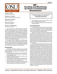
FIG. 1. Mean percentage <strong>of</strong> non-viable cells per well as<br />
determ<strong>in</strong>ed by <strong>the</strong> trypan blue exclusion test. iAEW: Isotonic<br />
acidic electrolysed water, PI: povidone iod<strong>in</strong>e. Mean1 S.D.,<br />
n 8 each, * P 005 aga<strong>in</strong>st all o<strong>the</strong>r groups. Statistical<br />
analysis was carried out by <strong>the</strong> one-way ANOVA test.<br />
analysis was performed us<strong>in</strong>g <strong>the</strong> one-way ANOVA<br />
test.<br />
Cytotoxicity <strong>of</strong> iAEW and povidone iod<strong>in</strong>e <strong>in</strong> vivo<br />
was also exam<strong>in</strong>ed by irrigat<strong>in</strong>g <strong>the</strong> ocular surface <strong>of</strong><br />
Alb<strong>in</strong>o Hartley gu<strong>in</strong>ea pigs with ei<strong>the</strong>r iAEW, 1%<br />
povidone iod<strong>in</strong>e or sal<strong>in</strong>e control for 15 sec. Corneal<br />
3. Results<br />
Bactericidal Effects and Cytotoxicity<br />
Preparation <strong>of</strong> AEW accord<strong>in</strong>g to <strong>the</strong> manufacturer’s<br />
protocol produces a hypotonic solution that<br />
may be detrimental to <strong>the</strong> mucosal tissue <strong>of</strong> <strong>the</strong> ocular<br />
surface. We have confirmed that modify<strong>in</strong>g AEW to an<br />
isotonic solution still meets <strong>the</strong> standards for pH and<br />
reduction potential specified by <strong>the</strong> maker, and fur<strong>the</strong>r<br />
sought to demonstrate <strong>the</strong> efficacy <strong>of</strong> isotonic iAEW <strong>in</strong><br />
vitro and <strong>in</strong> vivo. Table II shows <strong>the</strong> bactericidal effect<br />
<strong>of</strong> iAEW compared to that <strong>of</strong> serially diluted povidone<br />
iod<strong>in</strong>e solutions. Both iAEW and 1% povidone iod<strong>in</strong>e<br />
completely <strong>in</strong>hibited P. aerug<strong>in</strong>osa growth with<strong>in</strong> 5 sec<br />
<strong>of</strong> exposure. More dilute povidone iod<strong>in</strong>e solutions<br />
required longer exposure times for comparable effects.<br />
A cytotoxicity assay us<strong>in</strong>g a corneal epi<strong>the</strong>lial cell<br />
l<strong>in</strong>e was performed <strong>in</strong> order to compare <strong>the</strong> effects <strong>of</strong><br />
iAEW and serially diluted povidone iod<strong>in</strong>e solutions.<br />
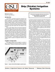
Figs 1 and 2 show <strong>the</strong> mean percentage <strong>of</strong> non-viable<br />
cells as determ<strong>in</strong>ed by <strong>the</strong> trypan blue exclusion<br />
test, and a representative photograph respectively.<br />
Although iAEW and 1% povidone iod<strong>in</strong>e were equally<br />
effective <strong>in</strong> bactericidal activity, <strong>the</strong> cytotoxicity assay<br />
(A)<br />
(B)<br />
FIG. 2. Typical micrographs <strong>of</strong> trypan blue sta<strong>in</strong>ed cultured corneal epi<strong>the</strong>lial cells treated with ei<strong>the</strong>r iAEW (A) or povidone<br />
iod<strong>in</strong>e (B). The confluent sheet <strong>of</strong> cells were <strong>in</strong>tact <strong>in</strong> both groups.
4 S. SHIMMURA ET AL.<br />
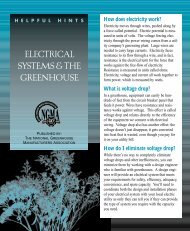
FIG. 3. The effects <strong>of</strong> iAEW, 1% povidone iod<strong>in</strong>e or sal<strong>in</strong>e<br />
alone on corneas <strong>of</strong> untreated gu<strong>in</strong>ea pigs. Signs <strong>of</strong><br />
<strong>in</strong>flammation were graded accord<strong>in</strong>g to <strong>the</strong> CDI scale, which<br />
did not reveal any signs <strong>of</strong> <strong>in</strong>flammation for up to 48 hr.<br />
showed significantly less damage <strong>in</strong> T-HCEC exposed<br />
to iAEW compared to cells treated with 1% povidone<br />
iod<strong>in</strong>e. The cytotoxicity study performed <strong>in</strong> vivo<br />
showed very little change <strong>in</strong> CDI scores follow<strong>in</strong>g<br />
irrigation <strong>of</strong> gu<strong>in</strong>ea pig corneas with iAEW, 1%<br />
povidone iod<strong>in</strong>e or sal<strong>in</strong>e (Fig. 3). Although iAEW was<br />
less cytotoxic <strong>in</strong> vitro, this difference was not demonstrated<br />
on a macroscopic level <strong>in</strong> vivo.<br />
Prophylactic Effects <strong>of</strong> iAEW <strong>in</strong> vivo<br />
Denud<strong>in</strong>g <strong>of</strong> <strong>the</strong> corneal epi<strong>the</strong>lium <strong>in</strong> <strong>the</strong> gu<strong>in</strong>ea<br />
pigs was performed to mimic <strong>the</strong> compromised<br />
condition <strong>of</strong> <strong>the</strong> ocular surface follow<strong>in</strong>g ocular surface<br />
and <strong>in</strong>traocular surgery. Bare stroma is an ideal<br />
substrate for <strong>the</strong> adhesion <strong>of</strong> microbes to <strong>in</strong>itiate a<br />
<strong>in</strong>fectious reaction that can lead to ulceration and<br />
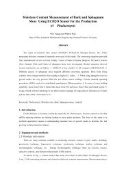
perforation <strong>of</strong> <strong>the</strong> cornea. Fig. 4(A) is a representative<br />
macroscopic view <strong>of</strong> a gu<strong>in</strong>ea pig 2 days follow<strong>in</strong>g<br />
<strong>in</strong>oculation with P. aerug<strong>in</strong>osa treated with control<br />
sal<strong>in</strong>e. Most corneas <strong>in</strong> <strong>the</strong> control group showed<br />
several abscess-like lesions <strong>in</strong> addition to corneal<br />
erosion and ground glass-like opacity up to day 5 [Fig.<br />
4(B)]. Conjunctival <strong>in</strong>jection and chemosis were more<br />
<strong>in</strong>tense <strong>in</strong> <strong>the</strong> control group. Five out <strong>of</strong> six control<br />
corneas showed improvement follow<strong>in</strong>g <strong>in</strong>itial<br />
changes, however, one cornea developed a severe<br />
ulcer similar to those observed when P. aerug<strong>in</strong>osa is<br />
directly <strong>in</strong>jected <strong>in</strong>to stromal tissue (Matsumoto et al.,<br />
1998). Corneas <strong>in</strong> <strong>the</strong> iAEW group showed early<br />
reduction <strong>in</strong> erosion size, but presented with ground<br />
glass-like opacity <strong>of</strong> <strong>the</strong> lesion 2 days after <strong>in</strong>oculation<br />
[Fig. 4(C)]. Most <strong>of</strong> <strong>the</strong> opacity as well as <strong>the</strong> erosion<br />
itself healed by day 5 [Fig. 4(D)]. One cornea developed<br />
a mild abscess-like lesion that healed by <strong>the</strong> end <strong>of</strong> <strong>the</strong><br />
study, leav<strong>in</strong>g a slightly opaque lesion <strong>in</strong> <strong>the</strong> subepi<strong>the</strong>lial<br />
area. CDI scores showed a statistically<br />
FIG. 4. Representative macroscopic view <strong>of</strong> corneas <strong>in</strong> control (sal<strong>in</strong>e-treated) and iAEW groups. Cornea <strong>in</strong> <strong>the</strong> control group<br />
(animal No. 2) on day 2 (A) and day 5 (B) show<strong>in</strong>g abscess-like lesions <strong>in</strong> addition to corneal erosion and ground glass-like<br />
opacity. Representative cornea <strong>in</strong> <strong>the</strong> iAEW group (animal No. 5) on day 2 (C) with milder opacity than control that healed<br />
by day 5 (D).
ACIDIC ELECTROLYSED WATER 5<br />
suggestive <strong>of</strong> endo<strong>the</strong>lial damage was observed <strong>in</strong> only<br />
one eye. In contrast, most corneas <strong>in</strong> <strong>the</strong> control group<br />
showed vary<strong>in</strong>g degrees <strong>of</strong> corneal swell<strong>in</strong>g and<br />
<strong>in</strong>flammatory cell <strong>in</strong>filtration [Fig. 6(B)]. Corneal<br />
architecture was damaged to various degrees, with <strong>the</strong><br />
most severe cornea show<strong>in</strong>g massive cellular <strong>in</strong>filtration<br />
<strong>in</strong> all layers <strong>of</strong> <strong>the</strong> stroma and loss <strong>of</strong> epi<strong>the</strong>lium<br />
and endo<strong>the</strong>lium as well as liquefactive necrosis [Fig.<br />
6(C)].<br />
FIG. 5. CDI scores <strong>of</strong> corneas <strong>in</strong> <strong>the</strong> iAEW group and<br />
control dur<strong>in</strong>g <strong>the</strong> 5 day study period. All corneas at <strong>the</strong><br />
beg<strong>in</strong>n<strong>in</strong>g <strong>of</strong> <strong>the</strong> study had CDI scores <strong>of</strong> 09, s<strong>in</strong>ce scor<strong>in</strong>g<br />
was performed after denud<strong>in</strong>g <strong>of</strong> <strong>the</strong> epi<strong>the</strong>lium.<br />
Mean1 S.D., n 6 each, * P 005, <strong>in</strong>ter-group analysis<br />
was assessed by <strong>the</strong> ANOVA test.<br />
significant difference at days 1, 2 and 3 (Fig. 5). All<br />
corneas at <strong>the</strong> beg<strong>in</strong>n<strong>in</strong>g <strong>of</strong> <strong>the</strong> study had scores <strong>of</strong> 09<br />
s<strong>in</strong>ce evaluation was done follow<strong>in</strong>g treatment with<br />
heptanol.<br />
Histopathology <strong>of</strong> corneas <strong>of</strong> <strong>the</strong> iAEW group<br />
performed at <strong>the</strong> end <strong>of</strong> <strong>the</strong> study revealed normal<br />
corneal architecture with no appreciable swell<strong>in</strong>g <strong>in</strong> 5<br />
out <strong>of</strong> 6 eyes [Fig. 6(A)]. Mild corneal swell<strong>in</strong>g<br />
4. Discussion<br />
Bacterial <strong>in</strong>fection <strong>of</strong> <strong>the</strong> ocular surface can cause a<br />
variety <strong>of</strong> cl<strong>in</strong>ical pictures rang<strong>in</strong>g from mild conjunctivitis<br />
to a rapidly progress<strong>in</strong>g, purulent ulcer that<br />
can lead to perforation <strong>of</strong> <strong>the</strong> globe and bl<strong>in</strong>dness. The<br />
<strong>in</strong>itial trigger <strong>in</strong> most cases is a compromised ocular<br />
surface with decreased barrier functions due to<br />
trauma, surgery, contact lens used or o<strong>the</strong>r pathologies.<br />
Bacteria that have evaded <strong>the</strong> anti-microbial<br />
barriers <strong>of</strong> <strong>the</strong> ocular surface adhere to subepi<strong>the</strong>lial<br />
tissue which <strong>the</strong>n becomes <strong>the</strong> focus <strong>of</strong> bacterial<br />
keratitis. Bacterial organisms commonly detected <strong>in</strong><br />
<strong>in</strong>fections <strong>of</strong> <strong>the</strong> ocular surface <strong>in</strong>clude P. aerug<strong>in</strong>osa,<br />
Streptococcus pneumoniae, Staphylococci and Moraxella<br />
lacunata. The degree <strong>of</strong> corneal damage depends on <strong>the</strong><br />
virulence <strong>of</strong> bacterial tox<strong>in</strong>s and <strong>the</strong> host response<br />
centered around PMNs (Kernacki and Berk, 1995)<br />
that can cause non-specific damage to tissue due to<br />
<strong>the</strong> release <strong>of</strong> proteases (Henson and Johnston, 1987)<br />
FIG. 6. (A) Hematoxyl<strong>in</strong> eos<strong>in</strong> sta<strong>in</strong><strong>in</strong>g <strong>of</strong> <strong>the</strong> same animal as Fig. 2(D) <strong>of</strong> <strong>the</strong> iAEW group. (B) Histology <strong>of</strong> control cornea<br />
<strong>in</strong> Fig. 2(B) with prom<strong>in</strong>ent stromal edema and cellular <strong>in</strong>filtration. (C) Massive cellular <strong>in</strong>filtration <strong>in</strong> all layers <strong>of</strong> <strong>the</strong> stroma<br />
and loss <strong>of</strong> epi<strong>the</strong>lium and endo<strong>the</strong>lium as well as liquefactive necrosis <strong>in</strong> animal susta<strong>in</strong><strong>in</strong>g <strong>the</strong> greatest damage <strong>in</strong> <strong>the</strong> control<br />
group.
6 S. SHIMMURA ET AL.<br />
and reactive oxygen species (Cross et al., 1987;<br />
Malech, 1987). Although AEW has been reported to<br />
be effective aga<strong>in</strong>st all species listed above as well as<br />
most o<strong>the</strong>r cl<strong>in</strong>ically relevant bacteria except for<br />
Bacillus sp (Iwasawa et al., 1993), P. aerug<strong>in</strong>osa was<br />
chosen as <strong>the</strong> pathogen <strong>in</strong> this study s<strong>in</strong>ce this species<br />
is especially notorious for caus<strong>in</strong>g a rapid progression<br />
<strong>of</strong> ulcers that may lead to irreversible damage and loss<br />
<strong>of</strong> vision. Prepar<strong>in</strong>g an isotonic solution was an<br />
additional measure to decrease cytotoxicity to <strong>the</strong><br />
ocular surface. Although <strong>the</strong> present study exam<strong>in</strong>ed<br />
<strong>the</strong> bactericidal effects <strong>of</strong> iAEW on P. aerug<strong>in</strong>osa only,<br />
we have confirmed that <strong>the</strong> parameters for def<strong>in</strong><strong>in</strong>g<br />
AEW activity (pH <strong>of</strong> 24 or less, a redox potential <strong>of</strong><br />
approximately 1100 mV) were unchanged follow<strong>in</strong>g<br />
isotonicity.<br />
Prophylactic measures are taken prior to surgery as<br />
a means to elim<strong>in</strong>ate pathogens from <strong>the</strong> surgical field<br />
and surgical staff, that may become a cause <strong>of</strong> <strong>in</strong>fection<br />
<strong>in</strong> <strong>the</strong> unhealed wound. Various antiseptics are<br />
available <strong>in</strong> <strong>the</strong> market for <strong>the</strong> purpose <strong>of</strong> steriliz<strong>in</strong>g<br />
tissue and surgical <strong>in</strong>struments. Povidone iod<strong>in</strong>e is<br />
one <strong>of</strong> <strong>the</strong> most commonly used solutions that has<br />
proven its efficacy over <strong>the</strong> years <strong>of</strong> use s<strong>in</strong>ce be<strong>in</strong>g<br />
<strong>in</strong>troduced <strong>in</strong> <strong>the</strong> market. Although <strong>the</strong> safety <strong>of</strong><br />
povidone iod<strong>in</strong>e is also well established, high concentrations<br />
applied to mucous membranes are reported to<br />
cause a 5–10-fold greater <strong>in</strong>crease <strong>in</strong> serum iod<strong>in</strong>e<br />
levels when compared to cutaneous absorption<br />
(Zamora, 1986). This has caused some concern as to<br />
<strong>the</strong> effects on thyroid function, especially <strong>in</strong> children<br />
(Lyen et al., 1982). Although <strong>the</strong> quantity <strong>of</strong> povidone<br />
iod<strong>in</strong>e used <strong>in</strong> ophthalmic surgery may not pose any<br />
problems systemically, it may contribute to corneal<br />
epi<strong>the</strong>lial damage that may affect <strong>the</strong> field <strong>of</strong> view<br />
dur<strong>in</strong>g <strong>in</strong>traocular surgery. The results <strong>of</strong> <strong>the</strong> trypan<br />
blue dye exclusion test (Fig. 1) shows that iAEW may<br />
be less cytotoxic <strong>in</strong> this regard.<br />
One <strong>of</strong> <strong>the</strong> drawbacks <strong>of</strong> iAEW is that its bactericidal<br />
effects are rapidly lost when exposed to organic<br />
material, <strong>in</strong>clud<strong>in</strong>g liv<strong>in</strong>g tissue. Although iAEW may<br />
temporarily elim<strong>in</strong>ate microbes on <strong>the</strong> ocular surface,<br />
bactericidal effects may be less effective for microbes<br />
with<strong>in</strong> <strong>the</strong> corneal stroma, Meibomian ducts and<br />
ciliary follicles. When iAEW was used <strong>in</strong> an experimental<br />
P. aerug<strong>in</strong>osa bacterial ulcer <strong>in</strong> gu<strong>in</strong>ea pigs<br />
as reported <strong>in</strong> our previous study (Matsumoto et al.,<br />
1998), <strong>the</strong>re was no difference <strong>in</strong> prognosis compared<br />
to sal<strong>in</strong>e control (data not shown). This is probably<br />
due to <strong>the</strong> fact that iAEW lost its bactericidal properties<br />
before reach<strong>in</strong>g pathogens <strong>in</strong> <strong>the</strong> deeper layers <strong>of</strong> <strong>the</strong><br />
stroma.<br />
Ano<strong>the</strong>r potential application for AEW <strong>in</strong> <strong>the</strong> field <strong>of</strong><br />
ophthalmology is <strong>the</strong> dis<strong>in</strong>fection <strong>of</strong> contact lens.<br />
Although <strong>the</strong> few commercially available devices for<br />
produc<strong>in</strong>g AEW are still costly and cumbersome, <strong>the</strong><br />
household use <strong>of</strong> AEW may become possible pend<strong>in</strong>g<br />
improvements <strong>in</strong> design and technology. S<strong>in</strong>ce AEW is<br />
a colorless, non-irritat<strong>in</strong>g solution, it has <strong>the</strong> potential<br />
<strong>of</strong> becom<strong>in</strong>g a major dis<strong>in</strong>fect<strong>in</strong>g agent <strong>of</strong> <strong>the</strong> sk<strong>in</strong> and<br />
mucosal tissue <strong>in</strong>clud<strong>in</strong>g <strong>the</strong> ocular surface. Our study<br />
has shown that adjust<strong>in</strong>g <strong>the</strong> osmolarity <strong>of</strong> AEW to<br />
physiological levels does not compromise its bactericidal<br />
activity aga<strong>in</strong>st P. aerug<strong>in</strong>osa, which fur<strong>the</strong>r<br />
improves <strong>the</strong> safety <strong>of</strong> us<strong>in</strong>g AEW <strong>in</strong> <strong>the</strong> ocular<br />
surface.<br />
References<br />
Araki-Sasaki, K., Ohashi, Y., Sasabe, T., Hayashi, K.,<br />
Watanabe, H., Tano, Y. and Handa, H. (1995). An SV<br />
40-immortalized human corneal epi<strong>the</strong>lial cell l<strong>in</strong>e and<br />
its characterization. Invest. Ophthalmol. Vis. Sci. 36,<br />
614–21.<br />
Cross, C. E., Halliwell, B. and Borish, E. T. (1987). Oxygen<br />
radicals and human disease. Ann. Intern. Med. 107,<br />
526–45.<br />
Henson, P. M. and Johnston, R. B. J. (1987). Tissue <strong>in</strong>jury<br />
<strong>in</strong> <strong>in</strong>flammation: oxidants, prote<strong>in</strong>ases, and cationic<br />
prote<strong>in</strong>s. J. Cl<strong>in</strong>. Invest. 79, 669–74.<br />
Iwasawa, A. and Nakamura, Y. (1996). Bactericidal effect <strong>of</strong><br />
acidic electrolyzed water-comparison <strong>of</strong> chemical acidic<br />
sodium hydrochloride (NaOCl) solution. Kansenshogaku<br />
Zasshi. 70, 915–22.<br />
Iwasawa, A., Nakamura, Y. and Mizuno, T. (1993).<br />
Bactericidal activity <strong>of</strong> aqua oxidation water aga<strong>in</strong>st<br />
cl<strong>in</strong>ically isolated stra<strong>in</strong>s. Nihon Kankyou Kansen Gakkaishi.<br />
8, 11–6.<br />
Kernacki, K. A. and Berk, R. S. (1995). Characterization and<br />
arachidonic acid metabolism and <strong>the</strong> polymorphonuclear<br />
leukocyte response <strong>in</strong> mice <strong>in</strong>fected <strong>in</strong>tracorneally<br />
with Pseudomonas aerug<strong>in</strong>osa. Invest.<br />
Ophthalmol. Vis. Sci. 36, 16–23.<br />
Lyen, K. R., F<strong>in</strong>egold, D., Orsoni, R., Herd, J. E. and Parks,<br />
J. S. (1982). Transient thyroid suppression associated<br />
with topically applied povidon-iod<strong>in</strong>e. Am. J. Dis. Child.<br />
136, 369–370.<br />
Malech, H. L. (1987). Neutrophils <strong>in</strong> human diseases. N.<br />
Engl. J. Med. 317, 687–94.<br />
Matsumoto, K., Shimmura, S., Goto, E., Saito, K., Takeuchi,<br />
T., Miyajima, S., Negi, A. and Tsubota, K. (1998).<br />
Lecith<strong>in</strong>-bound superoxide dismutase <strong>in</strong> <strong>the</strong> prevention<br />
<strong>of</strong> neutrophil-<strong>in</strong>duced damage <strong>of</strong> corneal tissue. Invest.<br />
Opthalmol. Vis. Sci. 39, 30–35.<br />
Shelanski, H. A. and Shelanski, M. V. (1956). PVP-iod<strong>in</strong>e:<br />
history, toxicity, and <strong>the</strong>rapeutic uses. J. Int. Coll. Surg.<br />
25, 727–734.<br />
Shimmura, S. and Tsubota, K. (1997). Ultraviolet B-<strong>in</strong>duced<br />
mitochondrial dysfunction is associated with decreased<br />
cell detachment <strong>of</strong> corneal epi<strong>the</strong>lial cells <strong>in</strong> vitro.<br />
Invest. Ophthalmol. Vis. Sci. 38, 620–6.<br />
Zamora, J. L. (1986). Chemical and microbiologic characteristics<br />
and toxicity <strong>of</strong> povidone iod<strong>in</strong>e solutions. Am. J.<br />
Surg. 151, 400–406.