Is Impedance Cardiography-Derived Systolic Time Ratio a Useful ...
Is Impedance Cardiography-Derived Systolic Time Ratio a Useful ...
Is Impedance Cardiography-Derived Systolic Time Ratio a Useful ...

Create successful ePaper yourself
Turn your PDF publications into a flip-book with our unique Google optimized e-Paper software.
<strong>Is</strong> <strong>Impedance</strong> <strong>Cardiography</strong>-<strong>Derived</strong> <strong>Systolic</strong> <strong>Time</strong> <strong>Ratio</strong> a<br />
<strong>Useful</strong> Method to Determine Left Ventricular<br />
<strong>Systolic</strong> Dysfunction in Heart Failure?<br />
Brenda Thompson, RN, MSN, Mark H Drazner, MD, MSc, Daniel L Dries, MD, MPH, Clyde W Yancy, MD.<br />
Cardiovascular Institute, University of Texas Southwestern Medical Center, Dallas, TX<br />
Presented at the Annual Scientific Meeting of the Heart Failure Society of America, September 14, 2004.<br />
Abstract published in the Journal of Cardiac Failure, 2004;10(suppl 4):S38.<br />
Introduction<br />
Left ventricular ejection fraction (EF) is the most<br />
common measure of ventricular function in patients<br />
with heart failure (HF). However, serial measurements<br />
of EF utilizing echocardiography or radionuclide<br />
ventriculography are not practical or cost effective for<br />
guiding frequent management decisions.<br />
<strong>Impedance</strong> cardiography (ICG) is a noninvasive method<br />
of obtaining hemodynamics. ICG utilizes the baseline<br />
and changes in electrical impedance to calculate<br />
hemodynamic parameters, and has been shown to<br />
be valid and reproducible in studies comparing ICG<br />
with the thermodilution method using a pulmonary<br />
artery catheter.<br />
ICG also allows measurement of electromechanical<br />
timing intervals, such as the systolic time ratio (STR),<br />
defined as the ratio of the ventricular isovolumetric<br />
contraction time, measured as the pre-ejection period<br />
(PEP), divided by the left ventricular ejection time<br />
(LVET). Theoretically, a higher STR indicates poorer<br />
heart function since the isovolumetric contraction time<br />
of the ventricles takes longer in relation to the ejection<br />
time of the ventricles. As heart function deteriorates,<br />
the pre-ejection period increases and the left ventricular<br />
ejection time decreases, increasing the STR.<br />
Objective<br />
The purpose of the study was to compare the<br />
relationship between ejection fraction (EF) and the ICG<br />
parameter STR in patients with known heart failure (HF).<br />
Methods<br />
Patients<br />
Retrospective chart review in consecutive patients<br />
enrolled in a comprehensive HF program. Patients with<br />
EF and STR measurements conducted within 14 days<br />
were included in the analysis.<br />
Ejection Fraction<br />
EF was derived from the multiple gated acquisition<br />
(MUGA) scan or echocardiogram (echo) method.<br />
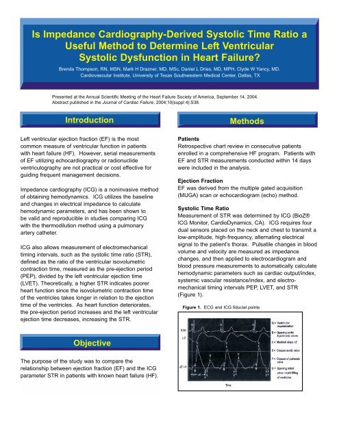
<strong>Systolic</strong> <strong>Time</strong> <strong>Ratio</strong><br />
Measurement of STR was determined by ICG (BioZ®<br />
ICG Monitor, CardioDynamics, CA). ICG requires four<br />
dual sensors placed on the neck and chest to transmit a<br />
low-amplitude, high-frequency, alternating electrical<br />
signal to the patient’s thorax. Pulsatile changes in blood<br />
volume and velocity are measured as impedance<br />
changes, and then applied to electrocardiogram and<br />
blood pressure measurements to automatically calculate<br />
hemodynamic parameters such as cardiac output/index,<br />
systemic vascular resistance/index, and electromechanical<br />
timing intervals PEP, LVET, and STR<br />
(Figure 1).<br />
Figure 1. ECG and ICG fiducial points
Statistical Methods<br />
Paired values of EF and STR were compared.<br />
Correlation was calculated using Pearson’s method.<br />
To evaluate STR as a diagnostic test for EF, a cut-off<br />
value of 0.50 was used. Values of EF and STR were<br />
compared to calculate sensitivity, specificity, and positive<br />
and negative predictive value of an STR > 0.50 to an EF<br />
< 50% or an STR < 0.50 to an EF > 50%.<br />
Results<br />
A total of 52 patients were evaluated. Baseline<br />
characteristics are shown in Table 1. MUGA EF was<br />
obtained on 23/52 (44.2%) and echo EF on 29/52<br />
(55.8%). Mean EF was 37.6 ± 20.2%, range<br />
10 to 80%. A total of 39/52 (75%) had EF < 50%.<br />
The mean time between EF and STR measurements<br />
was 3.54 ± 4.67 days.<br />
The overall correlation between EF and STR was 0.55<br />
(p < 0.001). For identifying an EF < 50%, STR > 0.50<br />
demonstrated a sensitivity of 92%, specificity of 85%,<br />
and positive and negative predictive values of 95% and<br />
79%, respectively. Overall accuracy was 90.4%. Of the<br />
five patients in which STR did not agree with the EF<br />
category, two were from MUGA EF and three were from<br />
echo EF.<br />
Table 1. Patient Characteristics<br />
N=52<br />
VARIABLE value (%)<br />
Gender<br />
Male 34 (65.4)<br />
Female 18 (34.6)<br />
Race<br />
White 34 (65.4)<br />
Black 16 (30.8)<br />
Hispanic 2 (3.8)<br />
HF etiology<br />
<strong>Is</strong>chemic 13 (25)<br />
Viral 6 (12)<br />
Pulmonary hypertension 7(13)<br />
Dilated cardiomyopathy 14 (27)<br />
Diastolic dysfunction 3 (6)<br />
Idiopathic 14 (27)<br />
NYHA class<br />
Class I 2 (3.8)<br />
Class II 17 (32.7)<br />
Class III 2 (3.8)<br />
Class IV 31 (59.6)<br />
Table 2. Results<br />
SYSTOLIC TIME RATIO (STR) VS.<br />
LEFT VENTRICULAR EJECTION FRACTION (EF)<br />
STR > 0.50 STR < 0.50<br />
EF < 0.50 36 3<br />
EF > 0.50 2 11<br />
Discussion<br />
In this retrospective analysis, STR demonstrated a<br />
strong relationship with EF and was able to reasonably<br />
distinguish EF above 50% from EF 50% or below.<br />
The management of HF necessitates the frequent<br />
assessment of a patient’s changing status.<br />
Measurement of EF is considered an important<br />
diagnostic tool to quantify left ventricular function.<br />
However, serial measurements of EF are not<br />
considered to be cost effective to guide more frequent<br />
evaluation of disease progression or improvement<br />
based on treatments that target neurohormonal or<br />
hemodynamic dysfunction.<br />
In contrast, the measurement of STR using ICG may<br />
offer such promise, as it is inexpensive and relatively<br />
simple to perform, taking only a few minutes in the<br />
outpatient or hospital setting. Decreases in STR could<br />
identify responses to therapy and increases in STR<br />
could signal potential decompensation.<br />
Limitations<br />
Because this study was retrospective in design, a<br />
prospective validation is suggested. EF and STR were<br />
not determined simultaneously, allowing for the<br />
possibility that changes in EF or STR may have<br />
occurred during the time between the two measurements.<br />
Conclusions<br />
The STR parameter has the potential to be a costeffective<br />
and reliable method of determining the<br />
presence of left ventricular dysfunction in chronic HF,<br />
and may aid in decision making of evidence-based HF<br />
treatment strategies.<br />
M453 Rev. B