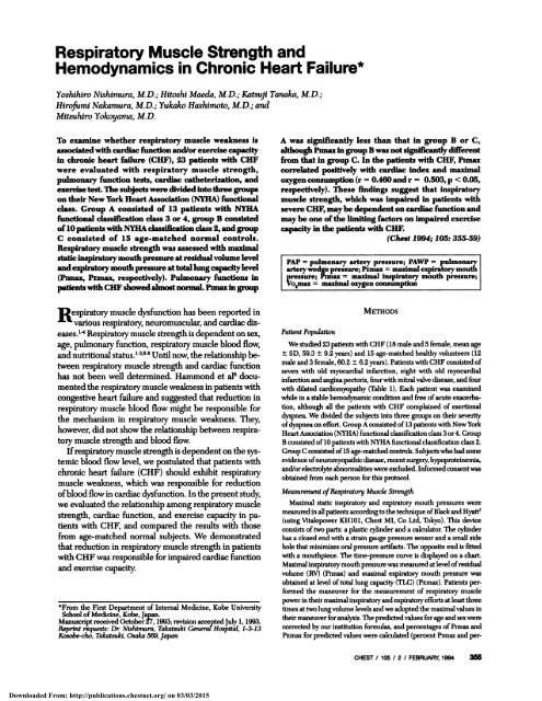
Respiratory Muscle Strength and
Respiratory Muscle Strength and
Respiratory Muscle Strength and

Create successful ePaper yourself
Turn your PDF publications into a flip-book with our unique Google optimized e-Paper software.
<strong>Respiratory</strong> <strong>Muscle</strong> <strong>Strength</strong> <strong>and</strong><br />
Hemodynamics in Chronic Heart Failure*<br />
Yoshihiro Nishimura, M.D.; Hitoshi Maeda, M.D.; Katsuji Tanaka, M.D.;<br />
Hirofumi Nakamura, M.D.; Yukako Hashimoto, M.D.; <strong>and</strong><br />
Mitsuhiro Yokoyama, M.D.<br />
To examine whether respiratory muscle weakness is<br />
associated with cardiac function <strong>and</strong>lor exercise capacity<br />
in chronic heart failure (CHF), 23 patients with CHF<br />
were evaluated with respiratory muscle strength,<br />
pulmonary function tests, cardiac catheterization, <strong>and</strong><br />
exercise test The subjects were divided lntothree groups<br />
on their New York Heart Association (NYHA) functional<br />
class. Group A consisted of 13 patients with NYHA<br />
functional classification class 3 or 4, group B consisted<br />
of 10 patients with NYllAclassification class 2, <strong>and</strong> group<br />
C consisted of 15 age-matched normal controls.<br />
<strong>Respiratory</strong> muscle strength was assessed with maximal<br />
static inspfratory mouth pressure at residual volume level<br />
<strong>and</strong>explratory mouthpressure at total lung capacity level<br />
(Pimax, PEmax, respectively). Pulmonary functions In<br />
entswith CHF showed almostnormaL Pimaxm group<br />
A was significantly less than that In group B or C,<br />
although Punaxin group B was not Significantly different<br />
from that in group C. In the patients with CHF, Pimax<br />
correlated positively with cardiac Index <strong>and</strong> maximal<br />
oxygen consumption (r = 0.460 <strong>and</strong> r = 0.503, p < 0.05,<br />
respectively). These findings suggest that lnspiratory<br />
muscle strength, which was Impaired In patients with<br />
severe CHF, may be dependent on cardiac function <strong>and</strong><br />
may be one ofthe limiting factors on Impaired exercise<br />
capacity In the patients with CUE<br />
(Chest 1994; 105: 355-59)<br />
PAP = pulmonary artery pressure; PAWP pulmona<br />
arterywedge pressure; Pasnax maximal expiratory mou<br />
pressure; Pimax meximal inspiratory mouth pressure;<br />
So5maz = mpima1 oxygen consumption<br />
R espiratory muscle dysfunction has been reported in<br />
various respiratory neuromuscular, <strong>and</strong> cardiac diseases.’4<br />
<strong>Respiratory</strong> muscle strength is dependent on sex,<br />
age, pulmonary function, respiratory muscle blood flow,<br />
<strong>and</strong> nutritional status.” Until now, the relationship between<br />
respiratory muscle strength <strong>and</strong> cardiac function<br />
has not been well determined. Hammond et al6 documented<br />
the respiratory muscle weakness in patients with<br />
congestive heart failure <strong>and</strong> suggested that reduction in<br />
respiratory muscle blood flow might be responsible for<br />
the mechanism in respiratory muscle weakness. They,<br />
however, did not show the relationship between respiratory<br />
muscle strength <strong>and</strong> blood flow.<br />
Ifrespiratory muscle strength is dependent on the systemic<br />
blood flow level, we postulated that patients with<br />
chronic heart failure (CHF) should exhibit respiratory<br />
muscle weakness, which was responsible for reduction<br />
ofblood flow in cardiac dysfunction. In the present study,<br />
we evaluated the relationship among respiratory muscle<br />
strength, cardiac function, <strong>and</strong> exercise capacity in patients<br />
with CHF, <strong>and</strong> compared the results with those<br />
from age-matched normal subjects. We demonstrated<br />
that reduction in respiratory muscle strength in patients<br />
with CHF was responsible for impaired cardiac function<br />
<strong>and</strong> exercise capacity.<br />
#{176}Fromthe First Department of Internal Medicine, Kobe University<br />
School ofMedicine, Kobe, Japan.<br />
Manuscript received October 27, 1993; revision acceptedJuly 1, 1993.<br />
Reprint requests: Dr Nishimura. Takatsuki GenerefHospital, 1-3-13<br />
Kosobe-cho, Takatsuki, Osaka 569, Japan<br />
Patient<br />
Population<br />
METHODS<br />
We studied 23patients with CHF (18 male <strong>and</strong> 5 female, mean age<br />
± SD, 59.3 ± 9.2 years) <strong>and</strong> 15 age-matched healthy volunteers (12<br />
male <strong>and</strong> 3 female, 60.2 ± 6.2 years). Patients with CHF consisted of<br />
seven with old myocardial infarction, eight with old myocardial<br />
infarction <strong>and</strong> anginapectoris, fourwith mitral valve disease, <strong>and</strong> four<br />
with dilated cardiomyopathy (Table 1). Each patient was examined<br />
while in a stable hemodynamic condition <strong>and</strong> free ofacute exacerbation,<br />
although all the patients with CHF complained of exertional<br />
dyspnea. We divided the subjects into three groups on their severity<br />
ofdyspneaon effort. Group A consisted ofl3 patients with New York<br />
Heart Association (NYHA) functional classffication class 3or4. Group<br />
B consisted oflOpatients with NYHA functional classification class 2.<br />
GroupC consistedofl5 age-matthedcontrob. Subjects whohad some<br />
evidence ofneuromyopathic disease, recent surgery, hypoproteinemia,<br />
<strong>and</strong>/or electrolyte abnormalities were excluded. Informed consent was<br />
obtained from each person for this protocol.<br />
Measurement of<strong>Respiratory</strong> <strong>Muscle</strong> <strong>Strength</strong><br />
Maximal static inspiratosy <strong>and</strong> expiratory mouth pressures were<br />
measuredin all patients accordingtothe technique ofBlack <strong>and</strong> Hyatt7<br />
(using Vitalopower KH1O1, Chest MI, Co Ltd. Tokyo). This device<br />
consists oftwo parts: a plastic cylinder <strong>and</strong> a calculator. The cylinder<br />
has a closed end with a strain gauge pressure sensor <strong>and</strong> a small side<br />
hole that minimizes oral pressure artifacts. The opposite end is fitted<br />
with a mouthpiece. The time-pressure curve is displayed on a chart.<br />
Maximal inspiratosymouth pressurewas measured at level of residual<br />
volume (RV) (Pimax) <strong>and</strong> maximal expiratory mouth pressure was<br />
obtained at level oftotal lung capacity (TLC) (Psmax). Patients performed<br />
the maneuver for the measurement of respiratory muscle<br />
powerin theirmaximal inspiratosy<strong>and</strong> expiratoryefforts atleast three<br />
times at two lung volume levels <strong>and</strong> we adopted the maximal values in<br />
their maneuver for analysis. The predicted values for age <strong>and</strong> sex were<br />
corrected by our institution formulas, <strong>and</strong> percentages of Pimax <strong>and</strong><br />
for predicted values were calculated (percent Pimax <strong>and</strong> per-<br />
CHEST I 105 I 2 I FEBRUAR 1994 355<br />
Downloaded From: http://publications.chestnet.org/ on 03/03/2015
No.<br />
Age, yr/<br />
Sex<br />
Table 1-Characteristics in fldients With<br />
Chronic Heart Thilure#{176}<br />
Height,<br />
cm<br />
V.ight,<br />
kg<br />
Clinical<br />
Diagnosis<br />
Myocardial<br />
Ischemia<br />
During<br />
Exercise<br />
GroupA .<br />
NYHA<br />
1167/F 144 46 MVD - 4<br />
2/69/M 155 45 MVD - 4<br />
3/66/M 162 68 OMI - 3<br />
4/48/F 158 55 DCM - 3<br />
5rwM 157 51 DCM - 3<br />
6/63/M 170 76 DCM - 3<br />
7/4WF 146 42 MVD - 3<br />
8/68/M 165 60 OMI,AP + 3<br />
93/M 162 63 OMI,AP + 3<br />
12/M 160 56 OMI, AP + 3<br />
1V6’F 158 67 OMI,AP + 3<br />
12/TIJM 165 63 OMI, #{192}Y + 3<br />
13/6WM 165 60 OMI,AP + 3<br />
Group B<br />
14162/M 158 58 OMI - 2<br />
15164/M 162 54 OMI - 2<br />
1&7/M 169 70 OMI - 2<br />
17/45/M 160 54 OMI - 2<br />
18/63’M 161 55 OMI - 2<br />
19/4S/M 165 63 OMI - 2<br />
20158/M 173 62 DCM - 2<br />
21151fF 153 42 MVD - 2<br />
2214WM 167 71 OMI,AP + 2<br />
23/6WM 152 62 OMI,AP + 2<br />
NYHA= New York Heart Association functional classification;<br />
M = male; F = female; OMI old myocardial infarction;<br />
AP= angina pectons; MVD = mitral valve disease; DCM = dilated<br />
cardiomyopathy.<br />
cent PEmax, respectively). The formulas established for Japanese<br />
healthy persons are as follows.5<br />
Male: PEmax 149 - 0.59 X AGE cm H2O(r = - 0.325,<br />
p , 0.05)-.(1)<br />
Pimax = 131 - 0.76 x AGE cm H2O(r = - 0.476,<br />
p < 0.01)-(2)<br />
Female: PEmax 93 - 0.33 X AGE cm H2O(r = - 0.277,<br />
p < 0.05)-(3)<br />
Pimax 102 - 0.69 X AGE cm H2O(r -0.438,<br />
p < 0.01)-(4)<br />
Pulmonanj Function Test<br />
Vital capacity (VC), forced vital capacity (FVC), forced expiratory<br />
volume in 1 S (FEy1), <strong>and</strong> TIC were measured using the spirometer<br />
ofcomputer processing (Autospirometer System-55, Minato Medical<br />
Science Co Ltd. Osaka, Japan) <strong>and</strong> the ratio of FEy1 for forced vital<br />
capacity (FEV1/FVC) was calculated.<br />
Cardiac Catheterization <strong>and</strong> Exercise Studies<br />
Mean pulmonary artery pressure (PAP) <strong>and</strong> mean pulmonary artery<br />
wedge pressure (PAWP) were measured with a Swan-Ganz catheter<br />
that was advanced into the right pulmonary artery. The direct<br />
Fick method was employed for measurement of the cardiac output<br />
from which the cardiac index (CI) was calculated.<br />
Exercise capacity was studied on an electrically braked bicycle ergometer<br />
in a supine position. An incremental exercise was performed<br />
to a symptom-limited maximum. The work load was increased by 25<br />
W every 3 mm. The expired gas was analyzed for fractions of oxygen<br />
<strong>and</strong> carbon dioxide by RM-300 (Minato Medical Science, Co. Ltd.,<br />
Osaka, Japan), <strong>and</strong> maximal oxygen consumption (Vo2max) was calculated.<br />
Statistical<br />
Analysis<br />
All values are given as mean ± SD. Statistical differences for vanables<br />
of respiratory muscle strength <strong>and</strong> pulmonary function were<br />
anal by analysis ofvaniance (ANOVA). Differences for hemodynamic<br />
data between two CHF groups were evaluated by Students t<br />
test. Relationships between respiratory muscle functions <strong>and</strong> hemodyn<br />
amic parameters were assessed by linear regression analysis. Values<br />
ofp < 0.05 were considered to be statistically significant.<br />
RESULTS<br />
The anthropometric data are shown in Table 2. Ages<br />
were not significantly different among group A, group<br />
B, <strong>and</strong> controls. Height <strong>and</strong> body weight did not differ<br />
significantly among three groups. Plasma albumin level<br />
in group A was not significantlylower than that in group<br />
B (3.7 ± 0.2 <strong>and</strong> 3.9 ± 0.3 g/dl, respectively). Six of 13<br />
patients with group A <strong>and</strong> 2 of 10 patients with group B<br />
showed myocardial ischemic response during exercise<br />
test.<br />
Mean values ofpulmonary function variables showed<br />
within normal limits in both group A <strong>and</strong> B. There were<br />
no significant differences in pulmonary function variables<br />
among three groups. In cardiac catheterization, PAP <strong>and</strong><br />
PAWP of group A were significantly higher than those<br />
ofgroup B. The CI <strong>and</strong> Vo2max ofgroup A were significantly<br />
lower than those ofgroup B.<br />
Figure 1 shows respiratory muscle strength in the patients<br />
with CHF <strong>and</strong> normal subjects. The mean value<br />
of Pimax in group A was 60 ± 15 cm H20, which was<br />
significantly smaller than the values in group B <strong>and</strong> group<br />
C (92 ± 32, 84 ± 24 cm H20, p < 0.05, respectively).<br />
Percent Pimax in group A was also significantly smaller<br />
than those in the other two groups. PEmaX <strong>and</strong> percent<br />
PEmax were not significantly different among three<br />
groups.<br />
There was a significant positive correlation between<br />
percent Pimax <strong>and</strong> CI in patients with CHF (r 0.460,<br />
p < 0.05) (Fig 2). Percent Plmax had a significant correlation<br />
with Vo2max (r 0.503, p < 0.05) in 15 patients<br />
with CHF without myocardial ischemic response during<br />
exercise test (Fig 3).<br />
DISCUSSION<br />
We have demonstrated that respiratory muscle<br />
strength exhibited reduction in patients with severe CHF<br />
<strong>and</strong> that the impairment in inspiratory muscle strength<br />
was intimately related to the deterioration in cardiac<br />
function <strong>and</strong> to the impaired exercise capacity. This may<br />
support our hypothesis that, if respiratory muscle<br />
strength is dependent on the cardiac dysfunction, the<br />
patients with CHF should exhibit respiratory muscle<br />
weakness related to cardiac output.<br />
Our present study showed that patients with severe<br />
CHF (group A) exhibited inspiratory muscle weakness<br />
compared with patients with mild heart failure (group<br />
356 <strong>Respiratory</strong> <strong>Muscle</strong> <strong>Strength</strong> In Chronic Heart Failure (Niahimura et aQ<br />
Downloaded From: http://publications.chestnet.org/ on 03/03/2015
Table 2-Anthropometric Data, IWmonary Function Thst Hemodynamics <strong>and</strong> <strong>Respiratory</strong> <strong>Muscle</strong> <strong>Strength</strong> in }btients<br />
With CUP <strong>and</strong> Age-Matched Normal Subjects<br />
CHF<br />
Age-Matched<br />
-<br />
Control<br />
Data Group A Group B Group C<br />
Anthropometnic data<br />
n 13 10 15<br />
Sex, maleffemale 9/4 9/1 12/3<br />
Age,yr 62.2±8.1 55.4±10.1 60.2±6.2<br />
Height,cm 159.0±7.1 162.0±6.4 160.2±8.7<br />
ight, kg 57.8±9.6 59.1 ±8.1 62.1 ± 10.4<br />
Pulmonary function test<br />
VC, L 3.03±0.68 3.38±0.57 3.39±0.73<br />
%pred 99.6±12.7 99.5±12.8 108.2±16.2<br />
FEY1, L 2.28±0.49 2.74±0.58 2.57±0.61<br />
FEVI/FVC, % 77.8±9.0 80.1±7.5 78.6±5.0<br />
TLC, % pred 92.3± 11.5 82.3± 14.2 96.1±26.5<br />
Hemodynamics<br />
PAI mm Hg 19.9±5.lt 12.8±2.6<br />
PAWP, mm Hg 12.2±4.9t 6.6± 2.2<br />
CI,LImin/m’ 2.49±O.73t 3.10±0.72<br />
Vo2max, mi/mm/kg 11.9±2.8t 18.6±3.2<br />
<strong>Respiratory</strong> muscle strength<br />
Punax, cm HO 60± 15fl 92±32 84±24<br />
%_ 80±2ltt 107±34 106±24<br />
Pnmax, cm HO 84± 18 104±34 106±34<br />
%_ 85±25 90±24 102±29<br />
0VC = vital capacity; FEV1 = forced expiratory volume in 1 5; PVC = forced vital capacity; PAP = mean pulmonary artery pressure;<br />
PAWP = mean pulmonary artery wedge pressure; CI = cardiac index; Vo2max = maximal oxygen consumption; Pimax = maximal inspiratory<br />
mouth pressure; % Pimax = percent of maximal inspiratory mouth pressure for predicted value5; Psmax = maximal expiratory mouth<br />
pressure; % PEmax = percent of maximal expiratory mouth pressure for predicted value.5 Values are mean ± SD.<br />
tp
%Plmax (%)<br />
200.<br />
150<br />
100.<br />
50.<br />
A-<br />
VO2max/BW<br />
(mI/mm/kg)<br />
8<br />
6<br />
4<br />
2<br />
0<br />
. #{149}11 #{149}. #{149}<br />
.<br />
1 3 5<br />
.<br />
Cl U2)<br />
FIGURE 2. Relationship between percent Pimax<strong>and</strong> CI in patients with<br />
CHF. The real line is the regression line. Percent Plmax is significantly<br />
correlated with CI (r = 0.460, p < 0.05).<br />
The second is generalized skeletal muscle weakness.<br />
Lecarpentier et al reported that reduction in respiratory<br />
muscle function in a rabbit model with CHF was<br />
associated with cardiac cachexia, suggesting that respiratory<br />
muscle weakness was related to the generalized<br />
muscle weakness. Although our anthropometric data<br />
showed no cachexic condition among three groups, generalized<br />
muscle weakness resulting from cardiac dysfunction<br />
plays an important role in part for inspiratory muscle<br />
weakness in patients with CHF.<br />
Pulmonary function abnormalities <strong>and</strong> nutritional status<br />
influence respiratory muscle function.5 Decreased<br />
TLC should cause reduction in expiratory muscle<br />
strength, <strong>and</strong> augmentation in RV should induce deterioration<br />
in inspiratory muscle strength. Furthermore,<br />
.<br />
#{149}#{149}<br />
0 50 100 150<br />
.<br />
%PI max (%)<br />
.<br />
malnutrition might be responsible for respiratory muscle<br />
weakness. There was, however, no significant difference<br />
of variables of pulmonary function tests among three<br />
groups, <strong>and</strong> no subject was included in this study who<br />
exhibited abnormalities of pulmonary function <strong>and</strong> nutritional<br />
status.<br />
In the present study, Vo2 max significantly correlated<br />
with Pimax. Sera’#{176} showed that patients with severe CHF<br />
exhibited much more increase in airway resistance <strong>and</strong><br />
decrease in dynamic pulmonary compliance during exercise<br />
than do patients with mild heart failure, suggesting<br />
that the respiratory muscle in patients with severe<br />
CHF might be loaded much more than in patients with<br />
mild CHF. In skeletal muscles, on the other h<strong>and</strong>, the<br />
perception of heaviness of a lifted object or of achieved<br />
force in an isometric contraction depends on sensing the<br />
grade of effort, or voluntarily generated input to monotones,<br />
rather than an afferent sensation signaling the<br />
muscular force achieved.” The sense ofeffort increased<br />
in relation to the augmentation of inspiratory loading<br />
pressure <strong>and</strong> the intensity of sense of effort in fatigued<br />
muscle was stronger than that in fresh muscle.’ It is suggested<br />
that respiratory muscle in patients with severe<br />
CHF plays a more important role in exercise capacity<br />
than it does in patients with mild CHF, <strong>and</strong> that inspiratory<br />
muscle function may be one of the limiting factors<br />
ofexercise capacity in patients with CHF, while myocardial<br />
ischemia should play an important role in exercise<br />
capacity in the ischemic patients.<br />
We used maximal inspiratoiy <strong>and</strong> expiratory mouth<br />
pressures to estimate inspiratory <strong>and</strong> expiratoiy muscle<br />
strength, because measurements ofmaximal mouth pressure<br />
could be performedwith an easy maneuver <strong>and</strong> with<br />
good reproducibility.5 As two components, the pressure<br />
generated by respiratory muscles <strong>and</strong> the recoil pressure<br />
ofthe respiratory system, were intimately associated with<br />
maximal mouth pressure, the pure mouth pressure generated<br />
with the respiratory muscles should be obtained<br />
aftercorrection forthe factor induced by recoil pressure.’3<br />
In this study, actual lung volume was not simultaneously<br />
measured while the maximal mouth pressure was measured.<br />
Therefore, both Pimax <strong>and</strong> PEmaX might be overestimated.<br />
We concluded that inspiratory muscle strength, which<br />
may be responsible for cardiac output, is significantly<br />
correlated with Vo2max in the patients with CHF without<br />
myocardial ischemia. This may suggest that inspiratory<br />
muscle may be dependent on cardiac function <strong>and</strong><br />
200 may be one of the limiting factors in impaired exercise<br />
capacity in the patients with CHF.<br />
FIGURE 3. Relationship between percent Pimax <strong>and</strong> Vo5max in the<br />
patients with CHF without myocardial ischemia during exercise. The<br />
real line is the regression line. Vo2max is significantly correlated with<br />
percent Plmax (r 0.503, p < 0.05).<br />
ACKNOWLEDGMENT: The authors thank H. Yamabe, M.D., <strong>and</strong><br />
A. Hashimoto, M.D., for invaluable help in exercise data collection.<br />
358 <strong>Respiratory</strong> <strong>Muscle</strong> <strong>Strength</strong> in Chroi* Heart Failure (Nishimura at al) 2<br />
Downloaded From: http://publications.chestnet.org/ on 03/03/2015
REFERENCES<br />
1 Nishimura Y, Hida W, Taguchi 0, Sakurai M, Ichinose M, Inoue<br />
H, et al. <strong>Respiratory</strong> muscle strength <strong>and</strong> gas exchange in<br />
neuromuscular disease: comparison with chronic pulmonary<br />
emphysema <strong>and</strong> idiopathic pulmonary fibrosis. Tohokuj Exp Med<br />
1989; 159:57-68<br />
2 Rochester DF, Arora NS, Braun NMT, Goldberg SK. The<br />
respiratory muscles in chronic obstructive pulmonary disease<br />
(COPD). Bull Eur Physiopathol Respir 1979; 15:951-75<br />
3 Arora NS, Rochester DF. Respiratoiy muscle strength <strong>and</strong> maximal<br />
ventilation in undernourished patients. Am Rev Respir Dis 1982;<br />
126:5-8<br />
4 Roussos C, Macklem PT. Diaphragmatic fatigue in man. J AppI<br />
Physiol 1977; 43:189-97<br />
5 Nishimura Y, Maeda H, Tanaka K, Hashimoto A, Hashimoto Y,<br />
Yokoyama M, et al. The effect of aging on respiratory muscle<br />
function. Jpn J Chest Dis 1991; 29:795-801 [English abstract]<br />
6 Hammond MD, Bauer KA, Sharp JT, Rocha RD. <strong>Respiratory</strong><br />
muscle strength in congestive heart failure. Chest 1990; 98:1091-<br />
94<br />
7 Black LF, Hyatt RE. Maximal respiratory pressures: normal values<br />
<strong>and</strong> relationship to age <strong>and</strong> sex. Am Rev Respir Dis 1969; 99:696-<br />
702<br />
8 Aubier M, Trippenbach T, Roussos C. <strong>Respiratory</strong> muscle fatigue<br />
during cardiogenic shock. J Appi Physiol Respir Environ Exercise<br />
Physiol 1981; 51:499-508<br />
9 Lecarpentier Y, Herve Ph, Villeneuve A, Lacroix P. Duroux P.<br />
Alterations of in vitro diaphragm function during chronic heart<br />
failure in rabbit. Am Rev Respir Dis 1989; 139:A162<br />
10 Sera K. Ventilatory mechanics in patients with cardiopulmonary<br />
disease: II. On congestive heart failure. Jpn CircJ 1961; 25:320-30<br />
11 G<strong>and</strong>evia SC, McCloskey DL. Changes in motor comm<strong>and</strong>s, as<br />
shown by changes in perceived heaviness, during curanzation <strong>and</strong><br />
peripheral anesthesia in man. J Physiol 1977; 272:673-89<br />
12 Supinski GS, Clary SJ, Bark H, Kelsen SG. Effect of inspiratory<br />
muscle fatigue on perception of effort during loaded breathing. J<br />
Appl Physiol 1987; 62:300-07<br />
13 Agostoni E, Mead J. Statics of the respiratory system. In: Fenn<br />
WO, Rahn H, ecis. H<strong>and</strong>book ofphysiology, section 3, respiration,<br />
vol 1. Washington, DC: American Physiological Society, 1964; 387-<br />
409<br />
First International Winter Meeting on Coagulation<br />
This program, to cover basic, laboratory <strong>and</strong> clinical aspects of venous thromboembolism, will<br />
be held March 9-12 at the Hotel Savoia, Cortina D’Ampezzo, Italy. Sponsors are the Centro<br />
Europeo per Ia Formazione, l’Aggiornamento e Ia Ricerca in Scienze Sanitane e in<br />
Biotecnologie, Istituto Scientifoci HS Raffaele, Milan; <strong>and</strong> IRCCS Policlinico San Matteo, Pavia.<br />
For information, contact G. Agosta, Biomedia sri, Via C Farini 70 20159 Milan, Italy (fax:<br />
02/69901311).<br />
CHEST I 105 I 2 I FEBRUARY, 1994 359<br />
Downloaded From: http://publications.chestnet.org/ on 03/03/2015