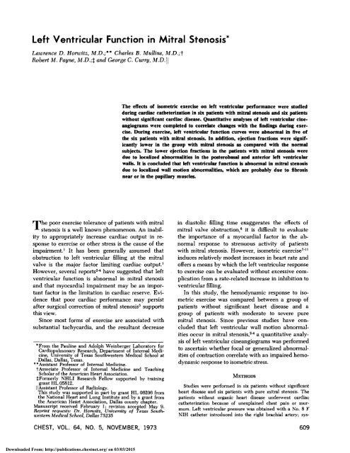
Left Ventricular Function in Mitral Stenosis*
Left Ventricular Function in Mitral Stenosis*
Left Ventricular Function in Mitral Stenosis*

You also want an ePaper? Increase the reach of your titles
YUMPU automatically turns print PDFs into web optimized ePapers that Google loves.
<strong>Left</strong> <strong>Ventricular</strong> <strong>Function</strong> <strong>in</strong> <strong>Mitral</strong> <strong>Stenosis*</strong>Lau:rence D. Horwitz, M.D.;"' Charles B. Mull<strong>in</strong>s, M.D.;?Robert M. Payne, M.D.;$ and George C. Curry, AI.D.1 IThe effects of isometric exercise on left ventricular performance were studieddur<strong>in</strong>g cardiac catheterization <strong>in</strong> six patients with mitral stenosis and six patientswithout significant cardiac disease. Quantitative analyses of left ventricular c<strong>in</strong>eangiogramswere completed to correlate changes with the f<strong>in</strong>d<strong>in</strong>gs dur<strong>in</strong>g exercise.Dur<strong>in</strong>g exercise, left ventricular function curves were abnormal <strong>in</strong> five ofthe six patients with mitral stenosis. In addition, ejection fractions were sipificantlylower <strong>in</strong> the group with mitral stenosis as compared with the normalsubjects. The lower ejection fractions <strong>in</strong> the patients with mitral stenosis weredue to localized abnormalities <strong>in</strong> the posterobasal and anterior left ventricularwalk. It is concluded that left ventricular function is abnormal <strong>in</strong> mitral stenosisdue to localized wall motion abnormalities, which are probably due to fibrosisnear or <strong>in</strong> the papillary muscles.he poor exercise tolerance of patients with mitralTstenosis is a well known phenomenon. An <strong>in</strong>abilityto appropriately <strong>in</strong>crease cardiac output <strong>in</strong> responseto exercise or other stress is the cause of theimpairment.' It has been generally assumed thatobstruction to left ventricular fill<strong>in</strong>g at the mitralvalve is the major factor limit<strong>in</strong>g cardiac output.'However, several reportsz4 have suggested that leftventricular function is abnormal <strong>in</strong> mitral stenosisand that myocardial impairment may be an importantfactor <strong>in</strong> the limitation <strong>in</strong> cardiac reserve. Evidencethat poor cardiac performance may persistafter surgical correction of mitral stenosis5 supportsthis view.S<strong>in</strong>ce most forms of exercise are associated withsubstantial tachycardia, and the resultant decrease'From the Paul<strong>in</strong>e and Adolph We<strong>in</strong>berger Laboratory forCardiopulmonary Research, Department of Internal Medic<strong>in</strong>e,University of Texas southwestern Medical School atDallas, Dallas, Texas."Assistant Professor of Internal Medic<strong>in</strong>e.+Associate Professor of Internal Medic<strong>in</strong>e and Teach<strong>in</strong>gScholar of the American Heart Association.$Formerly NHLI Research Fellow supported by tra<strong>in</strong><strong>in</strong>ggrant HL 05812.I IAssistant Professor of Radiology.This study was supported <strong>in</strong> part by grant HL 06296 fromthe National Heart and Lung Institute and by a rant fromthe American Heart Association, Dallas county ctapter.Manuscript received February 1; revision accepted May 9.Repr<strong>in</strong>t reqrrests: Dr. Howitz, University of Texas SotrthwesternMedical School, Dallas 75235<strong>in</strong> diastolic fill<strong>in</strong>g time exaggerates the effects ofmitral valve obstr~ction,~ it is difficult to evaluatethe importance of a myocardial factor <strong>in</strong> the abnormalresponse to strenuous activity of patientswith mitral stenosis. However, isometric exercise7-"<strong>in</strong>duces relatively modest <strong>in</strong>creases <strong>in</strong> heart rate andoffers a means by which the left ventricular responseto exercise can be evaluated without excessive complicationfrom a rate-related <strong>in</strong>crease <strong>in</strong> <strong>in</strong>hibition toventricular fill<strong>in</strong>g.In this study, the hemodynamic response to isometricexercise was compared between a group ofpatients without significant heart disease and agroup of patients with moderate to severe puremitral stenosis. S<strong>in</strong>ce previous studies have concludedthat left ventricular wall motion abnormalitiesoccur <strong>in</strong> mitral stenosi~,~~ a quantitative analysisof left ventricular c<strong>in</strong>eangiograms was performedto ascerta<strong>in</strong> whether focal or generalized abnormalitiesof contraction correlate with an impaired hemodynamicresponse to isometric stress.Studies were performed <strong>in</strong> six patients without significantheart disease and six patients with pure mitral stenosis. Thepatients without organic heart disease undenvent cardiaccatheterization because of unexpla<strong>in</strong>ed chest pa<strong>in</strong> or murmurs.<strong>Left</strong> ventricular pressure wa.5 obta<strong>in</strong>ed with a No. 8 FNIH catheter <strong>in</strong>troduced <strong>in</strong>to the right brachial artery; sys-CHEST, VOL. 64, NO. 5, NOVEMBER, 1973Downloaded From: http://publications.chestnet.org/ on 03/03/2015
FIGURE 1. Schematic illustration of method for quantitativeanalysis of left ventricular c<strong>in</strong>eangiograms. Length (L) andhemiaxes (H,-,) were measured at end-diastole (solid outerl<strong>in</strong>e) and end-systole (broken <strong>in</strong>ner l<strong>in</strong>e). H,H, denoteanterior hemiaxes, with H,-H, denot<strong>in</strong>g posterior.temic arterial pressure was monitored with a 6-<strong>in</strong> length of 5F polyv<strong>in</strong>yl tub<strong>in</strong>g <strong>in</strong>troduced percutaneously <strong>in</strong>to the leftbrachial artery. Cardiac output was measured by the <strong>in</strong>dicator-dilutionmethod us<strong>in</strong>g <strong>in</strong>docyan<strong>in</strong>e green dye <strong>in</strong>jected <strong>in</strong>tothe pulmonary artery and sampled from the left brachialartery.Isometric exercise was performed at the beg<strong>in</strong>n<strong>in</strong>g of thecardiac catheterization. Each patient's maximal voluntaryhandgrip contraction (MVC) was determ<strong>in</strong>ed with a handgripdynamometer (CH Stoelt<strong>in</strong>g Co.) held <strong>in</strong> the left hand.Control measurements of left ventricular and brachial artervpressures, heart rate, and cardiac output were obta<strong>in</strong>ed withthe patient at rest. A handgrip at 25 percent of MVC wasthen held for 3 4 m<strong>in</strong> and the measurements were repeated.After a 5-m<strong>in</strong> recovery period, the measurements were repeatedat 50 percent of MVC held for 1 m<strong>in</strong>. Care was takento assure that the patients breathed normally and did notperform Valsalva's maneuver dur<strong>in</strong>g the isometric exercise.<strong>Ventricular</strong> function curves were constructed by plott<strong>in</strong>gstroke work <strong>in</strong>dex as a function of left ventricular enddiastolicpressure at rest and at each level of handgripexercise. Stroke work <strong>in</strong>dex was calculated from:where:LVSWI = SV X ( LVS-LVEDP) X 1.36100 x BSATable 1Rest<strong>in</strong>g ~em&narnics <strong>in</strong>AgePAP S/DPatient, No. (Yrs) Sex (mm Hg)LVSWI = left ventricular stroke work <strong>in</strong>dex (g-m/M2);SV = stroke volume ( ml/beat );LVS = left ventricular mean ejection pressure (mm Hg);LVEDP = left ventricular end-diastolic pressure (mm Hg);BSA = body surface area ( M2).Approximately 15 m<strong>in</strong> after the isometric exercise, leftventricular c<strong>in</strong>eangiograms were performed <strong>in</strong> the 30° rightanterior oblique position us<strong>in</strong>g a fram<strong>in</strong>g sequence of 60frames per second. A power <strong>in</strong>jection of 40-50 ml of meglum<strong>in</strong>ediatrizoate and sodium diatrizoate (Renograf<strong>in</strong> 76 percent) was made over a 2-3 # sec period.The outl<strong>in</strong>e of the left ventricle at end-systole and enddiastolewas traced from <strong>in</strong>dividual 35-mm c<strong>in</strong>e frames. Itwas assumed that with<strong>in</strong> each <strong>in</strong>dividual cardiac cycle theframe with the largest left ventricular volume was enddiastoleand the frame with the lowest volume was endsystole.Premature beats and postextrasystolic beats wereexcluded. The magnification factor was obta<strong>in</strong>ed by film<strong>in</strong>g across-hatched grid placed 10 crn above the table top.End-systolic and end-diastolic left ventricular volumeswere determ<strong>in</strong>ed by the area-length method of Dodge et all2as modified by Kasser and Kennedy." Ejection fractionswere calculated by divid<strong>in</strong>g the difference between the enddiastolicand end-systolic volumes by the end-diastolic volume.<strong>Ventricular</strong> wall motion was analyzed by construct<strong>in</strong>gthree perpendiculars to the long axes at quarter length<strong>in</strong>tervals (Fig 1). The perpendiculars are bisected by thelong axis <strong>in</strong>to anterior wall hemiaxes ( and posteriorwall hemiaxes (H4.6). The percentage changes of eachhemiaxis and the long axis from end-diastole to end-systolewere calculated.4In the six patients with mitral stenosis, valve areas werecalculated as described by Gorl<strong>in</strong> and Corl<strong>in</strong>,l4 substitut<strong>in</strong>gthe pulmonary wedge pressure for left atrial pressure. Nomitral <strong>in</strong>sufficiency was present <strong>in</strong> any subject. Selectivecoronary angiography was performed <strong>in</strong> all of the normalpatients and four of the patients with mitral stenosis.The six control subjects ranged <strong>in</strong> age from 24 to58 years. The rest<strong>in</strong>g hemodynamics and selectivecoronary arteriograms were normal. Five patientshad heart catheterization because of unexpla<strong>in</strong>edchest pa<strong>in</strong>. Catheterization was performed on onepatient because of a murmur. A slight prolapse ofthe posterior mitral valve ledet was noted <strong>in</strong> onepatient, and a calcification of the mitral annuluswas noted <strong>in</strong> another patient. The rema<strong>in</strong>der had nodetectable cardiac abnormalities.The six subjects with mitral stenosis ranged <strong>in</strong> ageSix Patients with <strong>Mitral</strong> Stenosis-X PW LVEDP MVA M VI(mm Hg) (mm Hg) (em2) (cmVM9)Abbreviations: PAP = pulmonary artery pressure (S/D = systolic/diastolic) ; ;iZ PW = mean pulmonary wedge pressure; LVEDP =left ventricular end-diastolic pressure; MVA =mitral valve area; MVI =mitral valve <strong>in</strong>dex.CHEST, VOL. 64, NO. 5, NOVEMBER, 1973Downloaded From: http://publications.chestnet.org/ on 03/03/2015
-LEFT VENTRICULAR FUNCTION IN MITRAL STENOSIS 61 1Table 2--Hemdynamic Response to lrometric Exercise <strong>in</strong> Six Normal SubjectsRest25% of MVCLVEDP HR SV LVMEP SWI(mm Hg) (B/M<strong>in</strong>) (ml/B) (mm Hg) (R-m/MP)of MVC--- ~-- - -- -- - - - - -Abbreviations: MVC=maximal voluntary contraction; LVEDP=left ventricular end-diastolic pressure; HR=heart rate; SV=stroke volume; LVMEP=left ventricular mean ejection pressure; SWI =stroke work <strong>in</strong>dex. Values shown are mean f standarddeviation. p values are from a t test of significance compar<strong>in</strong>g each exercise value with its rest<strong>in</strong>g control. ns=p 1 0.05.from 21 to 48 years. No symptoms, physical f<strong>in</strong>d<strong>in</strong>gs, ventricular end-diastolic pressure <strong>in</strong>creased <strong>in</strong> eachor electrocardiographic changes suggestive of coro- subject dur<strong>in</strong>g the handgrip. Statistically significantnary artery disease were present. Selective coronary <strong>in</strong>creases <strong>in</strong> heart rate, cardiac output, and left venc<strong>in</strong>eangiographywas normal <strong>in</strong> two ( patients, 2,4 ),tricular mean ejection pressure occurred.and <strong>in</strong> two others the coronary arteries appeared The ventricular function curves for the controlnormal on aortogram (patients, 3,6). In one patient subjects are shown <strong>in</strong> Figure 2. All were considered(no. 5), selective coronary arteriograms showed an normal because of a progressive <strong>in</strong>crement <strong>in</strong> strokeisolated 50 percent stenosis of the middle portion of work as left ventricular end-diastolic pressure <strong>in</strong>theright coronary artery, but no wall motion ab- creased. This was <strong>in</strong>terpreted as an appropriate renonnalitiesor other f<strong>in</strong>d<strong>in</strong>gs attributable to this le- sponse to an <strong>in</strong>crease <strong>in</strong> preload, whereby augsionwere present. Three of the patients with mitral mentation <strong>in</strong> venous retm <strong>in</strong>creased left ventricularstenosis were <strong>in</strong> atrial fibrillation and tak<strong>in</strong>g a digi- end-diastolic pressure and volume and, through thetalis preparation ( patients 4,581 ; the rema<strong>in</strong>der Frank-Starl<strong>in</strong>g mechanism, resulted <strong>in</strong> an <strong>in</strong>creasedwere <strong>in</strong> normal s<strong>in</strong>us rhythm and were not tak<strong>in</strong>g force of contraction,15~16cardiac medications. The rest<strong>in</strong>g hemodynamics <strong>in</strong> The responses <strong>in</strong> the patients with mitral stenosisthe patients with mitral stenosis are shown <strong>in</strong> Table are shown <strong>in</strong> ~ ~ 3 and b ~i~~~~ l ~ 3. ~i~~~~ 3, the1. All had significant gradients between the pulmo- patients with atrial fibrillation are dist<strong>in</strong>guished bynary artery wedge and the left pressure the dashed l<strong>in</strong>es. As <strong>in</strong> the group, left vendur<strong>in</strong>gdiastole. No mitral regurgitation or lesions oftricular end-diastolic pressure rose <strong>in</strong> subjectother cardiac valves were present.dur<strong>in</strong>g handgrip exercise. <strong>Left</strong> ventricular meanejection pressure rose significantly. Heart rate andIsometric Exercisecardiac output tended to <strong>in</strong>crease, but these wereThe hemodynamic response to handgrip exercise not statistically sigmficant. In contrast to the control<strong>in</strong> the control group is summarized <strong>in</strong> Table 2. <strong>Left</strong> subjects, the changes <strong>in</strong> stroke work were variableand not significant.rAs shown <strong>in</strong> Figure 3, stroke work fell as leftventricular end-diastolic pressure rose <strong>in</strong> three of thepatients with mitral stenosis, a clearly abnormal response.In two others stroke work was only slightlySWIelevated over the rest<strong>in</strong>g value at 50 percent ofMVC, an equivocal response. Only one subjectwith mitral stenosis had a rise <strong>in</strong> stroke work comparable<strong>in</strong> amplitude to the control ventricular func-40tion curves.30Except for lower rest<strong>in</strong>g stroke work, there was nodifference between the isometric exercise response<strong>in</strong> the three patients with atrial fibrillation and those<strong>in</strong> normal s<strong>in</strong>us rhythm <strong>in</strong> the mitral stenosis group.Of the three patients with atrial fibrillation, one(patient 6) had a normal ventricular function curve,while the curves <strong>in</strong> the other two patients weremarkedly abnormal.20 0 5 10 15 20LV END-DIASTOLIC PRESSURE ,(mmHg1FIGURE 2. <strong>Left</strong> ventricular function curves <strong>in</strong> six control subjects.SWI <strong>in</strong>dicates stroke work <strong>in</strong>dex. Note all curves show<strong>in</strong>crease <strong>in</strong> stroke work as left ventricular pressure <strong>in</strong>creasesdur<strong>in</strong>g handgrip.CHEST, VOL. 64, NO. 5, NOVEMBER, 1973Downloaded From: http://publications.chestnet.org/ on 03/03/2015
Table %Hernodynamic Response to Isometric Exercise <strong>in</strong> Six Patients with <strong>Mitral</strong> Stenosis *LVEDP HR SV LVMEP SWI(mm Hg) (B/M<strong>in</strong>) (ml/B) (mm Hg) (g-m/MP)Rest 6+2 88 f 18 48 + 9 116 f 11 42 k 925% of MVC50% of MVC 11 +3 102 + 26 48 + 13 131 + 11 42 + 8p < 0.05 ns ns p < 0.01 ns*Abbreviations and symbols same as <strong>in</strong> Table 2.Angiocardwgraphic Analyses<strong>Left</strong> ventricular volume measurements are summarized<strong>in</strong> Table 4. The end-diastolic volumes weresimilar <strong>in</strong> the two groups, but the end-systolic volumeswere significantly greater <strong>in</strong> the group withmitral stenosis. Ejection fractions ranged from 64 to83 percent <strong>in</strong> the control subjects and from 37 to 62percent <strong>in</strong> the patients with mitral stenosis. Thereduced ejection fraction <strong>in</strong> the group with mitralstenosis was highly significant ( p < 0.001 ) .The results of the ventricular wall motion analysesare shown <strong>in</strong> Table 5. Shorten<strong>in</strong>g of the hemiaxes <strong>in</strong>the posterobasal (Hq and Hg) and <strong>in</strong> the mid anddistal anterior (Hz and Hs) walls was significantlydecreased <strong>in</strong> the group with mitral stenosis. Shorten<strong>in</strong>g<strong>in</strong> the anterobasal (HI) and posteroapical (Hs)regions was not significantly different <strong>in</strong> the twogroups. Representative angiograms from a normalsubject and a subject with mitral stenosis are shown<strong>in</strong> Figures 4,s.The nature of the changes <strong>in</strong> left ventricular enddiastolicpressure and dimensions <strong>in</strong> normal subjectsperform<strong>in</strong>g dynamic exercise has been clarified <strong>in</strong>SWI(gmm/mt )recent studies.17,18 When the exercise stress is severe,a Frank-Starl<strong>in</strong>g response with an <strong>in</strong>crease <strong>in</strong>left ventricular end-diastolic pressure and dimensionsoccurs.17.18 Lack of an <strong>in</strong>crease <strong>in</strong> end-diastolicpressure dur<strong>in</strong>g dynamic exercise is associatedwith very mild effort only.18 A similar <strong>in</strong>crease <strong>in</strong>left ventricular end-diastolic pressure occurs <strong>in</strong> subjectswith normal left ventricular function dur<strong>in</strong>gstrenuous isometric exercise as performed <strong>in</strong> thisst~dy.~,'l Occasional cases <strong>in</strong> which normal subjectshave been reported not to display an <strong>in</strong>crease <strong>in</strong> leftventricular end-diastolic pressure probably reflect alow level of exercise stress.It is well established that a Frank-Starl<strong>in</strong>g response,as <strong>in</strong>dicated by an <strong>in</strong>crease <strong>in</strong> left ventricularend-diastolic pressure, will, <strong>in</strong> a normal subject, result<strong>in</strong> improved ventricular performance, as <strong>in</strong>dicatedby an <strong>in</strong>crease <strong>in</strong> strokeThus,as left ventricular end-diastolic pressure <strong>in</strong>creasesdur<strong>in</strong>g isometric exercise, a normal left ventricularreserve is expressed as an <strong>in</strong>crease <strong>in</strong> stroke work,whereas a fill or lack of change <strong>in</strong> stroke work isevidence of impaired left ventricular function.In patients with mitral stenosis, left ventricularfill<strong>in</strong>g is seriously limited at high heart rates.6 Thus,the high heart rates dur<strong>in</strong>g dynamic exercise prohibitadequate diastolic <strong>in</strong>0ow to the left ventricleand limit cardiac output and stroke work on thisbasis. However, only small <strong>in</strong>creases <strong>in</strong> heart rateoccur with isometric exercise, and it is apparent thatthe failure to <strong>in</strong>crease stroke work <strong>in</strong> the patientswith mitral stenosis was due to abnor<strong>in</strong>al left ventricularfunction, rather than serious <strong>in</strong>hibition ofTable 4--<strong>Left</strong> <strong>Ventricular</strong> Volumes(N = Six <strong>in</strong> Each Group) *201 I 1 I 10 5 10 15 20LV END-DIASTOLIC PRESSURE(mmHg)FIGURE 3. <strong>Left</strong> ventricular function cnrves <strong>in</strong> six subjects withmitral stenosis. Solid l<strong>in</strong>es denote three patients <strong>in</strong> normals<strong>in</strong>us rhythm. Dashed l<strong>in</strong>es designate three patients <strong>in</strong> atrialfibrillation. SWI <strong>in</strong>dicates stroke work <strong>in</strong>dexEDV ESV EF(ml) (ml) (%INormal subjects 164 + 36 50 + 18 70 f 6<strong>Mitral</strong> stenosis 178 f 36 95 32 47 + 8ns p < 0.02 p < 0.001*Values are mean + standard deviation. p values were obta<strong>in</strong>edby a t test for unpaired variates. ns=p 2 0.05.EDV = end-diastolic volume; ESV =end-systolic volume;EF =ejection fraction.CHEST, VOL. 64, NO. 5, NOVEMBER, 1973Downloaded From: http://publications.chestnet.org/ on 03/03/2015
LEFT VENTRICULAR FUNCTION IN MITRAL STENOSISTable LAngimardiographic Analyru of Axia Shorten<strong>in</strong>gL HI Hz It I4 H, HsNormal Subjects, No.7 8 20 33 42 23 35 648 40 55 46 100 24 26 1009 18 67 42 47 n 38 5 110 21 67 35 49 42 52 0411 38 05 49 53 38 61 10012 28 67 31 50 35 54 63Mean 26 58 39 57 32 44 74SD 12 16 7 21 8 13 29Subjects with mitral stenosis, No.1 14 45 29 43 20 18 412 13 40 22 18 20 32 303 24 68 31 42 18 19 414 5 8 12 21 10 15 175 12 39 21 25 18 21 336 4 57 27 16 18 27 23Mean 12 43 25 28 17 20 31SD 7 20 7 12 4 7 10P
HORWITZ ET ALFIGURE 5. Photographs of RAO left ventricular c<strong>in</strong>eangiogramat end-diastole (a) and end-systole (b) <strong>in</strong> subject with mitralstenosis. There is decreased motion <strong>in</strong> distal portion ofanterior wall ( H,, H,) and <strong>in</strong> posterobasal region ( H,. H, ).study is a similar limitation <strong>in</strong> motion due to adherenceof the scarred, shortened anterior papillarymuscle to the contiguous ventricular wall.ACKNOWLEDChlENTS: We wish to acknowledge the valuabletechnical assistance of Miss Joyce Dillard, Mr. JamesHarper, and Mr. James Hirsch.1 Hugenholtz PC, Ryan TJ, Ste<strong>in</strong> SW, et al: The spectrumof pure mitral stenosis; hemodynamic studies <strong>in</strong> relationto cl<strong>in</strong>ical disability. Am J Cardiol 10:773, 19622 Heller SJ, Carleton RA: Abnormal left ventricular con-traction <strong>in</strong> patients with mitral stenosis. Circulation 42:1099, 19703 Feigenbaum H, Campbell RW, Wunsch CM, et al: Evaluationof the left ventricle <strong>in</strong> patients with mitral stenosis.Circillation 34 :462, 19664 Cilny CC, Elliott LP, Ramsey HW: Quantitative leftventricular angiocardiographic f<strong>in</strong>d<strong>in</strong>gs <strong>in</strong> mitral stenosis:Detailed analysis of the anterolateral wall of the leftventricle. Am J Cardiol29:621, 19725 Bergy GG, Bruce RA: Discrepancies between subjectiveand objective responses to mitral commissurotomy. NEngl J Med 253:887, 19556 Arani DT, Carleton RA: The deleterious role of tachycardia<strong>in</strong> mitral stenosis. Circulation 36:511, 19677 Mull<strong>in</strong>s CB, Lesh<strong>in</strong> SJ, Mierzwiak DS, et al: Isometricexercise (handgrip) as a stress test for evaluation of leftventricular function. Circulation 42: 111-122, 19708 Helfant RH, devilla MA, Meister SG: Effect of susta<strong>in</strong>edisometric handgrip exercise on left ventricular performance.Circulation 44:982, 19719 Kivowitz C, Parmley UrW, Donoso R, et al: Effects ofisometric exercise on cardiac performance: The grip test.Circulation 44:994. 197110 Bruce RA, L<strong>in</strong>d AR, Frankl<strong>in</strong> D, et al: The effects ofdigox<strong>in</strong> on fatigu<strong>in</strong>g static and dynamic exercise <strong>in</strong> man.Cl<strong>in</strong> Sci 34:29, 196811 Payne RM, Horwitz LD, Mull<strong>in</strong>s CB: Comparison ofisometric exercise and angiotens<strong>in</strong> <strong>in</strong>fusion as stress testfor evaluation of left ventricular function. Am J Cardiol31:428, 197312 Dodge HT, Sandler H, Ballew DW, et al: The use ofbiplane angiocardiography for the measurement of leftventricular volume <strong>in</strong> man. Am Heart J 60:762,196013 Kasser IS, Kennedy JW: Measurement of left ventricularvolumes <strong>in</strong> man by s<strong>in</strong>gle-plane c<strong>in</strong>eangiocardiography.Invest Radio1 4:83,196914 Gorl<strong>in</strong> R, Gorl<strong>in</strong> SG: Hydraulic formula for calculation ofthe area of the stenotic mitral valve, other cardiac valves,and central circulatory shunts. Am Heart J 41: 1, 195115 Sarnoff SJ. Berglund E: <strong>Ventricular</strong> function. I. Starl<strong>in</strong>g'slaw of the heart studied by means of simultaneous rightand left ventricular function curves <strong>in</strong> the dog. Circulation9:706, 195416 Ross J, Braunwald E: The study of left ventricular function<strong>in</strong> man by <strong>in</strong>creas<strong>in</strong>g resistance to ventricular ejectionwith angiotens<strong>in</strong>. Circulation 29:739, 196417 Erickson HH, Bishop VS, Kardon MB, et al: <strong>Left</strong> ventricular<strong>in</strong>ternal diameter and cardiac function dur<strong>in</strong>gexercise. J Appl Physiol30:473, 197118 Horwitz LD, Atk<strong>in</strong>s JM, Lesh<strong>in</strong> SJ: The role of the Frank-Starl<strong>in</strong>g mechanism <strong>in</strong> exercise. Cir Res 31:868, 197219 Brock RC: The surgical and pathological anatomy of themitral valve. Br Heart J 14:489, 195220 Grant RP: Architectonics of the heart. Am Heart J 46:405, 1953CHEST, VOL. 64, NO. 5, NOVEMBER, 1973Downloaded From: http://publications.chestnet.org/ on 03/03/2015