Strategies for Attaching Oligonucleotides to Solid Supports
Strategies for Attaching Oligonucleotides to Solid Supports
Strategies for Attaching Oligonucleotides to Solid Supports

Create successful ePaper yourself
Turn your PDF publications into a flip-book with our unique Google optimized e-Paper software.
<strong>Strategies</strong> <strong>for</strong> <strong>Attaching</strong> <strong>Oligonucleotides</strong> <strong>to</strong> <strong>Solid</strong> <strong>Supports</strong><br />
Contents<br />
1. Introduction ...................................................................................................................................... 1<br />
2. Two-dimensional Surfaces (Microarray slides) ................................................................................ 2<br />
2.1 Surface Treatment ...................................................................................................................... 3<br />
3. Three-dimensional Surfaces (Micro-spheres) .................................................................................. 4<br />
3.1 Handling Fluorescent Micro-spheres ......................................................................................... 5<br />
3.2 Attachment: ................................................................................................................................ 5<br />
3.3 Applications ................................................................................................................................ 6<br />
3.3.1 Liquid Arrays ........................................................................................................................ 6<br />
3.3.2 Detection of Single Nucleotide Polymorphisms .................................................................. 7<br />
4. Modifications .................................................................................................................................... 8<br />
4.1 I-Linker ........................................................................................................................................ 8<br />
4.2 Amino-modified <strong>Oligonucleotides</strong> ............................................................................................. 8<br />
4.2.1 5' Amino Modifiers .............................................................................................................. 8<br />
4.2.2 3' Amino Modifiers .............................................................................................................. 9<br />
4.2.3 Internal Amino Modification ............................................................................................... 9<br />
4.2.4 Labeling Amino Modified <strong>Oligonucleotides</strong> ........................................................................ 9<br />
4.2.5 <strong>Attaching</strong> Amino-Modified <strong>Oligonucleotides</strong> .................................................................... 10<br />
4.3 Thiol-Modified <strong>Oligonucleotides</strong> .............................................................................................. 13<br />
4.3.1 5' Thiol Modifier ............................................................................................................... 13<br />
4.3.2 3’ Thiol Modifier ............................................................................................................... 13<br />
4.3.3 S<strong>to</strong>ring and Reducing Thiol Modified <strong>Oligonucleotides</strong> .................................................... 14<br />
4.3.4 <strong>Attaching</strong> Thiol Modified <strong>Oligonucleotides</strong>....................................................................... 15<br />
5. Conclusion ...................................................................................................................................... 17<br />
6. References ...................................................................................................................................... 17<br />
1. Introduction<br />
Many important molecular applications, such as DNA oligonucleotide arrays, utilize synthetic<br />
oligonucleotides attached <strong>to</strong> solid supports. The most accessible approach <strong>for</strong> producing an<br />
oligonucleotide microarray is <strong>to</strong> synthesize individual oligonucleotides and subsequently<br />
immobilize them <strong>to</strong> a solid surface. For this immobilization <strong>to</strong> take place, the oligonucleotides<br />
must be modified with a functional group in order <strong>to</strong> have attachment <strong>to</strong> a reactive group on a<br />
solid surface.<br />
© 2014 (v5) Integrated DNA Technologies. All rights reserved. 1
<strong>Oligonucleotides</strong> can be attached <strong>to</strong> flat two-dimensional surfaces, such as glass slides, as well as<br />
<strong>to</strong> three-dimensional surfaces such as micro-beads and micro-spheres. Construction of arrays<br />
involves a number of parameters each of which must be optimized <strong>for</strong> efficient and effective<br />
experimental design.<br />
Here, we present in<strong>for</strong>mation on attachment <strong>to</strong> two- and three-dimensional surfaces as well as<br />
discuss the more commonly used chemistries available <strong>for</strong> solid support attachment.<br />
Modifications available at IDT include <strong>for</strong> oligonucleotide attachment include:<br />
I-Linker TM<br />
Amine-modified oligos covalently linked <strong>to</strong> an activated carboxylate group or succinimidyl<br />
ester<br />
Thiol-modified oligos covalently linked via an alkylating reagent such as an iodoacetamide<br />
or maleimide<br />
Digoxigenin NHS Ester<br />
Cholesterol-TEG<br />
Biotin-modified oligos captured by immobilized Streptavidin<br />
2. Two-dimensional Surfaces (Microarray slides)<br />
Substrates <strong>for</strong> arrays are usually silicon chips or glass microscope slides. Glass is a readily available<br />
and inexpensive support medium that has a relatively homogeneous chemical surface whose<br />
properties have been well studied and is amenable <strong>to</strong> chemical modification using very versatile<br />
and well developed silanization chemistry [1]. Most attachment pro<strong>to</strong>cols involve chemically<br />
modifying the glass surface <strong>to</strong> facilitate attachment of the oligo. Silianized oligonucleotides can<br />
also be covalently linked <strong>to</strong> an unmodified glass surface [1].<br />
Amino and thiol modifications have been routinely used <strong>to</strong> construct oligonucleotide arrays.<br />
Construction of arrays involves a number of parameters each of which must be optimized <strong>for</strong><br />
efficient and effective experimental design. One important parameter is the method of choice <strong>for</strong><br />
attachment of the synthetic oligonucleotides. Some of the issues <strong>to</strong> be considered in choosing an<br />
appropriate support and the associated attachment chemistry include: the level of scattering and<br />
fluorescence background inherent in the support material and added chemical groups; the<br />
chemical stability and complexity of the construct; the amenability <strong>to</strong> chemical modification or<br />
derivatization; surface area; loading capacity and the degree of non-specific binding of the final<br />
product [1]. Different modifications allow immobilization on<strong>to</strong> different surfaces:<br />
Modification<br />
NH2-modified oligos<br />
Succinylated oligos<br />
Surface treatment<br />
Epoxy silane or<br />
Isothiocyanate coated glass slide<br />
Aminophenyl or<br />
© 2014 (v5) Integrated DNA Technologies. All rights reserved. 2
Disulfide modified oligos<br />
Hydrazide (I-Linker TM )<br />
Aminopropyl-derivatized glass slide<br />
Mercap<strong>to</strong>silanized glass support<br />
Aldehyde or Epoxide<br />
2.1 Surface Treatment<br />
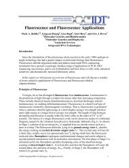
The two-dimensional surface is typically prepared by treating the glass or silicon surface with an<br />
amino silane which results in a uni<strong>for</strong>m layer of primary amines or epoxides (Figure 1) [1, 2]. These<br />
modifications meet several criteria [3]; the linkages are chemically stable, sufficiently long <strong>to</strong><br />
eliminate undesired steric interference from the support, and hydrophilic enough <strong>to</strong> be freely<br />
soluble in aqueous solution and not produce non-specific binding <strong>to</strong> the support [1]. Once these<br />
modifications have activated the surface, the efficiency of attaching the oligonucleotides depends<br />
largely on the chemistry used and how the oligonucleotide targets are modified [4].<br />
<strong>Oligonucleotides</strong> modified with an NH 2 group can be immobilized on<strong>to</strong> epoxy silane-derivatized [5]<br />
or isothiocyanate coated glass slides [1]. Succinylated oligonucleotides can be coupled <strong>to</strong><br />
aminophenyl- or aminopropyl-derivitized glass slides by peptide bonds [6], and disulfide-modified<br />
oligonucleotides can be immobilized on<strong>to</strong> a mercap<strong>to</strong>silanized glass support by a thiol/disulfide<br />
exchange reactions [7] or through chemical cross linkers.<br />
Figure 1. Surface modification. A. Amino modification with 3-<br />
aminopropyltrimethoxysilane. B. Epoxide modification with 3’ glycidoxy<br />
propyltrimethoxysilane.<br />
The surface density of the oligonucleotide probe is expected <strong>to</strong> be an important parameter of any<br />
array. A low surface coverage will yield a correspondingly low hybridization signal and decrease<br />
the hybridization rate.<br />
Conversely, high surface densities may result in steric interference between the covalently<br />
immobilized oligonucleotides, impeding access <strong>to</strong> the target DNA strand [1]. Guo et al. found an<br />
optimal surface coverage at which a maximum amount of a complementary PCR product is<br />
hybridized [1]. In a series of hybridization studies using PCR products as probes these authors<br />
determined the optimal surface coverage at which a maximum amount of the complementary PCR<br />
product is hybridized. For a 157 base probe, the optimum occurred at about 5.0 mM<br />
oligonucleotide concentration, which corresponded <strong>to</strong> a surface density of 500 Å2/molecule [1].<br />
The optimum <strong>for</strong> a longer 347 nucleotide fragment was found <strong>to</strong> be approximately 30% lower,<br />
probably reflecting the greater steric interference of the longer fragment.<br />
© 2014 (v5) Integrated DNA Technologies. All rights reserved. 3
The planar surface structure of glass slides or silicon chips can limit the loading capacity of<br />
oligonucleotides. This limitation has been addressed by applying acrylamide gels <strong>to</strong> glass slides <strong>to</strong><br />
construct a three dimensional surface, greatly increasing the surface area per spot [8, 9]. The<br />
increased surface area permits relatively large amounts of DNA <strong>to</strong> be linked <strong>to</strong> the surface<br />
resulting in strong signal intensities and a good dynamic range. This effect is partly offset by a<br />
higher background produced mainly by the same structural characteristic (the large surface area<br />
per spot) responsible <strong>for</strong> the high loading capacity [10].<br />
3. Three-dimensional Surfaces (Micro-spheres)<br />
Molecular biologists and chemists are discovering that micro-spheres are ideal substrates <strong>for</strong> an<br />
ever-increasing number of applications that use synthetic oligo-nucleotides. In these microsphere-based<br />
assays, each oligonucleotide is attached <strong>to</strong> a micro-sphere. The micro-spheres can<br />
be individually assayed, usually with a flow cy<strong>to</strong>meter, or isolated based on the physical<br />
characteristics of the bead. They are amenable <strong>to</strong> multiple assay <strong>for</strong>mats, multiplexing, rapid assay<br />
development and have distinct cost advantages over two-dimensional glass or silicon chip arrays<br />
<strong>for</strong> characterizing populations of nucleic acids. Several different types of micro-spheres are<br />
available:<br />
Polystyrene micro-spheres can be fluorescently labeled through internal dye entrapment or<br />
surface attachment [11]. Surface labeling is preferred if the environmental responsiveness of the<br />
dye is important or if the particles are <strong>to</strong> be used in a nonaqueous solvent. In most applications,<br />
however, internally labeled micro-spheres are favored. Internal labeling leaves surface groups<br />
available <strong>for</strong> coupling reactions. Additionally, internally labeled fluorescent micro-spheres are<br />
generally brighter, less susceptible <strong>to</strong> pho<strong>to</strong> bleaching, and are available in a larger range of colors<br />
than surface-labeled micro-spheres. To entrap the fluorescent dye, the polymeric micro-spheres<br />
are swelled in an organic solvent or dye solution. The water-insoluble dye diffuses in<strong>to</strong> the<br />
polymer matrix and is trapped when the solvent is removed from the micro-spheres. With these<br />
techniques, scientists at Luminex have been able <strong>to</strong> create a set of 100 unique fluorescent<br />
polystyrene micro-spheres simply by using precise ratios of two spectrally distinct fluorophores.<br />
Magnetic micro-spheres have been used <strong>for</strong> many years in radioimmunoassays, ELISAs, and cell<br />
separation assays. Magnetic micro-spheres are synthesized by dispersing ferrite crystals in a<br />
suspension of styrene/divinylbenzene monomers and polymerizing this cocktail in<strong>to</strong> microspheres.<br />
A popular use <strong>for</strong> this type of micro-sphere has been <strong>to</strong> capture mRNA from a cell lysate<br />
using beads coupled <strong>to</strong> oligo dT. These micro-spheres are often used <strong>to</strong> shorten the processing<br />
time in au<strong>to</strong>mated high-throughput systems [12]. The magnetic beads must be encapsulated <strong>for</strong><br />
most molecular biology applications <strong>to</strong> ensure that the iron doesn’t interfere with polymerases<br />
and other enzymes. Polystyrene magnetic beads are available with both carboxylic acid and amine<br />
surface chemistries.<br />
Silica micro-spheres have a relatively homogeneous chemical surface that can easily be modified<br />
using versatile and well developed silanization chemistry [1]. Their use in molecular biology has<br />
© 2014 (v5) Integrated DNA Technologies. All rights reserved. 4
een primarily in solid phase reversible immobilization (SPRI) pro<strong>to</strong>cols <strong>for</strong> nucleic acid<br />
purification. Nucleic acids can be isolated and purified from a cell lysate or other solution by<br />
capture on<strong>to</strong> silica beads in the presence of a chaotropic agent (KI, NI, or NaSCN). The negatively<br />
charged nucleic acid adsorbs <strong>to</strong> the bead through an ionic interaction and is later eluted in low<br />
ionic strength buffer. SPRI purification is easy <strong>to</strong> au<strong>to</strong>mate and is often the method of choice in<br />
high-throughput sequencing facilities [13]. Silica micro-spheres are denser than polystyrene or<br />
latex micro-spheres and can easily be separated from liquid phase by gravity sedimentation or<br />
gentle centrifugation.<br />
3.1 Handling Fluorescent Micro-spheres<br />
S<strong>to</strong>re fluorescent micro-spheres as aqueous suspensions at 2–8 o C and in the dark <strong>to</strong> minimize<br />
pho<strong>to</strong>bleaching. Use aseptic techniques where practicable. Internally dyed micro-spheres should<br />
not be exposed <strong>to</strong> organic solvents since this will cause swelling of the polymer matrix and<br />
leaching of the dye. If aggregation is a problem, a surfactant such as Tween 20 may be added <strong>to</strong><br />
the suspension.<br />
3.2 Attachment:<br />
Nucleic acids can be covalently attached <strong>to</strong> micro-spheres with any of several methods. Carboxyl<br />
and amino groups are the most common reactive groups <strong>for</strong> attaching ligands <strong>to</strong> surfaces. These<br />
groups are very stable over time, and their chemistries have been widely explored. Listed below<br />
are several reactive groups that can be incorporated on the micro-sphere surface <strong>for</strong> covalent<br />
coupling.<br />
Micro-sphere Surface Chemistries <strong>for</strong><br />
Covalent Coupling<br />
-COOH<br />
-RNH 2<br />
-ArNH 2<br />
-ArCH 2 Cl<br />
-CONH 2<br />
-CONHNH 2<br />
-CHO<br />
-OH<br />
-SH<br />
-COC-<br />
Carboxylic acid<br />
Primary aliphatic amine<br />
Aromatic amine<br />
Chloromethyl (vinyl benzyl<br />
chloride)<br />
Amide<br />
Hydrazide<br />
Aldehyde<br />
Hydroxyl<br />
Thiol<br />
Epoxy<br />
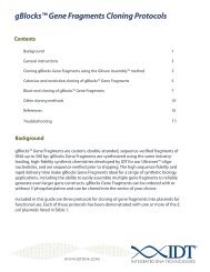
<strong>Attaching</strong> an amino group <strong>to</strong> the 5' or 3' end of an oligonucleotide or a PCR primer is<br />
straight<strong>for</strong>ward and inexpensive. These amine-modified oligos can then be reacted with<br />
carboxylate-modified micro-spheres with carbodiimide chemistry in a one-step process at pH 6–8<br />
(Figure 2). One typical water-soluble carbodiimide is 1-ethyl-3-(3-dimethylaminoproply)carbodiimide<br />
hydrochloride (EDAC). The chief advantage of EDAC is that it provides<br />
one-step coupling. Un<strong>for</strong>tunately, EDAC is indiscriminate; it can crosslink any two primary amines<br />
© 2014 (v5) Integrated DNA Technologies. All rights reserved. 5
in the reaction. Using this reagent may yield a matrix of crosslinked oligos and micro-spheres.<br />
Another option is <strong>to</strong> bind biotin-labeled oligos <strong>to</strong> avidin-coated beads.<br />
3.3 Applications<br />
3.3.1 Liquid Arrays<br />
Figure 2. Covalent coupling of an amine-modified oligonucleotide <strong>to</strong> a carboxylatemodified<br />
micro-sphere with water-soluble carbodiimide.<br />
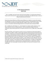
Figure 3. Quantitation of multiple target<br />
hybridization events using uniquely labeled<br />
fluorescent micro-spheres.<br />
© 2014 (v5) Integrated DNA Technologies. All rights reserved. 6
The availability of large sets of fluorescent microspheres<br />
that can be used <strong>to</strong>gether allows <strong>for</strong><br />
highly multiplexed assays and efficient sample<br />
processing. For example, using a set of 100<br />
distinct fluorescent beads in a multiplex <strong>for</strong>mat<br />
could generate almost 10,000 unique data points<br />
from a single 96-well plate which is a scale<br />
normally associated with microarrays. One<br />
plat<strong>for</strong>m that exploits this potential is the<br />
Luminex LabMAP TM system. A capture probe or<br />
target-specific oligonucleotide is coupled <strong>to</strong> the<br />
surface of a fluorescent micro-sphere. A specific<br />
fluorescent dye combination is used <strong>to</strong> uniquely<br />
label each micro-sphere and serves <strong>to</strong> encode<br />
bead identity. These spectral "bar codes" are<br />
equivalent <strong>to</strong> the x,y positioning in<strong>for</strong>mation on a 2-<br />
D solid array. The target (another oligo, a PCR<br />
product, cDNA, or protein) is labeled <strong>to</strong> allow<br />
fluorescent detection and quantitation.<br />
Hybridization takes place in solution between the<br />
capture probe and denatured target.<br />
Hybridization in solution is much more efficient<br />
than solid-phase hybridization and this efficiency<br />
one of the advantages of micro-sphere-based<br />
assays. Following hybridization, the micro-spheres<br />
are assayed by flow cy<strong>to</strong>metric methods. Two<br />
signals are assayed: the unique fluorescent signal<br />
that identifies the probe and the fluorescent<br />
signal that indicates hybridization has occurred.<br />
Relative fluorescence intensity of the<br />
hybridization signal is used <strong>to</strong> quantitate the<br />
target (Figure 3). Yang et al. recently described a<br />
bead-based liquid array <strong>for</strong> detecting gene<br />
expression in a high throughput assay [14].<br />
Defoort and colleagues used bead-based flow<br />
cy<strong>to</strong>metric methods and reverse transcription–<br />
PCR <strong>to</strong> simultaneously identify HIV, hepatitis B,<br />
and hepatitis C in a single plasma sample[15].<br />
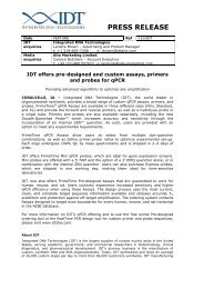
Figure 4. Schematic representation of the<br />
micro-sphere-based ASPE assay. In ASPE,<br />
reactions <strong>for</strong> each SNP a pair of probes are<br />
designed which differ from each other at<br />
their extreme 3' nucleotide (the<br />
polymorphic site). In the presence of DNA<br />
polymerase, dNTPs and a small portion of<br />
the biotin labeled dCTP, a labeled<br />
extension product of the 3' portion of the<br />
primer is obtained only if the template<br />
includes the target sequence.<br />
3.3.2 Detection of Single Nucleotide<br />
Polymorphisms<br />
Single-nucleotide polymorphisms (SNPs) are the most common <strong>for</strong>m of sequence variation<br />
between individuals [16]. Analysis of this variation offers an opportunity <strong>to</strong> understand the genetic<br />
basis of disease and is a driving <strong>for</strong>ce behind modern pharmacogenomics. Accurate, high<br />
throughput, and cost effective methods <strong>to</strong> analyze SNPs is crucial <strong>to</strong> fully utilize the medical value<br />
© 2014 (v5) Integrated DNA Technologies. All rights reserved. 7<br />
is
of the DNA sequence data that has been generated in the human genome project. Several<br />
methods <strong>for</strong> discerning SNPs have been described including oligonucleotide ligation assay (OLA)<br />
[16], single base chain extension assay [17, 18], and allele-specific oligonucleotide hybridization<br />
(ASO) [19]. Each of these assays can be adapted <strong>to</strong> multiplex-analysis using arrays of<br />
oligonucleotides attached <strong>to</strong> fluorescently encoded mircrospheres [20]. An example of one of<br />
these bead-based adaptations, Allele specific primer extension (ASPE) is illustrated in Figure 4.<br />
4. Modifications<br />
4.1 I-Linker<br />
I-Linker TM is a proprietary covalent attachment chemistry <strong>for</strong> oligonucleotides that was developed<br />
at IDT. The modifier is attached <strong>to</strong> the 5’-end of the oligo. I-Linker TM can be substituted <strong>for</strong> amino<br />
modifications in many applications. In addition, I-Linker TM expands the range of reactive groups<br />
that can be used <strong>for</strong> conjugation, including aldehyde- and ke<strong>to</strong>ne-modified ligands or surfaces.<br />
4.2 Amino-modified <strong>Oligonucleotides</strong><br />
A primary amine can be used <strong>to</strong> covalently attach a variety of products <strong>to</strong> an oligonucleotide,<br />
including fluorescent dyes [21, 22], biotin [23], alkaline phosphatase [24], EDTA [25], or <strong>to</strong> a solid<br />
surface. An amino modifier can be place at the 5'-end, 3'-end or internally using and amino-dC or<br />
amino-dT modified base.<br />
4.2.1 5' Amino Modifiers<br />
Various amino-modifiers can be added <strong>to</strong> the 5'-terminus of a target oligonucleotide. Because<br />
conventional au<strong>to</strong>mated synthesis proceeds from 3' <strong>to</strong> 5', addition of the 5'-amino-mod is the last<br />
step in synthesis. As a result, any failed products which are truncated and capped will not receive<br />
the 5'-amino-modification and will not participate in subsequent chemical reactions involving the<br />
primary amine.<br />
5' amino modifiers are -cyanoethyl phosphoramidites which, when activated with 1H tetrazole,<br />
can couple <strong>to</strong> the 5' terminus of the oligonucleotide in the same time frame and with similar<br />
efficiency as nucleoside phosphoramidites. A number of 5' amino modifiers are available from IDT,<br />
these include simple amino groups with a six or twelve carbon spacers, a Uni-Link Amino modifier<br />
or amino modified thymidine or cy<strong>to</strong>sine.<br />
The shorter carbon chain linkers (Amino C6 and Uni-Link) may be used <strong>to</strong> attach compounds<br />
where proximity <strong>to</strong> the oligonucleotide poses no problem. The longer carbon chain linker (Amino<br />
C12) is recommend whenever steric or charge considerations require greater distance between<br />
the oligo and ligand or surface; examples include some affinity chroma<strong>to</strong>graphy applications<br />
where the oligonucleotide must be adequately spaced from the surface, labeling with biotin [23],<br />
or certain fluorescent tags where interaction with the oligonucleotide, or the duplex it <strong>for</strong>ms, may<br />
partially quench fluorescence [21, 22]. Sometimes even greater distance is needed than can be<br />
achieved by the amino-C12 modifier. In this case, one or more internal spacer modifiers (such as<br />
the S18 spacer) can be used <strong>to</strong> further separate the 5'-terminal amino-modifier from the oligo.<br />
© 2014 (v5) Integrated DNA Technologies. All rights reserved. 8
4.2.2 3' Amino Modifiers<br />
3'-amino-modifiers contain branched linkers in which the amino group is protected with the<br />
fluorenylmethoxycarbonyl (Fmoc) group which was originally popularized <strong>for</strong> use in peptide<br />
chemistry [26, 27]. The Fmoc group is stable yet can be removed specifically from the support <strong>to</strong><br />
allow solid-phase addition of an amino linked modification. However, during handling, some Fmoc<br />
groups can be lost; in this setting the free amino group is capped with acetic anhydride during<br />
synthesis and results in decreased yields.<br />
3'-Amino-Modifier CPGs (the starting support <strong>for</strong> oligonucleotide synthesize) otherwise support<br />
oligo synthesis with the same efficiency as the standard nucleoside CPGs. After deprotection, the<br />
finished oligonucleotide has a free primary amine at the 3'-terminus. An oligo with a 3'-amino-mod<br />
can be labeled at the 5'-end with various phosphoramidite modifier groups (like fluorescent dyes,<br />
biotin, etc.) during synthesis. Alternatively, the oligo can be left with a free 5'-OH, which can later<br />
be labeled with 32 P using a polynucleotide kinase. The 3'-amino-modifier eliminates the native 3'-<br />
OH group from the oligo, which functionally blocks this oligo from participating as a primer in DNA<br />
synthesis, sequencing, or PCR.<br />
4.2.3 Internal Amino Modification<br />
Amino-modifier C6 dT and the recently introduced amino-modifier C6 dC are available <strong>for</strong> internal<br />
labeling (Figure 5). Addition of the amino-modifier itself does not adversely affect oligo<br />
hybridization characteristics; however subsequent addition of a bulky hydrophobic dye or other<br />
ligand can lower T m or interfere with primer function. There<strong>for</strong>e, it is preferable <strong>to</strong> attach large<br />
bulky groups <strong>to</strong> the 5'-end of the oligo, separated by spacers as needed. Amino-modifier dT and dC<br />
couple with similar efficiency as normal phosphoramidite monomers and their trifluoracetylprotecting<br />
group is removed during the standard ammonium hydroxide deprotection step.<br />
Figure 5. Internal amino modifications C6 dT phosphoramidite (on the left) and<br />
C6 dC phosphoramidite (on the right).<br />
4.2.4 Labeling Amino Modified <strong>Oligonucleotides</strong><br />
This general procedure can be used <strong>to</strong> conjugate amino-modified oligonucleotides with active<br />
succinimidyl ester or isothiocyanate derivatives of various ligands, such as fluorescent dyes. At pH<br />
9.0 the conjugation reaction occurs virtually exclusively at the free primary amine and does not<br />
involve the exocyclic amino groups of the nucleosides.<br />
1. For a 250 nmole scale synthesis, resuspend the amino-modified oligonucleotide (i.e.<br />
approximately 100 nmoles of primary reactive amine) in 0.7 mL of sterile distilled water.<br />
2. Add 100 of 10X conjugation buffer (1.0 M NaHCO3)/Na2CO3, pH 9.0).<br />
© 2014 (v5) Integrated DNA Technologies. All rights reserved. 9
3. Freshly prepare a 10 mg/mL solution of active ester in DMF. Add 200 of the solution <strong>to</strong><br />
the reaction mixture.<br />
4. Allow the mixture <strong>to</strong> stand at least 2 hours.<br />
5. Remove the unreacted ligand by gel filtration (such as Sephadex G-25).<br />
6. Depending on the reactivity of the NHS-ester used, coupling efficiency can range from 20-<br />
80%. Labeled-oligo can be purified from unreacted oligo by preparative RP-HPLC<br />
purification.<br />
4.2.5 <strong>Attaching</strong> Amino-Modified <strong>Oligonucleotides</strong><br />
The attachment of an amino-modified oligonucleotide <strong>to</strong> a surface or another molecule requires<br />
an acylating reagent. Depending on which acylating reagent is used, carboxamides, sulfonamides,<br />
ureas or thioureas are <strong>for</strong>med upon reaction with the amine moiety. The kinetics of the reaction<br />
depends on the reactivity and concentration of both the acylating reagent and the amine. Buffers<br />
that contain free amines such as Tris and glycine must be avoided when using any amine-reactive<br />
reagent.<br />
Attachment chemistries currently in use <strong>for</strong> amino modified oligonucleotides <strong>for</strong> linkage <strong>to</strong><br />
molecule or surface:<br />
Acylating Agent Linkage Features<br />
Carbodiimide Carbonyl amide Most common method, stable<br />
attachment<br />
Isothiocyanate Thiourea Stable covalent attachments<br />
Sulfonyl chloride sulfonamide Sulfonyl chloride is unstable<br />
in water, but once conjugated<br />
<strong>to</strong> the oligo the sulfonamide<br />
bond is very stable<br />
Succinimidyl esters (NHSester)<br />
carboxamide<br />
Carboxamide bond <strong>for</strong>med is<br />
very stable<br />
© 2014 (v5) Integrated DNA Technologies. All rights reserved. 10
The most significant fac<strong>to</strong>rs affecting the reactivity of an amine are its class and its basicity.<br />
<strong>Oligonucleotides</strong> with an amino modification have a free aliphatic amine that is moderately basic<br />
and reactive with most acylating reagents. However, the concentration of the free base <strong>for</strong>m of<br />
aliphatic amines that are below<br />
pH 8 is very low; thus, the<br />
kinetics of acylation reactions of<br />
amines by isothiocyanates,<br />
succinimidyl esters and other<br />
reagents are strongly pH<br />
dependent. A pH of 8.5-9.5 is<br />
usually optimal <strong>for</strong><br />
oligonucleotide conjugation.<br />
Aromatic amines, which are<br />
present within each base, are<br />
very weak bases and, thus, are<br />
not pro<strong>to</strong>nated at pH 7 or<br />
above.<br />
Most attachment chemistries<br />
currently in use <strong>for</strong> amino<br />
modified oligonucleotides<br />
utilize a carbodiimide mediated<br />
acylation. Acylation of a<br />
carboxyl group generates a<br />
stable amide carbonyl as<br />
illustrated in Figure 6.<br />
Isothiocyanates <strong>for</strong>m thioureas<br />
upon reaction with amines that<br />
are relatively stable covalent<br />
Figure 6. Carbodiimide-mediated amino attachment. The first<br />
attachments (Figure 7). A very step is addition of the carboxylic acid <strong>to</strong> a C=N bond of the<br />
common example of this<br />
carbodiimide <strong>to</strong> generate an O-acylated derivative of urea. This is a<br />
reaction is the coupling of reactive acylating agent as there is a strong preference <strong>for</strong><br />
fluorescein isothiocyanate eliminating the urea unit. The result is <strong>for</strong>mation of a stable amide<br />
(FITC) with amino-labeled oligos carbonyl group.<br />
<strong>to</strong> <strong>for</strong>m a fluorescent probe.<br />
Succinimidyl esters (NHS-esters) are<br />
excellent reagents <strong>for</strong> amine<br />
modification because the amide<br />
bonds they <strong>for</strong>m (Figure 7) are very<br />
stable. These reagents are can be<br />
s<strong>to</strong>red long term if frozen or<br />
desiccated. NHS-esters are very<br />
reactive with primary amines and<br />
have low reactivity with secondary<br />
amines, alcohols, phenols (including<br />
© 2014 (v5) Integrated DNA Technologies. All rights reserved. 11<br />
Figure 7. Amino attachment chemistries.
tyrosine) and histidine. Succinimidyl esters will also react with thiols in organic solvents <strong>to</strong> <strong>for</strong>m<br />
thioesters. Succinimidyl ester hydrolysis can compete with conjugation, but this side reaction is<br />
usually slow below pH 9. Some succinimidyl esters derivatives, such as certain fluorescent dyes,<br />
have poor solubility in aqueous solution and must be resuspended in an organic solvent.<br />
Sulfonyl chlorides are also reactive with primary amines. However, these reagents are quite<br />
unstable in water, especially at the higher pH required <strong>for</strong> reaction with aliphatic amines, and as<br />
such are not commonly used in oligonucleotide attachment pro<strong>to</strong>cols (Figure 7). Once conjugated,<br />
however, the sulfonamides that are <strong>for</strong>med are extremely stable. Sulfonyl chlorides can also react<br />
with phenols (including tyrosine), aliphatic alcohols (including polysaccharides), thiols and<br />
imidazoles.<br />
As noted, a number of methods <strong>for</strong> attachment of 5'-amino-modified oligonucleotides <strong>to</strong> solid<br />
supports have been described. One method involves using an epoxide opening reaction <strong>to</strong><br />
generate a covalent linkage between 5'-amino-modified oligonucleotides and epoxy silanederivatized<br />
glass [5, 28]. Attachment of 5' amino-modified oligonucleotides on epoxy derivatized<br />
slides is relatively thermostable, but the sensitivity of the epoxy ring <strong>to</strong> moisture is a major<br />
drawback and leads <strong>to</strong> reproducibility problems [29]. More commonly, attachment of aminomodified<br />
oligos involves reacting the surface bound amino groups with excess p-phenylene 1,4<br />
diisothiocyanate (PDC) <strong>to</strong> convert the support's bound primary amines <strong>to</strong> amino-reactive<br />
phenylisothiocyanate groups. Coupling of 5' amino-modified oligos reaction <strong>to</strong> the<br />
phenylisothiocyanate groups follows, resulting in the covalent attachment of the oligonucleotide<br />
(Figure 8) [1].<br />
Figure 8. Amino attachment via phenylisothiocyanate.<br />
Modifications on this theme have included using homobifunctional crosslinking agents such as<br />
disuccinimidylcarbonate (DCS), disuccinimidyloxalte (DSO), and dimethylsuberimidate (DMS).<br />
These have all been used <strong>to</strong> covert glass bound amino groups in<strong>to</strong> reactive isothiocyanates, N-<br />
hydroxysuccimimidy-esters (NHS-esters) or imidoesters, respectively. Activation with PDITC, DMS,<br />
DSC and DSO work well <strong>for</strong> the attachment of oligonucleotides, while immobilization of aminolinked<br />
PCR fragments cross-linking with PDITC or DMS is superior because they are less labile <strong>to</strong><br />
the Tris buffer reagent in PCR amplification buffers [10].<br />
The chemistry of 1-ethyl=3-(3-<br />
dimethylaminopropl)-carbodiimide<br />
hydrochloride (EDC, Figure 9) has been<br />
© 2014 (v5) Integrated DNA Technologies. All rights reserved. 12<br />
Figure 9. 1-ethyl=3-(3-dimethylaminopropyl)-<br />
carbodiimide hydrochloride (EDC).
employed with other supports such as amino controlled-pore glass [30], latex beads [31, 32],<br />
dextran supports [33], and polystyrene [34]. Heterobifunctional cross-linkers such as EDC are<br />
designed <strong>to</strong> <strong>for</strong>m stable covalent links in aqueous solution and their use in binding<br />
oligonucleotides on<strong>to</strong> glass surfaces by their 3' [35] or 5' [36] ends has been widespread.<br />
4.3 Thiol-Modified <strong>Oligonucleotides</strong><br />
The thiol (SH) modifier enables covalent attachment of an oligo <strong>to</strong> a variety of ligands. A SHmodifier<br />
can be placed at either the 5'-end or 3'-end of an oligo. It can be used <strong>to</strong> <strong>for</strong>m reversible<br />
disulfide bonds (ligand-S-S-oligo) or irreversible bonds with a variety of activated accepting groups.<br />
Options include active esters or isothiocyanate derivatives, as are commonly used <strong>for</strong> tagging free<br />
amino-modified oligonucleotides. Maleimide, bromide, iodide, or sulphonyl derivatives are<br />
suitable <strong>for</strong> tagging thiol-linked oligonucleotides with a variety of groups such as fluorescent dyes<br />
[21, 22], biotin [23] and alkaline phosphatase [24]. Conjugation of oligonucleotides <strong>to</strong> enzymes,<br />
such as alkaline phosphatase or horseradish peroxidase, is commonly employed in commercial<br />
probe systems. The thiol modification also enables attachment <strong>to</strong> solid surfaces (e.g. CPG-SH;<br />
Pierce) via a disulphide bond [37] or maleimide linkages.<br />
Thiol-modifiers can be incorporated at the start of synthesis by placing the reactive SH-group at<br />
the 3'-end using a thiol-CPG and can also be incorporated as the last step of synthesis using a thiolphosphoramidite<br />
which places the reactive SH-group at the 5'-end of the oligo.<br />
4.3.1 5' Thiol Modifier<br />
The incorporation of a thiol group at the 5' end of an oligo is achieved with S-trityl-6-<br />
mercap<strong>to</strong>hexyl derivatives (Figure 10) [21, 38]. IDT recommends HPLC purification of thiolmodified<br />
oligonucleotides.<br />
4.3.2 3’ Thiol Modifier<br />
© 2014 (v5) Integrated DNA Technologies. All rights reserved. 13
The 3'-thiol modifier C3 S-S CPG is used <strong>to</strong> introduce a thiol group <strong>to</strong> the 3'-terminus of a target<br />
oligonucleotide. A 3' thiol group can be particularly useful <strong>for</strong> post-synthesis modification in cases<br />
where a second 5' modification is already in place. Special precautions are taken during the<br />
oxidation steps of the synthesis <strong>to</strong> avoid oxidative cleavage of the disulfide linkage. Deprotection<br />
with a cocktail containing DTT is used <strong>to</strong> remove the base protecting groups and cleave the<br />
disulfide linkage <strong>to</strong> generate the<br />
reactive 3'-thiol.<br />
Oligos from IDT are provided in a<br />
disulfide <strong>for</strong>m (Figure 11). However,<br />
even when shipped in the disulfide<br />
<strong>for</strong>m, spontaneous cleavage will<br />
occur <strong>to</strong> some variable fraction of<br />
the molecules. This is normal and<br />
will cause disulfide dimers <strong>to</strong><br />
accumulate over time. Because of<br />
this, all thiol-oligos need <strong>to</strong> be<br />
reduced in preparation prior <strong>to</strong><br />
conjugation immediately be<strong>for</strong>e use<br />
(see pro<strong>to</strong>cols below).<br />
A.<br />
B.<br />
A.<br />
4.3.3 S<strong>to</strong>ring and Reducing Thiol<br />
Modified <strong>Oligonucleotides</strong><br />
Thiol-modified oligonucleotides<br />
should be s<strong>to</strong>red frozen. To ensure<br />
full reactivity, thiol-modified oligos<br />
should be reduced immediately<br />
be<strong>for</strong>e use. In general, the oligo is<br />
treated with a reducing agent (like<br />
DTT) and this agent is fully removed<br />
prior <strong>to</strong> coupling. Specific pro<strong>to</strong>cols<br />
are provided below.<br />
B.<br />
4.3.3a Reduction Pro<strong>to</strong>cols:<br />
Figure 10. A. Thiol modifier C6 S-S phosphoramidite. B. Reaction<br />
Pro<strong>to</strong>col 1. Treatment with solidphase<br />
DTT<br />
oligonucleotide.<br />
scheme <strong>for</strong> adding a 5’ thiol modifier <strong>to</strong> a synthetic<br />
DTT is available immobilized on<br />
acrylamide resin (Reductacryl, Calbiochem<br />
Inc. Cat. No. 233157). The Reductacryl<br />
reagent can be used in a batch technique <strong>to</strong><br />
reduce the disulfide bonds.<br />
1. Resuspend oligo plus resin in TE (or<br />
similar buffer which is neutral or<br />
slightly alkaline such as pH 7.5).<br />
© 2014 (v5) Integrated DNA Technologies. All rights reserved. 14<br />
Figure 11. A. Thiol modifier C6 S-S Controlled Pore<br />
Glass (CPG). B. Reaction scheme <strong>for</strong> adding a 3’ thiol<br />
modifier <strong>to</strong> a synthetic oligonucleotide.
2. Use a ratio of 1 mg oligo with 50 mg resin <strong>to</strong> ensure complete reduction.<br />
3. Stir or agitate at room temperature <strong>for</strong> 15 minutes.<br />
4. Remove Reductacryl TM by filtration (e.g., pass through a syringe filter)<br />
5. Activated oligo can be directly injected in<strong>to</strong> the coupling reaction or can be s<strong>to</strong>red <strong>for</strong> brief<br />
periods of time be<strong>for</strong>e use.<br />
Pro<strong>to</strong>col 2. Treatment with DTT in liquid phase<br />
The oligo can be treated with DTT or s<strong>to</strong>red in DTT, which must be removed immediately be<strong>for</strong>e<br />
use.<br />
1. Make a solution of oligonucleotide in TE plus DTT. We use 100 M oligo in 10 mM DTT in 1x<br />
TE.<br />
2. Pass oligo through a large bed volume Sephadex column <strong>to</strong> remove DTT. Note that small<br />
bed volume spin columns can allow trace DTT <strong>to</strong> remain with the oligo, which can interfere<br />
with subsequent coupling reactions. As an alternative, use the extraction procedure<br />
outlined below.<br />
Pro<strong>to</strong>col 3. Bulk Reduction<br />
Reconstitute the oligonucleotide (up <strong>to</strong> 1 mg of oligo can be used) in 100 L of 2% TEA<br />
(triethylamine), 50 mM DTT and allow <strong>to</strong> stand at room temperature <strong>for</strong> 10 min. Remove DTT<br />
using one of the 3 methods outlined below:<br />
1. Extraction/PPT: extract 4x using 400 l of ethyl acetate (layers readily separate and the DTT<br />
will partition with the ethyl acetate and the DNA will partition in the aqueous phase).<br />
2. Recover oligo by ace<strong>to</strong>ne precipitation or gel filtration.<br />
1. Add five volumes of ace<strong>to</strong>ne solution (2% LiClO 4 w/w in ace<strong>to</strong>ne) <strong>to</strong> one volume of<br />
the oligo solution in a 14 mL tube.<br />
2. Chill the resulting solution at -20 o C <strong>for</strong> 15 minutes.<br />
3. Centrifuge the sample at 2500 - 5000 RPM <strong>for</strong> 10 - 5 minutes respectively.<br />
4. Remove the supernatant.<br />
5. Dry the sample under vacuum <strong>to</strong> remove trace ace<strong>to</strong>ne.<br />
6. To remove LiClO 4 and other salts, the sample can be washed with 2-3 mL of n-<br />
butanol centrifuged again followed by removal of the butanol supernatant.<br />
3. Size exclusion or gel filtration chroma<strong>to</strong>graphy<br />
1. Load the sample of oligonucleotide on a Sephadex G25F column that has been<br />
thoroughly washed with distilled water.<br />
2. Elute the column with water by gravity flow and collect fractions.<br />
3. Measure the UV absorbance at 260 nm. The first eluting peak at the void volume is<br />
the oligonucleotide.<br />
4. Concentrate fractions using a SpeedVac evapora<strong>to</strong>r.<br />
Any oligonucleotide that is not used immediately should be s<strong>to</strong>red frozen. Over time, the oligo will<br />
oxidize and the above procedure will need <strong>to</strong> be repeated be<strong>for</strong>e coupling.<br />
4.3.4 <strong>Attaching</strong> Thiol Modified <strong>Oligonucleotides</strong><br />
As a class, the cross-linkers used <strong>to</strong> attach thiol-modified oligonucleotides <strong>to</strong> solid supports are<br />
her<strong>to</strong>bifunctional, meaning that they posses functional groups capable of reaction with two<br />
© 2014 (v5) Integrated DNA Technologies. All rights reserved. 15
Figure 12. Succinimidyl 4-[malemidophenyl]butyrate (SMPB) used <strong>to</strong> link a<br />
thiol-modified synthetic oligonucleotide <strong>to</strong> an amine-derivatized solid support.<br />
chemically distinct functional groups, e.g. amines and thiols. The linkers serve two purposes: <strong>to</strong><br />
covalently bind two distinct chemical entities which otherwise would remain un-reactive <strong>to</strong>ward<br />
each other and as a physical spacer which provides greater accessibility and or freedom <strong>to</strong> each of<br />
the linked biomolecules [35].<br />
A number of heterobifunctional cross linkers have been developed <strong>for</strong> covalent attachment of<br />
thiol-modified DNA oligomers <strong>to</strong> aminosilane monolayer films [35]. These cross linkers combine<br />
groups reactive <strong>to</strong>ward amines such as N-hydroxy-succinimidyl esters and groups reactive <strong>to</strong>ward<br />
thiols such as maleimide or alpha-haloacetyl moieties. Figure 12 illustrates the use of one such<br />
cross linker, succinimidyl 4-[maleimidophenyl]butyrate (SMPB) <strong>to</strong> link a thiol-modified oligo <strong>to</strong> an<br />
amine derivatized solid support.<br />
The use of SMPB <strong>to</strong> cross link thiol modified oligos and aminosilanized glass slides has resulted in a<br />
surface density of approximately 20 pmoles of bound DNA/cm 2 [35]. This density was significantly<br />
greater than that reported <strong>for</strong> DNA films <strong>for</strong>med on similar substrates prepared using aminosilane<br />
films with a diisothiocyanate cross linker [1] or epoxysilane films [5].<br />
© 2014 (v5) Integrated DNA Technologies. All rights reserved. 16
5. Conclusion<br />
<strong>Solid</strong> support attachment of oligonucleotides, including microarray technology, provides a<br />
powerful set of <strong>to</strong>ols <strong>for</strong> molecular biologists. In order <strong>to</strong> insure that the data available from these<br />
applications are accurate, precise, and reliable, considerable ef<strong>for</strong>t in choosing and designing<br />
oligonucleotides is essential. Success begins with choosing the best method of attachment <strong>for</strong> the<br />
synthetic oligonucleotides. <strong>Oligonucleotides</strong> with amino or thiol modifications have been the most<br />
reliable <strong>to</strong> date.<br />
6. References<br />
1. Guo Z, Guilfoyle RA, et al. (1994) Direct fluorescence analysis of genetic polymorphisms by<br />
hybridization with oligonucleotide arrays on glass supports. Nucleic Acids Res, 22(24):<br />
54565465.<br />
2. Kumar A, Larsson O, et al. (2000) Silanized nucleic acids: a general plat<strong>for</strong>m <strong>for</strong> DNA<br />
immobilization. Nucleic Acids Res, 28(14): E71.<br />
3. Maskos U and Southern EM. (1992) Oligonucleotide hybridizations on glass supports: a<br />
novel linker <strong>for</strong> oligonucleotide synthesis and hybridization properties of oligonucleotides<br />
synthesised in situ. Nucleic Acids Res, 20(7): 16791684.<br />
4. Lindroos K, Liljedahl U, et al. (2001) Minisequencing on oligonucleotide microarrays:<br />
comparison of immobilisation chemistries. Nucleic Acids Res, 29(13): E699.<br />
5. Lamture JB, Beattie KL, et al. (1994) Direct detection of nucleic acid hybridization on the<br />
surface of a charge coupled device. Nucleic Acids Res, 22(11): 21212125.<br />
6. Joos B, Kuster H, and Cone R. (1997) Covalent attachment of hybridizable oligonucleotides<br />
<strong>to</strong> glass supports. Anal Biochem, 247(1): 96101.<br />
7. Rogers YH, Jiang-Baucom P, et al. (1999) Immobilization of oligonucleotides on<strong>to</strong> a glass<br />
support via disulfide bonds: A method <strong>for</strong> preparation of DNA microarrays. Anal Biochem,<br />
266(1): 2330.<br />
8. Mirzabekov AD. (1994) DNA sequencing by hybridization--a megasequencing method and a<br />
diagnostic <strong>to</strong>ol? Trends Biotechnol, 12(1): 2732.<br />
9. Proudnikov D, Timofeev E, and Mirzabekov A. (1998) Immobilization of DNA in<br />
polyacrylamide gel <strong>for</strong> the manufacture of DNA and DNA-oligonucleotide microchips. Anal<br />
Biochem, 259(1): 3441.<br />
10. Beier M and Hoheisel JD. (1999) Versatile derivatisation of solid support media <strong>for</strong> covalent<br />
bonding on DNA-microchips. Nucleic Acids Res, 27(9): 19701977.<br />
11. Arshady R. (1993) Microspheres <strong>for</strong> biomedical applications: preparation of reactive and<br />
labelled microspheres. Biomaterials, 14(1): 515.<br />
12. Skowronski EW, Armstrong N, et al. (2000) Magnetic, microplate-<strong>for</strong>mat plasmid isolation<br />
pro<strong>to</strong>col <strong>for</strong> high-yield, sequencing-grade DNA. Biotechniques, 29(4): 786788, 790, 792.<br />
13. Engelstein M, Aldredge TJ, et al. (1998) An efficient, au<strong>to</strong>matable template preparation <strong>for</strong><br />
high throughput sequencing. Microb Comp Genomics, 3(4): 237241.<br />
© 2014 (v5) Integrated DNA Technologies. All rights reserved. 17
14. Yang L, Tran DK, and Wang X. (2001) BADGE, Beads Array <strong>for</strong> the Detection of Gene<br />
Expression, a high-throughput diagnostic bioassay. Genome Res, 11(11): 18881898.<br />
15. Defoort JP, Martin M, et al. (2000) Simultaneous detection of multiplex-amplified human<br />
immunodeficiency virus type 1 RNA, hepatitis C virus RNA, and hepatitis B virus DNA using<br />
a flow cy<strong>to</strong>meter microsphere-based hybridization assay. J Clin Microbiol, 38(3):<br />
10661071.<br />
16. Iannone MA, Taylor JD, et al. (2000) Multiplexed single nucleotide polymorphism<br />
genotyping by oligonucleotide ligation and flow cy<strong>to</strong>metry. Cy<strong>to</strong>metry, 39(2): 131140.<br />
17. Chen J, Iannone MA, et al. (2000) A microsphere-based assay <strong>for</strong> multiplexed single<br />
nucleotide polymorphism analysis using single base chain extension. Genome Res, 10(4):<br />
549557.<br />
18. Cai H, White PS, et al. (2000) Flow cy<strong>to</strong>metry-based minisequencing: a new plat<strong>for</strong>m <strong>for</strong><br />
high-throughput single-nucleotide polymorphism scoring. Genomics, 66(2): 135143.<br />
19. Ye F, Li MS, et al. (2001) Fluorescent microsphere-based readout technology <strong>for</strong><br />
multiplexed human single nucleotide polymorphism analysis and bacterial identification.<br />
Hum Mutat, 17(4): 305316.<br />
20. Armstrong B, Stewart M, and Mazumder A. (2000) Suspension arrays <strong>for</strong> high throughput,<br />
multiplexed single nucleotide polymorphism genotyping. Cy<strong>to</strong>metry, 40(2): 102108.<br />
21. Connolly BA and Rider P. (1985) Chemical synthesis of oligonucleotides containing a free<br />
sulphydryl group and subsequent attachment of thiol specific probes. Nucleic Acids Res,<br />
13(12): 44854502.<br />
22. Zuckermann R, Corey D, and Schultz P. (1987) Efficient methods <strong>for</strong> attachment of thiol<br />
specific probes <strong>to</strong> the 3'-ends of synthetic oligodeoxyribonucleotides. Nucleic Acids Res,<br />
15(13): 53055321.<br />
23. Sproat BS, Beijer B, et al. (1987) The synthesis of protected 5'-mercap<strong>to</strong>-2',5'-<br />
dideoxyribonucleoside-3'-O-phosphoramidites; uses of 5'-mercap<strong>to</strong>oligodeoxyribonucleotides.<br />
Nucleic Acids Res, 15(12): 48374848.<br />
24. Li P, Medon PP, et al. (1987) Enzyme-linked synthetic oligonucleotide probes: nonradioactive<br />
detection of entero<strong>to</strong>xigenic Escherichia coli in faecal specimens. Nucleic Acids<br />
Res, 15(13): 52755287.<br />
25. Dreyer GB and Dervan PB. (1985) Sequence-specific cleavage of single-stranded DNA:<br />
oligodeoxynucleotide-EDTA X Fe(II). Proc Natl Acad Sci U S A, 82(4): 968972.<br />
26. Nelson PS, Sherman-Gold R, and Leon R. (1989) A new and versatile reagent <strong>for</strong><br />
incorporating multiple primary aliphatic amines in<strong>to</strong> synthetic oligonucleotides. Nucleic<br />
Acids Res, 17(18): 71797186.<br />
27. Nelson PS, Kent M, and Muthini S. (1992) Oligonucleotide labeling methods. 3. Direct<br />
labeling of oligonucleotides employing a novel, non-nucleosidic, 2-aminobutyl-1,3-<br />
propanediol backbone. Nucleic Acids Res, 20(23): 62536259.<br />
28. Beattie WG, Meng L, et al. (1995) Hybridization of DNA targets <strong>to</strong> glass-tethered<br />
oligonucleotide probes. Mol Biotechnol, 4(3): 213225.<br />
29. Adessi C, Mat<strong>to</strong>n G, et al. (2000) <strong>Solid</strong> phase DNA amplification: characterisation of primer<br />
attachment and amplification mechanisms. Nucleic Acids Res, 28(20): E87.<br />
30. Ghosh SS and Musso GF. (1987) Covalent attachment of oligonucleotides <strong>to</strong> solid supports.<br />
Nucleic Acids Res, 15(13): 53535372.<br />
© 2014 (v5) Integrated DNA Technologies. All rights reserved. 18
31. Wolf SF, Haines L, et al. (1987) Rapid hybridization kinetics of DNA attached <strong>to</strong> submicron<br />
latex particles. Nucleic Acids Res, 15(7): 29112926.<br />
32. Lund V, Schmid R, et al. (1988) Assessment of methods <strong>for</strong> covalent binding of nucleic acids<br />
<strong>to</strong> magnetic beads, Dynabeads, and the characteristics of the bound nucleic acids in<br />
hybridization reactions. Nucleic Acids Res, 16(22): 1086110880.<br />
33. Gingeras TR, Kwoh DY, and Davis GR. (1987) Hybridization properties of immobilized<br />
nucleic acids. Nucleic Acids Res, 15(13): 53735390.<br />
34. Rasmussen SR, Larsen MR, and Rasmussen SE. (1991) Covalent immobilization of DNA on<strong>to</strong><br />
polystyrene microwells: the molecules are only bound at the 5' end. Anal Biochem, 198(1):<br />
138142.<br />
35. Chrisey LA, Lee GU, and O'Ferrall CE. (1996) Covalent attachment of synthetic DNA <strong>to</strong> selfassembled<br />
monolayer films. Nucleic Acids Res, 24(15): 30313039.<br />
36. O'Donnell MJ, Tang K, et al. (1997) High-Density, Covalent Attachment of DNA <strong>to</strong> Silicon<br />
Wafers <strong>for</strong> Analysis by MALDI-TOF Mass Spectrometry. Anal. Chem., 69(13): 24382443.<br />
37. Bischoff R, Coull JM, and Regnier FE. (1987) Introduction of 5'-terminal functional groups<br />
in<strong>to</strong> synthetic oligonucleotides <strong>for</strong> selective immobilization. Anal Biochem, 164(2):<br />
336344.<br />
38. Sinha ND and Cook RM. (1988) The preparation and application of functionalised synthetic<br />
oligonucleotides: III. Use of H-phosphonate derivatives of protected amino-hexanol and<br />
mercap<strong>to</strong>-propanol or -hexanol. Nucleic Acids Res, 16(6): 26592669.<br />
© 2014 (v5) Integrated DNA Technologies. All rights reserved. 19