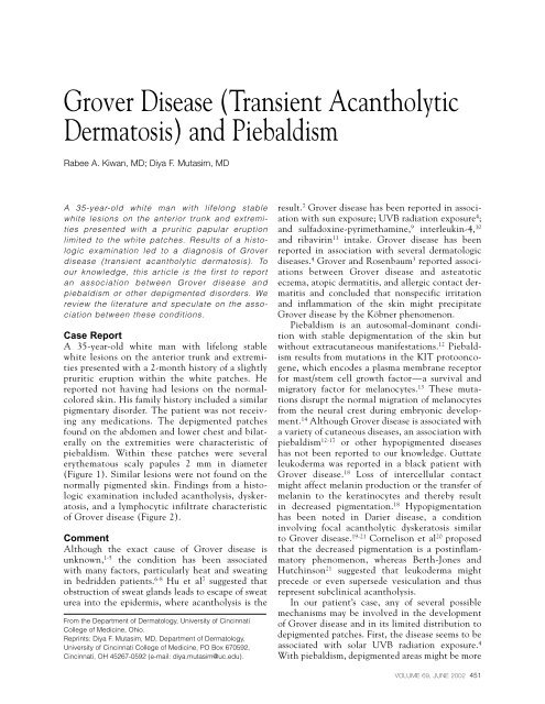
Grover Disease (Transient Acantholytic Dermatosis ... - Ob.Gyn. News
Grover Disease (Transient Acantholytic Dermatosis ... - Ob.Gyn. News
Grover Disease (Transient Acantholytic Dermatosis ... - Ob.Gyn. News

You also want an ePaper? Increase the reach of your titles
YUMPU automatically turns print PDFs into web optimized ePapers that Google loves.
<strong>Grover</strong> <strong>Disease</strong> (<strong>Transient</strong> <strong>Acantholytic</strong><br />
<strong>Dermatosis</strong>) and Piebaldism<br />
Rabee A. Kiwan, MD; Diya F. Mutasim, MD<br />
A 35-year-old white man with lifelong stable<br />
white lesions on the anterior trunk and extremities<br />
presented with a pruritic papular eruption<br />
limited to the white patches. Results of a histologic<br />
examination led to a diagnosis of <strong>Grover</strong><br />
disease (transient acantholytic dermatosis). To<br />
our knowledge, this article is the first to report<br />
an association between <strong>Grover</strong> disease and<br />
piebaldism or other depigmented disorders. We<br />
review the literature and speculate on the association<br />
between these conditions.<br />
Case Report<br />
A 35-year-old white man with lifelong stable<br />
white lesions on the anterior trunk and extremities<br />
presented with a 2-month history of a slightly<br />
pruritic eruption within the white patches. He<br />
reported not having had lesions on the normalcolored<br />
skin. His family history included a similar<br />
pigmentary disorder. The patient was not receiving<br />
any medications. The depigmented patches<br />
found on the abdomen and lower chest and bilaterally<br />
on the extremities were characteristic of<br />
piebaldism. Within these patches were several<br />
erythematous scaly papules 2 mm in diameter<br />
(Figure 1). Similar lesions were not found on the<br />
normally pigmented skin. Findings from a histologic<br />
examination included acantholysis, dyskeratosis,<br />
and a lymphocytic infiltrate characteristic<br />
of <strong>Grover</strong> disease (Figure 2).<br />
Comment<br />
Although the exact cause of <strong>Grover</strong> disease is<br />
unknown, 1-5 the condition has been associated<br />
with many factors, particularly heat and sweating<br />
in bedridden patients. 6-8 Hu et al 7 suggested that<br />
obstruction of sweat glands leads to escape of sweat<br />
urea into the epidermis, where acantholysis is the<br />
From the Department of Dermatology, University of Cincinnati<br />
College of Medicine, Ohio.<br />
Reprints: Diya F. Mutasim, MD, Department of Dermatology,<br />
University of Cincinnati College of Medicine, PO Box 670592,<br />
Cincinnati, OH 45267-0592 (e-mail: diya.mutasim@uc.edu).<br />
result. 7 <strong>Grover</strong> disease has been reported in association<br />
with sun exposure; UVB radiation exposure 4 ;<br />
and sulfadoxine-pyrimethamine, 9 interleukin-4, 10<br />
and ribavirin 11 intake. <strong>Grover</strong> disease has been<br />
reported in association with several dermatologic<br />
diseases. 4 <strong>Grover</strong> and Rosenbaum 3 reported associations<br />
between <strong>Grover</strong> disease and asteatotic<br />
eczema, atopic dermatitis, and allergic contact dermatitis<br />
and concluded that nonspecific irritation<br />
and inflammation of the skin might precipitate<br />
<strong>Grover</strong> disease by the Köbner phenomenon.<br />
Piebaldism is an autosomal-dominant condition<br />
with stable depigmentation of the skin but<br />
without extracutaneous manifestations. 12 Piebaldism<br />
results from mutations in the KIT protooncogene,<br />
which encodes a plasma membrane receptor<br />
for mast/stem cell growth factor—a survival and<br />
migratory factor for melanocytes. 13 These mutations<br />
disrupt the normal migration of melanocytes<br />
from the neural crest during embryonic development.<br />
14 Although <strong>Grover</strong> disease is associated with<br />
a variety of cutaneous diseases, an association with<br />
piebaldism 12-17 or other hypopigmented diseases<br />
has not been reported to our knowledge. Guttate<br />
leukoderma was reported in a black patient with<br />
<strong>Grover</strong> disease. 18 Loss of intercellular contact<br />
might affect melanin production or the transfer of<br />
melanin to the keratinocytes and thereby result<br />
in decreased pigmentation. 18 Hypopigmentation<br />
has been noted in Darier disease, a condition<br />
involving focal acantholytic dyskeratosis similar<br />
to <strong>Grover</strong> disease. 19-21 Cornelison et al 20 proposed<br />
that the decreased pigmentation is a postinflammatory<br />
phenomenon, whereas Berth-Jones and<br />
Hutchinson 21 suggested that leukoderma might<br />
precede or even supersede vesiculation and thus<br />
represent subclinical acantholysis.<br />
In our patient’s case, any of several possible<br />
mechanisms may be involved in the development<br />
of <strong>Grover</strong> disease and in its limited distribution to<br />
depigmented patches. First, the disease seems to be<br />
associated with solar UVB radiation exposure. 4<br />
With piebaldism, depigmented areas might be more<br />
VOLUME 69, JUNE 2002 451
<strong>Grover</strong> <strong>Disease</strong> and Piebaldism<br />
Figure 1. Papules of transient acantholytic<br />
dermatosis within the depigmented patches<br />
of piebaldism.<br />
Figure 2. Findings from a<br />
histologic examination of<br />
a biopsy specimen included<br />
focal intraepidermal acantholysis,<br />
dyskeratosis of<br />
superficial epidermal<br />
cells, parakeratosis, and a<br />
superficial lymphocytic<br />
infiltrate (H&E, original<br />
magnification 200).<br />
452 CUTIS ®
<strong>Grover</strong> <strong>Disease</strong> and Piebaldism<br />
sensitive to UVB radiation, and this higher sensitivity<br />
might facilitate the development of <strong>Grover</strong> disease<br />
in these areas. Second, the Köbner phenomenon<br />
has been implicated in several cutaneous diseases<br />
(eg, vitiligo, <strong>Grover</strong> disease). Whether piebald areas<br />
are more likely to be affected by the Köbner phenomenon<br />
and consequently to be more prone to<br />
acantholysis is unclear. Third, sweating has been<br />
strongly associated with <strong>Grover</strong> disease. Fourth, in<br />
piebald areas deprived of melanocytes, which are of<br />
neuroectodermal origin, an associated abnormal sympathetic<br />
regulation of sweat glands predisposes to<br />
increased sweating. Our patient reported no excessive<br />
sweating in the depigmented patches.<br />
In summary, we report a case of <strong>Grover</strong> disease in<br />
which lesions were limited to depigmented patches of<br />
piebaldism. Similar observations in further studies<br />
may help explain the etiology of this association.<br />
REFERENCES<br />
1. <strong>Grover</strong> RW. <strong>Transient</strong> acantholytic dermatosis. Arch<br />
Dermatol. 1970;101:426-434.<br />
2. Heenan PJ, Quirk CJ. <strong>Transient</strong> acantholytic dermatosis<br />
(<strong>Grover</strong>’s disease). In: Fitzpatrick TB, Eisen AZ, Wolff K, et<br />
al, eds. Dermatology in General Medicine. New York, NY:<br />
McGraw-Hill; 1993:553-556.<br />
3. <strong>Grover</strong> RW, Rosenbaum R. The association of transient<br />
acantholytic dermatosis with other skin diseases. J Am<br />
Acad Dermatol. 1984;11:253-256.<br />
4. Parsons JM. <strong>Transient</strong> acantholytic dermatosis (<strong>Grover</strong>’s<br />
disease): a global perspective. J Am Acad Dermatol.<br />
1996;35:653-666.<br />
5. Davis MD, Dinneen AM, Landa N, et al. <strong>Grover</strong>’s disease:<br />
clinicopathologic review of 72 cases. Mayo Clin Proc.<br />
1999;74:229-234.<br />
6. Manteaux AM, Rapini RP. <strong>Transient</strong> acantholytic dermatosis<br />
in patients with cancer. Cutis. 1990;46:488-490.<br />
7. Hu CH, Michel B, Farber EM. <strong>Transient</strong> acantholytic<br />
dermatosis (<strong>Grover</strong>’s disease): a skin disorder related to<br />
heat and sweating. Arch Dermatol. 1985;121:1439-1441.<br />
8. French LE, Piletta PA, Etienne A, et al. Incidence of transient<br />
acantholytic dermatosis (<strong>Grover</strong>’s disease) in a hospital<br />
setting. Dermatology. 1999;198:410-411.<br />
9. Ott A. Persistent acantholytic dermatosis with increased<br />
light sensitivity. Z Hautkr. 1987;62:369-378.<br />
10. Mahler SJ, DeVillez RL, Pulitzer DR. <strong>Transient</strong> acantholytic<br />
dermatosis induced by recombinant human interleukin<br />
4. J Am Acad Dermatol. 1993;29:206-209.<br />
11. Antunes I, Azevedo F, Mesquita-Guimaraes J, et al.<br />
<strong>Grover</strong>’s disease secondary to ribavirin. Br J Dermatol.<br />
2000;142:1257-1258.<br />
12. Mosher DB, Fitzpatrick TB. Piebaldism. Arch Dermatol.<br />
1988;124:364-365.<br />
13. Geibel LB, Spritz RA. Mutation of the KIT (mast/stem cell<br />
growth factor receptor) protooncogene in human piebaldism.<br />
Proc Natl Acad Sci U S A. 1991;88:8696-8699.<br />
14. Boissy RE, Nordlund JJ. Molecular basis of congenital<br />
hypopigmentary disorders in humans: a review. Pigment Cell<br />
Res. 1997;10:12-24.<br />
15. Cooke JV. Familial white spotting (piebaldness)(“partial<br />
albinism”) with white forelock. J Pediatr. 1952;41:1-12.<br />
16. Campbell B, Swift S. Partial albinism: nine cases in six generations.<br />
JAMA. 1962;18:1103-1106.<br />
17. Ortonne JP. Piebaldism, Waardenburg’s syndrome, and<br />
related disorders: “neural crest depigmentation syndromes.”<br />
Dermatol Clin. 1988;6:205-216.<br />
18. Rowley MJ, Nesbitt LT Jr, Carrington PR, et al. Hypopigmented<br />
macules in acantholytic disorders. Int J Dermatol.<br />
1995;34:390-392.<br />
19. Goddal JW, Richmond QM. A case of Darier’s disease. Br J<br />
Clin Pract. 1965;19:475-476.<br />
20. Cornelison RL, Smith EB, Knox JM. Guttate leukoderma<br />
in Darier’s disease. Arch Dermatol. 1970;102:447-450.<br />
21. Berth-Jones J, Hutchinson PE. Darier’s disease with perifollicular<br />
depigmentation. Br J Dermatol. 1989;120:827-830.<br />
VOLUME 69, JUNE 2002 453