Tufted Hair Folliculitis After Scalp Injury
Tufted Hair Folliculitis After Scalp Injury
Tufted Hair Folliculitis After Scalp Injury

Create successful ePaper yourself
Turn your PDF publications into a flip-book with our unique Google optimized e-Paper software.
<strong>Tufted</strong> <strong>Hair</strong> <strong>Folliculitis</strong> <strong>After</strong> <strong>Scalp</strong> <strong>Injury</strong><br />
José Carlos Fernandes, MD, Porto, Portugal<br />
Teresa Martine Correia, MD, Porto, Portugal<br />
Filomena Azevedo, MD, Porto, Portugal<br />
José Mesquita-Guimarães, MD, Porto, Portugal<br />
We describe the case of a 38-year-old epileptic<br />
man with tufted hair folliculitis. The condition<br />
started 5 years ago after a scalp laceration that<br />
had been sustained 3 months earlier during an<br />
epileptic crisis. There then appeared a circumscribed<br />
inflammatory bulging lesion (with exudation<br />
and crusts) that evolved to scarring alopecia<br />
with tufts of 20 to 30 apparently normal hair shafts.<br />
Results of bacteriologic examination of pus extruding<br />
from the dilated follicular ostia revealed<br />
Staphylococcus aureus. The cutaneous pathologic<br />
examination showed polymorphous inflammatory<br />
exudate in the upper and mid dermis, which was<br />
mostly perifollicular, and the presence of normal<br />
and independent follicles in the deep dermis,<br />
which, while ascending, converged to a common<br />
dilated follicular channel. The patient was treated<br />
successively with oral flucloxacillin, erythromycin,<br />
ciprofloxacin, and amoxicillin/clavulanic acid and<br />
with topical application of erythromycin, clindamycin,<br />
povidone iodine, and ketoconazole.<br />
Transient improvement was followed by recurrence<br />
and enlargement of the affected area.<br />
<strong>Tufted</strong> hair folliculitis is a rare condition, described<br />
for the first time by Tagami in 1970 as<br />
“numerous multiple hairs.” 1 The term tufted hair<br />
folliculitis was first used by Smith and Sanderson. 2 Nineteen<br />
new cases have been published subsequently. 3-12<br />
<strong>Tufted</strong> hair folliculitis is characterized by the<br />
appearance of 1 or more inflammatory and exudating<br />
plaque lesions on the scalp that evolve slowly with<br />
peripheral extension. They result in scarring alopecia<br />
with sclerotic, erythematous, shining skin, from<br />
which tufts of 5 to 30 apparently normal hair shafts<br />
emerge through dilated follicular openings.<br />
It occurs in patients of both genders, with a malefemale<br />
ratio of 2.7:1. In the cases described, patients<br />
Drs. Fernandes, Correia, Azevedo, and Mesquita-Guimarães are<br />
from the Department of Dermatology and Venereology, Hospital<br />
de São João, Porto, Portugal.<br />
Reprints not available from the author.<br />
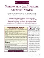
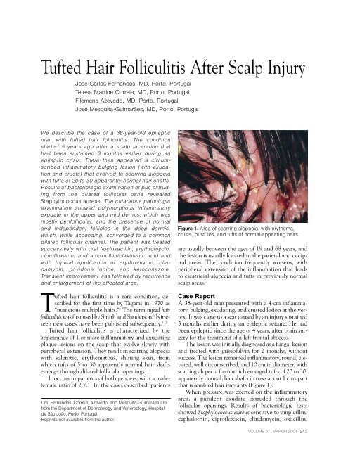
Figure 1. Area of scarring alopecia, with erythema,<br />
crusts, pustules, and tufts of normal-appearing hairs.<br />
are usually between the ages of 19 and 68 years, and<br />
the lesion is usually located in the parietal and occipital<br />
areas. The condition frequently worsens, with<br />
peripheral extension of the inflammation that leads<br />
to cicatricial alopecia and tufts in previously normal<br />
scalp areas. 7<br />
Case Report<br />
A 38-year-old man presented with a 4-cm inflammatory,<br />
bulging, exudating, and crusted lesion at the vertex.<br />
It was close to a scar caused by an injury sustained<br />
3 months earlier during an epileptic seizure. He had<br />
been epileptic since the age of 4 years, after brain surgery<br />
for the treatment of a left frontal abscess.<br />
The lesion was initially diagnosed as a fungal kerion<br />
and treated with griseofulvin for 2 months, without<br />
success. The lesion remained inflammatory, round, elevated,<br />
well circumscribed, and 10 cm in diameter, with<br />
scarring alopecia from which emerged tufts of 20 to 30,<br />
apparently normal, hair shafts in rows about 1 cm apart<br />
that resembled hair implants (Figure 1).<br />
When pressure was exerted on the inflammatory<br />
area, a purulent exudate extruded through the<br />
follicular openings. Results of bacteriologic tests<br />
showed Staphylococcus aureus sensitive to ampicillin,<br />
cephalothin, ciprofloxacin, clindamycin, oxacillin,<br />
VOLUME 67, MARCH 2001 243
TUFTED HAIR FOLLICULITIS<br />
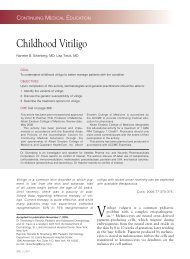
Figure 2. Convergence of several follicles into a common<br />
dilated infundibulum (H&E).<br />
tetracycline, trimethoprim sulfamethoxazole, and<br />
vancomycin. Several fungal cultures were negative.<br />
The remainder of the scalp was normal; there<br />
were no enlarged cervical lymph nodes, and the<br />
patient was in generally good health. Examination of<br />
a skin biopsy specimen showed an inflammatory<br />
process in the perifollicular dermis, with neutrophils,<br />
lymphocytes, plasma cells, and eosinophils. There<br />
was interfollicular fibrosis in the upper dermis, and<br />
the hairs merged into a common follicular ostium<br />
(Figure 2). The general analytical study was normal<br />
or negative.<br />
For the 5 years since the lesions had appeared, the<br />
patient was treated systemically with flucloxacillin,<br />
erythromycin, ciprofloxacin, and amoxicillin/<br />
clavulanic acid and topically with erythromycin,<br />
clindamycin, povidone iodine, and ketoconazole.<br />
However, because the patient experienced a worsening<br />
of epileptic seizures when taking antibiotics, he<br />
discontinued the medication prematurely. As a result,<br />
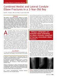
the affected area grew centrifugally until it covered<br />
the entire central portion of the scalp, leaving a normal<br />
rim of about 5 cm (Figure 3).<br />
Recently, with the addition of vigabatrin to carbamazepine<br />
and phenobarbital (the antiepileptic<br />
agents he had been taking previously), better control<br />
of the epilepsy was achieved, allowing the patient to<br />
Figure 3. Transition between the central affected area<br />
and the peripheral, normal-appearing scalp.<br />
continue with systemic amoxicillin/clavulanic acid<br />
(625 mg every 8 h) for 3 weeks and then with flucloxacillin<br />
(500 mg every 8 h) for 4 weeks. A marked<br />
reduction of the inflammatory signs was observed but<br />
only during treatment periods.<br />
Comment<br />
The cause of tufted hair folliculitis is unknown, but<br />
several explanations have been proposed. It is considered<br />
by some authors as a variant of folliculitis<br />
decalvans. 1,13 Since S aureus is almost always isolated<br />
in this situation, it may be a recurrent staphylococal<br />
folliculitis with fibrosis in the interfollicular<br />
areas and consequent approximation of the follicles,<br />
with hairs emerging in tufts. 2,8 In addition, it<br />
has been suggested that tuft formation is due to<br />
telogenic hairs being retained around an anagenic<br />
follicle, 6 but it has since been demonstrated that<br />
most follicles in a tuft are anagenic. 8 It also may be<br />
a localized nevoid malformation, with tufts present<br />
since birth but becoming apparent only when infection<br />
occurs with the destruction of some of the<br />
follicles. 5 The compound follicles are more prone to<br />
chronic infection by S aureus, 14 but this theory does<br />
not explain the centrifugal nature of the lesions nor<br />
the appearance of tufts in previously normal scalp<br />
areas. Pujol et al 15 suggest that hair tufting may be a<br />
244 CUTIS ®
TUFTED HAIR FOLLICULITIS<br />
nonspecific secondary phenomenon that may occur<br />
in several exudative inflammatory diseases of the<br />
scalp, including dissecting cellulitis of the scalp,<br />
folliculitis decalvans, and folliculitis keloidalis.<br />
In our patient, the lesion started shortly after he<br />
sustained a scalp injury. In 4 clinical cases described<br />
previously, there was also a reference to traumatic or<br />
surgical injury to the scalp sometime before the onset<br />
of dermatosis. 8,11 However, most authors give little<br />
importance to this fact and mention it without further<br />
consideration. The diagnosis of tufted hair folliculitis<br />
is usually late, and minor trauma occurring<br />
sometime before might not be mentioned by the<br />
patient. As a result, its frequency may not be appreciated.<br />
It seems reasonable that injuries may allow<br />
the installation of a staphylococcal infection that, for<br />
reasons not yet understood, become chronic and<br />
could be caused by host factors, such as an immunologic<br />
defect that results in greater susceptibility to<br />
S aureus infection. 7<br />
The most significant fact in the pathology of<br />
tufted hair folliculitis is the convergence of several<br />
follicles toward a common follicular duct. The lowest<br />
portions of the follicles are normal, each with a papilla<br />
and independent internal and external root<br />
sheaths, which differentiate them from pili multigemini.<br />
16 In the superior and mid dermis, there is an<br />
inflammatory infiltrate with neutrophils, eosinophils,<br />
lymphocytes, and plasma cells, mostly perifollicular.<br />
Rupture of the follicular wall and the presence of hair<br />
debris in the macrophage cytoplasm and in multinucleate<br />
giant cells have been reported 3,6-8,11 but were not<br />
seen in our patient.<br />
Antibiotics administered systemically and topically<br />
have been the most commonly used treatment.<br />
However, complete cures are rare. 8,11 As a rule, the<br />
antibiotics allow reasonable control of the inflammatory<br />
signs, but discontinuing their use leads—after a<br />
period from weeks to months—to the reappearance<br />
of lesions. 7<br />
Isotretinoin, 7 zinc sulfate, 7,8 and rifampin 8 are used<br />
without great success. Surgery, while technically feasible,<br />
5 seems to be effective only at an early stage,<br />
which is why early diagnosis is essential.<br />
REFERENCES<br />
1. Tagami H. Numerous multiple hairs. Arch Dermatol. 1970;<br />
102:309-312.<br />
2. Smith NP, Sanderson KV. <strong>Tufted</strong> folliculitis of the scalp. J<br />
R Soc Med. 1978;71:606-608.<br />
3. Metz J, Metz G. Navoide bundelhaare beim menschen.<br />
Hautarzt. 1978;29:586-589.<br />
4. Oakley A, Scollay D. <strong>Hair</strong> bundles. A presentation of folliculitis.<br />
Aust J Dermatol. 1985;26:139-143.<br />
5. Tong AKF, Baden HP. <strong>Tufted</strong> hair folliculitis. J Am Acad<br />
Dermatol. 1989;21:1096-1099.<br />
6. Dalziel KL, Telfer NR, Wilson CL, et al. <strong>Tufted</strong> folliculitis: a<br />
specific bacterial disease? Am J Dermatopathol. 1990;12:37-41.<br />
7. Pujol RM, Matias-Guíu X, Garcia-Patos V, et al. <strong>Tufted</strong>hair<br />
folliculitis. Clin Exp Dermatol. 1991;16:199-201.<br />
8. Luelmo-Aguilar J, Gonzalez-Castro U, Castells-Rodellas A.<br />
<strong>Tufted</strong> hair folliculitis: a study of four cases. Br J Dermatol.<br />
1993;128:454-457.<br />
9. Offidani A, Cellini A, Giangiacomi M, et al. Folliculite<br />
épilante de quinquaud et cheveux en touffes. Ann Dermatol<br />
Venereol. 1994;121:319-321.<br />
10. Secch T, Balme B, Thomas L, et al. Folliculites en touffes<br />
du cuir chevelu. Ann Dermatol Venereol. 1994;121:479-481.<br />
11. Veraldi S, Grimalt R, Cappio F, et al. <strong>Tufted</strong> hair folliculitis.<br />
Eur J Dermatol. 1995;5:125-127.<br />
12. Lombardo M, Collina G, Giroloni G. Guess what! (tufted<br />
hair folliculitis). Eur J Dermatol. 1996;6:75-76.<br />
13. Champion RH, Burton JL, Ebling FJG. In: Rook/Wilkinson/<br />
Ebling: Textbook of Dermatology. 5th ed. Oxford, England:<br />
Blackwell Scientific Publications; 1992:2600-2601.<br />
14. Loewenthal LJA. “Compound” and grouped hairs of the human<br />
scalp: their connection with follicular infections. J Invest<br />
Dermatol. 1947;8:263-273.<br />
15. Pujol RM, García-Patos V, Ravella-Mateu A, et al. <strong>Tufted</strong><br />
hair folliculitis: a specific disease? Br J Dermatol. 1994;130:<br />
259-260.<br />
16. Pinkus H. Multiple hairs: report of two cases of pili multigemini<br />
and discussion of some other anomalies of the pilary<br />
complex. J Invest Dermatol. 1951;17:291-301.<br />
VOLUME 67, MARCH 2001 245