Hemolysis - Emory University School of Medicine
Hemolysis - Emory University School of Medicine
Hemolysis - Emory University School of Medicine

You also want an ePaper? Increase the reach of your titles
YUMPU automatically turns print PDFs into web optimized ePapers that Google loves.
Diseases Presenting<br />
With RBC Breakup<br />
By Allan Platt, PA-C, MMSc<br />
Allan Platt is on the faculty at the <strong>Emory</strong> <strong>University</strong> <strong>School</strong> <strong>of</strong> <strong>Medicine</strong> PA<br />
program in Atlanta.<br />
Red blood cells (RBCs) normally have a 120-day lifespan; the<br />
premature breaking apart <strong>of</strong> RBCs is called hemolysis. When<br />
RBC destruction outpaces bone marrow RBC production,<br />
hemolytic anemia occurs. Causes <strong>of</strong> hemolytic anemia include defects<br />
within the RBCs or from elements in the circulation. 1 Many systemic<br />
diseases present with hemolysis, such as hereditary red cell disorders,<br />
systemic infections, cancer, leukemia, immune disorders, liver disease,<br />
enzyme deficiencies and clotting disorders. 1 This article discusses the<br />
pathophysiology <strong>of</strong> hemolysis, the diagnostic workup and the treatment<br />
<strong>of</strong> diseases that cause hemolytic anemia. Clinicians can use clues<br />
from the history, physical examination and laboratory blood tests to<br />
help diagnose the cause <strong>of</strong> hemolysis. Patients should be referred to<br />
hematologists if the cause is not self-limiting.<br />
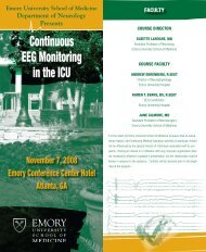
Pathophysiology<br />
RBCs are made in the bone marrow from stem cells<br />
under the stimulation <strong>of</strong> the kidney-produced<br />
hormone erythropoietin. Materials for RBC production,<br />
including iron, B 12<br />
, folic acid, protein<br />
and lipids, are transported from the gut or<br />
storage to the bone marrow factory. As the<br />
RBC matures, it manufactures hemoglobin<br />
and extrudes its nucleus before being released<br />
into the blood circulation as a reticulocyte.<br />
The reticulocyte count is the best indicator <strong>of</strong><br />
the response <strong>of</strong> the bone marrow factory to<br />
RBC loss; the count should be elevated in<br />
the presence <strong>of</strong> hemolysis, during which<br />
the bone marrow responds by making<br />
new red cells.<br />
Anemia is defined as low hemoglobin,<br />
low hematocrit or low red cell<br />
mass on the complete blood count<br />
(CBC). Anemia occurs if bone marrow<br />
production cannot keep pace with the<br />
hemolytic destruction. RBCs are recycled<br />
in the spleen, where hemoglobin<br />
is broken down into indirect (unconjeffrey<br />
leeser, scott frymoyer
liver or spleen may be the source <strong>of</strong> red cell destruction in lymphoma, metastatic<br />
cancer, portal hypertension and mononucleosis. 1,2<br />
jugated) bilirubin, iron and protein. <strong>Hemolysis</strong><br />
causes an elevated indirect bilirubin, and if the level<br />
is greater than 3 mg/dL, jaundice occurs. The enzyme lactate dehydrogenase<br />
(LDH) is present in abundance in RBCs. The combination <strong>of</strong> anemia, high<br />
reticulocyte count, high indirect bilirubin and high LDH is the classic biomarker<br />
pr<strong>of</strong>ile <strong>of</strong> hemolysis. 1,2<br />
<strong>Hemolysis</strong> occurs from within the red cell in the presence <strong>of</strong> genetic mutations<br />
in the hemoglobin structure, red cell membrane problems, enzyme deficiencies<br />
or parasite invasion. Mechanical destruction occurs outside the red cell<br />
in the circulation from fibrin threads, disseminated intravascular coagulation<br />
(DIC), thrombotic thrombocytopenic purpura (TTP) or hemolytic uremic syndrome<br />
(HUS), prosthetic heart valves or immune attack from antigen-antibody<br />
reactions. The patient’s history, physical examination, laboratory values and<br />
certain radiologic findings can help guide the differential diagnosis. 1<br />
Clues From the Medical History<br />
Anemic patients complain <strong>of</strong> nonspecific symptoms, including increased<br />
fatigue, generalized weakness, shortness <strong>of</strong> breath, non-vertigo dizziness<br />
and palpitations. Jaundiced sclera should prompt a hemolysis workup.<br />
Fever and recent travel to malaria-, Bartonella- or Babesia-endemic areas<br />
should prompt specific testing. A history <strong>of</strong> lifelong anemia, early gallstones<br />
and a positive family history point to genetic causes such as sickle<br />
cell disease, thalassemia, spherocytosis or elliptocytosis. <strong>Hemolysis</strong> after<br />
exposure to a sulfa medication, antimalarials or fava beans suggests<br />
glucose-6-phosphate dehydrogenase (G6PD) deficiency. 1,2<br />
Exposure to toxigenic Escherichia coli from contaminated undercooked<br />
meat or water can cause HUS. Pregnancy and hemolysis might indicate<br />
preeclampsia and HELLP syndrome (hemolytic anemia, elevated liver<br />
enzymes and low platelet count). Mechanical heart valve placement also<br />
might be the cause <strong>of</strong> hemolysis. 1,2<br />
Clues From the Physical Examination<br />
Physical examination may reveal jaundiced sclera and pallor in the<br />
palpebral conjunctiva, nail beds or palmar creases. The pulse rate may be<br />
increased. Fever indicates systemic infection or inflammation. Petechiae or<br />
skin bruises indicate low or dysfunctional platelets, DIC, HUS or TTP. 1,2<br />
Elevated blood pressure in pregnancy indicates preeclampsia/eclampsia,<br />
and very high blood pressure indicates malignant hypertension. An enlarged<br />
Clues From Lab Values<br />
The first-pass laboratory workup <strong>of</strong> hemolysis should include urinalysis,<br />
CBC, reticulocyte count, red cell morphology, complete metabolic pr<strong>of</strong>ile<br />
(CMP), direct and indirect Coombs test, hemoglobin electrophoresis, Heinz<br />
body stain and osmotic fragility test (OFT). Analyzing these results in combination<br />
<strong>of</strong>fer clues to the systemic cause. 1,2 Free hemoglobin may pass through<br />
the glomeruli, causing a red-brown cola-colored urine that tests positive for<br />
hemoglobin, but with no RBCs seen. 1<br />
A high reticulocyte count with an elevated LDH level and an elevated<br />
indirect bilirubin indicates active hemolysis. An abnormal hemoglobin<br />
electrophoresis with decreased hemoglobin A, increased hemoglobin F and<br />
hemoglobin A 2<br />
and microcytic anemia with an increased red cell count all<br />
indicate beta thalassemia. Target cells also may be seen on the peripheral<br />
smear.<br />
Hemoglobin electrophoresis also will identify hereditary hemoglobinopathies<br />
such as sickle cell disease. The Heinz body stain will be positive in G6PD<br />
deficiency. The direct Coombs test will be positive if there is antibody on the<br />
red cells, and the indirect Coombs will be positive if there is antibody in the<br />
serum. A thin and thick blood smear should be scanned for red cell morphology<br />
and intracellular parasites such as malaria and babesiosis, depending on<br />
the travel history. The OFT is helpful to identify red cell membrane problems<br />
such as spherocytosis, ovalocytosis and elliptocytosis. Low platelets, elevated<br />
prothrombin time (PT) and increased activated partial thromboplastin time<br />
(aPTT) may indicate HUS, DIC or TTP. Erythrocyte enzyme assays will help<br />
diagnose enzyme deficiencies. 1,2<br />
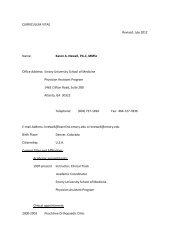
A blood smear read by a trained technologist will give the red cell morphology<br />
and can <strong>of</strong>fer significant clues to the cause <strong>of</strong> hemolysis; thus, it<br />
should be among the first tests ordered to narrow the cause. 3 This test is not<br />
a part <strong>of</strong> the CBC and must be requested. A suspicion <strong>of</strong> red cell parasites<br />
should be communicated to the hematology laboratory so that thick smears<br />
can be performed to scan more red cells. Table 1 lists the different red cell<br />
shapes and their significance.<br />
Radiologic findings <strong>of</strong> bone marrow expansion indicate chronic hemolysis,<br />
and one <strong>of</strong> the hereditary causes should be suspected.<br />
Differential Diagnosis<br />
The differential diagnosis <strong>of</strong> diseases presenting with hemolysis can be<br />
remembered with the mnemonic HEMATOLOGIST.<br />
Hemoglobinopathy. Sickle cell disease is one <strong>of</strong> the most common hemoglobinopathies.<br />
It is a family <strong>of</strong> hemoglobin phenotypes including types SS, SC,<br />
S beta + thalassemia, S beta 0 thalassemia, SD, SE, SS with persistent fetal, SG<br />
and SOArab. The definitive test is quantitative hemoglobin electrophoresis.<br />
Depending on phenotype, there is a wide spectrum <strong>of</strong> disease manifestations<br />
from mild to very severe. Patients with complications should be<br />
Editor’s Note<br />
ADVANCE for Physician Assistants and the Eugene Applebaum<br />
College <strong>of</strong> Pharmacy and Health Sciences, and<br />
Wayne State <strong>University</strong> <strong>School</strong> <strong>of</strong> <strong>Medicine</strong>, are pleased<br />
to <strong>of</strong>fer this continuing education opportunity.<br />
The Wayne State <strong>University</strong> <strong>School</strong> <strong>of</strong> <strong>Medicine</strong> is<br />
accredited by the Accreditation Council for Continuing<br />
Medical Education to provide continuing medical education<br />
for physicians.<br />
The Wayne State <strong>University</strong> <strong>School</strong> <strong>of</strong> <strong>Medicine</strong> designates<br />
this educational activity for a maximum <strong>of</strong> one AMA<br />
PRA Category 1 Credit(s). Physicians should only claim<br />
credit commensurate with the extent <strong>of</strong> their participation<br />
in the activity.<br />
To receive 1 hour <strong>of</strong> AMA PRA Category 1 CME credit,<br />
read this article and follow the directions on the answer<br />
form at the end <strong>of</strong> the article.<br />
Learning Objectives<br />
1. Discuss the pathophysiology <strong>of</strong> hemolysis.<br />
2. Describe the clues from the medical history that point<br />
to hemolysis.<br />
3. Understand the laboratory workup for a patient with<br />
hemolysis.<br />
4. Review the differential diagnosis <strong>of</strong> hemolysis.<br />
Disclosure <strong>of</strong> Conflict <strong>of</strong> Interest<br />
Allan Platt indicates no relationships to disclose related<br />
to the contents <strong>of</strong> this article.<br />
The CME coordinator for ADVANCE for Physician Assistants,<br />
John McGinnity, MS, PA-C, discloses receiving<br />
honoraria from Novartis and Omron Healthcare and that<br />
he is on the speakers’ bureau for Pfizer.<br />
www.advanceweb.com/pa • January-February 2009 • Advance for Physician Assistants 21
Hematology CME<br />
Table 1<br />
RBC Morphology From a Peripheral Smear<br />
Red Cell Morphology<br />
Burr cells<br />
Spur cells<br />
Stomatocytes<br />
Spherocytes<br />
Schistocytes<br />
(helmet cells)<br />
Elliptocyte<br />
Teardrop cells<br />
Sickle cells<br />
Target cells<br />
Parasites<br />
Bite cells<br />
Significance<br />
managed by comprehensive sickle cell centers if<br />
available. All children should be on prophylactic<br />
daily penicillin and screened for stroke risk<br />
using transcranial Doppler ultrasound (TCD).<br />
Bone marrow transplant has been curative but<br />
carries 10% mortality, and there are few patients<br />
with sibling matches. Hydroxyurea is an effective<br />
preventive medication that decreases pain crisis,<br />
blood transfusions and hospital admissions. It<br />
has been shown to prolong life and is effective in<br />
children in preventing complications. 4<br />
Hemoglobinuria. Paroxysmal nocturnal hemoglobinuria<br />
manifestations include episodic intravascular<br />
hemolysis, hemoglobinuria, abdominal<br />
pain, thrombotic episodes and increased infections.<br />
Red cells are more susceptible to complement-mediated<br />
injury, causing hemolysis and<br />
burgundy-colored urine. <strong>Hemolysis</strong> can occur<br />
at any time <strong>of</strong> day, but urine findings are most<br />
noticeable in the morning, when the urine is<br />
concentrated. Diagnosis is based on the presence<br />
<strong>of</strong> positive sucrose and acid hemolysis tests.<br />
Treatment is reserved for those with severe symptoms<br />
and includes folate for red cell production<br />
Uremia, liver disease, low potassium, posttransfusion state, stomach<br />
cancer, bleeding peptic ulcers<br />
Abetalipoproteinemia, postsplenectomy state, alcoholic liver disease,<br />
malabsorptive states<br />
Hereditary, alcoholism, cirrhosis, obstructive liver disease<br />
Hereditary, immune hemolytic anemia, posttransfusion state, hemolytic<br />
anemia, water dilution hemolysis, fragmentation hemolysis<br />
Thrombotic thrombocytopenic purpura, disseminated intravascular<br />
coagulation, vasculitis, glomerulonephritis, renal graft rejection,<br />
carcinomatosis, heart valve hemolysis, burns<br />
Hereditary, thalassemia, iron deficiency<br />
Myelophthistic anemias, thalassemia<br />
Sickle cell disease<br />
Obstructive liver disease, hemoglobinopathies, thalassemias, iron<br />
deficiency, postsplenectomy state<br />
Malaria, babesiosis, bartonella<br />
G6PD deficiency<br />
Adapted in part from: Bull BS. Morphology <strong>of</strong> the erythron. In: Lichtman MA, Beutler E, Kipps TJ, Seligsohn U,<br />
Kaushansky K, Prchal JT, eds. Williams Hematology. 7th ed. New York, NY: McGraw-Hill; 2005:369-385.<br />
and eculizumab, an FDA-approved humanized<br />
monoclonal antibody against complement protein<br />
C5 that inhibits terminal complement activation. 5<br />
Enzyme deficiency. Inherited pyruvate kinase<br />
(PK) deficiency is the most common cause <strong>of</strong><br />
hereditary nonspherocytic hemolytic anemia.<br />
There is a history <strong>of</strong> lifelong anemia and complications<br />
similar to those <strong>of</strong> hereditary hemolytic<br />
anemia. There is no specific treatment, only supportive<br />
care including transfusion for symptomatic<br />
anemia. Other rare red cell enzyme deficiencies<br />
include pyrimidine-5’-nucleotidase deficiency,<br />
phosph<strong>of</strong>ructokinase deficiency, phosphoglycerate<br />
kinase deficiency, aldolase deficiency and<br />
triosephosphate isomerase deficiency. These are<br />
diagnosed by performing erythrocyte enzyme<br />
assays. These patients should be referred to a<br />
hematologist for lifelong management. 6<br />
Membrane problems. Hereditary spherocytosis<br />
(HS) is caused by the inheritance <strong>of</strong> red cell membrane<br />
protein abnormalities that result in sphere<br />
formation, membrane budding and increased<br />
permeability to sodium. The red cells appear as<br />
round red balls instead <strong>of</strong> the normal biconcave<br />
disk shape with an area <strong>of</strong> central pallor. 7<br />
Hereditary elliptocytosis is caused by an abnormal<br />
structural protein, which causes a defective<br />
red cell membrane. Elliptocytes resemble Good &<br />
Plenty candy pieces on a peripheral smear.<br />
The shape <strong>of</strong> ovalocytes is somewhere between<br />
the elongated elliptocytes and the beach ball<br />
appearance <strong>of</strong> spherocytes. Mutations in a number<br />
<strong>of</strong> distinct genes account for hereditary<br />
spherocytosis and elliptocytosis, while a single<br />
genetic defect accounts for all cases <strong>of</strong> hereditary<br />
ovalocytosis. 8<br />
Antibodies. In immune acquired hemolytic anemia,<br />
hemolysis is caused by antibodies or complement<br />
proteins attached to the red cell mem brane.<br />
The direct Coombs test is positive in the presence<br />
<strong>of</strong> antibody on the red cell membrane, and the<br />
indirect Coombs test is positive in the presence <strong>of</strong><br />
antibody in the patient’s serum. Causes include<br />
alloimmune, autoimmune and drug-induced<br />
hemolytic anemia. 9,10<br />
Warm-reacting IgG antibodies occur secondary<br />
to lymphoproliferative syndrome (30%), collagen<br />
vascular diseases (20%), other tumors (20%) or<br />
as idiopathic disease (20%). Laboratory findings<br />
are anemia with an increased reticulocyte count,<br />
and blood smear shows microspherocytes. Direct<br />
Coombs test is positive for IgG or IgG and C’. This<br />
is respon sive to steroids and/or splenectomy. 9,10<br />
Cold-reacting IgM antibodies occur secondary<br />
to viral and mycoplasma infections, with lymphoproliferative<br />
disease and as idiopathic disorders<br />
in the elderly. Patients are usually unresponsive<br />
to treatment with steroids or splenectomy. 9,10<br />
Trauma to the red cells. Artificial heart valves<br />
can cause mechanical destruction <strong>of</strong> RBCs as<br />
they pass through the valve. <strong>Hemolysis</strong> usually<br />
is mild and subclinical but is severe in up to 15%<br />
<strong>of</strong> patients with certain prostheses, such as ballcage<br />
and bileaflet valves. It is uncommon with<br />
tissue valves, although hemolytic anemia may be<br />
the initial presentation <strong>of</strong> porcine valve failure. 11<br />
The peripheral smear shows variable numbers<br />
<strong>of</strong> schistocytes and smaller red cell fragments.<br />
<strong>Hemolysis</strong> also can occur during vigorous sports<br />
activities or running on hard surfaces. This usually<br />
is self-limited and does not require treatment. 12<br />
Overactive clotting system. When clotting occurs<br />
in multiple sites in the vascular system, the fibrin<br />
threads act like razor wire to slice red cells as they<br />
speed through. This causes a hemolytic picture,<br />
and schistocyte or helmet cells are seen in the<br />
peripheral blood smear. Lab results may show<br />
22 Advance for Physician Assistants • January-February 2009 • www.advanceweb.com/pa
Hematology CME<br />
elevated PT and aPTT. Among the conditions that<br />
can cause this are TTP, HUS, DIC, preeclampsia,<br />
HELLP syndrome and malignant hypertension. A<br />
hematology consultation is recommended. 13,14<br />
Liver disease. Severe liver disease from any<br />
cause may be associated with abnormal lipid<br />
loading in red cell membranes, causing spur cell<br />
formation and a shortened red cell survival. Zieve<br />
syndrome is associated with alcoholic fatty liver<br />
and cirrhosis, severe upper abdominal and right<br />
upper quadrant pain, jaundice, hyperlipidemia<br />
and hemolytic anemia. The condition improves<br />
when alcohol consumption is stopped. 15<br />
Oral ingestions and other agents. Lead poisoning<br />
can cause central nervous system symptoms,<br />
hepatitis, renal insufficiency, hypertension,<br />
abdominal pain and hemolytic anemia. Acute<br />
and occasionally severe hemolytic anemia is a<br />
rare complication <strong>of</strong> therapy with interferon alfa.<br />
Amyl nitrite and butyl nitrite, primarily via inhalation,<br />
can cause hemolysis. Topical anesthesia<br />
with benzocaine or lidocaine, either as a spray<br />
or cream, can cause severe methemoglobinemia<br />
with cyanosis and dyspnea. Dapsone, used in<br />
the treatment <strong>of</strong> leprosy, can cause hemolysis<br />
and methemoglobinemia. Ribavirin used to treat<br />
hepatitis C infection has been reported to cause<br />
hemolytic anemia. 16<br />
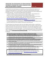
G6PD. The inheritance <strong>of</strong> G6PD deficiency is<br />
X‐linked. Males who inherit the deficient gene<br />
are fully affected. Homozygous females also are<br />
fully affected. Drugs or other oxidants may cause<br />
increased oxidant stress within the red cell, causing<br />
precipitation <strong>of</strong> hemoglobin into Heinz bodies<br />
and thereby causing hemolysis. Demonstration<br />
<strong>of</strong> Heinz bodies with methyl violet stain is the<br />
most useful test during acute hemolysis. G6PD<br />
deficiency can be confirmed by enzyme assay<br />
in two to three months when the erythrocytes<br />
have a more normal age distribution. With the<br />
Mediterranean variant, activity usually is low<br />
during the acute hemolytic episode. The patient<br />
should be educated to avoid medications listed<br />
in Table 2. 1,17,18<br />
Infection. Intra-erythrocytic parasites (malaria<br />
and babesiosis) lyse red cells as they emerge.<br />
These parasites can be identified on examination<br />
<strong>of</strong> a thick smear slide. Clostridium perfringens septicemia<br />
may cause massive hemolysis. 19 Bartonella,<br />
or Oroya fever, is caused by Bartonella bacilliformis<br />
and transmitted by sand flies <strong>of</strong> the genus<br />
Phlebotomus upon travel to the Peruvian Andes. 20<br />
Another tropical parasite that causes hemolysis is<br />
Table 2<br />
Causes <strong>of</strong> <strong>Hemolysis</strong> in G6PD Deficiency<br />
Aspirin (high dose, >3 g/day)<br />
Dapsone/chlorproguanil<br />
TMP-SMX<br />
Furazolidone<br />
Isobutyl nitrite<br />
Methylene blue<br />
Nalidixic acid<br />
Adapted from: Dhaliwal G, Cornett PA, Tierney LM Jr. Hemolytic anemia. Am Fam Physician. 2004;69(11):2599-<br />
2606; Cappellini MD, Fiorelli G. Glucose-6-phosphate dehydrogenase deficiency. Lancet. 2008;371(9606):64-74;<br />
and Frank JE. Diagnosis and management <strong>of</strong> G6PD deficiency. Am Fam Physician. 2005;72(7):1277-1282.<br />
Leishmania donovani. Fever, hemolysis and travel<br />
to an endemic area should prompt testing.<br />
Splenic destruction. Splenomegaly and hypersplenism<br />
can occur secondary to infections, portal<br />
hypertension, infiltration with leukemia or<br />
lymphoma and collagen vascular diseases. The<br />
spleen is the primary site <strong>of</strong> red cell recycling.<br />
When the spleen is congested, red cell destruction<br />
can be accelerated, causing hemolytic anemia. The<br />
underlying cause should be treated, or the spleen<br />
can be removed surgically. 21<br />
Thalassemias. Thalassemias must be considered<br />
in a hemolytic anemia with a low mean corpuscular<br />
volume. Thalassemia, the most common<br />
inherited cause <strong>of</strong> anemia worldwide, is caused<br />
by the inheritance <strong>of</strong> genetic abnormalities that<br />
decrease synthesis <strong>of</strong> alpha or beta chains, leading<br />
to decreased production <strong>of</strong> hemoglobin A. This<br />
causes a lifelong microcytic anemia with hemolysis.<br />
Clues from the CBC include low hemoglobin<br />
and an increased red cell count; the mean corpuscular<br />
hemoglobin concentration is low, and the<br />
red blood cell distribution width is normal. The<br />
peripheral smear may show target cells. 22 <br />
Nitr<strong>of</strong>urantoin<br />
Niridazole<br />
Phenazopyridine<br />
Phenylhydrazine<br />
Pamaquine<br />
Primaquine<br />
Sulfacetamide<br />
Sulfapyridine<br />
Thiazole sulfone<br />
Toluidine blue<br />
Trinitrotoluene<br />
Fava beans<br />
Ascorbic acid (>1 g)<br />
Vitamin K analogs<br />
References<br />
1. Dhaliwal G, Cornett PA, Tierney LM Jr. Hemolytic anemia.<br />
Am Fam Physician. 2004;69(11):2599-2606.<br />
2. Tefferi A. Anemia in adults: a contemporary approach to<br />
diagnosis. Mayo Clin Proc. 2003;78(10):1274-1280.<br />
3. Ryan DH. Examination <strong>of</strong> the blood. In: Lichtman MA,<br />
Beutler E, Kipps TJ, Seligsohn U, Kaushansky K, Prchal JT,<br />
eds. Williams Hematology. 7th ed. New York, NY: McGraw-Hill;<br />
2005:11-20.<br />
4. Frenette PS, Atweh GF. Sickle cell disease: old discoveries,<br />
new concepts, and future promise. J Clin Invest. 2007;117(4):850-<br />
858.<br />
5. Brodsky RA. Narrative review: paroxysmal nocturnal<br />
hemoglobinuria: the physiology <strong>of</strong> complement-related hemolytic<br />
anemia. Ann Intern Med. 2008;148(8):587-595.<br />
6. Zanella A, Fermo E, Bianchi P, Valentini G. Red cell<br />
pyruvate kinase deficiency: molecular and clinical aspects. Br J<br />
Haematol. 2005;130(1):11-25.<br />
7. Bolton-Maggs PH, Stevens RF, Dodd NJ, Lamont G,<br />
Tittensor P, King MJ; General Haematology Task Force <strong>of</strong> the<br />
British Committee for Standards in Haematology. Guidelines for<br />
the diagnosis and management <strong>of</strong> hereditary spherocytosis. Br J<br />
Haematol. 2004;126(4):455-474.<br />
8. An X, Mohandas N. Disorders <strong>of</strong> red cell membrane. Br J<br />
Haematol. 2008;141(3):367-375.<br />
9. Gertz MA. Cold agglutinin disease and cryoglobulinemia.<br />
Clin Lymphoma. 2005;5(4):290-293.<br />
10. Packman CH. Hemolytic anemia due to warm autoantibodies.<br />
Blood Rev. 2008;22(1):17-31.<br />
11. Ismeno G, Renzulli A, Carozza A, et al. Intravascular<br />
hemolysis after mitral and aortic valve replacement with different<br />
types <strong>of</strong> mechanical prostheses. Int J Cardiol. 1999;69(2):179-<br />
183.<br />
12. Abarbanel J, Benet AE, Lask D, Kimche D. Sports hematuria.<br />
J Urol. 1990;143(5):887-890.<br />
13. Razzaq S. Hemolytic uremic syndrome: an emerging<br />
health risk. Am Fam Physician. 2006;74(6):991-996.<br />
14. Sibai BM. Imitators <strong>of</strong> severe preeclampsia. Obstet Gynecol.<br />
2007;109(4):956-966.<br />
15. Shiraishi K, Matsuzaki S, Itakura M, Ishida H. Abnormality<br />
in membrane fatty acid compositions <strong>of</strong> cells measured on erythrocyte<br />
in alcoholic liver disease. Alcohol Clin Exp Res. 1996;20(1<br />
suppl):56A-59A.<br />
16. Schrier SL. Extrinsic nonautoimmune hemolytic anemia<br />
due to drugs and toxins. UpToDate. Updated March 6, 2008.<br />
Accessed January 9, 2009.<br />
17. Cappellini MD, Fiorelli G. Glucose-6-phosphate dehydrogenase<br />
deficiency. Lancet. 2008;371(9606):64-74.<br />
18. Frank JE. Diagnosis and management <strong>of</strong> G6PD deficiency.<br />
Am Fam Physician. 2005;72(7):1277-1282.<br />
19. Schrier SL. Extrinsic nonimmune hemolytic anemia due<br />
to systemic disease. UpToDate. Updated June 4, 2008. Accessed<br />
January 9, 2009.<br />
20. Eremeeva ME, Gerns HL, Lydy SL, et al. Bacteremia,<br />
fever, and splenomegaly caused by a newly recognized bartonella<br />
species. N Engl J Med. 2007;356(23):2381-2387.<br />
21. Caro J. Hypersplenism and hyposplenism. In: Lichtman<br />
MA, Beutler E, Kipps TJ, Seligsohn U, Kaushansky K, Prchal JT,<br />
eds. Williams Hematology. 7th ed. New York, NY: McGraw-Hill;<br />
2005:773-778.<br />
22. Cunningham MJ. Update on thalassemia: clinical care<br />
and complications. Pediatr Clin North Am. 2008;55(2):447-460.<br />
www.advanceweb.com/pa • January-February 2009 • Advance for Physician Assistants 23
Hematology CME<br />
Self-Assessment Questions: <strong>Hemolysis</strong><br />
1. Which one <strong>of</strong> the following is the normal<br />
lifespan <strong>of</strong> a red blood cell?<br />
a. 35 days b. 75 days<br />
c. 120 days d. 170 days<br />
2. Red blood cells are produced made in the bone<br />
marrow from which one <strong>of</strong> the following cells?<br />
a. Mast cells b. Parietal cells<br />
c. Stem cells d. Glial cells<br />
3. Which one <strong>of</strong> the following structures recycles<br />
red blood cells and is where hemoglobin<br />
is broken down into indirect bilirubin, iron,<br />
and protein?<br />
a. The liver b. The spleen<br />
c. The ileum d. The duodenum<br />
4. <strong>Hemolysis</strong> causes an elevated indirect<br />
bilirubin, and if the level is greater than 3<br />
mg/dL, jaundice occurs.<br />
a. True b. False<br />
5. <strong>Hemolysis</strong> after exposure to all <strong>of</strong> the<br />
following substances except which one suggests<br />
glucose-6-phosphate dehydrogenase<br />
(G6PD) deficiency?<br />
a. A sulfa medication b. Erythromycin<br />
c. Antimalarials d. Fava beans<br />
6. First-pass laboratory workup <strong>of</strong> hemolysis<br />
should include all <strong>of</strong> the following except<br />
Registration/Answer Form<br />
For accuracy, please print clearly.<br />
which one?<br />
a. Urinalysis<br />
b. Direct and indirect Coombs test<br />
c. Reticulocyte count<br />
d. Liver function tests<br />
To receive 1 hour <strong>of</strong> AMA PRA Category 1 CME credit, read the preceding article and<br />
mark your responses on this form. You must complete all questions to receive credit.<br />
Then return this form and the $10 fee to the address below. Make checks payable to<br />
Wayne State <strong>University</strong> <strong>School</strong> <strong>of</strong> <strong>Medicine</strong>. A certificate awarding one hour <strong>of</strong> Category<br />
1 CME credit will be sent to you by mail. This CME evaluation form must be postmarked<br />
within 6 months <strong>of</strong> the last day <strong>of</strong> the month <strong>of</strong> the date <strong>of</strong> this issue <strong>of</strong> ADVANCE for<br />
Physician Assistants (e.g., January-February issue must be postmarked no later than<br />
August 31, 2009). Please allow up to 4 weeks for your certificate to arrive.<br />
7. Which one <strong>of</strong> the following indicates G6PD<br />
deficiency?<br />
a. Elevated reticulocyte count<br />
b. Positive Heinz body stain<br />
c. Presence <strong>of</strong> IgM antibodies<br />
d. Red-brown cola-colored urine<br />
8. Which one <strong>of</strong> the following is the most<br />
common cause <strong>of</strong> hereditary nonspherocytic<br />
hemolytic anemia?<br />
a. Pyruvate kinase deficiency<br />
b. Bartonella<br />
c. Zieve syndrome<br />
d. Malaria<br />
9. Which one <strong>of</strong> the following hemolysis<br />
causes is transmitted by sand flies in the<br />
Peruvian Andes?<br />
a. Pyruvate kinase deficiency<br />
b. Bartonella<br />
c. Zieve syndrome<br />
d. Malaria<br />
10. Which one <strong>of</strong> the following is associated<br />
with alcoholic fatty liver and cirrhosis, severe<br />
Test<br />
1.<br />
2.<br />
3.<br />
4.<br />
5.<br />
Shade circles like this: Not like this: ✓<br />
A B C D<br />
upper abdominal and right upper quadrant pain,<br />
jaundice, hyperlipidemia and hemolytic anemia?<br />
a. Pyruvate kinase deficiency<br />
b. Bartonella<br />
c. Zieve syndrome<br />
d. Malaria<br />
Activity Evaluation<br />
6.<br />
7.<br />
8.<br />
9.<br />
10.<br />
1. The content and level <strong>of</strong> material was appropriate<br />
for your needs.<br />
a. Strongly agree d. Disagree<br />
b. Agree e. Strongly disagree<br />
c. Neutral<br />
2. The educational objectives were achieved.<br />
a. Strongly agree d. Disagree<br />
b. Agree e. Strongly disagree<br />
c. Neutral<br />
3. Information provided was practical and can<br />
be applied to your pr<strong>of</strong>essional needs.<br />
a. Strongly agree d. Disagree<br />
b. Agree e. Strongly disagree<br />
c. Neutral<br />
4. All information provided was fair, balanced,<br />
free <strong>of</strong> commercial bias and fully supported by<br />
scientific evidence.<br />
a. Strongly agree d. Disagree<br />
b. Agree e. Strongly disagree<br />
c. Neutral<br />
A B C D<br />
X<br />
Evaluation<br />
1.<br />
2.<br />
3.<br />
4.<br />
A B C D E<br />
First Name MI Last Name<br />
Comments:<br />
Street Address<br />
City State Zip Code<br />
Area Code Telephone Number Ext.<br />
Which topics would you like covered in future CME articles?<br />
Social Security Number (required and confidential)<br />
Medical Specialty<br />
Credit Card Number Exp. Date visa MC<br />
Signature<br />
Statement <strong>of</strong> Completion<br />
I attest to having completed the CME activity.<br />
Signature<br />
Date<br />
Send the completed CME Evaluation form to<br />
Wayne State <strong>University</strong> <strong>School</strong> <strong>of</strong> <strong>Medicine</strong>, Attn: PA<br />
101 E. Alexandrine, Lower Level, Detroit, MI 48201<br />
or fax to (313) 577-7554<br />
Questions: Phone (313) 577-1453<br />
This CME activity expires August 31, 2009<br />
24 Advance for Physician Assistants • January-February 2009 • www.advanceweb.com/pa