AJR 2007:187 - Emory University School of Medicine
AJR 2007:187 - Emory University School of Medicine
AJR 2007:187 - Emory University School of Medicine

Create successful ePaper yourself
Turn your PDF publications into a flip-book with our unique Google optimized e-Paper software.
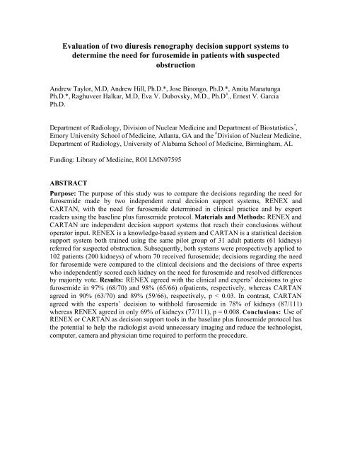
Evaluation <strong>of</strong> two diuresis renography decision support systems to<br />
determine the need for furosemide in patients with suspected<br />
obstruction<br />
Andrew Taylor, M.D, Andrew Hill, Ph.D.*, Jose Binongo, Ph.D.*, Amita Manatunga<br />
Ph.D.*, Raghuveer Halkar, M.D, Eva V. Dubovsky, M.D., Ph.D # ., Ernest V. Garcia<br />
Ph.D.<br />
Department <strong>of</strong> Radiology, Division <strong>of</strong> Nuclear <strong>Medicine</strong> and Department <strong>of</strong> Biostatistics * ,<br />
<strong>Emory</strong> <strong>University</strong> <strong>School</strong> <strong>of</strong> <strong>Medicine</strong>, Atlanta, GA and the # Division <strong>of</strong> Nuclear <strong>Medicine</strong>,<br />
Department <strong>of</strong> Radiology, <strong>University</strong> <strong>of</strong> Alabama <strong>School</strong> <strong>of</strong> <strong>Medicine</strong>, Birmingham, AL<br />
Funding: Library <strong>of</strong> <strong>Medicine</strong>, ROI LMN07595<br />
ABSTRACT<br />
Purpose: The purpose <strong>of</strong> this study was to compare the decisions regarding the need for<br />
furosemide made by two independent renal decision support systems, RENEX and<br />
CARTAN, with the need for furosemide determined in clinical practice and by expert<br />
readers using the baseline plus furosemide protocol. Materials and Methods: RENEX and<br />
CARTAN are independent decision support systems that reach their conclusions without<br />
operator input. RENEX is a knowledge-based system and CARTAN is a statistical decision<br />
support system both trained using the same pilot group <strong>of</strong> 31 adult patients (61 kidneys)<br />
referred for suspected obstruction. Subsequently, both systems were prospectively applied to<br />
102 patients (200 kidneys) <strong>of</strong> whom 70 received furosemide; decisions regarding the need<br />
for furosemide were compared to the clinical decisions and the decisions <strong>of</strong> three experts<br />
who independently scored each kidney on the need for furosemide and resolved differences<br />
by majority vote. Results: RENEX agreed with the clinical and experts’ decisions to give<br />
furosemide in 97% (68/70) and 98% (65/66) <strong>of</strong>patients, respectively, whereas CARTAN<br />
agreed in 90% (63/70) and 89% (59/66), respectively, p < 0.03. In contrast, CARTAN<br />
agreed with the experts’ decision to withhold furosemide in 78% <strong>of</strong> kidneys (87/111)<br />
whereas RENEX agreed in only 69% <strong>of</strong> kidneys (77/111), p = 0.008. Conclusions: Use <strong>of</strong><br />
RENEX or CARTAN as decision support tools in the baseline plus furosemide protocol has<br />
the potential to help the radiologist avoid unnecessary imaging and reduce the technologist,<br />
computer, camera and physician time required to perform the procedure.
INTRODUCTION<br />
In the evaluation <strong>of</strong> suspected ureteral obstruction, an international consensus panel<br />
recommended baseline imaging with Tc-99m mercaptoacetyltriglycine (MAG3) followed<br />
by furosemide administration and 15 minutes <strong>of</strong> additional imaging [1]. Some<br />
investigators have recommended strategies to eliminate the need for furosemide in<br />
selected patients [2]. The authors <strong>of</strong> the consensus report did note that baseline imaging<br />
might be sufficient to exclude obstruction in experienced hands but this option was not<br />
their general recommendation [1]. Nevertheless, if the baseline examination can exclude<br />
obstruction, the patient can be spared the administration <strong>of</strong> furosemide and additional<br />
imaging time and there will also be a cost savings due to the reduction <strong>of</strong> camera,<br />
computer and technologist time required to complete the furosemide component <strong>of</strong> the<br />
study and physician time required to interpret it.<br />
In spite <strong>of</strong> these advantages, it may be challenging for a general radiologist to acquire the<br />
experience and expertise in diuresis renography to determine with confidence whether or<br />
not a kidney is obstructed or when furosemide is not required. Radiologists are required to<br />
master an ever-expanding knowledge base while the hours available to master this<br />
knowledge base and apply it to specific tasks are steadily shrinking due to the pressure to<br />
increase the number <strong>of</strong> studies each radiologist interprets. The convergence <strong>of</strong> an<br />
expanding knowledge base and escalating time constraints inevitably increases the risk <strong>of</strong><br />
physician error. This is particularly true <strong>of</strong> low volume studies such as diuresis<br />
renography where radiologists may have had limited training and experience. In fact, a<br />
large percentage <strong>of</strong> the estimated 590,000 renal scans performed annually in the United<br />
States are interpreted at sites that perform less than three studies per week [3]. For these<br />
reasons, it is particularly important to develop and implement decision support tools to<br />
assist physicians in interpreting low volume studies so that they can be interpreted at a<br />
faster rate and at a higher level <strong>of</strong> expertise.<br />
The digital and dynamic nature <strong>of</strong> Tc-99m MAG3 renal scan data has made it possible to<br />
quantify a variety <strong>of</strong> functional renal parameters to produce an expanded knowledge<br />
base. Two independent renal decision support systems (RENEX and CARTAN) are being<br />
developed to use this expanded knowledge base to determine the need for furosemide and to<br />
detect renal obstruction in patients referred for diuresis renography [4, 5]. RENEX (renal<br />
expert) uses a set <strong>of</strong> rules obtained from human experts to analyze the quantitative<br />
parameters obtained from MAG3 scintigraphy whereas CARTAN (classification and<br />
regression tree analysis) is a statistical method that grows and prunes a decision tree based<br />
on an analysis <strong>of</strong> these quantitative parameters in a training data set [6]. As an<br />
intermediate step to developing a decision support system to detect obstruction, this<br />
prospective study was performed to compare the decisions regarding the need for<br />
furosemide made by a heuristic approach (RENEX) and an analytic approach (CARTAN)<br />
with the need for furosemide determined in clinical practice and by expert readers.
SUBJECTS AND METHODS<br />
This study was performed under the purview and with the approval <strong>of</strong> <strong>Emory</strong>’s Internal<br />
Review Board. Data collection and database use were compliant with the terms <strong>of</strong> the<br />
Health Insurance Portability and Accountability Act.<br />
Acquisition Protocol and Data Collection:<br />
Patients were hydrated with approximately 10 ounces <strong>of</strong> water on arrival in the<br />
department. Imaging was performed with the patient supine and the scintillation camera<br />
detector placed under the table. A three-phase dynamic acquisition was begun at the time<br />
<strong>of</strong> injection <strong>of</strong> approximately 10 mCi <strong>of</strong> Tc-99m MAG3. Phase one consisted <strong>of</strong> twentyfour<br />
2-second frames, phase two was sixteen 15-second frames, and phase three was forty<br />
30-second frames. All patient studies were processed using the QuantEM 2.0 renal<br />
quantification program to generate the input parameters for RENEX and CART. The<br />
QuantEM TM s<strong>of</strong>tware, developed specifically for Tc-99m MAG3 [7], has been validated in<br />
a multicenter trial [8], incorporates several quality control procedures to improve<br />
reproducibility and generates specific quantitative parameters recommended for scan<br />
interpretation.<br />
To process the baseline renogram, a static image is summed from the 2-3 minute postinjection<br />
frames. Using a filtered version <strong>of</strong> this image, whole kidney, background and<br />
cortical regions <strong>of</strong> interest (ROIs) are automatically defined. The user can override any <strong>of</strong><br />
these automatic ROIs and replace them with manual ROIs. Background-subtracted whole<br />
kidney and cortical curves are generated and 47 quantitative parameters are generated<br />
including patient demographics (height, weight, age, sex, body surface area), curve<br />
parameters (time to peak counts, and 20 min to count ratio for both whole kidney and<br />
cortical ROIs), voiding indices (post-void to pre-void and post-void to maximum count<br />
ratios), relative uptake and the MAG3 clearance. The MAG3 clearance is calculated from<br />
the 1-2.5 minute whole kidney uptake <strong>of</strong> MAG3 corrected for renal depth and attenuation<br />
and the pre-injection and post-injection images <strong>of</strong> the dose syringe [7-10].<br />
Protocol Survey: To estimate the frequency <strong>of</strong> use <strong>of</strong> the baseline plus furosemide<br />
protocol, one <strong>of</strong> the authors asked for a show <strong>of</strong> hands or key pad response from<br />
radiologists and nuclear medicine physicians attending renal lectures at the Educational<br />
Symposia Nuclear <strong>Medicine</strong> course in March, 2005, the <strong>Emory</strong> Course in nuclear<br />
medicine in July 2005, the 2005 Northeast Regional Meeting <strong>of</strong> the Society <strong>of</strong> Nuclear<br />
<strong>Medicine</strong> and a 2005 educational course at the RSNA.<br />
RENEX: The architecture <strong>of</strong> RENEX was inspired by two previously developed expert<br />
systems, MYCIN [11] and PERFEX [12], and is the subject <strong>of</strong> a separate publication<br />
[5]. MYCIN is a pioneering rule-based expert system developed in the 1970s to help
physicians determine the appropriate antibiotic for patients with infections; the name<br />
‘MYCIN’ was chosen because many <strong>of</strong> the available antibiotics included “mycin” in the<br />
name <strong>of</strong> the antibiotic. PERFEX is a commercially available imaging expert system<br />
developed to assist physicians in the interpretation <strong>of</strong> myocardial perfusion SPECT<br />
studies [13].<br />
Normal limits were established for 47 quantitative parameters extracted from the Tc-99m<br />
MAG3 scans <strong>of</strong> 106 potential renal donors [14, 15]. These 47 parameters consisted <strong>of</strong> 7<br />
demographic patient parameters, 20 left kidney parameters and 20 corresponding right<br />
kidney parameters. From these data an expert in radionuclide scintigraphy (domain<br />
expert) used his experience and published data [15] to estimate 5 boundary conditions for<br />
each parameter: (1) definitely abnormal, (2) probably abnormal, (3) equivocal, (4)<br />
probably normal and (5) definitely normal. A sigmoid-like fit constrained to these 5<br />
boundary conditions was then generated to create a parameter knowledge library to be<br />
used for converting the value <strong>of</strong> any individual quantitative parameter to a certainty factor<br />
(alternative to conditional probability) regarding normality or abnormality. For example,<br />
the certainty factor value <strong>of</strong> + 1 indicates that the parameter is “definitely abnormal” and<br />
the value <strong>of</strong> - 1 indicates the parameter is “definitely normal”; the cut-<strong>of</strong>f certainty factor<br />
values between abnormal and equivocal and between normal and equivocal are + 0.2 and<br />
– 0.2, respectively (Figs 1A and 1B). Sixty heuristic rules (IF A THEN B) were extracted<br />
from the domain expert to generate the knowledge base for detecting obstruction; twelve<br />
<strong>of</strong> these 60 rules are specifically applied to the baseline study to determine the need for a<br />
furosemide administration. Each rule uses the certainty factors describing the degree or<br />
abnormality or normality for each parameter that the rule evaluates to generate a certainty<br />
factor regarding the need for furosemide to exclude obstruction. These applied rules that<br />
are chained together by a forward chaining inference engine. An inference engine is<br />
s<strong>of</strong>tware that selects and executes the rules; the design <strong>of</strong> the RENEX inference engine<br />
follows the MYCIN inference engine by approximating Bayes theorem to combine the<br />
certainty factors generated by the relevant<br />
rules to reach a conclusion (combined certainty factor) regarding the need for furosemide;<br />
the combined certainty factor can range from “definitely needs furosemide (+ 1.0)” to<br />
“definitely does not need furosemide (-1.0)”. A meta-rule states that patients whose study<br />
has a combined certainty factor in the equivocal range (+ 0.2 to – 0.2) should also receive<br />
furosemide. A s<strong>of</strong>tware component called a justification engine was implemented to<br />
record the sequence <strong>of</strong> each rule that was fired and the certainty factor value <strong>of</strong> all input<br />
and output parameters at the time <strong>of</strong> instantiation in order to track and justify the logic <strong>of</strong><br />
the conclusions [4, 5]. The justification engine allows a user to query RENEX to<br />
determine the rules and parameter values that “justify” or explain the s<strong>of</strong>tware’s<br />
conclusion regarding the need for furosemide.<br />
To determine the need for furosemide, the entire system was trained using a pilot group <strong>of</strong><br />
31 patients (61 kidneys), 10 males and 21 females with a mean age <strong>of</strong> 58.8 +/- 17.6 years.<br />
Patients in the training set were obtained from our renal database <strong>of</strong> patients referred for<br />
diuresis renography and they were specifically selected to include a wide range <strong>of</strong>
esponses in order to develop a complete set <strong>of</strong> rules for determining the need for<br />
furosemide. Based on the decision <strong>of</strong> an expert panel (majority opinion <strong>of</strong> three <strong>of</strong> the<br />
authors, two institutions), 34 kidneys required furosemide and 27 kidneys did not require<br />
furosemide. The entire decision support system was fine tuned and optimized to agree<br />
with the expert decisions regarding each kidney in the training set. Processing time per<br />
patient is practically instantaneous using a 3.0 GHz PC programmed using IDL (Research<br />
Systems, Inc., Boulder, CO).<br />
CARTAN: CARTAN used the same pilot group <strong>of</strong> 31 patients (61 kidneys), the same 47<br />
parameters as used by RENEX and analyzed each kidney separately. Because there were a<br />
large number <strong>of</strong> parameters (47) relative to the number <strong>of</strong> kidneys (61), a statistical<br />
procedure known as CART (classification and regression trees) was implemented to<br />
classify kidneys and patients regarding the need for furosemide [6, 15, 16]. Computations<br />
were performed using the ‘rpart’ library in the R freeware statistical analysis package to<br />
grow and prune decision trees based on the pilot dataset [17]. A classification tree was<br />
constructed by first identifying a single parameter that determined the best separation<br />
between kidneys that required furosemide and kidneys that were not obstructed and did<br />
not require furosemide. Next, a second parameter was identified that determined the best<br />
separation between the two branches <strong>of</strong> the previous parameter. This process was<br />
continued and combined with cross-validation and pruning back <strong>of</strong> branches to limit the<br />
size <strong>of</strong> the decision tree and avoid over-fitting the data.<br />
To reduce dependency on the training data and to stabilize the algorithm, 1001<br />
classification trees were constructed by the common statistical technique <strong>of</strong> bootstrapping<br />
the training data [17, 19]. Bootstrap aggregation, or bagging, is a computationally<br />
intensive re-sampling technique that produces a different classification tree for each<br />
bootstrap <strong>of</strong>ten involving different dichotomized parameters.<br />
Each <strong>of</strong> the 1001 classification trees was subsequently applied to the prospective set <strong>of</strong><br />
102 patients (200 kidneys) to predict the need for furosemide. The results <strong>of</strong> each<br />
prediction algorithm (furosemide needed or not needed) <strong>of</strong> each bootstrap sample were<br />
tallied and the conclusion was based on a simple majority vote regarding the need for<br />
furosemide for each <strong>of</strong> the kidneys in the prospective group.<br />
Determination <strong>of</strong> the need for furosemide in clinical practice: To determine the<br />
percentage <strong>of</strong> patients referred for suspected obstruction who required and who did not<br />
require furosemide, a retrospective review <strong>of</strong> renal scans was conducted from January,<br />
1994 through August, 2002 A total <strong>of</strong> 711 MAG3 renal scans were performed for<br />
suspected obstruction between these dates; obstruction was excluded by the baseline scan<br />
alone in 221/711 studies (3 1%) and 490 patients (69%) received furosemide. These<br />
percentages were used to select patients in the furosemide and no furosemide categories<br />
for the prospective study so that the distribution <strong>of</strong> patients in the prospective study would<br />
match the distribution in clinical practice.
Prospective study population and expert panel review: The prospective population<br />
consisted <strong>of</strong> 102 patients (200 kidneys), 56 males and 46 females with a mean age <strong>of</strong> 54.6 ±<br />
16.9 years. None <strong>of</strong> the 102 patients were included in the training set. To obtain a<br />
representative sample, 102 patients were randomly selected; 70 patients were randomly<br />
selected from a database <strong>of</strong> patients referred for suspected obstruction who were given<br />
furosemide and 32 additional patients were randomly selected from a database <strong>of</strong> patients<br />
for whom furosemide was consider unnecessary. The percentage <strong>of</strong> patients receiving<br />
furosemide (69%) was selected to match the need for furosemide observed in clinical<br />
practice (see above). Using only data provided on the scans, three experts (three <strong>of</strong> the<br />
authors) from 2 different institutions independently scored each kidney on the need for<br />
furosemide and resolved differences by majority vote. Results from RENEX and<br />
CARTAN were prospectively compared with the experts’ decision and the actual clinical<br />
decision to give or withhold furosemide; results for the left kidney were compared to<br />
results for the right kidney. In addition, the experts’ decision was compared with the<br />
clinical decision to give or withhold furosemide.<br />
Statistical Analysis: Statistical analysis was based on the exact form <strong>of</strong> McNemar’s test for<br />
correlated proportions [19]. Due to some small sample sizes and generally good<br />
coverage, the Wilson score method was used for calculating confidence intervals for<br />
proportions [21, 22]. Significance was set at a p value < 0.05.<br />
RESULTS<br />
The baseline plus furosemide protocol recommended in the 1996 consensus report on<br />
diuresis renography appears to be in common use [1]. Based on a show <strong>of</strong> hands or key<br />
pad response, 50%, 90%, 85% and 80% <strong>of</strong> the participants attending the 2005 Educational<br />
Symposia nuclear medicine course, the 2005 <strong>Emory</strong> Course, the 2005 Northeast Regional<br />
SNM Meeting and a 2005 RSNA review course, respectively, used the standard protocol<br />
as either two separate acquisitions or a continuous acquisition with furosemide<br />
administered approximately after 20 minutes.<br />
In regard to the need for furosemide, RENEX agreed with the clinical decision to give<br />
furosemide in 97% <strong>of</strong> patients (68/70) whereas CARTAN agreed in 90% <strong>of</strong> patients<br />
(63/70), p = 0.03 (Table 1). Similarly, RENEX agreed with the experts’ decision to give<br />
furosemide in 98% <strong>of</strong> patients (65/66) whereas CARTAN agreed with 89% (59/66), p =<br />
0.01 (Table 1). When the data were analyzed by kidney rather than by patient, RENEX<br />
agreed with the experts’ decision to give furosemide in 94% <strong>of</strong> kidneys (84/89) compared to<br />
82% (73/89) for CARTAN, p = 0.002. Sample studies are illustrated in Figs 2 and 3. The<br />
experts’ decision agreed with the clinical decision to give furosemide in 94% <strong>of</strong> patients<br />
(66/70).<br />
There was no difference between RENEX and CARTAN when the clinicians and<br />
experts deemed that furosemide was not necessary, p = 0.18 and 0.32, respectively (Table
2). However, when the data were analyzed by individual kidneys (Table 2), CARTAN<br />
agreed with the experts’ decision in 78% <strong>of</strong> kidneys (87/111) whereas RENEX agreed in<br />
69% <strong>of</strong> kidneys (77/111), p = 0.008. The experts’ decision agreed with the clinical<br />
decision to withhold furosemide in 100% (32/32) <strong>of</strong> patients.<br />
Overall, RENEX agreed with the clinical decision in 89% (91/102) <strong>of</strong> patients,<br />
with the experts’ decision in 87% (89/102) <strong>of</strong> patients and with the experts’ decision in<br />
81% (16 1/200) <strong>of</strong> kidneys. CARTAN agreed with the clinical decision in 87% (89/102)<br />
<strong>of</strong> patients, with the experts’ decision in 83% (85/102) <strong>of</strong> patients and with the experts’<br />
decision in 80% (160/200) <strong>of</strong> kidneys. The experts’ decision agreed with the clinical<br />
decision to give or withhold furosemide in 96% (98/102) <strong>of</strong> patients.<br />
The performance <strong>of</strong> RENEX and CARTAN for the right kidney was compared<br />
to the performance in the left kidney. RENEX agreed with experts’ decision to<br />
administer furosemide in 93% (40/43) <strong>of</strong> left kidneys and 96% (44/46) <strong>of</strong> right<br />
kidneys, p = NS (Table 3). Comparable results were obtained for CARTAN (Table<br />
3). Similarly, there was no significant difference between the right and left kidney for<br />
RENEX and CARTAN when the results were analyzed based on the experts’<br />
decisions to withhold furosemide (Table 4).<br />
DISCUSSION<br />
Over the past several years, artificial intelligence methods have been investigated as a<br />
way to develop tools to improve diagnostic performance. Examples from nuclear<br />
medicine include neural networks [23-25] and case-based reasoning techniques [26] to<br />
provide computer-assisted diagnosis <strong>of</strong> planar and SPECT myocardial perfusion studies.<br />
In knowledge-based expert systems, a knowledge base <strong>of</strong> heuristic rules is obtained from<br />
human experts capturing how they make their interpretations. These rules are usually<br />
expressed in the form <strong>of</strong> IF A THEN B expressions. One major advantage <strong>of</strong> a<br />
knowledge-based expert system is that the system does not require the same large<br />
numbers <strong>of</strong> studies as are required to develop neural nets or case based reasoning<br />
approaches. A second advantage, especially from a learning perspective, is that it is<br />
possible to query a knowledge-based system to learn the rules that led to a specific<br />
conclusion. For example, RENEX disagreed with the experts in regard to the need for<br />
furosemide in the right kidney (Fig 3) but it is possible to query RENEX to determine the<br />
reasons for its decision. The primary reason that RENEX concluded furosemide was<br />
needed was the fact that the T ½ was abnormal (20.8 minutes); RENEX recognized that<br />
there was moderate evidence that furosemide was not needed because the post-void to<br />
maximum ratio was 0.2 (upper limits <strong>of</strong> normal <strong>of</strong> 0.16, [15]) but RENEX gave greater<br />
weight to the abnormal T ½. The experts were not queried but clearly gave greater<br />
weight to the images and the low post-void to max ratio. RENEX can be improved by<br />
comparing results and “reasoning” with expert decisions and adding new rules and/or<br />
adjusting the weighting factors. In this case, RENEX should be tested giving greater<br />
weight to the post-void to maximum count ratio than the T ½.<br />
We chose to implement a purely statistical method (CARTAN) for the second
decision support approach because the combined approach <strong>of</strong> classification and regression<br />
tree (CART) analysis with bootstrapping could handle a relatively large number <strong>of</strong><br />
variables as well as arrive at a prediction algorithm with a relatively small training set.<br />
CART analysis, but not bagging, has been used to help distinguish between healthy and<br />
diseased kidneys using magnetic resonance data that includes measurements <strong>of</strong> renal<br />
arterial blood flow and parenchymal perfusion [27]; to our knowledge, this is the first time<br />
CART analysis and bagging has been applied to a problem in nuclear medicine. CART<br />
agreed with the expert decisions illustrated in Fig 3 but CART cannot provide the<br />
rationale for these decisions; however CART can use a large sample <strong>of</strong> test patients to<br />
determine which parameters and what parameter cut-<strong>of</strong>f values provide the best separation<br />
in distinguishing when furosemide is needed or not needed or whether or not a kidney is<br />
obstructed. These parameters and cut-<strong>of</strong>f values can be used to guide clinicians and<br />
RENEX in determining the need for furosemide and the presence or absence <strong>of</strong><br />
obstruction.<br />
Our long term goal is to develop decision support systems to determine if a kidney is<br />
obstructed. The dual statistical and knowledge based approaches have provided<br />
complementary information and helped target features that can be transferred or<br />
emphasized to improve the overall performance <strong>of</strong> both systems. At present, data are<br />
insufficient to determine if one approach is inherently superior to the other. Analysis <strong>of</strong><br />
the baseline study to determine the need for furosemide may be useful for the 50-90% <strong>of</strong><br />
institutions that use a baseline plus furosemide protocol; these decision support systems<br />
have the potential to serve as a second opinion to support a clinician’s preliminary<br />
decision regarding the need for furosemide. Importantly, this study has also provided<br />
important data and direction for developing decision support systems to diagnose<br />
obstruction.<br />
The focus <strong>of</strong> this research effort is to develop statistical and knowledge based decision<br />
support systems that will have a diagnostic performance in diuresis renography<br />
comparable to that <strong>of</strong> experts. This is not a trivial task; however, it could be argued that a<br />
better goal would be to develop decision support systems that use the clinical outcome as<br />
the gold standard rather than expert readers. This is an attractive goal but it is confounded<br />
by the fact that an obstructed or non-obstructed scan interpretation has a major impact on<br />
the clinical outcome and, consequently, this gold standard becomes biased. Additional<br />
problems <strong>of</strong> data analysis can be illustrated by a patient who had a pyeloplasty to relieve<br />
obstruction 6-24 months after a diuresis renography scan was interpreted as no<br />
obstruction. In this example, did the scan miss obstruction, was the study interpreted<br />
incorrectly or did the patient become obstructed 6-24 months following the scan? Using<br />
patient outcome as a gold standard can be an important goal but interpretation <strong>of</strong> the<br />
results is not straightforward. Moreover, regardless <strong>of</strong> the type <strong>of</strong> study, clinical practice<br />
should be improved if radiologists can provide an interpretation equivalent to that <strong>of</strong><br />
expert readers.<br />
The study has several limitations. The training set was relatively small; this is
probably more <strong>of</strong> a disadvantage for a statistical system such as CARTAN than for a<br />
knowledge based system but both <strong>of</strong> these systems could potentially be improved by<br />
increasing the size <strong>of</strong> the training set. This study addressed the diuresis renography<br />
protocol recommended by the international consensus report where baseline data are<br />
obtained followed by the administration <strong>of</strong> furosemide and an additional period <strong>of</strong><br />
imaging [1]. There are other protocols in which furosemide is given 15 minutes before<br />
the radiopharmaceutical, at the same time as the radiopharmaceutical or 5 to 10 minutes<br />
later [1, 28, 29]. Obviously, the systems we describe do not apply to these protocols<br />
Finally, QuantEM 2.0 cannot detect and correct for patient motion and, at this time, the<br />
s<strong>of</strong>tware cannot distinguish between diffuse retention with slow washout due to impaired<br />
function and focal pelvic retention with slow washout due to possible obstruction.<br />
Algorithms to detect and correct for motion and to distinguish between diffuse retention in<br />
a kidney and retention in a dilated renal collecting system need to be designed,<br />
implemented and tested.<br />
CONCLUSIONS<br />
RENEX performed better than CARTAN when clinicians and experts determined that<br />
furosemide was necessary to evaluate obstruction. When furosemide was not required,<br />
CARTAN performed better than RENEX when the results were analyzed by the<br />
individual kidneys; there was no difference when the results were analyzed by patients.<br />
Use <strong>of</strong> RENEX or CARTAN as decision support tools in the baseline plus furosemide<br />
protocol has the potential to help avoid unnecessary imaging, minimize patient<br />
inconvenience and reduce costs by saving technologist, camera, computer and physician<br />
time. Importantly, the results obtained from this study will assist in the more complex<br />
task <strong>of</strong> developing decision support systems for diuresis renography to evaluate<br />
obstruction.<br />
Acknowledgements: We would like to thank Russell D. Folks, CNMT, Angela Hunsche,<br />
MD and Meghna Krishnan, MD for their assistance in collecting and archiving the data<br />
used in this study.
REFERENCES<br />
1. O’Reilly P, Aurell M, Britton K, Kletter K, Rosenthal L, Testa T. Consensus on<br />
diuresis renography for investigating the dilated upper urinary tract. J Nucl Med 1996;<br />
37:<strong>187</strong>2-<strong>187</strong>6<br />
2. Kuyvenhoven J, Piepsz A, Ham H. When could the administration <strong>of</strong><br />
furosemide be avoided. Clin Nucl Med 2003 ;28:732-737<br />
3. IMV Medical information division. 2003 Nuclear <strong>Medicine</strong> Census Marke<br />
Summary Report., IMV, Limited, Des Plaines, IL, IV (7-11)<br />
4. Garcia EV, Halkar R, Folks R, Krishna M, Taylor A. RENEX: An expert system<br />
for the interpretation <strong>of</strong> Tc-99m MAG3 scans to detect renal obstruction. J Nucl<br />
Med 2005; 46:205P-206P<br />
5. Garcia EV, Taylor A, Halkar R et al. RENEX: An expert system for the interpretation<br />
<strong>of</strong> Tc-99m MAG3 scans to detect renal obstruction. J Nucl Med 2006;47:320-329<br />
6. Breiman L, Friedman J, Olshen R and Stone C. Classification and Regression Trees,<br />
Wadsworth, Belmont, CA, 1984<br />
7. Taylor A Jr, Corrigan PL, Galt J, et al. Measuring technetium-99m-MAG3<br />
clearance with an improved camera-based method. J Nucl Med 1995; 36:1689-<br />
1695<br />
8. Taylor A, Manatunga A, Morton K, et al. Multicenter trial validation <strong>of</strong> a camerabased<br />
method to measure Tc-99m mercaptoacetyltriglycine, or Tc-99m MAG3,<br />
clearance. Radiology 1997; 204:47-54<br />
9. Taylor A, Lewis C, Giacometti A et al. Improved formulas for the estimation <strong>of</strong><br />
renal depth in adults. J Nucl Med 1993;34:1766-1769<br />
10. Taylor A. Formulas to estimate renal depth in adults. J Nucl Med<br />
1994;35:2054- 2055<br />
11. Shortliffe EH. Computer-Based Medical Consultations: MYCIN. Elsevier<br />
scientific publishing company, Amsterdam, Netherlands 1976, pp264.<br />
12. Garcia EV, Cooke CD, Folks RD, Santana CA, Krawczynska EG, De Braal L,<br />
Ezquerra NF: Diagnostic Performance Of An Expert System For The Interpretation Of<br />
Myocardial Perfusion SPECT Studies. J Nucl Med 2001; 42:1185-1191.<br />
13. Ezquerra N, Mullick R, Cooke D, Krawczynska E, Garcia E. PERFEX: An expert<br />
system for interpreting 3d myocardial perfusion. Expert Systems with Applications,<br />
1993, 6:459-468.<br />
14. Esteves FP, Halkar RK, Issa MM, Grant S, Taylor A. Comparison <strong>of</strong> camera – based<br />
Tc-99m MAG3 and 24-hour creatinine clearances for evaluation <strong>of</strong> kidney function.<br />
<strong>AJR</strong> 2006; <strong>187</strong> :W3 1 6-W3 19<br />
15. Esteves F, Taylor A, Manatunga A, Folks R, Krishnan M, Garcia E. Normal values<br />
for camera-based 99mTc-MAG3 clearance, MAG3 curve parameters, excretory<br />
parameters and residual urine volume. <strong>AJR</strong>: 2006, <strong>187</strong>:W1-W8<br />
16. Venables WN, Ripley BD. Modern Applied Statistics with S-Plus, pp 303-310, 3rd<br />
edition, Springer-Verlag, New York
17. Hastie T, Tibshirani R, Friedman J. The Elements <strong>of</strong> Statistical Learning: Data<br />
Mining, Inference, and Prediction, section 9.2 and pp 246-250 Springer, New York<br />
2001<br />
18. Comprehensive R Archive Network (CRAN): http://www.r-project.org<br />
19. Efron B, Tibshirani R. An Introduction to the Bootstrap, Chapman and Hall, London,<br />
1993<br />
20. McNemar Q. Note on the sampling error <strong>of</strong> the difference between correlated<br />
proportions or percentages. Ps ychometrika 1947;12:153-157<br />
21. Newcombe RG. Two-sided confidence intervals for the single proportion:<br />
comparison <strong>of</strong> seven methods. Statistics in <strong>Medicine</strong> 1998;17:857-872<br />
22. Agresti A, Coull BA. Approximate is better than “exact” for interval estimation <strong>of</strong><br />
binominal proportions. The American Statistician 1998;52: 119-126<br />
23. Fujita H, Katafuchi T, Uehara T, Nishimura T. Application <strong>of</strong> neural network to<br />
computer-aided diagnosis <strong>of</strong> coronary artery disease in myocardial SPECT Bull’s-eye<br />
images. J Nucl Med. 1992; 33:272-276.<br />
24. Porenta G, Dorffner G, Kundrat S, Petta P, Duit-Schedlmayer J, Sochor H. Automated<br />
interpretation <strong>of</strong> planar thallium-201-dipyridamole stress-redistribution scintigrams<br />
using artificial neural networks. J Nucl Med. 1994; 35:2041-2047.<br />
25. Hamilton D, Riley PJ, Miola UJ, Amro AA. A feed forward neural network for<br />
classification <strong>of</strong> bull’s-eye myocardial perfusion images. Eur J Nucl Med. 1995;<br />
22:108-1 15.<br />
26. Haddad M, Adlassnig KP, Porenta G. Feasibility analysis <strong>of</strong> a case-based reasoning<br />
system for automated detection <strong>of</strong> coronary heart disease from myocardial<br />
scintigrams. Artificial Intelligence in <strong>Medicine</strong>. 1997; 9:61-78.<br />
27. Michaely HJ, Schoenberg SO, Carina I, Dikow R, Bock M, Guenther M. Renal<br />
disease: Value <strong>of</strong> functional magnetic resonance imaging with flow and perfusion<br />
measurements. Invest Radiol 2004;39:698-705<br />
28. Sfakianakis GN, Cohen DJ, Braunstein RH et al: MAG3-F0 scintigraphy in decision<br />
making for emergency intervention in renal colic after helical CT positive for a<br />
urolith. J Nucl Med 2000;41:1813-1822, 2000<br />
29. Wong DC, Rossleight MA, Farnsworth RH. F + 0 diuresis renography in infants<br />
and children. J Nucl Med 1999:40:1805-1811
FIGURES<br />
Fig 1A. A knowledge library was generated for each parameter to convert any given<br />
parameter value into a certainty factor. Fig 1A illustrates the parameter knowledge library<br />
to convert any time to half maximum count value for the left kidney (whole kidney region<br />
<strong>of</strong> interest) to a certainty factor value. The boundary values (see Methods for more detail)<br />
were 8, 11, 13, 15, and 20 minutes respectively.<br />
Fig 1B: The figure shows the parameter knowledge library to convert any 20<br />
minute/maximum count ratio for the left kidney (cortical region <strong>of</strong> interest) to a certainty<br />
factor value. The boundary values were 0.17, 0.27, 0.32, 0.43 and 0.92 respectively.
Fig 2A: A 50 year old female was referred for a MAG3 scan because <strong>of</strong> suspected<br />
obstruction. The baseline scan showed no infiltration; the MAG3 clearance was reduced.<br />
The right kidney showed prompt uptake and excretion and was not obstructed. The left<br />
kidney showed a reduction in relative function (18%). There was uniform uptake in the<br />
left kidney with slow washout but, importantly, there was no retention <strong>of</strong> the tracer in the<br />
collecting system. Clinically, the study was interpreted as not obstructed and furosemide<br />
was not administered. RENEX, CARTAN and the experts agreed with this decision.
Fig 2B: An expanded review display shows the patient values for the MAG3 clearance,<br />
void/max ratio for whole kidney and cortical ROIs as well as the normal ranges for each<br />
<strong>of</strong> these values. The expanded review page also shows an enlarged parenchymal image<br />
obtained at 2-3 min, an enlarged display <strong>of</strong> the 19-20 min image and quality control<br />
images showing the pre and post-injection syringe counts and time <strong>of</strong> the bolus arrival in<br />
the kidneys. The post-void/maximum count ratio <strong>of</strong> the left kidney is slightly elevated at<br />
23% but this ratio tells the clinician that almost 80% <strong>of</strong> the maximal activity has washed<br />
out <strong>of</strong> the left kidney by the conclusion <strong>of</strong> the study and such a high washout percentage<br />
is strong evidence against obstruction.
Fig 3A: A 40 year old male was referred for a MAG3 scan because <strong>of</strong> suspected<br />
obstruction. The baseline scan showed no infiltration; the camera-based MAG3 clearance<br />
was normal. The left kidney showed rapid uptake and washout <strong>of</strong> the tracer in the<br />
sequential 2-minute images (center); the relative uptake <strong>of</strong> the left kidney was 56%, the<br />
whole kidney T ½ was 10.8 minutes and post-void to maximum ratio was normal (Fig 1B)<br />
excluding obstruction. The relative function <strong>of</strong> the right kidney was 46% and the right<br />
kidney showed dilatation <strong>of</strong> the renal pelvis which can be appreciated on the 2-min image<br />
as well as on the enlarged 2-3 min image (Fig 3B). The T ½ for the whole kidney ROI<br />
was prolonged at 20.3 minutes and the 20 min to maximum ratio was elevated at 0.58. On<br />
the other hand, visually the pelvis empties after voiding and the post-void to maximum<br />
ratio was 0.20 (Fig 3B). Although a post-void to maximum ratio is slightly elevated (0.16<br />
is the upper limit <strong>of</strong> normal[ 15]), the experts and the clinical decision was that furosemide<br />
was not indicated. CARTAN agreed with the experts but RENEX<br />
concluded that furosemide was needed to exclude obstruction <strong>of</strong> the right kidney (see<br />
Discussion).
Fig 3B: An expanded review display shows the patient values for the MAG3 clearance,<br />
residual urine volume, percent relative uptake and the Tmax, 20 min/max, T ½ and postvoid/max<br />
ratio for whole kidney and cortical ROIs as well as the normal ranges for each<br />
<strong>of</strong> these values. The expanded review page also shows an enlarged parenchymal image<br />
obtained at 2-3 min, an enlarged display <strong>of</strong> the 19-20 min image and quality control<br />
images showing the pre and post-injection syringe counts and time <strong>of</strong> the bolus arrival in<br />
the kidneys. In particular, the enlarged 2-3 minute image shows the dilated renal pelvis<br />
and the post-void to maximum ratio for the right kidney is slightly elevated.
Table 1: Performance <strong>of</strong> RENEX and CARTAN compared with<br />
expert consensus and clinical decisions to administer furosemide<br />
Clinical<br />
Decision<br />
(Patients)<br />
Consensus<br />
(Patients)<br />
Consensus<br />
(Kidneys)<br />
*P = 0.03<br />
~P = 0.01<br />
#P = 0.002<br />
Number<br />
requiring<br />
furosemide<br />
RENEX<br />
Correct<br />
proportion and<br />
95% confidence<br />
interval<br />
CARTAN<br />
Correct proportion<br />
and 95%<br />
confidence interval<br />
70 68 0.97 (0.90-0.99) 63 0.90* (0.81-0.95)<br />
66 65<br />
0.98 (0.92-1.00)<br />
59 0.89~ (0.80-0.95)<br />
89 84 0.94 # (0.88-0.98) 73 0.82 # (0.73-0.89)<br />
Table 2: Performance <strong>of</strong> RENEX and CARTAN compared with expert<br />
consensus and clinical decision that furosemide was not necessary<br />
Correct<br />
Number<br />
proportion<br />
Correct proportion<br />
not<br />
RENEX and 95% CARTANand 95% confidence<br />
requiring<br />
confidence<br />
interval<br />
furosemide<br />
interval<br />
Clinical<br />
Decision<br />
(Patients)<br />
32 23 0.72 (0.55-0.84) 26 0.81* (0.65-0.91)<br />
Consensus<br />
(Patients)<br />
36 24 0.67 (0.50-0.80) 26 0.72~ (0.56-0.84)<br />
Consensus<br />
111 77 0.69 # (0.60-0.77) 87 0.78 # (0.70-0.85)<br />
(Kidneys)<br />
*P = 0.18 ~P = 0.32<br />
#P = 0.008
Table 3: Performance <strong>of</strong> RENEX and CARTAN compared with expert<br />
consensus to administer furosemide for the right and left kidneys<br />
Left<br />
Number<br />
requiring<br />
furosemide RENEX<br />
Correct proportion<br />
and 95% confidence<br />
interval<br />
CARTAN<br />
Correct<br />
proportion<br />
and 95%<br />
confidence<br />
interval<br />
43 40 0.93 (0.81-0.98) 35 0.81 (0.67-0.90)<br />
Right 46 44 0.96 (0.85-0.99) 38 0.83 (0.69-0.91)<br />
Table 4: Performance <strong>of</strong> RENEX and CARTAN compared with expert<br />
consensus that furosemide was not necessary for the right and left kidneys<br />
Number<br />
Correct proportion<br />
not<br />
and 95% confidence<br />
requiring<br />
furosemide RENEX interval<br />
CARTAN<br />
Correct<br />
proportion<br />
and 95%<br />
confidence<br />
interval<br />
Left 55 39 0.71 (0.58-0.81) 45 0.82 (0.70-0.90)<br />
Right 56 38 0.68 (0.55-0.79) 42 0.80 (0.62-0.84)