Vertebroplasty - Regal Medical Group
Vertebroplasty - Regal Medical Group
Vertebroplasty - Regal Medical Group

You also want an ePaper? Increase the reach of your titles
YUMPU automatically turns print PDFs into web optimized ePapers that Google loves.
CLINICAL<br />
PRACTICE<br />
GUIDELINE<br />
REVIEW<br />
WORKSHEET<br />
Procedure:<br />
Page#: 1 of 72<br />
Guideline Review Cycle: 2010<br />
Reviewed By:<br />
<strong>Vertebroplasty</strong><br />
Tuan Phan, MD<br />
Review Date: December 2010<br />
Committee Approval Date: December 16, 2010<br />
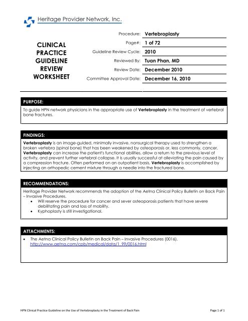
PURPOSE:<br />
To guide HPN network physicians in the appropriate use of <strong>Vertebroplasty</strong> in the treatment of vertebral<br />
bone fractures.<br />
FINDINGS:<br />
<strong>Vertebroplasty</strong> is an image-guided, minimally invasive, nonsurgical therapy used to strengthen a<br />
broken vertebra (spinal bone) that has been weakened by osteoporosis or, less commonly, cancer.<br />
<strong>Vertebroplasty</strong> can increase the patient's functional abilities, allow a return to the previous level of<br />
activity, and prevent further vertebral collapse. It is usually successful at alleviating the pain caused by<br />
a compression fracture. Often performed on an outpatient basis, <strong>Vertebroplasty</strong> is accomplished by<br />
injecting an orthopedic cement mixture through a needle into the fractured bone.<br />
RECOMMENDATIONS:<br />
Heritage Provider Network recommends the adoption of the Aetna Clinical Policy Bulletin on Back Pain<br />
– Invasive Procedures.<br />
Will reserve the procedure for cancer and sever osteoporosis patients that have severe<br />
debilitating pain and loss of mobility.<br />
Kyphoplasty is still investigational.<br />
ATTACHMENTS:<br />
The Aetna Clinical Policy Bulletin on Back Pain – Invasive Procedures (0016).<br />
http://www.aetna.com/cpb/medical/data/1_99/0016.html<br />
HPN Clinical Practice Guideline on the Use of <strong>Vertebroplasty</strong> in the Treatment of Back Pain Page 1 of 1
Back Pain - Invasive Procedures<br />
ttp://www.aetna.com/cpb/medical/data/1_99/0016.html<br />
Page 1 of 71<br />
11/10/2010<br />
Close Window<br />
Aetna.com Home | Help | Contact Us<br />
Search<br />
Go<br />
Clinical Policy Bulletin:<br />
Back Pain - Invasive Procedures<br />
Number: 0016<br />
Policy<br />
Aetna considers any of the following injections or procedures medically necessary for the treatment<br />
of back pain; provided, however, that only one invasive modality or procedure will be considered<br />
medically necessary at a time.<br />
Policy History<br />
Last Review: 05/14/2010<br />
Effective: 07/31/1995<br />
Next Review: 01/13/2011<br />
Review History<br />
Definitions<br />
Additional Information<br />
Clinical Policy Bulletin<br />
Notes<br />
I.<br />
II.<br />
Facet joint injections are considered medically necessary in the diagnosis of facet pain in<br />
persons with chronic back or neck pain (pain lasting more than 3 months despite appropriate<br />
conservative treatment).<br />
Facet joint injections are considered experimental and investigational as therapy for back<br />
and neck pain and for all other indications.<br />
A set of facet joint injections means up to 6 injections per sitting, and this can be<br />
repeated once to establish the diagnosis. It is not considered medically necessary to repeat<br />
facet joint injections more frequently than once every 7 days. Additional sets of facet<br />
injections are considered experimental and investigational because they have no proven<br />
value.<br />
Trigger point injections of corticosteroids and/or local anesthetics, are considered medically<br />
necessary for treating members with chronic neck or back pain or myofascial pain<br />
syndrome, when all of the following selection criteria are met:<br />
A. Trigger points have been identified by palpation, and<br />
B. Symptoms have persisted for more than 3 months, and<br />
C. Conservative therapies such as bed rest, exercises, heating or cooling modalities,<br />
massage, and pharmacotherapies such as non-steroidal anti-inflammatory drugs,<br />
muscle relaxants, non-narcotic analgesics, should have been tried and failed; and<br />
D.<br />
Trigger point injections are not administered in isolation, but are provided as part of a<br />
comprehensive pain management program, including physical therapy, patient<br />
education, psychosocial support, and oral medication where appropriate.<br />
Trigger point injections are considered experimental and investigational for all other<br />
indications.<br />
A trigger point is defined as a specific point or area where, if stimulated by touch or<br />
pressure, a painful response will be induced. A set of trigger point injections means
Back Pain - Invasive Procedures<br />
ttp://www.aetna.com/cpb/medical/data/1_99/0016.html<br />
Page 2 of 71<br />
11/10/2010<br />
III.<br />
IV.<br />
injections in several trigger points in one sitting. It is not considered medically necessary to<br />
repeat injections more frequently than every 7 days. Up to four sets of injections are<br />
considered medically necessary to diagnose the origin of a patient's pain and achieve a<br />
therapeutic effect; additional sets of trigger point injections are not considered medically<br />
necessary if no clinical response is achieved. Once a diagnosis is established and a<br />
therapeutic effect is achieved, it is rarely considered medically necessary to repeat trigger<br />
point injections more frequently than once every two months. Repeated injections extending<br />
beyond 12 months may be reviewed for continued medical necessity.<br />
Sacroiliac joint injections are considered medically necessary to relieve pain associated with<br />
lower lumbosacral disturbances in members who meet both of the following criteria:<br />
A. Member has back pain for more than 3 months; and<br />
B. The injections are not used in isolation, but are provided as part of a comprehensive<br />
pain management program, including physical therapy, patient education,<br />
psychosocial support, and oral medication where appropriate.<br />
Sacroiliac joint injections are considered experimental and investigational for all other<br />
indications.<br />
Up to two sacroiliac injections are considered medically necessary to diagnose the patient's<br />
pain and achieve a therapeutic effect. It is not considered medically necessary to repeat<br />
these injections more frequently than once every 7 days. If the member experiences no<br />
symptom relief or functional improvement after two sacroiliac joint injections, additional<br />
sacroiliac joint injections are not considered medically necessary. Once the diagnosis is<br />
established, it is rarely medically necessary to repeat sacroiliac injections more frequently<br />
than once every two months. Repeat injections extending beyond 12 months may be<br />
reviewed for continued medical necessity.<br />
Epidural injections of corticosteroid preparations (e.g., Depo-Medrol), with or without added<br />
anesthetic agents, are considered medically necessary in the outpatient setting for<br />
management of back or neck pain when all of the following are met:<br />
A. Intraspinal tumor or other space-occupying lesion, or non-spinal origin for pain, has<br />
been ruled out as the cause of pain; and<br />
B. Member has failed to improve after two or more weeks of conservative measures<br />
(e.g., rest, systemic analgesics and/or physical therapy); and<br />
C.<br />
Epidural injections beyond the first set of three injections are provided as part of a<br />
comprehensive pain management program, which includes physical therapy, patient<br />
education, psychosocial support, and oral medications, where appropriate.<br />
Epidural injections of corticosteroid preparations, with or without added anesthetic agents,<br />
are considered experimental and investigational for all other indications.<br />
Repeat epidural injections beyond the first set of three injections are considered medically<br />
necessary when provided as part of a comprehensive pain management program, which<br />
includes physical therapy, patient education, psychosocial support, and oral medications,<br />
where appropriate. Repeat epidural injections more frequently than every 7 days are not<br />
considered medically necessary. Up to three epidural injections are considered medically<br />
necessary to diagnose a member's pain and achieve a therapeutic effect; if the member<br />
experiences no pain relief after three epidural injections, additional epidural injections are<br />
not considered medically necessary. Once a therapeutic effect is achieved, it is rarely<br />
medically necessary to repeat epidural injections more frequently than once every two<br />
months. In selected cases where more definitive therapies (e.g., surgery) cannot be
Back Pain - Invasive Procedures<br />
ttp://www.aetna.com/cpb/medical/data/1_99/0016.html<br />
Page 3 of 71<br />
11/10/2010<br />
V.<br />
tolerated or provided, additional epidural injections may be considered medically necessary.<br />
Repeat injections extending beyond 12 months may be reviewed for continued medical<br />
necessity.<br />
See also CPB 722 - Selective Nerve Root Blocks<br />
Chymopapain chemonucleolysis is considered medically necessary for the treatment of<br />
sciatica due to a herniated disc when all of the following are met:<br />
A. Member has leg pain worse than low back pain; and<br />
B. Member has radicular symptoms reproduced by sciatic stretch tests; and<br />
C. Member has only a single level herniated disc with nerve root impingement at<br />
clinically suspected level demonstrated by MRI, CT, or myelography; and<br />
D. Member has objective neurologic deficit (e.g., diminished DTR, motor weakness, or<br />
hypalgesia in dermatomal distribution); and<br />
E.<br />
Pain not relieved by at least 6 weeks of conservative therapy.<br />
Chymopapain chemonucleolysis is considered experimental and investigational for all other<br />
indications, including the following:<br />
A.<br />
B.<br />
C.<br />
D.<br />
E.<br />
F.<br />
G.<br />
H.<br />
I.<br />
J.<br />
K.<br />
L.<br />
M.<br />
Acute low back pain alone<br />
When performed with chondroitinase ABC or agents other than chymopapain<br />
For herniated thoracic or cervical discs<br />
Cauda equina syndrome<br />
Sequestered disc fragment<br />
Multiple back operations (failed back surgery syndrome)<br />
Neurologic disease (e.g., multiple sclerosis)<br />
Pregnancy<br />
Profound or rapidly progressive neurologic deficit<br />
Severe spondylolisthesis<br />
Spinal cord tumor<br />
Spinal instability<br />
Severe spinal stenosis.<br />
VI.<br />
Percutaneous lumbar discectomy, manual or automated, is considered medically necessary<br />
for treatment of herniated lumbar discs when all of the following are met:<br />
A.<br />
B.<br />
C.<br />
D.<br />
Member is otherwise a candidate for open laminectomy; and<br />
Member has failed 6 months of conservative management; and<br />
Diagnostic studies show that the nuclear bulge of the disc is contained within the<br />
annulus (i.e., the herniated disc is contained); and<br />
Member has no previous surgery or chemonucleolysis of the disc to be treated; and<br />
VII.<br />
E.<br />
Member must have typical clinical symptoms of radicular pain corresponding to the<br />
level of disc involvement.<br />
Percutaneous lumbar diskectomy is considered experimental and investigational for all other<br />
indications.<br />
Note: Clinical studies have not established any clinically significant benefit of use of a laser<br />
over use of a scalpel for percutaneous lumbar diskectomy.<br />
Nonpulsed radiofrequency facet denervation (also known as facet neurotomy, facet<br />
rhizotomy, or articular rhizolysis) is considered medically necessary for treatment of
Back Pain - Invasive Procedures<br />
ttp://www.aetna.com/cpb/medical/data/1_99/0016.html<br />
Page 4 of 71<br />
11/10/2010<br />
members with intractable cervical or back pain with or without sciatica in the outpatient<br />
setting when all of the following are met:<br />
A. Member has experienced severe pain limiting activities of daily living for at least 6<br />
months; and<br />
B. Member has had no prior spinal fusion surgery; and<br />
C. Neuroradiologic studies are negative or fail to confirm disc herniation; and<br />
D. Member has no significant narrowing of the vertebral canal or spinal instability<br />
requiring surgery; and<br />
E. Member has tried and failed conservative treatments such as bed rest, back<br />
supports, physiotherapy, correction of postural abnormality, as well as<br />
pharmacotherapies (e.g., anti-inflammatory agents, analgesics and muscle<br />
relaxants); and<br />
F.<br />
Trial of facet joint injections has been successful in relieving the pain.<br />
Nonpulsed radiofrequency facet denervation is considered experimental and investigational<br />
for all other indications.<br />
Only 1 treatment procedure per level per side is considered medically necessary in a 6-<br />
month period.<br />
VIII.<br />
IX.<br />
See also CPB 735 - Pulsed Radiofrequency.<br />
Implantable infusion pumps are considered medically necessary when used to administer<br />
opioid drugs (e.g., morphine) intrathecally or epidurally for treatment of severe chronic<br />
intractable pain of malignant or non-malignant origin in members with life expectancies of<br />
more than 3 months who have proven unresponsive to less invasive medical therapy as<br />
determined by the following criteria:<br />
A.<br />
B.<br />
Member's history must indicate that he/she would not respond adequately to noninvasive<br />
methods of pain control, such as systemic opioids (including attempts to<br />
eliminate physical and behavioral abnormalities which may cause an exaggerated<br />
reaction to pain); and<br />
A preliminary trial of intraspinal opioid drug administration must be undertaken with a<br />
temporary intrathecal/epidural catheter to substantiate adequately acceptable pain<br />
relief, the degree of side effects (including effects on the activities of daily living), and<br />
member's acceptance.<br />
Implantable infusion pumps for intrathecal or epidural infusion of opioids are considered<br />
experimental and investigational for all other indications.<br />
Pedicle screws for spinal fixation are considered medically necessary for the following<br />
indications:<br />
A. Spondylolisthesis -- grades I-IV<br />
B. Spinal trauma of all types including fractures and dislocations<br />
C. Thoracic fractures<br />
D. Scoliosis and kyphosis requiring spinal instrumentation<br />
E. Segmental defects or loss of posterior elements following tumor resection<br />
F. Fusion after decompression<br />
G. Pseudoarthrosis repair<br />
H. Revision lumbar disc surgery requiring instrumentation because of instability at the<br />
previous level of surgery
Back Pain - Invasive Procedures<br />
ttp://www.aetna.com/cpb/medical/data/1_99/0016.html<br />
Page 5 of 71<br />
11/10/2010<br />
I. Fusion adjacent to prior lumbar fusion.<br />
Pedicle screw fixation is considered experimental and investigational for all other indications,<br />
including the following:<br />
A.<br />
B.<br />
C.<br />
D.<br />
E.<br />
F.<br />
First time intervertebral disc herniation<br />
Degenerative disc disease<br />
Single level discectomy<br />
Isolated low back pain without spinal instability or neurologic deficits<br />
Failed lumbar surgery without documentation of instability pattern or pseudarthrosis<br />
Decompressive laminectomy for spinal stenosis without evidence of instability.<br />
X.<br />
XI.<br />
Intervertebral body fusion devices (spine cages) (e.g., BAK Interbody Fusion System, Ray<br />
Threaded Fusion Cage, STALIF stand-alone anterior lumbar fusion cage) are considered<br />
medically necessary for use with autogenous bone graft in members who meet criteria for<br />
spinal fusion as outlined in CPB 743 Spinal Surgery: Laminectomy and Fusion. Spine cages<br />
are considered experimental and investigational for all other indications.<br />
Percutaneous polymethylmethacrylate vertebroplasty (PPV) or kyphoplasty is considered<br />
medically necessary for members with persistent, debilitating pain in the cervical, thoracic or<br />
lumbar vertebral bodies resulting from any of the following:<br />
A.<br />
B.<br />
C.<br />
D.<br />
E.<br />
F.<br />
G.<br />
Primary malignant neoplasm of bone or bone marrow; or<br />
Secondary osteolytic metastasis, excluding sacrum and coccyx; or<br />
Multiple myeloma; or<br />
Painful and/or aggressive hemangiomas; or<br />
Painful, debilitating osteoporotic collapse/compression fractures (e.g., Kummell's<br />
disease); or<br />
Steroid-induced fractures; or<br />
Painful vertebral eosinophilic granuloma.<br />
AND all of the following criteria have been met:<br />
A.<br />
B.<br />
Severe debilitating pain or loss of mobility that cannot be relieved by optimal medical<br />
therapy (e.g., acetaminophen, NSAIDS, narcotic analgesics, braces, physical<br />
therapy, etc.); and<br />
Other causes of pain such as herniated intervertebral disk have been ruled out by<br />
computed tomography or magnetic resonance imaging; and<br />
C.<br />
The affected vertebra has not been extensively destroyed and is at least one-third of<br />
its original height.<br />
XII.<br />
Lateral (including extreme, extra and direct lateral) interbody fusion is considered an<br />
acceptable method of performing a medically necessary anterior interbody fusion. See CPB<br />
743 Spinal Surgery: Laminectomy and Fusion.<br />
Experimental and Investigational Interventions<br />
Aetna considers any of the following injections or procedures experimental and investigational:<br />
I. Radiofrequency lesioning of dorsal root ganglia for back pain;<br />
II. Radiofrequency lesioning of terminal (peripheral) nerve endings for back pain;<br />
III. Epiduroscopy (also known as epidural spinal endoscopy, spinal endoscopy, myeloscopy,<br />
and epidural myeloscopy) for the diagnosis and treatment of intractable low back pain or<br />
other indications;
Back Pain - Invasive Procedures<br />
ttp://www.aetna.com/cpb/medical/data/1_99/0016.html<br />
Page 6 of 71<br />
11/10/2010<br />
IV.<br />
V.<br />
VI.<br />
VII.<br />
VIII.<br />
IX.<br />
X.<br />
XI.<br />
XII.<br />
XIII.<br />
XIV.<br />
XV.<br />
XVI.<br />
XVII.<br />
XVIII.<br />
XIX.<br />
Epidural injections of lytic agents (e.g., hypertonic saline, hyaluronidase) or mechanical lysis<br />
in the treatment of epidural fibrosis, adhesive arachnoiditis, failed back syndrome, or other<br />
indications;<br />
Percutaneous endoscopic diskectomy with or without laser (PELD) (also known as<br />
arthroscopic microdiskectomy or Yeung Endoscopic Spinal Surgery System (Y.E.S.S.));<br />
Microsurgical anterior foraminotomy for cervical spondylotic myelopathy or other indications;<br />
Sacroiliac fusion for the treatment of low back pain due to sacroiliac joint syndrome; Note:<br />
Sacroiliac fusion may be medically necessary for sacroiliac pain due to severe traumatic<br />
injury, where a trial of an external fixator is successful in providing pain relief.<br />
Sacroplasty for osteoporotic sacral insufficiency fractures and other indications;<br />
Racz procedure (epidural adhesiolysis with the Racz catheter) for the treatment of members<br />
with epidural adhesions, adhesive arachnoiditis, failed back syndrome from multiple<br />
previous surgeries for herniated lumbar disk, or other indications;<br />
Microendoscopic discectomy (MED) procedure for decompression of lumbar spine stenosis,<br />
lumbar disc herniation, or other indications;<br />
Dynamic stabilization (e.g., Dynesys Spinal System and the Stabilimax NZ Dynamic Spine<br />
Stabilization System);<br />
Inter-spinous distraction (e.g., X-Stop device, Coflex inter-spinous stabilization spinal<br />
implant, ExtenSure bone allograft inter-spinous spacer, the Eclipse inter-spinous distraction<br />
device, and the TOPS System) for spinal stenosis or other indications;<br />
Endoscopic laser foraminoplasty;<br />
Piriformis muscle resection;<br />
Xclose Tissue Repair System;<br />
Radiofrequency denervation for sacroiliac joint pain;<br />
Coccygeal ganglion (ganglion impar) block for pelvic pain;<br />
Facet joint implantation;<br />
Vesselplasty (e.g., Vessel-X).<br />
See also CPB 602 - Thermal Intradiscal Procedure.<br />
Reimbursement Notes:<br />
Laser: Clinical studies have not established a clinically significant benefit of use of a laser<br />
over a scalpel in spinal surgery. No additional benefit will be provided for the use of a laser<br />
in spinal surgery.<br />
Microscope and endoscope: Use of a microscope or endoscope is considered an integral<br />
part of the spinal surgery and not separately reimbursable.<br />
Background<br />
Epidural Steroids<br />
An epidural steroid injection is an injection of long lasting steroid in the epidural space – that is the<br />
area which surrounds the spinal cord and the nerves coming out of it. The efficacy of epidurally<br />
administered steroids has been demonstrated without adverse consequence in a large number of<br />
patients with reproducible results. In a large number of studies, long-term relief of pain (greater<br />
than 3 months) can be achieved in at least 10-30% of patients, while short-term relief (less than 1<br />
month) can be achieved in 60-100% of patients. Results for cervical pain are somewhat lower than<br />
those for lumbar pain. Such therapy is considered under accepted guidelines to be indicated in<br />
patients with low back and cervical pain that has not resolved after only a short period of more<br />
conservative measures since studies have shown a better response to therapy in patients whose<br />
pain is of shorter duration. Even if pain relief is temporary, it may have long-term benefit because it<br />
allows initiation of physical therapy or other rehabilitative measures at an earlier stage. Most
Back Pain - Invasive Procedures<br />
ttp://www.aetna.com/cpb/medical/data/1_99/0016.html<br />
Page 7 of 71<br />
11/10/2010<br />
authors indicate that a limit on number of injections is appropriate, and that most patients will<br />
respond with three or fewer injections.<br />
The American Academy of Neurology's assessment on the use of epidural steroid injections in the<br />
treatment of radicular lumbosacral pain (Armond, et al., 2007) concluded that:<br />
Epidural steroid injections may result in some improvement in radicular lumbosacral pain<br />
when determined between 2 and 6 weeks following the injection, compared to control<br />
treatment (Level C, Class I–III evidence). The average magnitude of effect is small, and the<br />
generalizability of the observation is limited by the small number of studies, limited to highly<br />
selected patient populations, the few techniques and doses studied, and variable<br />
comparison treatments.<br />
In general, epidural steroid injections for radicular lumbosacral pain have shown no impact<br />
on average impairment of function, on need for surgery, or on long-term pain relief beyond 3<br />
months. Their routine use for these indications is not recommended (Level B, Class I–III<br />
evidence).<br />
Data on use of epidural steroid injections to treat cervical radicular pain are inadequate to<br />
make any recommendation (Level U).<br />
Trigger Point Injections<br />
Trigger point injections are injections of local anesthetic medication, saline, and/or steroids into<br />
trigger points. A myofascial trigger point is a discrete focal tenderness, 2-5 mm in diameter that is<br />
located in distinct tight bands or knots of skeletal muscle (AHFMR, 2002). When palpated, these<br />
hyper-irritable areas cause pain in distant areas, or referred pain zones, which are specific for each<br />
trigger point. Trigger point injection, or direct wet needling, involves injection of fluid directly into the<br />
trigger point located in the taut muscle band. The main objective of trigger point injection is fast<br />
pain relief and elimination of muscle spasm in order to break the pain cycle. This facilitates physical<br />
therapy aimed at reducing muscle contracture and increasing range of motion. Trigger point<br />
injection is rarely used in isolation but is generally part of a multi-disciplinary approach aimed at<br />
treating both the trigger points and reducing all contributing factors (Scott & Guo, 2005; AHFMR,<br />
2002; Sanders, et al., 1999). Thus, treatment may also include patient education, psychosocial<br />
support, oral medications, and physical therapy to improve the strength and flexibility of the<br />
affected musculoskeletal systems. An assessment conducted by the Alberta Heritage Foundation<br />
for <strong>Medical</strong> Research (Scott & Guo, 2005) found that the evidence for the effectiveness of trigger<br />
point injections when used as the sole treatment for patients with chronic head, neck, and shoulder<br />
pain and whiplash syndrome was inconclusive, regardless of whether sterile water, saline, or<br />
botulinum toxin is injected. The assessment found that the combined use of dry needling and<br />
trigger point injection with procaine offers no obvious clinical benefit in the treatment of chronic<br />
craniofacial pain, while the effectiveness of trigger point injection for the treatment of cervicogenic<br />
headache is unknown. In contrast, the assessment found that trigger point injection with lidocaine<br />
may be useful in the treatment of joint pain caused by osteoarthritis (Scott & Guo, 2005). The<br />
assessment found no proof that triggers point injection is more effective than other less invasive<br />
treatments, such as physical therapy and ultrasound, in achieving pain relief, and there is some<br />
suggestion that the only advantage of injecting anesthetic into trigger points is that it reduces the<br />
pain of the needling process (Scott & Guo, 2005). Usually, approximately three treatments are<br />
necessary to abolish a trigger point completely (AHFMR, 2002). A number of trigger points may be<br />
injected in one session, but rarely more than five. Repeated injections in a particular muscle are not<br />
recommended if two or three previous attempts have been unsuccessful (Alvarez & Rockwell,<br />
2002; Sanders, et al., 1999). The pain relief may last for the duration of the anesthetic to many<br />
months, depending on the chronicity and severity of the trigger points and the concomitant<br />
treatment of perpetuating factors. According to available guidelines, use of trigger point injections<br />
should be short term and part of a comprehensive rehabilitation program. Available guidelines
Back Pain - Invasive Procedures<br />
ttp://www.aetna.com/cpb/medical/data/1_99/0016.html<br />
Page 8 of 71<br />
11/10/2010<br />
indicate that, while there are a number of uncontrolled case studies using trigger point injections in<br />
more acute pain presentations, there is virtually no consistent evidence for its application with<br />
chronic non-malignant pain syndrome patients to date (Sanders, et al., 1999; AHFMR, 2002).<br />
Lumbar Laminectomy with or without Fusion<br />
Most individuals with acute low back problems spontaneously recover activity tolerance within 4-6<br />
weeks of conservative therapy (AHCPR, 1994). Conservative therapy for acute low back pain<br />
includes:<br />
Limited bed rest with gradual return to normal activities<br />
Low impact exercise as tolerated (e.g., walking, swimming, stationary bike)<br />
Avoidance of activities that aggravate pain<br />
Cognitive support and reassurance that recovery is expected<br />
Heat/cold modalities for home use<br />
Chiropractic manipulation in the first 4 weeks if no radiculopathy<br />
Non-narcotic analgesics<br />
Pharmacotherapy (e.g., non-narcotic analgesics, NSAIDs (as second-line choices), avoid<br />
muscle relaxants, or only use during the first week, avoid narcotics)<br />
Exercise program<br />
Education regarding spine biomechanics.<br />
If conservative therapy fails to relieve symptoms of sciatica and radiculopathy and there is strong<br />
evidence of dysfunction of a specific nerve root confirmed at the corresponding level by findings<br />
demonstrated by CT/MRI, lumbar laminectomy may be proposed as a treatment option. The goal of<br />
lumbar laminectomy is to provide decompression of the affected nerve root to relieve the<br />
individual's symptoms. It involves the removal of all or part of the lamina of a lumbar vertebra. The<br />
addition of fusion with or without instrumentation is considered when there are concerns about<br />
instability.<br />
Decompression with or without Discectomy for Cauda Equine Syndrome<br />
Cauda equina ("horse's tail") is the name given to the lumbar and sacral nerve roots within the<br />
dural sac caudal to the conus medullaris. Cauda equina syndrome is usually the result of a<br />
ruptured, midline intervertebral disk, most commonly occurring at the L4-L5 level. However, tumors<br />
and other compressive masses may also cause the syndrome. Individuals generally present with<br />
progressive symptoms of fecal or urinary incontinence, impotence, distal motor weakness, and<br />
sensory loss in a saddle distribution. Muscle stretch reflexes may also be reduced. The presence of<br />
urinary retention is the single most consistent finding (Perron and Huff, 2002).<br />
In acute cauda equine syndrome, surgical decompression as soon as possible is recommended. In<br />
a more chronic presentation with less severe symptoms, decompression could be performed when<br />
medically feasible and should be delayed to optimize the patient's medical condition; with this<br />
precaution, decompression is less likely to lead to irreversible neurological damage (Dawodu,<br />
2005).<br />
Cervical Laminectomy with or without Fusion<br />
A cervical laminectomy (may be combined with an anterior approach) is sometimes performed<br />
when acute cervical disc herniation causes central cord syndrome or in cervical disc herniations<br />
refractory to conservative measures. Studies have shown that an anterior discectomy with fusion is<br />
the recommended procedure for central or anterolateral soft disc herniation, while a posterior<br />
laminotomy-foraminotomy may be considered when technical limitations for anterior access exist<br />
(e.g., short thick neck) or when the individual has had prior surgery at the same level (Windsor,<br />
2006).
Back Pain - Invasive Procedures<br />
ttp://www.aetna.com/cpb/medical/data/1_99/0016.html<br />
Page 9 of 71<br />
11/10/2010<br />
Discectomy alone is regarded as a technique that most frequently results in spontaneous fusion (70<br />
-80%). Additional fusion techniques include the use of bone grafts (autograft, allograft or artificial)<br />
with or without cages and/or the use of an anterior plate. A Cochrane systematic review (2004)<br />
reported the results of fourteen studies (n=939) that evaluated three comparisons of different fusion<br />
techniques for cervical degenerative disc disease and concluded that discectomy alone has a<br />
shorter operation time, hospital stay, and post-operative absence from work than discectomy with<br />
fusion with no statistical difference for pain relief and rate of fusion. The authors concluded that<br />
more conservative techniques (discectomy alone, autograft) perform as well or better than allograft,<br />
artificial bone, and additional instrumentation; however, the low quality of the trials reviewed<br />
prohibited extensive conclusions and more studies with better methodology and reporting are<br />
needed.<br />
Chemonucleolysis<br />
Chemonucleolysis is a procedure that involves the dissolving of the gelatinous cushioning material<br />
in an intervertebral disk by the injection of chymopapain or other enzyme. The AHCPR evidencebased<br />
guideline on the management of acute back pain and the medical literature supports the use<br />
of chemonucleolysis (CNL) with chymopapain as a safe and effective alternative to surgical disc<br />
excision in the majority of patients who are candidates for surgery for intractable sciatica due to<br />
herniated nucleus pulposus (HNP). Chemonucleolysis involves the enzymatic degradation of the<br />
nucleus pulposus, and has been shown to be more effective than percutaneous discectomy since it<br />
can be successfully performed for protruded and extruded discs, just as long as the herniated disc<br />
material is still in continuity with the disc of its origin. Following CNL, in many cases, relief of<br />
sciatica is immediate; however, in up to 30% of patients, maximal relief of symptoms may take up<br />
to 6 weeks. The overall success rate for CNL in long-term follow-up (7-20 years) in 3,130 patients<br />
from 13 contributors averaged 77% (range 71-93%), the same as that reported for surgical<br />
discectomy. In the United States, CNL is approved by the FDA for use in the lumbar spine only.<br />
Facet Blocks<br />
A facet block is an injection of local anesthetic and/or steroids into the facet joint of the spine.<br />
Degenerative changes in the posterior lumber facet joints have been established as a source of low<br />
back pain that may radiate to the leg. Pain impulses from the medial branches of lumbar dorsal<br />
rami can be interrupted by blocking this nerve with anesthetic (facet block) or coagulating it nerve<br />
with a radiofrequency wave (radiofrequency facet denervation). Typically, facet joint blocks are<br />
performed as a part of a workup for back or neck pain (Wagner, 2003). Pain relief following a<br />
precise intra-articular injection of local anesthetic confirms the facet joint as the source of<br />
pain. Based on the outcome of a facet joint nerve block, if the patient gets sufficient relief of pain<br />
but the pain recurs, denervation of the facet joint may be considered.<br />
A number of uncontrolled studies have suggested positive effects of facet injections on chronic<br />
back pain (Wagner, 2003). However, randomized controlled clinical trials have failed to<br />
demonstrated a benefit. A well-designed trial (n = 101) of patients who responded to a local<br />
anesthetic injection into the facet joint published in the New England Journal of Medicine found no<br />
difference in the likelihood of pain relief following randomization to glucocorticoid or saline facet<br />
joint injection at either one or three months post injection (Carette, et al., 1991). A higher proportion<br />
of patients in the steroid injection group reported marked improvement after six months (46 versus<br />
15 percent), but the benefit was attenuated after controlling for cointerventions used in the steroid<br />
group, and there is no biologic explanation for a delayed benefit from steroids. A second, smaller<br />
trial found no differences between steroid and/or bupivacaine injection compared to placebo (Lilius,<br />
et al., 1989).<br />
A number of systematic evidence reviews and evidence-based guidelines have evaluated the<br />
literature on facet injections for chronic back pain. Guidelines from the American Pain Society
Back Pain - Invasive Procedures<br />
ttp://www.aetna.com/cpb/medical/data/1_99/0016.html<br />
Page 10 of 71<br />
11/10/2010<br />
(Chou, et al., 2009) stated: "We found good or fair evidence that ... facet joint injection ... are not<br />
effective." Guidelines from the American Association of Neurological Surgeons (Resnick, et al.,<br />
2005) state: "Facet injections are not recommended as long-term treatment for chronic low-back<br />
pain." Guidelines from the American College of Occupational and Environmental Medicine<br />
(Hegmann, 2007) state that therapeutic facet joint injections for acute, subacute, chronic low back<br />
pain or radicular pain syndrome are "not recommended." An assessment by the Canadian Agency<br />
for Drugs and Technologies in Health (Zakaria, et al., 2007) concluded: "According to the RCTs<br />
[randomized controlled trials] completed to date, FJIs [facet joint injections] with local anesthetics or<br />
steroids have not been proven to be superior to placebo for the treatment of chronic LBP [low back<br />
pain]. Steroid FJIs have not been proven to be superior to local anesthetic FJIs in the treatment of<br />
chronic neck pain secondary to a motor vehicle accident. The studies are limited. ..." An<br />
assessment for BMJ Clinical Evidence (McIntosh & Hall, 2007) concluded that facet injections for<br />
chronic back pain are of "unknown effectiveness." A Cochrane systematic evidence review found<br />
no clear differences between facet joint glucocorticoid and placebo injections (Staal, et al., 2008). A<br />
review in UpToDate (Chou, 2009) stated: "Evidence is unavailable, unreliable, or contradictory<br />
regarding the effectiveness of glucocorticoid injections for other sites, including ... facet joint<br />
injections .... We suggest not performing these procedures for chronic low back pain."<br />
Radiofrequency Facet Denervation<br />
Facet joints of the spine have joint capsules that are supplied by a branch of the posterior ramus of<br />
the spinal nerve. Percutaneous radiofrequency facet denervation, also known as radiofrequency<br />
facet joint rhizotomy or facet neurotomy, involves selective denervation using radiofrequency under<br />
fluoroscopic guidance. As a method of neurolysis, radiofrequency facet denervation has been<br />
shown to be a very safe procedure and can offer relief for many patients with mechanical low back<br />
pain in whom organic pathology, most commonly a herniated lumbar disc, has been eliminated.<br />
According to the literature, it offers advantages over conventional neurolytic agents (e.g., phenol,<br />
alcohol, and hypertonic saline) because of its long lasting effects, the relative lack of discomfort,<br />
and its completely local action without any random diffusion of the neurolytic agent. Because there<br />
are no reliable clinical signs that confirm the diagnosis, successful relief of pain by injections of an<br />
anesthetic agent into the joints are necessary before proceeding with radiofrequency facet<br />
denervation. Results from many studies have shown that radiofrequency facet denervation results<br />
in significant (excellent or good) pain relief, reduced use of pain medication, increased return-towork,<br />
and is associated with few complications. Success rate, however, depends on a careful<br />
selection of patients.<br />
Pedicle Screw Fixation<br />
Pedicle screw fixation systems consist of steel or titanium plates that are longitudinally<br />
interconnected and anchored to adjacent vertebrae using bolts, hooks, or screws. Pedicle screw<br />
fixation in the spine is used to produce a rigid connection between two or more adjacent vertebrae<br />
in order to correct deformity and to stabilize the spine, thereby reducing pain and any neurological<br />
deficits. It is most often used in the lumbosacral spine from L1 though S1, and may also be used in<br />
the thoracic spine. Excision of tissues compressing the spinal cord (posterior decompression) is a<br />
common treatment for patients with herniated or subluxed vertebrae (spondylolisthesis),<br />
degenerative intervertebral discs, certain types of vertebral fractures, or spinal tumors. Spinal<br />
instability following decompression may be sufficiently severe to require stabilization by bony fusion<br />
(arthrodesis) of affected and adjacent vertebrae using implanted autologous bone grafts. Following<br />
placement of the graft, sufficient mechanical stability to allow its incorporation may be provided by<br />
combinations of various surgically implanted hooks, rods, or wires. However, severe instability may<br />
require surgical implantation of plates or rods anchored to vertebral pedicles using screws (pedicle<br />
screw fixation systems) in order to provide rigid three-column fixation and minimize the risk of<br />
incomplete fusion (pseudoarthrosis or pseudarthrosis) or loss of alignment during fusion. The<br />
current medical literature suggests that rigid fixation of the lumbar spine with pedicle screws
Back Pain - Invasive Procedures<br />
ttp://www.aetna.com/cpb/medical/data/1_99/0016.html<br />
Page 11 of 71<br />
11/10/2010<br />
improves the chances of successful fusion as compared with patients with lumbar spine fusion not<br />
supplemented with internal fixation. Internal fusion and fixation are major operative procedures with<br />
significant risks and according to the available literature should be reserved for patients with spinal<br />
instability associated with neurological deficits, major spinal deformities, spinal fracture, spinal<br />
dislocation or complications of tumor. Spinal fusion and pedicle screw fixation has been shown not<br />
to be effective for the treatment of isolated chronic back pain, and surgery is not advocated to treat<br />
this diagnosis in the absence of instability or neurological deficits. In July 1998, the FDA<br />
reclassified into Class II the pedicle screw spinal systems intended to provide immobilization and<br />
stabilization of spinal segments in skeletally mature patients as an adjunct to fusion in the treatment<br />
of the following acute or chronic instabilities or deformities of the thoracic, lumbar, and sacral spine:<br />
degenerative spondylolisthesis with objective evidence of neurological impairment, fracture,<br />
dislocation, scoliosis, kyphosis, spinal tumor, and failed previous fusion (pseudarthrosis). Pedicle<br />
screw systems intended for any other uses are considered post-amendment Class III devices for<br />
which pre-market approval is required.<br />
Intervertebral Body Fusion Devices (Spine Cages)<br />
A spine cage, also known as an interbody cage, is a small hollow cylindrical device, usually made<br />
of titanium, with perforated walls. The device is placed in the disc space between two vertebrae to<br />
restore lost disc height resulting from a collapsed disc and to relieve pressure on nerve roots.<br />
Currently, there are two intervertebral body fusion devices approved by the FDA: the BAK<br />
Interbody Fusion System (Spine-Tech, Inc.), and the Ray Threaded Fusion Cage (Surgical<br />
Dynamics, a subsidiary of United States Surgical Corporation). The BAK (Bagley and Kuslich)<br />
Interbody Fusion System and the Ray Threaded Fusion Cage (TFC) are hollow cylinders made of<br />
titanium, which may be implanted by anterior or posterior approach. Unlike pedicle screws, both of<br />
these fusion devices are permanent implants, as the literature describes bone growing into and<br />
through the implant. The safety and effectiveness of these fusion devices have not been<br />
established in three or more levels to be fused, previous fusion attempt at the involved level(s),<br />
spondylolisthesis or retrolisthesis of Grade II or greater. Although the BAK has received FDA<br />
approval for implantation laparoscopically, studies performed for FDA approval demonstrated<br />
significantly greater incidence of complications from anterior spinal reconstructive surgery using a<br />
laparoscopic approach than using an open approach. Furthermore, patients with laparoscopically<br />
implanted BAK fusion devices were followed for only 6 months; thus, the long-term stability of<br />
laparoscopically implanted BAK cages is unknown. Thus, coverage of laparoscopic (endoscopic)<br />
implantation of the BAK should be denied as experimental and investigational. (See discussion of<br />
anterior endoscopic spinal reconstructive surgery above.)<br />
<strong>Vertebroplasty</strong><br />
Percutaneous polymethylmethacrylate vertebroplasty (PPV) is a therapeutic, interventional<br />
radiologic procedure, which consists of the injection of a bone cement (usually methyl<br />
methacrylate) into a cervical, thoracic or lumbar vertebral body lesion for the relief of pain and the<br />
strengthening of bone. This procedure is being used for patients with lytic lesions due to bone<br />
metastases, aggressive hemangiomas, or multiple myeloma, and for patients who have medically<br />
intractable debilitating pain resulting from osteoporotic vertebral collapse. Results from two<br />
uncontrolled prospective studies and several case series reports, including one with 187 patients,<br />
indicated that percutaneous vertebroplasty can produce significant pain relief and increase mobility<br />
in 70 percent to 80 percent of patients with osteolytic lesions in the vertebrae. In these reports, pain<br />
relief was apparent within one to two days after injection, and appeared to persist for at least<br />
several months up to several years. While experimental studies and preliminary clinical results<br />
suggest that percutaneous vertebroplasty can also strengthen the vertebral bodies and increase<br />
mobility, it remains to be proven whether this procedure can prevent additional fractures in the<br />
injected vertebrae. In addition, the duration of effect was not known; there were no long-term follow<br />
-up data on most of these patients, and these data may be difficult to obtain and interpret in
Back Pain - Invasive Procedures<br />
ttp://www.aetna.com/cpb/medical/data/1_99/0016.html<br />
Page 12 of 71<br />
11/10/2010<br />
patients with an underlying malignant process because disease progression may confound<br />
evaluation of the treatment effect. Complications were relatively rare, although some studies<br />
reported a high incidence of clinically insignificant leakage of bone cement into the paravertebral<br />
tissues. In a few cases, the leakage of polymer caused compression of spinal nerve roots or<br />
neuralgia. Several instances of pulmonary embolism were also reported.<br />
The U.S. Food and Drug Administration (FDA, 2004) notified healthcare professionals about<br />
complications related to the use of polymethylmethacrylate bone cement to treat osteoporotic<br />
compression fractures of the spine using vertebroplasty and kyphoplasty. Reported complications,<br />
such as soft tissue damage and nerve root pain and compression, are related specifically to the<br />
leakage of bone cement. Other reported complications include pulmonary embolism, respiratory<br />
and cardiac failure, and death.<br />
Percutaneous vertebroplasty is an in-patient procedure because it may cause compression of<br />
adjacent structures and require emergency decompressive surgery. In addition, radiation therapy or<br />
concurrent surgical interventions, such as laminectomy, may also be required in patients with<br />
compression of the spinal cord due to ingrowth of a tumor. An assessment of percutaneous<br />
vertebroplasty by the National Institute for Clinical Excellence (NICE) (2003) concluded that<br />
"current evidence on the safety and efficacy of percutaneous vertebroplasty appears adequate."<br />
However, two subsequently published randomized controlled clinical trials published in the New<br />
England Journal of Medicine have found no significant benefit with vertebroplasty. In the<br />
Investigational <strong>Vertebroplasty</strong> Safety and Efficacy Trial (INVEST), Kallmes, et al. (2009) reported<br />
that pain and disability outcomes at 1 month in a group of patients who underwent vertebroplasty<br />
were similar to those in a control group that underwent a sham procedure. In the other trial,<br />
Buchbinder et al. (2009) measured pain, quality of life, and functional status at 1 week and at 1, 3,<br />
and 6 months after sham and active vertebroplasty and found there were no significant betweengroup<br />
differences at any time point. As in INVEST, patients in the two study groups had<br />
improvement in pain.<br />
The Society for Interventional Radiology (SIR, 2009) had identified a number of issues in<br />
interpreting these studies, including potential biases in patient selection, the use of vertebroplasty<br />
in older (greater than 3 months) fractures, and a potentially inadequate amount of PMMA that was<br />
injected into the vertebrae. The SIR concluded: "We recognize the value of randomized controlled<br />
trials and evidence-based medicine. But based on the above-discussed weakness in the studies<br />
and the degree of discordance between the outcomes of these studies, prior studies and<br />
experience, we believe it is premature—and possibly incorrect—to conclude that vertebroplasty is<br />
no better than a control sham procedure (trigger point, facet injection). We suggest waiting for the<br />
results of the VERTOSS 2 trial to be published and encourage larger clinical trials to address the<br />
weaknesses of the two New England Journal of Medicine articles."<br />
In a retrospective study, He, et al. (2008) examined if a repeat percutaneous vertebroplasty (PV) is<br />
effective on pain-relief at the vertebral levels in patients who had previously undergone PV. Of the<br />
334 procedures of PV performed in 242 patients with osteoporotic vertebral compression fractures<br />
from October 2000 to June 2006 in the authors' institute, 15 vertebrae in 15 patients with unrelieved<br />
pain in 4 to 32 days after an initial PV were treated with a repeat vertebroplasty. The clinical<br />
outcomes were assessed by measurements of VAS, and the imaging features were analyzed preand<br />
post-procedure. The mean volume of polymethylmethacrylate injected in each vertebra was<br />
4.0 ml (range of 1.5 to 9 ml) in the repeat PV. During the first month of follow-up after repeat PV in<br />
this series, a mean VAS scores of the pain level was reduced from 8.6 (range of 7 to 10) preprocedure<br />
to 1.67 points (range of 0 to 4) post-procedure, with a mean reduction of 6.93 points<br />
(range of 4 to 8). Complete and partial pain relief were reached in 11 (73 %) and 4 patients (27 %),<br />
respectively in a mean follow-up of 15 months. No serious complications related to the procedures<br />
occurred, however asymptomatic polymethylmethacrylate leakage around vertebrae was
Back Pain - Invasive Procedures<br />
ttp://www.aetna.com/cpb/medical/data/1_99/0016.html<br />
Page 13 of 71<br />
11/10/2010<br />
demonstrated on radiograph or computed tomography in 2 patients. The authors concluded that the<br />
outcomes of this series suggested that repeat PV is effective at the same vertebral levels in<br />
patients without pain-relief who underwent previous PV. Absent or inadequate filling of cement in<br />
the unstable fractured areas of the vertebral body may be responsible for the unrelieved pain after<br />
the initial PV.<br />
An accompanying editorial by Kallmes (2008) of the afore-mentioned article stated that "[u]<br />
nfortunately, limitations in the current study likely preclude definitive answers, but still the series<br />
may help focus future studies". The editorialist also noted that while the authors found insufficient<br />
or absent filling in 100% of the failed cases, they did not provide any information regarding the<br />
frequency in which they had insufficient or absent filling in the other 227 (successful) cases.<br />
Furthermore, Kallmes is still somewhat concerned about the safety of the repeat procedure.<br />
The BlueCross BlueShield Association Technology Evaluation Center, the Washington State<br />
Health Technology Assessment Program, and the California Technology Assessment Forum are<br />
conducting reassessments of the vertebroplasty procedure.<br />
Kyphoplasty<br />
Kyphoplasty (also known as balloon-assisted vertebroplasty) is a minimally-invasive orthopedic<br />
procedure, which has been developed to restore bone height lost due to painful osteoporotic<br />
compression fractures. It involves the insertion of one or two balloon devices into the fractured<br />
vertebral body. Once inserted, the surgeon inflates the balloon(s) to create a cavity and to compact<br />
the deteriorated bone with the intent to restore vertebral height. The balloon(s) are then removed<br />
and the newly created cavity is filled with the surgeon's choice of bone filler material, creating an<br />
internal cast for the fractured area.<br />
An assessment of balloon kyphoplasty by the National Institute for Health and Clinical Excellence<br />
(NICE, 2006) concluded that "[c]urrent evidence on the safety and efficacy of balloon kyphoplasty<br />
for vertebral compression fractures appears adequate to support the use of this procedure provided<br />
that normal arrangements are in place for consent, audit and clinical governance." The NICE<br />
assessment reviewed three non-randomised studies, two of which compared balloon kyphoplasty<br />
with conventional medical care (physical and analgesic therapy) and one which compared the<br />
procedure with vertebroplasty. All three studies found that patients who had undergone balloon<br />
kyphoplasty had improved pain scores compared with the control group at a maximum follow-up of<br />
24 months. The assessment noted that the specialist advisors to NICE expressed uncertainties<br />
about whether the improvements following balloon kyphoplasty (reduced pain and height<br />
restoration) are maintained in the long term. In clinical studies, the most common complication<br />
following balloon kyphoplasty was cement leakage, occurring in up to 11 percent of patients. Other<br />
potential complications of kyphoplasty include infection, allergy, and spinal cord or nerve root injury<br />
caused by incorrect needle placement.<br />
Based on the results of an assessment, the Ontario Ministry of Health and Long Term Care (2004)<br />
reached the following conclusions about balloon kyphoplasty: "There are currently two methods of<br />
cement injection for the treatment of osteoporotic VCFs. These are vertebroplasty and balloon<br />
kyphoplasty. Although no RCT has been conducted to compare the two techniques, the existing<br />
evidence shows that balloon kyphoplasty is a reasonable alternative to vertebroplasty, given the<br />
lower reported peri-operative and long-term complications of balloon kyphoplasty."<br />
Wardlaw, et al. (2009) reported positive results with kyphoplasty compared with nonsurgical care in<br />
a nonblinded, randomized controlled multicenter clinical study. The investigators randomly<br />
assigned 300 adults with one to three acute vertebral fractures to kyphoplasty (n=149) or<br />
nonsurgical care (n=151). At one month, mean SF-36 Physical Component Score (PCS) improved<br />
by 7.2 points (95% confidence interval 5.7 to 8.8) in the kyphoplasty group, and by 2.0 points (95%<br />
confidence interval 0.4 to 3.6) in the
Back Pain - Invasive Procedures<br />
ttp://www.aetna.com/cpb/medical/data/1_99/0016.html<br />
Page 14 of 71<br />
11/10/2010<br />
non-surgical group, a difference between groups that was statistically significant (p
Back Pain - Invasive Procedures<br />
ttp://www.aetna.com/cpb/medical/data/1_99/0016.html<br />
Page 15 of 71<br />
11/10/2010<br />
The average pain score before treatment was 8.72 +/- 1.25 (SD), whereas the average pain score<br />
after treatment was 3.38 +/- 2.35. The average mobility score before treatment was 2.31 +/- 1.94,<br />
whereas the average mobility score after treatment was 0.59 +/- 1.05 (p < 0.001). The average<br />
analgesic use score before treatment was 3.07 +/- 1.46, whereas it was 1.86 +/- 1.90 after<br />
treatment (p < 0.001). There was no evidence of clinical complications. The authors concluded<br />
that vesselplasty offers statistically significant benefits in improvements of pain, mobility, and the<br />
need for analgesia in patients with symptomatic VCFs, thus providing a safe alternative in the<br />
treatment of these fractures.<br />
While vesselplasty appears to be a promising new technique for VCFs, there is insufficient<br />
evidence of its safety and effectiveness. Prospective, randomized, controlled studies with a larger<br />
number of patients and long-term follow-up are needed.<br />
Epiduroscopy<br />
Epiduroscopy involves insertion of a fiberoptic camera through the sacral hiatus into the lower<br />
epidural space, which is then guided upwards towards the lower lumbar discs and nerve roots.<br />
Epidural adhesions can be released and anesthetic and steroid injected around nerve roots. In<br />
September 1996, the epiduroscope (myeloscope) was cleared by the FDA for visualization of the<br />
epidural space. It has been used in the outpatient setting for the diagnosis and treatment of<br />
intractable low back pain. Insertion of this miniature fiberoptic scope into the epidural space allows<br />
direct visualization of scarring and placement of a catheter through which fluid is injected under<br />
pressure to break down scar tissue and lyse adhesions. Although a number of pain treatment<br />
centers advertise the availability of this technique and claim it to be successful, there is insufficient<br />
scientific evidence in the peer-reviewed medical literature to support the clinical utility of this<br />
technique for diagnosis or therapy in patients with spinal pain syndromes, including those with<br />
failed back surgery syndromes. Moreover, currently available noninvasive technologies allow<br />
adequate visualization of the epidural space to confirm pathology contained therein. An<br />
assessment of epiduroscopy for the Australian Safety and Efficacy Register of New Interventional<br />
Procedures (ASERNIP-S, 2003) concluded that "[t]here is little high-quality evidence available on<br />
the safety and efficacy of epiduroscopically guided surgery/drug delivery... More studies are<br />
needed to compare the safety and efficacy of epiduroscopy relative to other procedures." An<br />
assessment by the National Institute for Clinical Excellence (NICE, 2004) concluded that "current<br />
evidence on the safety and efficacy of endoscopic epidural procedures does not appear adequate<br />
for these procedures to be used without special arrangements for consent and for audit or<br />
research." The NICE assessment found that "The studies identified were small and uncontrolled.<br />
Some measures used in these studies to assess outcomes, such as scores of pain and function,<br />
were of unknown validity."<br />
Racz Catheter<br />
The Racz catheter is a small caliber, flexible catheter that is introduced into the sacral hiatus and<br />
into the lubrosacral epidural space. The Racz catheter is used to release adhesions deliver steroids<br />
and anesthetics into the epidural space. There is no evidence from adequate well-designed<br />
randomized controlled clinical trials in the peer-reviewed medical literature supporting the safety<br />
and effectiveness of manipulation of an indwelling epidural Racz catheter or epidural injections of<br />
hypertonic saline or hyaluronidase to relieve back pain in patients with epidural adhesions,<br />
adhesive arachnoiditis, or failed back syndrome from multiple previous surgeries for herniated<br />
lumbar disk. The Racz epidural catheter was cleared by the FDA based on a 510(k) premarket<br />
notification (PMN) due to FDA's judgment that the device was "substantially equivalent" to devices<br />
that were marketed prior to the 1976 <strong>Medical</strong> Device Amendments to the Food, Drug and Cosmetic<br />
Act; thus, the manufacturer was not required to provide the evidence of effectiveness that is<br />
necessary to support a premarket approval (PMA) application. Most of the reported studies of the<br />
Racz catheter are retrospective (Racz & Holubec, 1989; Manchikanti, et al., 2001; Manchikanti, et
Back Pain - Invasive Procedures<br />
ttp://www.aetna.com/cpb/medical/data/1_99/0016.html<br />
Page 16 of 71<br />
11/10/2010<br />
al., 1999) or lacking a control group (Racz, et al., 1999). Manchikanti, founder and president of the<br />
American Society of Interventional Pain Physicians (ASIPP), is a leading advocate of the use of the<br />
Racz catheter (Manchikanti, et al., 1999; Manchikanti & Bakhit, 2000; Manchikanti & Singh, 2002).<br />
He is lead author of ASIPP guidelines which incorporate the Racz catheter into the management of<br />
chronic spinal pain (Manchikanti, et al., 2003). Manchikanti, et al. (2001, 2004) has reported the<br />
results of two controlled clinical studies of the Racz catheter in the ASIPP's official journal Pain<br />
Physician. One of these studies involved 45 patients with chronic low back pain, 30 of whom<br />
received Racz catheter treatment, and a control group of 15 patients who did not receive Racz<br />
catheter treatment. The study was unblinded and utilized a biased control group, as control group<br />
subjects were patients who refused Racz catheter treatment, either because coverage was denied<br />
by their insurer or for other reasons (Manchikanti, et al., 2001). In another study, subjects with<br />
chronic low back pain were randomized to a sham control group or two treatment groups (n = 25 in<br />
each group). Nineteen of 25 subjects in the control group were unblinded or lost to follow-up before<br />
completion of the 12-month study (Manchikanti, et al., 2004). Both of these controlled clinical<br />
studies involve small groups of patients and are from the same group of investigators from a single<br />
private practice, raising questions about the generalizability of the findings (Manchikanti, et al.,<br />
2001: Manchikanti, et al., 2004). The small sample sizes of these studies do not allow adequate<br />
evaluation of potential adverse outcomes that may occur with the procedure (Fibuch, 1999). A Joint<br />
Health Technology Assessment of the German <strong>Medical</strong> Association and the German National<br />
Association of Statutory Health Insurance Physicians (KBV, 2003) concluded that, "due to<br />
insufficient evaluation and lack of empirical data, at present there is no convincing evidence for the<br />
efficacy or effectiveness of the Racz treatment procedure."<br />
Epidural Lysis of Adhesions<br />
The National Institute for Clinical Excellence (NICE, 2004) assessed mobilization and division of<br />
epidural adhesions, and concluded that "[c]urrent evidence on the safety and efficacy of<br />
endoscopic division of epidural adhesions does not appear adequate for this procedure to be used<br />
without special arrangements for consent and for audit or research." The assessment noted that<br />
studies of epidural lysis of adhesions are "small and uncontrolled." In addition, NICE noted that "[s]<br />
ome measures used in the studies to assess outcomes, such as scores of pain and function, were<br />
of unknown validity." NICE stated that the main safety concerns are infection, bleeding,<br />
neurological damage, epidural hematoma, and damage to the nerve roots or cauda equina.<br />
Veihelmann et al (2006) examined if epidural neuroplasty is superior to conservative treatment with<br />
physiotherapy in treating patients with chronic sciatica with or without low back pain. A total of 99<br />
patients with chronic low back pain were enrolled in this study and randomly assigned into either a<br />
group with physiotherapy (n = 52) or a second group undergoing epidural neuroplasty (n = 47).<br />
Patients were assessed before and 3, 6, and 12 months after treatment by a blinded investigator.<br />
After 3 months, the VAS score for back and leg pain was significantly reduced in the epidural<br />
neuroplasty group, and the need for pain medication was reduced in both groups. Furthermore, the<br />
VAS for back and leg pain as well as the Oswestry disability score were significantly reduced until<br />
12 months after the procedure in contrast to the group that received conservative treatment. The<br />
authors concluded that epidural neuroplasty results in significant alleviation of pain and functional<br />
disability in patients with chronic low back pain and sciatica based on disc protrusion/prolapse or<br />
failed back surgery on a short-term basis as well as at 12 months of follow-up. Moreover, these<br />
investigators stated that further prospective randomized double-blinded studies are needed to<br />
prove the effectiveness of epidural neuroplasty in comparison to placebo and in comparison to<br />
open discectomy procedures.<br />
Microsurgical Anterior Foraminotomy<br />
Microsurgical anterior foraminotomy has been developed to improve the treatment of intractable<br />
cervical radiculopathy. This new technique provides direct anatomical decompression of
Back Pain - Invasive Procedures<br />
ttp://www.aetna.com/cpb/medical/data/1_99/0016.html<br />
Page 17 of 71<br />
11/10/2010<br />
compressed nerve roots by removing the compressive spondylotic spur or disc fragments through<br />
the holes of unilateral anterior foraminotomies. Using microsurgical instruments, the surgical<br />
approach exposes the lateral aspect of the spinal column through a small incision at the front of the<br />
neck in a naturally occurring crease. The affected nerve root is exposed, and a herniated disc or<br />
bone spur is removed to decompress the nerve. By removing only the herniated portion of the disc,<br />
the procedure is intended to preserve normal disc function and avoid bone fusion. As it utilizes a<br />
microsurgical technique that minimizes laminectomy and facet trauma, this technique does not<br />
require bone fusion or postoperative immobilization. However, there is a paucity of clinical studies<br />
to validate the effectiveness of this approach. The studies reported in the medical literature involve<br />
a small number of patients, are published by just one author, and a considerable portion of each<br />
article discusses only the technical aspects of the procedure.<br />
Sacroiliac Fusion<br />
Sacroiliac fusion involves bony fusion of the sacroiliac joint for stabilization. There is insufficient<br />
scientific evidence to support use of sacroiliac fusion in treating low back pain due to sacroiliac joint<br />
syndrome.<br />
In the 1920's, sacroiliac dysfunction was a common diagnosis and fusion of this joint was the most<br />
common form of back surgery. However, there is little evidence that the sacroiliac joint is a<br />
common source of back pain. European guidelines on the diagnosis and treatment of pelvic girdle<br />
pain (Vleeming, et al., 2004) recommend against the fusion of sacroiliac joints. The guidelines note<br />
that severe traumatic cases of pelvic girdle pain can be an exception to this recommendation, but<br />
only when other non-operative treatment modalities have failed. In that case, preoperative<br />
assessment with an external fixator for three weeks to evaluate longer lasting effects of fixation, is<br />
recommended (Wahlheim, 1984; Slätis and Eskola, 1989; Sturesson, et al., 1999). The authors<br />
identified no controlled trials of sacroiliac fusion. Available evidence consists of cohort studies (level<br />
D evidence) (Smith-Petersen and Rogers, 1926; Gaenslen, 1927; Hagen, 1974; Olerud &<br />
Wahlheim, 1984; Waisbrod, et al., 1987; Moore, 1995; Keating, 1995; Belanger and Dall, 2001;<br />
Berthelot, et al., 2001; van Zwienen, et al,, 2004; Giannikas, et al., 2004). The guidelines note that,<br />
in all reports of fusion surgery, an operation took place only on patients in whom non-operative<br />
treatment had been unsuccessful. The cohort studies included from 2 to 77 patients and the results<br />
were assessed by the authors as fair to excellent in 50 to 89% of the patients. However, controlled<br />
studies are necessary to reach firm conclusions about the effectiveness of this procedure in the<br />
treatment of back pain.<br />
Guidelines on treatment of low back pain from the Colorado Department of Labor and Employment<br />
(2005) state that sacroiliac joint fusion is of limited use in trauma and is considered to be under<br />
investigation for patients with typical mechanical low back pain: "Until the efficacy of this procedure<br />
for mechanical low back pain is determined by an independent valid prospective outcome study,<br />
this procedure is not recommended for mechanical low back pain."<br />
Endoscopic Diskectomy<br />
There is insufficient evidence from clinical studies proving additional benefits from using an<br />
endoscope for performing disc decompression (such as in percutaneous endoscopic diskectomy or<br />
endoscopic laser percutaneous diskectomy (LASE)). At this time there are no reliable clinical<br />
studies of endoscopic spinal surgery that have included an adequate comparison group of patients<br />
receiving open procedures. In addition, there is limited evidence on the long-term outcomes<br />
resulting from these endoscopic procedures. Gibson, et al. (2002), reporting on the results of a<br />
systematic review of studies on surgery for lumbar disc prolapse, explained that "[t]here is currently<br />
no evidence supporting endoscopic... treatment of disc prolapse."<br />
Yeung Endoscope Spine Surgery (Arthroscopic Microdiskectomy, Percutaneous Endoscopic<br />
Diskectomy with or without Laser (PELD))
Back Pain - Invasive Procedures<br />
ttp://www.aetna.com/cpb/medical/data/1_99/0016.html<br />
Page 18 of 71<br />
11/10/2010<br />
Yeung Endoscopic Spinal Surgery (YESS) (also known as arthroscopic microdiskectomy or<br />
percutaneous endoscopic diskectomy (PELD)) is an endoscopic approach to lumbar disc surgery<br />
that involves a multichannel scope and special access cannulae that allow spinal probing in a<br />
conscious patient, diagnostic endoscopy, and "minimally invasive surgery" (Yeung & Porter, 2002).<br />
The Yeung Endoscope Spine System (Y.E.S.S.) (Richard Wolf Surgical Instrument Corp., Vernon<br />
Hills, IL) or similar specialized instruments may be used to perform these procedures. The spinal<br />
endoscope is used to direct probing and targeted fragmentectomy of disc herniations. In<br />
addition, the approach may be used for foraminoplasty, where an endoscope-assisted laser is used<br />
to widen the exit route foramina of the lumbar spine and ablate any protruding portions of the<br />
intervertebral disk. Typically, procedures are performed at several levels of the spine, either<br />
simultaneously or in close temporal succession. Other adjunctive therapeutic procedures may<br />
be performed such as applying chemonucleolytic agents, lasers, radiofrequency technology,<br />
electrothermal energy, flexible mechanical instruments or intradiscal steroids. Supporters of<br />
arthroscopic microdiskectomy state that it provides visualization at the same time as application of<br />
therapeutic services. In addition, they argue that the ability to provoke pain while the patient is in<br />
the aware state and able to communicate during surgery allows the surgeon to better identify and<br />
treat the source of the patient's back pain. However, there is inadequate evidence to determine<br />
whether the results of arthroscopic microdiskectomy are as durable or as effective as open spinal<br />
surgery. A particular concern is whether this microendoscopic approach allows for adequate<br />
visualization of the spine during surgery. Literature to date on arthroscopic microdiskectomy has<br />
been limited to review articles and reports of retrospective case series. There are no published<br />
prospective, randomized controlled clinical studies of arthroscopic microdiskectomy., and there are<br />
no prospective studies with long-term follow up. In addition, the studies of Y.E.S.S. that have been<br />
published thus far have been from a single investigator group, raising questions about the<br />
generalization of the findings. Thus, arthroscopic microdiskectomy does not meet Aetna's criteria.<br />
Other centers have developed similar approaches; these approaches are not supported by reliable<br />
evidence in the peer reviewed published medical literature. These centers typically advertise their<br />
"unique" methods of performing endoscopic spine surgery through very small portals using<br />
specialized instruments that have been developed by the centers themselves. These procedures<br />
are performed while the patient is conscious under moderate sedation. Typically, several surgical<br />
procedures are performed at multiple levels simultaneously or on successive days until the patient<br />
reports pain relief or surgery is exhausted. Proponents argue that these procedures involve fewer<br />
anesthetic risks, a smaller incision, reduced blood loss, faster postoperative recovery and<br />
performance of surgery in an outpatient setting.<br />
An important concern about this minimally invasive approach is the limited visualization of the<br />
spine, such that the surgeon cannot reliably identify and ensure complete removal all bone spurs<br />
and other structures impinging on nerves. In addition, the performance of several surgical<br />
procedures in close temporal succession does not allow adequate evaluation of the outcomes of<br />
one surgical procedure before subsequent surgical procedures are performed.<br />
One center advertises that they manufacture special instruments and develop new techniques to<br />
perform complex microscopic laser spinal surgeries through portals of 1/4 to 1/2 of an inch under<br />
conscious sedation. They state that they have developed "unique" methods of performing<br />
endoscopic surgeries. The center states that they are the only facility that performs endoscopic<br />
spinal joint surgery, thoracic laser discectomy, endoscopic sacroiliac joint surgery, endoscopic<br />
hardware removal, or endoscopic bio-absorbable fusions or intradiscal stem cell therapy. The<br />
center also asserts that their unique minimally invasive spine surgery techniques are so advanced<br />
that patients who have failed other minimally invasive or conventional spine surgeries may benefit<br />
from their procedures. The center advertises that they have performed over 7000 of these<br />
minimally invasive spinal surgeries. Although they state that they regularly publish their findings in<br />
peer reviewed journals, what evidence they have published is limited to small, uncontrolled case
Back Pain - Invasive Procedures<br />
ttp://www.aetna.com/cpb/medical/data/1_99/0016.html<br />
Page 19 of 71<br />
11/10/2010<br />
series focusing on short-term followup (Haufe, et al., 2008; Haufe & Mork, 2007; Haufe & Mork,<br />
2006; Haufe & Mork, 2005; Haufe & Mork, 2004).<br />
Another center makes similar claims for the effectiveness of unique endoscopic laser spinal<br />
surgical procedures performed under conscious sedation with patented instruments. The center<br />
performs spinal procedures using videoendoscopy and specially adapted surgical probes.<br />
Procedures include specialized methods of laser diskectomy, laser lumbar facet debridement, laser<br />
foraminoplasty, and laser debridement of spinal processes. The center's website includes<br />
testimonials and a list of abstracts presented at meetings, but the center has not published the<br />
results of their procedures in peer-reviewed publications. The center recently announced initiation<br />
of an outcome study to evaluate outcomes of their procedures in persons with failed back<br />
syndrome.<br />
Another center offers unique endoscopic laser methods of performing surgery for back and neck<br />
pain. The primary procedures include foraminotomy, laminotomy, percutaneous endoscopic<br />
discectomy, and facet thermal ablation. The center advertises the ability to complete all necessary<br />
evaluation, preoperative preparation, surgery, and postoperative physical therapy within one week.<br />
The center advertises that advantages of their method of minimally invasive surgery includes no<br />
general anesthesia, no hospitalization, minimally invasive surgery, minimal scar tissue formation,<br />
and the availability of outpatient procedures. The center states that the most prominent difference<br />
between their techniques and that of other spinal centers is the endoscopic method in which they<br />
enter the body to minimize trauma, scar tissue formation, and healing times. The center states that<br />
their surgeons have performed approximately ten thousand surgeries collectively for over a<br />
decade. Their website includes testimonials. However, they have not submitted their results for<br />
peer-review publication.<br />
Laser diskectomy<br />
Laser diskectomy, or laser-assisted disc decompression (LADD), involves the use of a laser to<br />
vaporize a small portion of the nucleus pulposus in order to decompress a herniated disc. In laser<br />
diskectomy, the surgeon places a laser through a delivery device that has been directed under<br />
radiographic control to the disc, and removes the disc material using the laser. It uses many of the<br />
same techniques used in automated percutaneous discectomy. An endoscope may be used in<br />
conjunction with this procedure to visualize the disc space and nucleus pulposus, or the procedure<br />
may be done percutaneously. By contrast, percutaneous disc decompression uses an x-ray to<br />
localize the tip of the needle/trocar to ensure that it is in the appropriate level and location. The<br />
mechanism of action for pain relief in LADD is not well understood; most believe that the primary<br />
mechanism of pain reduction after LADD is its decrease in intradiscal pressure. According to the<br />
literature, laser-assisted disc decompression appears to be a safe procedure, but studies have not<br />
compared it to open surgical alternatives or other percutaneous methods. Randomized controlled<br />
trials are needed to compare current standard alternatives to both LADD and conservative<br />
treatment. A Cochrane review of surgical procedures for lumbar disc herniation concluded that "[t]<br />
here is currently no evidence supporting endoscopic (micro-suction) or laser treatment of disc<br />
prolapse" (Gibson, et al., 2002). A systematic review of the literature on percutaneous endoscopic<br />
laser discectomy for the Royal Australasian College of Surgeons (Boult, et al., 2000) reached<br />
similar conclusions: "Given the extremely low level of evidence available for this procedure it was<br />
recommended that the procedure be regarded as experimental until the results are available from a<br />
controlled clinical trial, ideally with random allocation to an intervention and control group."<br />
An assessment of laser lumbar diskectomy conducted for the National Institute of Clinical<br />
Excellence (NICE, 2003) concluded that current evidence on the safety and efficacy of laser lumbar<br />
discectomy does not appear adequate to support the use of this procedure without special<br />
arrangements for consent and for audit or research. A systematic evidence review by Jordan, et al.<br />
(2003) similarly concluded that the effectiveness of laser diskectomy is "unknown."
Back Pain - Invasive Procedures<br />
ttp://www.aetna.com/cpb/medical/data/1_99/0016.html<br />
Page 20 of 71<br />
11/10/2010<br />
Microdiscectomy<br />
Microdiscectomy refers to removal of protruding disc material, using an operating microscope to<br />
guide surgery. Dent (2001) recently assessed the evidence supporting the use of microdiscectomy<br />
for prolapsed intervertebral disc, and found no evidence of differences in clinical outcomes between<br />
microdiscectomy and standard open discectomy. A Cochrane review found evidence that<br />
microdiscectomy takes longer to perform than standard open discectomy (Gibson, et al., 2002).<br />
The review found no evidence of difference in short or long-term symptom relief or complications,<br />
or length of inpatient stay. Similarly, a systematic assessment of the literature by Jordan, et al.<br />
(2003) concluded that microdiskectomy has not been shown to be more effective than standard<br />
diskectomy.<br />
MicroEndoscopic Discectomy<br />
MicroEndoscopic Discectomy (MED) procedure combines conventional lumbar microsurgical<br />
techniques with endoscopy and is performed at an outpatient setting. It is employed for the<br />
treatment of lumbar spine stenosis and lumbar disc herniation. It has been suggested that MED is<br />
less invasive (no damage to muscle, bone or soft tissue) compared with traditional open<br />
microdiscectomy. Moreover, MED allegedly allows an early return to work. However, this<br />
endoscopic procedure is difficult because of the limited exposure and 2-dimensional video display.<br />
The potential injury of the nerve root and prolonged surgical time remain as matters of serious<br />
concern. Currently, there is insufficient evidence to support the clinical value of this procedure<br />
especially its long-term effectiveness.<br />
Muramatsu, et al. (2001) examined if MED was minimally invasive with respect to the nerve roots,<br />
cauda equina, and paravertebral muscles by comparing the post-operative magnetic resonance<br />
imaging findings in patients treated by MED and the conventional Love's method. The authors<br />
concluded that MED had an effect on the nerve roots and cauda equina that was comparable with<br />
that of Love's method. The magnetic resonance images of the route of entry failed to show that<br />
MED is appreciably less invasive with respect to the paravertebral muscles. Furthermore, in a<br />
review on the various minimally invasive procedures available for the treatment of lumbar disc<br />
disease, Maroon (2002) stated that although all percutaneous techniques (including MED) have<br />
been reported to yield high success rates, to date no studies have demonstrated any of these to be<br />
superior to microsurgical discectomy, which continues to be regarded as the standard with which all<br />
other techniques must be compared.<br />
Dynamic Stabilization<br />
Failed back surgery syndrome (FBSS) is reported to occur in 5 to 50 % of cases of lumbar spine<br />
operation. A marked rise in the number of performed spinal procedures has also led to an increase<br />
in the number of FBSS cases, which is the consequence of biological, psychological, social, and/or<br />
economical causes. Patient selection and correct indications are of key importance for successful<br />
surgical intervention of this syndrome. Surgical interventions that have been used for FBSS<br />
treatment include decompression, stabilization and fusion, as well as dynamic<br />
stabilization/neutralization procedures (Chrobok, et al., 2005).<br />
The use of rigid instrumentation in the treatment of degenerative spinal disorders seems to<br />
increase the fusion rate of the lumbar spine. However, rigid devices are associated with adverse<br />
effects such as pseudoarthrosis and adjacent segment degeneration. The use of semi-rigid and<br />
dynamic devices has been advocated to decrease such adverse effects of rigid fixation and thereby<br />
to attain a more physiological bony fusion (Korovessis, et al., 2004). Dynamic stabilization systems<br />
(e.g., the Dynesys Spinal System) are intended to restrict segmental motion and thus prevent<br />
further degeneration of the lumbar spine. The Dynesys, a non-fusion pedicle screw stabilization<br />
system (a flexible posterior stabilization system), was developed in an attempt to overcome the
Back Pain - Invasive Procedures<br />
ttp://www.aetna.com/cpb/medical/data/1_99/0016.html<br />
Page 21 of 71<br />
11/10/2010<br />
inherent disadvantages of rigid instrumentation and fusion. It uses flexible materials threaded<br />
through pedicle screws rather than rigid rods or bone grafts alone as an adjunct to fusion. The<br />
Dynesys is installed posteriorly, and does not require bone to be taken from the hip, as is required<br />
in other fusion procedures. It is designed to prevent over-loading the disc, but it restricts extension<br />
and loses lordosis (Sengupta and Mulholland, 2005; Putzier, et al., 2005).<br />
The Dynesys Spinal System (Centerpulse Spine-Tech, Inc., Minneapolis, MN) was cleared by the<br />
United States Food and Drug Administration (FDA) via a 510(k) pre-market notification in March<br />
2004. According to the product labeling, it is indicated to provide stabilization of spinal segments in<br />
skeletally mature patients as an adjunct to fusion in the treatment of the chronic instabilities or<br />
deformities of the thoracic, lumbar and sacral spine: degenerative spondylolisthesis with objective<br />
evidence or neurological impairment, kyphosis; and failed previous fusion (pseudoarthrosis). In<br />
addition, the product labeling states that the Dynesys system is intended for use in persons who<br />
meet all of the following criteria:<br />
Patients who are receiving fusions with autologous graft only; and<br />
Patients who are having the device attached to the lumbar or sacral spine; and<br />
Patients who are having the device removed after the development of a solid fusion mass.<br />
Although the Dynesys has been in clinical use for several years, there is insufficient evidence<br />
demonstrating that implantation of this device results in improved health outcomes compared to<br />
standard treatments.<br />
In a randomized controlled study, Korovessis, et al. (2004) examined the short-term effects of rigid<br />
versus semi-rigid and dynamic instrumentation on the global and segmental lumbar spine profile,<br />
subjective evaluation of the result, and the associated complications. The study did not examine<br />
objective functional outcomes. They compared 3 equal groups of 45 adult patients, who underwent<br />
primary decompression and stabilization for symptomatic degenerative lumbar spinal stenosis.<br />
Patients were randomly selected and received either the rigid (<strong>Group</strong> A), or semi-rigid (<strong>Group</strong> B), or<br />
dynamic (<strong>Group</strong> C) spinal instrumentation with formal decompression and fusion. The mean ages<br />
for the 3 groups were 65 +/- 9, 59 +/- 16, and 62 +/- 10 years, respectively. All patients had detailed<br />
roentgenographical study including computed tomography (CT) scan and magnetic resonance<br />
imaging (MRI) before surgery to the latest follow-up observation. The following roentgenographical<br />
parameters were measured and compared in all spines: lumbar lordosis (L1 - S1), total lumbar<br />
lordosis (T12 - S1), sacral tilt, distal lordosis (L4 - S1), segmental lordosis, vertebral inclination, and<br />
disc index. The SF-36 health survey and visual analog scale (VAS) was used before surgery to the<br />
latest evaluation. All patients were evaluated after a mean follow-up of 47 +/- 14 months. Both<br />
lumbar and total lordosis correction did not correlate with the number of the levels instrumented in<br />
any group. Total lordosis was slightly decreased after surgery (3 %, p < 0.05) in <strong>Group</strong> C. The<br />
segmental lordosis L2 - L3 was increased after surgery by 8.5 % (p < 0.05) in <strong>Group</strong> C, whereas<br />
the segmental lordosis L4 - L5 was significantly decreased in <strong>Group</strong>s A and C by 9.8 % (p = 0.01)<br />
and 16.2 % (p < 0.01), respectively. The disc index L2 - L3 was decreased after surgery in <strong>Group</strong>s<br />
A and C by 17 % (p < 0.05) and 23.5 % (p < 0.05), respectively. The disc index L3 - L4 was<br />
increased in <strong>Group</strong> C by 18.74 % (p < 0.01). After surgery, the disc index L4 - L5 was decreased in<br />
all 3 groups: <strong>Group</strong> A by 21 % (p = 0.01), <strong>Group</strong> B by 13 % (p < 0.05), and <strong>Group</strong> C by 13.23 % (p<br />
< 0.05). The disc index L5 - S1 was significantly decreased in <strong>Group</strong> B by 13 % (p < 0.05). The<br />
mean pre-operative scores of the SF-36 before surgery were 11, 14, and 13 for <strong>Group</strong>s C, B, and<br />
A, respectively. In the first year after surgery, there was a significant increase of the pre-operative<br />
SF-36 scores to 65, 61, and 61 for <strong>Group</strong>s C, B, and A, respectively, that represents an<br />
improvement of 83 %, 77 %, and 79 %, respectively. In the second year after surgery and<br />
thereafter, there was a further increase of SF-36 scores of 19 %, 23 %, and 21 % for <strong>Group</strong>s C, B,<br />
and A, respectively. The mean pre-operative scores of VAS for low back pain (LBP) for <strong>Group</strong>s C,<br />
B, and A were 5, 4.5, and 4.3, respectively, and decreased after surgery to 1.9, 1.5, and 1.6,
Back Pain - Invasive Procedures<br />
ttp://www.aetna.com/cpb/medical/data/1_99/0016.html<br />
Page 22 of 71<br />
11/10/2010<br />
respectively. The mean pre-operative scores of the VAS for leg pain for <strong>Group</strong>s C, B, and A were<br />
7.6, 7.1, and 6.9, respectively, and decreased after surgery to 2.5, 2.5, and 2.7, respectively. All<br />
fusions healed radiologically within the expected time in all three groups without pseudoarthrosis or<br />
malunion. Delayed hardware failure (1 screw and 2 rod breakages) without radiological<br />
pseudoarthrosis was observed in 2 patients in <strong>Group</strong> C 1 year and 18 months following surgery.<br />
There was no adjacent segment degeneration in any spine until the last evaluation. These<br />
investigators concluded that all three instrumentations applied over a short area for symptomatic<br />
degenerative spinal stenosis almost equally maintained the pre-operative global and segmental<br />
sagittal profile of the lumbosacral spine and was followed by similarly significant improvement of<br />
both self-assessment and pain scores. Hardware failure occurred at a low rate following dynamic<br />
instrumentation solely without radiologically visible pseudoarthrosis or loss of correction. These<br />
researchers further noted that because of the similar clinical and radiological data in all three<br />
groups and the relative small number of patients that were included in each group, it is difficult to<br />
make any recommendation in favor of any instrumentation.<br />
Putzier, et al. (2005) examined the effect of dynamic stabilization on the progression of segmental<br />
degeneration after nucleotomy. A total of 84 patients underwent nucleotomy of the lumbar spine for<br />
the treatment of symptomatic disc prolapse. Additional dynamic stabilization (the Dynesys system)<br />
was performed in 35 subjects. All patients showed signs of initial disc degeneration (Modic Type I -<br />
changes in the vertebral end plate are frequently associated with degenerative disc disease. Type<br />
1 changes include decreased signal intensity on T1-weighted and increased signal intensity on T2-<br />
weighted MRI). Evaluation was carried out before surgery, 3 months after surgery, and at follow-up.<br />
The mean duration of follow-up was 34 months. Examinations included radiographs, MRI, physical<br />
examination, and subjective patient evaluation using Oswestry score and VAS. Clinical symptoms,<br />
Oswestry score, and VAS improved significantly in both groups after 3 months. At follow-up, a<br />
significant increase in the Oswestry score and in the VAS was seen only in the non-stabilized<br />
group. In the dynamically stabilized group, no progression of disc degeneration was noted at followup,<br />
while radiological signs of accelerated segmental degeneration existed in the solely<br />
nucleotomized group. There were no implant-associated complications. These investigators<br />
concluded that the Dynesys system is useful to prevent progression of initial degenerative disc<br />
disease of lumbar spinal segments following nucleotomy. Moreover, the same group of researchers<br />
noted that the Dynesys system seems not to be indicated for treating marked deformities or if<br />
osseous decompression needs to be performed (Putzier, et al., 2004).<br />
In contrast to the observation of Korovessis, et al. (2004) and Putzier, et al. (2005), a number of<br />
investigators have questioned whether the Dynesys Spinal System offers any clinical advantages<br />
over rigid instrumentation (Hopf, et al., 2004; Grob, et al., 2005; Schwarzenbach, et al., 2005).<br />
In a clinical trial, Hopf, et al. (2004) compared the use of artificial disc replacement with dynamic<br />
stabilization procedure (Dynesys' method) in the treatment of patients with LBP. Indications for the<br />
operation were unsuccessful conservative treatment for over 6 months, segmental pain, age of less<br />
than 45 years, evidence of mono- or bi-segmental disc degeneration, with or without disc prolapse,<br />
demonstrated by MRI, exclusion of psychogenic disease and positive pre-operative, diagnostic<br />
measures such as facet joint infiltration and discography. These investigators stated that in younger<br />
patients with mono- or bi-segmental disc degeneration there is an indication for the implantation of<br />
an artificial disc. Contraindications for the operation are facet joint arthrosis and age of over 45<br />
years. The investigators commented that the indication in subjects with a classic FBSS is still<br />
unclear, the improvement of the instrumentation and a further adaptation of the systems to the<br />
known biomechanics of the lumbar spine are mandatory as is an intensive discussion of the<br />
operative procedure in the case of revision operations. These authors further noted that the<br />
Dynesys' method, with the inherent danger of segmental kyphozitation, a published, significant<br />
revision quota combined with a reduction of motility, does not fulfill this criterion.
Back Pain - Invasive Procedures<br />
ttp://www.aetna.com/cpb/medical/data/1_99/0016.html<br />
Page 23 of 71<br />
11/10/2010<br />
In a retrospective study, Grob and colleagues (2005) assessed patient-oriented outcome after<br />
implantation of the Dynesys Spinal System. A total of 50 consecutive patients instrumented with the<br />
Dynesys over the preceding 40 months were invited to complete a postal, patient-oriented follow-up<br />
questionnaire. The data from 31 of these subjects (11 men and 20 women; mean age of 50 years),<br />
with at least 2 years' follow-up, were analyzed. The primary indication for surgery was degenerative<br />
disease (disc/stenosis) with associated "instability"; 11 of 31 (35 %) patients had had prior spinal<br />
surgery. One-level instrumentation was performed in 32 % cases, 2-level instrumentation in 52 %<br />
cases, 3-level in 13 % cases, and 4-level in 3 % cases. Thirteen of 31 (42 %) patients underwent<br />
additional decompression. Within the 2-year follow-up period, 6 of 31 (19 %) patients had needed<br />
or were scheduled for another surgical intervention. At follow-up, mean back and leg pain (0 to10<br />
VAS) were 4.7 and 3.8, respectively. The following global outcomes were reported: (i) back<br />
symptoms -- 67 % improved, 30 % same, 3 % worse; (ii) leg symptoms - 64 % improved, 21 %<br />
same, 14 % worse; (iii) ability to do physical activities/sports - 40 % improved, 33 % same, 27 %<br />
worse; (iv) quality of life - 50 % improved, 37 % same, 13 % worse; (v) how much the operation<br />
helped - 29 % helped a lot, 23 % helped, 10 % only helped a little, 35 % didn't help, 3 % made<br />
things worse. These investigators concluded that their findings indicated that both back and leg<br />
pain are, on average, still moderately high 2 years following instrumentation with the Dynesys<br />
Spinal System. Only half of the patients declared that the operation had helped and had improved<br />
their overall quality of life; less than half reported improvements in functional capacity. The reoperation<br />
rate following implantation of the Dynesys was relatively high. The investigators<br />
concluded that these results provide no support for the notion that semi-rigid fixation of the lumbar<br />
spine resulted in better patient-oriented outcomes than those typical of fusion.<br />
In a recent review on posterior dynamic stabilization systems, Schwarzenbach, et al. (2005) stated<br />
that their experience with the Dynesys has shown that this method has limitations in "elderly<br />
patients with osteoporotic bone or in patients with a severe segmental macro-instability combined<br />
with degenerative spondylolisthesis and advanced disc degeneration. Such cases have an<br />
increased risk of failure. Only future randomized evaluations will be able to address the potential<br />
reduction of accelerated adjacent segment degeneration. The few posterior dynamic stabilization<br />
systems that have had clinical applications so far have produced clinical outcomes comparable with<br />
fusion. No severe adverse events caused by these implants have been reported. Long-term followup<br />
data and controlled prospective randomized studies are not available for most of the cited<br />
implants but are essential to prove the safety, efficacy, appropriateness, and economic viability of<br />
these methods".<br />
In a review on dynamic stabilization in the surgical management of painful lumbar spinal disorders,<br />
Nockels (2005) concluded that posterior dynamic stabilization systems may provide benefit<br />
comparable to fusion techniques, but without the elimination of movement. Moreover, the author<br />
also noted that further study (well-designed prospective, randomized, controlled trial) is needed to<br />
ascertain optimal design and clinical indications.<br />
In a systematic evidence review on non-rigid stabilization procedures for the treatment of LBP, the<br />
National Institute for Health and Clinical Excellence (NICE, 2005) stated that "current evidence on<br />
the safety of these procedures is unclear and involves a variety of different devices and outcome<br />
measures. Therefore, these procedures should not be used without special arrangements for<br />
consent and for audit or research". Additionally, the specialist advisors to the Institute's<br />
Interventional Procedures Advisory Committee noted that these procedures may be undertaken<br />
concurrently with disc decompression or discectomy. Thus, it is difficult to ascertain what clinical<br />
benefit is derived from the implants themselves. The specialist advisors noted that the reported<br />
adverse events include infection, malpositioned or broken screws leading to nerve root damage,<br />
cerebrospinal fluid leak, failure of the bone/implant interface, and failure to control pain. The<br />
theoretical risks with the techniques include: device failure (particularly long term), increased<br />
lordosis, and root damage caused by loose or misaligned screws.
Back Pain - Invasive Procedures<br />
ttp://www.aetna.com/cpb/medical/data/1_99/0016.html<br />
Page 24 of 71<br />
11/10/2010<br />
Welch and colleagues (2007) presented the preliminary clinical outcomes of dynamic stabilization<br />
with the Dynesys spinal system as part of a multi-center randomized prospective FDA<br />
investigational device exemption (IDE) clinical trial. This study included 101 patients from 6 IDE<br />
sites (no participants were omitted from the analysis) who underwent dynamic stabilization of the<br />
lumbar spine with the Dynesys construct. Patient participation was based on the presence of<br />
degenerative spondylolisthesis or retrolisthesis (Grade I), lateral or central spinal stenosis, and their<br />
physician's determination that the patient required decompression and instrumented fusion for 1 or<br />
2 contiguous spinal levels between L1 and S1. Subjects were evaluated pre-operatively, postoperatively<br />
at 3 weeks, and then at 3-, 6-, and 12-month intervals. The 100-mm VAS was used to<br />
score both lower-limb and back pain. Patient functioning was evaluated using the ODI, and the<br />
participants' general health was assessed using the Short Form-12 questionnaire. Overall, patient<br />
satisfaction was also reported. One hundred one patients (53 women and 48 men) with a mean<br />
age of 56.3 years (range of 27 to 79 years) were included. The mean pain and function scores<br />
improved significantly from the baseline to 12-month follow-up evaluation, as follows: leg pain<br />
improved from 80.3 to 25.5, back pain from 54 to 29.4, and ODI score from 55.6 to 26.3 %. The<br />
authors concluded that the early clinical outcomes of treatment with Dynesys are promising, with<br />
lessening of pain and disability found at follow-up review. Dynesys may be preferable to fusion for<br />
surgical treatment of degenerative spondylolisthesis and stenosis because it decreases back and<br />
leg pain while avoiding the relatively greater tissue destruction and the morbidity of donor site<br />
problems encountered in fusion. However, long-term follow-up care is still recommended.<br />
In a prospective case series, Kumar et al (2008) examined the radiological changes in the<br />
intervertebral disc after Dynesys dynamic stabilization. A total of 32 patients who underwent<br />
Dynesys procedure and have completed 2-year follow-up MRI scans were included in this study.<br />
Pre-operative and 2-year post-operative lumbar MRI scans were evaluated by 2 independent<br />
observers. T2-weighted mid-sagittal images were used and disc degeneration were classified<br />
according to the Woodend classification of disc degeneration. Anterior and posterior intervertebral<br />
disc heights were also measured. Of the 32 patients, 20 patients underwent Dynesys procedure<br />
alone and 12 underwent additional fusion at 1 or more levels. A total of 70 levels were operated<br />
on, of which 13 levels were fused. There was a statistically significant increase in the mean<br />
Woodend score at the operated levels in the Dynesys alone group, a change from 1.95 before<br />
surgery to 2.52 after surgery (p < 0.001). The mean Woodend scores changed from 1.27 preoperative<br />
to 1.55 post-operative (p = 0.066) at the proximal adjacent levels, and from 1.37 to 1.62<br />
at the distal levels (p = 0.157). There was good inter-observer agreement (weighted k score of<br />
0.819). The anterior intervertebral disc height reduced by 2 mm from 9.25 to 7.17 (p < 0.001). The<br />
posterior disc height increased by 0.14 mm but this change insignificant. The authors concluded<br />
that disc degeneration at the bridged and adjacent segment seems to continue despite Dynesys<br />
dynamic stabilization.<br />
The Stabilimax NZ Dynamic Spinal Stabilization System is an investigational device that is being<br />
evaluated for the treatment of patients with symptomatic spinal stenosis. The Stabilimax NZ is<br />
inserted and fixed to the vertebra by means of pedicle screws in exactly the same manner a fusion<br />
device is inserted and attached. The only difference is that for the Stabilimax NZ no bone graft will<br />
be placed around or between the vertebra to promote bone growth for fusion. It should be noted<br />
that a clinical trial sponsored by Applied Spine Technologies to evaluate if the Stabilimax NZ is at<br />
least as safe and effective as the control therapy of fusion in patients receiving decompression<br />
surgery for the treatment of clinically symptomatic spinal stenosis at one or two contiguous<br />
vertebral levels from L1 to S1 has been suspended (Applied Spine Technologies, 2008); the reason<br />
for this suspension is unclear.<br />
In summary, despite some preliminary evidence that dynamic stabilization systems (e.g., the<br />
Dynesys) have produced clinical outcomes comparable to that of fusion, the clinical value of
Back Pain - Invasive Procedures<br />
ttp://www.aetna.com/cpb/medical/data/1_99/0016.html<br />
Page 25 of 71<br />
11/10/2010<br />
dynamic stabilization awaits the findings of prospective, randomized, controlled trials, which are an<br />
essential requirement for practice of evidence-based medicine.<br />
Inter-Spinous Distraction Procedures<br />
Lumbar spinal stenosis (LSS) refers to narrowing of the lumbar spinal canal, lateral recess, or<br />
foramen resulting in neurovascular compression that may lead to pain. Spinal stenosis may be<br />
classified by etiology (e.g., congenital or acquired) or symptomatology (e.g., radiculopathy,<br />
neurogenic claudication, or mechanical back pain). It can also be classified radiographically, by the<br />
location of the stenosis (e.g., central canal, lateral recess, or intervertebral foramen) or by the<br />
presence of deformity such as spondylolisthesis or scoliosis. Overlapping in the classification of<br />
LSS can occur in that central stenosis with thecal sac compression usually leads to neurogenic<br />
claudication, while lateral recess compression is associated with compression of an individual<br />
nerve root, thus resulting in radiculopathy. Although symptoms may arise from narrowing of the<br />
spinal canal, not all patients with narrowing develop symptoms. The reason why some patients<br />
develop symptomatic stenosis and others do not is still unknown. Therefore, LSS doe not refer to<br />
the pathoanatomical finding of spinal canal narrowing. It is a clinical syndrome of lower extremity<br />
pain caused by mechanical compression on neural elements or their vascular supply (Truumees,<br />
2005).<br />
Non-surgical treatments (e.g., activity modification, medications such as non-steroidal antiinflammatory<br />
drugs, physical therapy that focuses on flexion-based exercises, as well as epidural<br />
steroid injections) are usually the first treatment choice for patients suffering from neurogenic<br />
intermittent claudication (NIC) secondary to LSS. If symptoms failed to improve with non-surgical<br />
treatments, decompressive surgery (e.g., laminectomy, facetectomy, multi-level laminotomies,<br />
fenestration, distraction laminoplasty, and microscopic decompression), with or without fusion, may<br />
be necessary. Moreover, several studies reported that surgical treatment produces better outcomes<br />
than non-surgical treatment in the short-term; however, the results tend to deteriorate with time<br />
(Yuan, et al., 2005).<br />
While fusion operations have traditionally been used to manage many disorders of the lumbar<br />
spine related to instability, pain, or deformity, concern over the long-term effects of fusion on<br />
adjacent spinal segments has led to the development of new approaches such as inter-spinous<br />
distraction procedures. The X-Stop Inter-Spinous Process Distraction/Decompression System (St.<br />
Francis <strong>Medical</strong> Technologies, Inc., Alameda, CA) was developed to provide an alternative<br />
therapeutic. The principal behind the X-Stop (eXtension-Stop) is that by decompressing the<br />
affected spinal segment and maintaining it in a slightly flexed position (and also preventing<br />
extension) the symptoms of LSS can be relieved. Additionally, it allows the patient to resume their<br />
normal posture rather than flex the entire spine. The X-Stop is made of titanium alloy and is<br />
available in five sizes -- 6, 8, 10, 12, and 14 mm in diameter. It consists of two major parts: (i) the<br />
universal wing, and (ii) the main body (with oval spacer and tissue expander). The wings prevent<br />
anterior and lateral movement while the supraspinous ligament prevents posterior displacement.<br />
The oval spacer swivels, making it self-aligning relative to the uneven surface of the spinous<br />
process. This ensures that no sharp edges come into contact with the spinous process and that<br />
compressive loads are distributed equally on the surface of the bone.<br />
The X-Stop Inter-Spinous Process Distraction/Decompression System gained FDA's pre-market<br />
approval (PMA) in November 2005 for use in alleviating the symptoms of patients with LSS. The X-<br />
Stop is intended to be used in patients with symptomatic LSS at one or two levels who have failed<br />
at least 6 months of conservative treatment. Under local anesthesia, the implant is inserted<br />
between the spinous processes of the affected level(s), and prevents extension at those levels.<br />
Talwar, et al. (2005) stated that patients with lower bone mineral density must be approached with<br />
more caution during insertion of the inter-spinous process implant.
Back Pain - Invasive Procedures<br />
ttp://www.aetna.com/cpb/medical/data/1_99/0016.html<br />
Page 26 of 71<br />
11/10/2010<br />
According to SFMT Europe B.V., a subsidiary of St. Francis <strong>Medical</strong> Technologies, the X-Stop is<br />
indicated for any of the following conditions:<br />
Neurogenic intermittent claudication due to central and/or lateral-recess LSS; or<br />
Spondylolisthesis up to grade 1.5 (of 4) (about 35 %), with NIC; or<br />
Baastrup's syndrome (also known as kissing spines); or<br />
Axial-load induced back pain; or<br />
Facet syndrome; or<br />
Degenerative and/or iatrogenic (post-discectomy) disc syndrome; or<br />
Contained herniated nucleus pulposus; or<br />
Unloading of disc adjacent to a lumbar fusion procedure, primary or secondary.<br />
There is a scarcity of randomized controlled studies on the clinical value of the X-Stop for the<br />
indications listed above, especially its long-term (over 2 years) benefits. Currently, available<br />
evidence on this device is mainly from J.F. Zucherman and K.Y. Hsu (developers of this<br />
technology), and their associates.<br />
Verhoof and colleagues (2008) stated that the X-Stop inter-spinous distraction device has been<br />
reportedto be an alternative to conventional surgical procedures in the treatment of symptomatic<br />
degenerative lumbar spinal stenosis. However, the effectiveness of the X-Stop in symptomatic<br />
degenerative lumbar spinal stenosis caused by degenerative spondylolisthesis is not known. A<br />
cohort of 12 consecutive patients with symptomatic lumbar spinal stenosis caused by degenerative<br />
spondylolisthesis were treated with the X-Stop inter-spinous distraction device. All patients had<br />
LBP, neurogenic claudication and radiculopathy. Pre-operative radiographs revealed an average<br />
slip of 19.6 %. Magnetic resonance imaging of the lumbo-sacral spine showed a severe stenosis. In<br />
10 patients, the X-Stop was placed at the L4 to L5 level, whereas 2 patients were treated at both,<br />
L3 to L4 and L4 to L5 level. The mean follow-up was 30.3 months. In 8 patients, a complete relief of<br />
symptoms was observed post-operatively, whereas the remaining 4 patients experienced no relief<br />
of symptoms. Recurrence of pain, neurogenic claudication, and worsening of neurological<br />
symptoms was observed in 3 patients within 24 months. Post-operative radiographs and MRI did<br />
not show any changes in the percentage of slip or spinal dimensions. Finally, secondary surgical<br />
treatment by decompression with postero-lateral fusion was performed in 7 patients (58 %) within<br />
24 months. The authors concluded that the X-Stop inter-spinous distraction device showed an<br />
extremely high failure rate, defined as surgical re-intervention, after short term follow-up in patients<br />
with spinal stenosis caused by degenerative spondylolisthesis. They do not recommend the X-Stop<br />
for the treatment of spinal stenosis complicating degenerative spondylolisthesis.<br />
Lindsey, et al. (2003) examined the kinematics of the instrumented lumbar spine and adjacent<br />
levels due to the insertion of the X-Stop. Seven lumbar spines (L2 - L5) were tested in flexionextension,<br />
lateral bending, and axial rotation. Images were taken during each test to determine the<br />
kinematics of each motion segment. The X-Stop was inserted at the L3 - L4 level, and the test<br />
protocol was repeated. These researchers found that the X-Stop does not significantly alter the<br />
kinematics of the motion segments adjacent to the instrumented level.<br />
In a study using 7 cadaveric spines (L2 - L5), Fuchs, et al. (2005) noted that the X-Stop may be<br />
used in conjunction with a unilateral medial facetectomy or unilateral total facetectomy. However, it<br />
should not be used in conjunction with bilateral total facetectomy. In another cadaveric L2 - L5<br />
spine study (n = 7), Wiseman, et al. (2005) reported that inter-spinous process decompression by<br />
placing the X-Stop between the L3 - L4 spinous processes will unlikely cause adjacent level facet<br />
pain or accelerated facet joint degeneration. Furthermore, pain induced from pressure originating in<br />
the facets and/or posterior anulus of the lumbar spine may be relieved by inter-spinous process<br />
decompression. Richards, et al. (2005) quantified the effect of the X-Stop on the dimensions of the<br />
spinal canal and neural foramina during flexion and extension. By means of a positioning frame, 8
Back Pain - Invasive Procedures<br />
ttp://www.aetna.com/cpb/medical/data/1_99/0016.html<br />
Page 27 of 71<br />
11/10/2010<br />
specimens (L2 - L5) were positioned to 15 degrees of flexion and 15 degrees of extension. Each<br />
specimen was assessed sing magnetic resonance imaging (MRI), with and without the X-Stop,<br />
placed between the L3 - L4 spinous processes. Canal and foramina dimensions were compared<br />
between the intact and implanted specimens. These investigators concluded that the X-Stop<br />
prevents narrowing of the spinal canal and foramina in extension.<br />
Lee and colleagues (2004) reported their preliminary findings on the use of the X-Stop for LSS in<br />
elderly patients (n = 10). Subjects were evaluated post-operatively by MRI and the Swiss Spinal<br />
Stenosis Questionnaire. Cross-sectional areas of the dural sac and intervertebral foramina at the<br />
stenotic level were measured post-operatively and compared with the pre-operative values. After<br />
implantation of the X-Stop, the cross-sectional area of the dural sac increased 16.6 mm2 (22.3 %)<br />
and intervertebral foramina increased 22 mm2 (36.5 %). The intervertebral angle as well as the<br />
posterior disc height changed significantly. A total of 70 % of the patients stated that they were<br />
satisfied with the surgical outcome.<br />
In a multi-center, prospective, randomized, controlled trial, Zucherman and colleagues (2005)<br />
compared the outcomes of X-Stop treated NIC patients (n = 100) with their non-operatively treated<br />
counterparts (n = 91). The primary outcomes measure was the Zurich Claudication Questionnaire<br />
(ZCQ) -- a patient-completed, validated instrument for NIC. At every follow-up visit, X-Stop treated<br />
patients had significantly better outcomes in each domain of the ZCQ. At 2 years, the X-Stop<br />
treated patients improved by 45.4 % over the mean baseline Symptom Severity score compared<br />
with 7.4 % in the control group; the mean improvement in the Physical Function domain was 44.3<br />
% in the X-Stop group and -0.4 % in the control group. In the X-Stop group, 73.1 % patients were<br />
satisfied with their treatment compared with 35.9 % of control patients.<br />
Siddiqui, et al. (2007) reported on the one year results of a prospective observational study of the X<br />
Stop interspinous implant for the treatment of lumbar spinal stenosis. Forty consecutive patients<br />
were enrolled and surgically treated with X Stop implantation. The X Stop device was implanted at<br />
the stenotic segment, which was either at 1 or 2 levels in each patient. Sixteen of 40 patients failed<br />
to complete all clinical questionnaires at each of the specified time intervals and were excluded<br />
from the study. The investigators reported that, by 12 months after surgery, 54 percent of the 24<br />
remaining patients reported clinically significant improvement in their symptoms, 33 reported<br />
clinically significant improvement in their physical function, and 71 percent expressed satisfaction<br />
with the procedure. Twenty-nine percent of patients required caudal epidural after 12 months for<br />
recurrence of their symptoms of neurogenic claudication. The investigators noted that, although<br />
this study indicates that the X Stop offers significant short-term improvement, these results were<br />
less favorable than the previous randomized clinical study. Limitations of this study include the lack<br />
of a control group, short duration of follow-up, and high proportion of dropouts.<br />
In a literature review, Christie, et al. (2005) evaluated the mechanisms of action and effectiveness<br />
of inter-spinous distraction devices in managing symptomatic lumbar spinal pathology. They stated<br />
that these devices continue to be evaluated in clinical trials; and that although the use of interspinous<br />
implants is still experimental, the early results are promising, and it is likely that future<br />
studies will establish a niche for them in the management of lumbar spinal pathology.<br />
Bono and Vaccaro (2007) reviewed interspinous process devices for the lumbar spine, and stated<br />
that, although some clinical data exist for some of these devices, defining the indications for these<br />
minimally invasive procedures will be crucial. "Indications should emerge from thoughtful<br />
consideration of data from randomized controlled studies."<br />
Based upon a systematic evidence review on inter-spinous distraction procedures for spinal<br />
stenosis causing neurogenic claudication in the lumbar spine, the National Institute for Health and<br />
Clinical Excellence (NICE, 2006) concluded that "evidence of efficacy is limited and is confined to<br />
the medium and short term. These procedures should only be used in the context of special<br />
arrangements for consent, audit and research". Additionally, the specialist advisors to the Institute's
Back Pain - Invasive Procedures<br />
ttp://www.aetna.com/cpb/medical/data/1_99/0016.html<br />
Page 28 of 71<br />
11/10/2010<br />
Interventional Procedures Advisory Committee noted that given the fluctuating symptoms<br />
associated with this condition, the assessment of outcomes in clinical studies may be unreliable.<br />
Furthermore, some advisors questioned the long-term effectiveness of the procedure.<br />
The questions regarding the long-term effectiveness of the X-Stop raised by Christie, et al. (2005)<br />
as well as some specialist advisors of the National Institute for Health and Clinical Excellence's<br />
Interventional Procedures Advisory Committee (2006) are congruous with those raised by<br />
documents released by the FDA in 2004 prior to a public hearing on the product. The FDA's PMA<br />
review stated that "although the device can be inserted with a minimally invasive operative<br />
technique as an outpatient procedure with generally a local anesthetic a decision as to the safety<br />
and effectiveness of this device is based solely on 24 month data because information on the<br />
patient outcomes after 24 months is not available. This information becomes important when<br />
looking at pain relief and return to function. Even though the goal of the study was accomplished<br />
showing a significant, statistical difference between the investigational and control groups, more<br />
patients report improvement at 12 months than at 24 months. Contrary to what has been observed<br />
in spinal fusion studies, in this study, a percentage of patients whose symptoms improved at 6 and<br />
12 months show a trend of regression of pain and function symptoms toward baseline levels. There<br />
appears to be a trend with early pain relief but the data suggests that in about 15 % of patients<br />
initially successfully treated by the X-stop had only temporary relief".<br />
On August 31, 2004, the FDA's Orthopaedic and Rehabilitation Devices Panel voted five to three to<br />
recommend a "not approvable" decision on the premarket approval application (PMA) for the X-<br />
Stop. The Panel cited concern with the need to identify the patient population that is most likely to<br />
benefit from the device, noting that overall effectiveness was not demonstrated in a majority of the<br />
clinical study population. The Panel also cited concerns with the longer term effectiveness of the<br />
device (longer than two years), with potential bias in the clinical study, and with the need for<br />
radiographic or other objective evidence of the device's mechanism of effect on the spine in<br />
patients.<br />
As a condition of approval, the FDA has required the manufacturer to conduct a postmarketing<br />
study of the long-term safety and effectiveness of the X-Stop in patients who received the X-Stop<br />
under the Investigational Device Exemption (IDE). The FDA has required the manufacturer to<br />
conduct an additional post-approval study involving 240 patients at up to 8 clinical sites.<br />
Recently published guidelines from the North American Spine Society (2007) concluded that there<br />
was insufficient evidence to support the use of the XSTOP in persons with lumbar spinal stenosis.<br />
The NASS guidelines noted: "Although the study cited in support of this recommendation is a level I<br />
study, it is a single study. Therefore, until further evidence is published there remains insufficient<br />
evidence to make a recommendation [about the use of the XSTOP in lumbar spinal stenosis]".<br />
In summary, the clinical value of X-Stop for patients with LSS is still uncertain. In particular,<br />
whether its reported benefit will decline over time will require more research with longer-term<br />
evaluation. Additionally, further randomized controlled studies are needed to compare these interspinous<br />
process implants with traditional surgical interventions such as laminectomy and/or fusion.<br />
In December 2004, the FDA granted 510(k) approval for ExtenSure bone allograft inter-spinous<br />
spacer device, which is a cylindrically fashioned piece of allograft bone intended to effect<br />
distraction, restore and maintain the space between 2 adjacent spinous processes and indirectly<br />
decompress a stenotic spinal canal at 1 or 2 levels. The procedure promotes fusion of the allograft<br />
to the spinous process above, while allowing motion between the allograft and the spinous process<br />
below. It is thought that this would provide a long-term solution to implant stability while retaining<br />
segmental motion. It may also be used to facilitate fusion between 2 or more adjacent spinous<br />
processes. This is similar to the action of the X-Stop device. However, there is a lack of clinical<br />
studies demonstrating effectiveness of the ExtenSure device.
Back Pain - Invasive Procedures<br />
ttp://www.aetna.com/cpb/medical/data/1_99/0016.html<br />
Page 29 of 71<br />
11/10/2010<br />
The TOPS System, a total posterior arthroplasty implant, is an alternative to spinal fusion that is<br />
designed to stabilize but not fuse the affected vertebral level following decompression surgery to<br />
alleviate pain stemming from lumbar spinal stenosis while maintaining range of motion. It is<br />
indicated for patients with lower back and leg pain resulting from moderate-to-severe lumbar spinal<br />
stenosis at a single level between L3 and L5 that may be accompanied by facet arthrosis or<br />
degenerative spondylolisthesis. The TOPS System is not available for commercial use in the<br />
United States. Enrollment for an FDA investigational device exemption study commenced in May<br />
2008.<br />
Piriformis Muscle Resection<br />
Piriformis syndrome is believed to be a condition in which the piriformis muscle, a narrow muscle<br />
located in the buttocks, compresses or irritates the sciatic nerve. There is debate within the medical<br />
community whether this is a discrete condition, since it lacks objective evidence, and thus can not<br />
be reliably evaluated. Pain associated with piriformis syndrome is exacerbated in prolonged sitting.<br />
Specific physical findings are tenderness in the sciatic notch and buttock pain in flexion, adduction,<br />
and internal rotation of the hip. Imaging modalities are rarely helpful. Physical therapy is a mainstay<br />
of conservative treatment; and is usually enhanced by local injections (Papadopoulos and Khan,<br />
2004). There is insufficient evidence regarding the effectiveness of section of the piriformis muscle<br />
as a treatment for piriformis syndrome.<br />
Endoscopic Laser Foraminoplasty<br />
Endoscopic laser foraminoplasty (decompression) is primarily employed to treat patients with back<br />
pain caused by a prolapsed intervertebral disc. This endoscope-assisted laser technique is used to<br />
widen the lumbar exit route foramina in the spine. A laser is inserted to ablate portions of the<br />
intervertebral disc that have protruded. Hafez and associates (2001) noted that laser ablation of<br />
bone and ligament for nerve root decompression using the Ho: YAG laser may offer substantial<br />
advantages, but the risk of serious complication may only be avoided if the technique is combined<br />
with saline irrigation.<br />
Knight and colleagues (2001) reported that the complication rate of endoscopic laser<br />
foraminoplasty is significantly lower than that reported following conventional spinal surgery. From<br />
these results, these investigators concluded that endoscopic laser foraminoplasty as a treatment<br />
for chronic LBP and sciatica presents less risk to a patient than conventional methods of spinal<br />
surgery. On the other hand, the National Institute for Clinical Excellence's (2003) guidance on this<br />
procedure stated that current evidence on the safety and effectiveness of endoscopic laser<br />
foraminoplasty does not appear adequate to support the use of this procedure without special<br />
arrangements for consent and for audit or research. Moreover, the Specialist Advisors believed the<br />
effectiveness of this procedure to be unproven; and they also noted a number of potential<br />
complications including nerve injury and infection. Takeno, et al. (2006) stated that percutaneous<br />
lumbar disc decompression is associated with significant risk of disc, end-plate, and nerve root<br />
injuries, contrary to the general belief that the procedure is minimally invasive. Their findings<br />
highlight the need for careful diagnosis and sufficient technical skill when selecting percutaneous<br />
lumbar disc decompression as a treatment option.<br />
Percutaneous Discectomy<br />
Percutaneous disc decompression is a procedure specifically for a herniated disc in which the core<br />
of the disc has not broken through the disc wall. Performed through a needle in the skin, it is a form<br />
of surgery in which small bits of disc are removed to relieve pressure on the nerves surrounding the<br />
disc. The procedure may be performed with a cutting instrument or laser. Although the literature<br />
indicates that open laminectomy is an acceptable and, at times, necessary method of treatment for<br />
herniated intervertebral discs, percutaneous discectomy has emerged as a method of treatment for
Back Pain - Invasive Procedures<br />
ttp://www.aetna.com/cpb/medical/data/1_99/0016.html<br />
Page 30 of 71<br />
11/10/2010<br />
contained and non-migrated sequestered herniated discs. It has taken on two different forms: the<br />
selective removal of nucleus pulposus from the herniation site with various manual and automated<br />
instruments under endoscopic control (percutaneous nucleotomy with discoscopy, arthroscopic<br />
microdiscectomy, percutaneous endoscopic discectomy); the other is the removal of nucleus<br />
pulposus from the center of the disc space with one single automated instrument (automated<br />
percutaneous lumbar discectomy) to achieve an intradiscal decompression. Automated<br />
percutaneous discectomy refers to techniques using minimal skin incisions (generally several, all<br />
less than 3–5 mm) to allow small instruments to be inserted, using radiography to visualize these<br />
instruments, and using extensions for the surgeon to reach the operative site without having to<br />
dissect tissues. Lasers to vaporize the nucleus pulposus have become an additional percutaneous<br />
option. Proponents of percutaneous lumbar discectomy cite several potential advantages over<br />
open discectomy procedures, including reduced morbidity, less potential for perineural scarring,<br />
less intraoperative blood loss, fewer complications of epidural fibrosis, transverse myelitis or disc<br />
space infection, reduced patient recovery times, and a faster return to normal activity. Initial case<br />
series focusing on lumbar disc disease reported encouraging results and the technique was widely<br />
adopted (Onik, 1990; Fiume, et al., 1994; Ohnmeiss, et al., 1994; Kotilainen and Valtonen, 1998).<br />
However, controlled trials reported less impressive results.<br />
An interventional guidance on laser lumbar discectomy issued by the National Institute for Health<br />
and Clinical Excellence (NICE, 2003) stated that "Current evidence on the safety and efficacy of<br />
laser lumbar discectomy does not appear adequate to support the use of this procedure without<br />
special arrangements for consent and for audit or research." The guidance noted that in an<br />
uncontrolled study of 348 patients with chronic back pain, 210 (60%) patients reported good or<br />
excellent results at one year, however, the validity of the studies on this procedure were<br />
compromised by high rates of loss to follow-up and the lack of long-term data on efficacy<br />
outcomes.<br />
A review of minimally invasive procedures for disorders of the lumbar spine (Deen, et al., 2003)<br />
stated that "Percutaneous lumbar diskectomy techniques hold considerable promise; however,<br />
lumbar microdiskectomy is the gold standard for surgical treatment of lumbar disk protrusion with<br />
radiculopathy."<br />
A National Institute for Health and Clinical Excellence (NICE, 2005) guidance on automated<br />
percutaneous mechanical lumbar discectomy stated that "Current evidence suggests that there are<br />
no major safety concerns associated with automated percutaneous mechanical lumbar discectomy.<br />
There is limited evidence of efficacy based on uncontrolled case series of heterogeneous groups of<br />
patients, but evidence from small randomised controlled trials shows conflicting results. In view of<br />
the uncertainties about the efficacy of the procedure, it should not be used without special<br />
arrangements for consent and for audit or research."<br />
A Cochrane review on surgical interventions for lumbar disc prolapse (Gibson and Waddell, 2007)<br />
examined the evidence on automated percutaneous discectomy and laser discectomy. The<br />
reviewers found four trials on automated percutaneous discectomy that met their inclusion criteria:<br />
two trials that compared automated percutaneous discectomy with chymopapain (Revel, 1993;<br />
Krugluger, 2000) and two that compared automated percutaneous discectomy with<br />
microdiscectomy (Chatterjee, 1995; Haines, 2002). The reviewers reported that the results from<br />
these four trials suggested that automated percutaneous discectomy produced inferior results to<br />
either more established procedure. The reviewers found two trials that met their inclusion criteria on<br />
laser discectomy: one trial compared the effects of a Nd-YAG-laser with that of a diode laser (Paul<br />
and Hellinger, 2000) and reported slight vaporization with both lasers and excellent shrinkage of<br />
disc tissue, however, no comparative outcome results were published; the other trial compared<br />
chemonucleolysis with laser discectomy (Steffen and Wittenberg, 1997) and reported that the study<br />
results favored chemonucleolysis. The reviewers concluded that while microdiscectomy gives<br />
broadly comparable results to open discectomy, the evidence on other minimally invasive
Back Pain - Invasive Procedures<br />
ttp://www.aetna.com/cpb/medical/data/1_99/0016.html<br />
Page 31 of 71<br />
11/10/2010<br />
techniques remains unclear (with the exception of chemonucleolysis using chymopapain, which is<br />
no longer widely available).<br />
Nezer and Hermoni (2007) reviewed the evidence for percutaneous discectomy and percutaneous<br />
intradiscal radiofrequency thermocoagulation from four leading evidence-based databases: the<br />
National Institute for Clinical Excellence (NICE), which is an independent organization responsible<br />
for providing national guidance on treatments, the Cochrane Library, which is the largest library<br />
world-wide for systematic reviews and randomized controlled trials, the Center for Review and<br />
Dissemination at the University of York, which undertakes reviews of research about the effects of<br />
interventions in health and social care and finally, a search via Medline. The authors concluded<br />
that "The results from those systematic reviews and randomized trials show that, at present, unless<br />
or until better scientific evidence is available, automated percutaneous discectomy and laser<br />
discectomy should be regarded as research techniques."<br />
Goupille, et al. (2007) reviewed the literature on percutaneous laser disc decompression for<br />
treating lumbar disc herniation and stated that "Experimental and clinical studies have investigated<br />
the modality of percutaneous laser disc decompression, but no consensus exists on the type of<br />
laser to use, the wavelength, duration of application, or appropriate energy applied. Studies have<br />
evaluated the impact of different techniques on the amount of disc removed, intradisc pressure,<br />
and damage to neighboring tissue. Several open studies have been published, but their<br />
methodology and conclusions are questionable, and no controlled study has been performed." The<br />
authors concluded that "Although the concept of laser disc nucleotomy is appealing, this treatment<br />
cannot be considered validated for disc herniation-associated radiculopathy resistant to medical<br />
treatment."<br />
A California Technology Assessment (2008) reviewed the scientific evidence for percutaneous<br />
laser disc decompression in the treatment of symptomatic lumbar disc herniation and found no<br />
published randomized, concurrently controlled, blinded trials comparing outcomes of percutaneous<br />
laser disc decompression with conventional conservative measures or open discectomy or<br />
laminectomy. The authors reported that the published articles concerning percutaneous laser disc<br />
decompression are almost all uncontrolled case series: two nonrandomized comparative trials<br />
(Ohnmeiss, et al., 1994, Tassi, 2006) and one systematic review (Boult, et al., 2000) of<br />
percutaneous laser disc decompression have been published. The assessment stated that "The<br />
published data are not sufficient to conclude that the efficacy and safety of the percutaneous laser<br />
disc decompression procedure have been established in the investigational setting, let alone under<br />
conditions of usual medical practice. Percutaneous laser disc decompression requires further<br />
evaluation in a randomized controlled trial to assess its efficacy as an alternative treatment for<br />
symptomatic lumbar disc herniation."<br />
An assessment by the National Institute for Health and Clinical Excellence (NICE, 2008) of<br />
percutaneous endoscopic laser lumbar diskectomy concluded that "[c]urrent evidence on the safety<br />
and efficacy of percutaneous endoscopic laser lumbar discectomy is inadequate in quantity and<br />
quality. Therefore this procedure should only be used with special arrangements for clinical<br />
governance, consent, and audit or research." The specialist advisors to NICE considered<br />
theoretical adverse events to include a higher risk of nerve or dural injury because of the poor<br />
visual field and disorientation, and a higher probability of missed fragments. One specialist advisor<br />
stated that there had been cases of heat damage to the cauda equine when laser was used for<br />
lumbar discectomy with concomitant foraminoplasty.<br />
An assessment by NICE (2008) reached similar conclusions about the unproven status of<br />
percutaneous endoscopic laser cervical diskectomy. The NICE assessment concluded that "[c]<br />
urrent evidence on the safety and efficacy of percutaneous endoscopic laser cervical diskectomy is<br />
inadequate in quantity and quality. Available evidence reviewed by NICE was limited to<br />
uncontrolled case series. The specialist advisors to NICE considered the most important theoretical
Back Pain - Invasive Procedures<br />
ttp://www.aetna.com/cpb/medical/data/1_99/0016.html<br />
Page 32 of 71<br />
11/10/2010<br />
risk of the procedure to be heat damage to nerve roots or to the spinal cord, potentially leading to<br />
quadriplegia. One specialist advisor stated that neurological damage had occurred in a patient as a<br />
result of using laser in the spine. The NICE review committee noted that the extent to which laser<br />
ablation was used instead of, or in addition to, mechanical methods of removing prolapsed disc<br />
material was unclear in much of the published evidence.<br />
All of the trials reviewed above focused on lumbar disc herniation. There were no clinical trials of<br />
percutaneous discectomy of cervical or thoracic disc herniation.<br />
Xclose Tissue Repair System<br />
The Xclose TM Tissue Repair System (Anulex Technologies, Inc., Minnetonka, MN) has received 510<br />
(k) clearance for use in soft tissue approximation for procedures such as general and orthopedic<br />
surgery. It is being investigated as a method of soft tissue re-approximation of the anulus fibrosus<br />
after a lumbar discectomy procedure. However, there is insufficient evidence of the clinical<br />
effectiveness of the Xclose Tissue Repair System following a lumbar discectomy procedure.<br />
Randomized controlled studies are needed to determine whether closing the anulus following a<br />
lumbar discectomy procedure will result in improved clinical outcomes (i.e., decrease in reherniation<br />
rates). To evaluate the benefits of anulus fibrosis repair utilizing the Xclose Tissue<br />
Repair system, Anulex is sponsoring a prospective, controlled, randomized study that will compare<br />
discectomy patients who receive anular repair using the Xclose Tissue Repair System to those who<br />
receive a standard discectomy without using the Xclose. However, results from this study have not<br />
yet been published in the peer-reviewed medical literature.<br />
Radiofrequency Denervation for Sacroiliac Joint Pain<br />
Cohen et al (2008) carried out a randomized placebo-controlled study in 28 patients with injectiondiagnosed<br />
sacroiliac joint pain. Fourteen patients received L4-L5 primary dorsal rami and S1-S3<br />
lateral branch radiofrequency (RF) denervation using cooling-probe technology after a local<br />
anesthetic block, and 14 patients received the local anesthetic block followed by placebo<br />
denervation. Patients who did not respond to placebo injections crossed-over and were treated<br />
with RF denervation using conventional technology. One, 3, and 6 months after the procedure, 11<br />
(79 %), 9 (64 %), and 8 (57 %) RF-treated patients experienced pain relief of 50 % or greater and<br />
significant functional improvement. In contrast, only 2 patients (14 %) in the placebo group<br />
experienced significant improvement at their 1-month follow-up, and none experienced benefit 3<br />
months after the procedure. In the cross-over group (n = 11), 7 (64 %), 6 (55 %), and 4 (36 %)<br />
experienced improvement 1, 3, and 6 months after the procedure. One year after treatment, only 2<br />
patients (14 %) in the treatment group continued to demonstrate persistent pain relief. The authors<br />
concluded that these results provide preliminary evidence that L4 and L5 primary dorsal rami and<br />
S1-S3 lateral branch RF denervation may provide intermediate-term pain relief and functional<br />
benefit in selected patients with suspected sacroiliac joint pain. They stated that larger, multicentered<br />
studies with long-term follow-up and comprehensive outcome measures are needed to<br />
confirm these results, further establish safety and determine the optimal candidates and treatment<br />
parameters.<br />
Drawbacks of this study, albeit a randomized controlled one, include small number of patients as<br />
well as "poor" long-term results (only 14 % in the treatment group showed continued pain relief<br />
after 1 year). In addition, a systematic review on sacroiliac joint interventions (Hansen et al, 2007)<br />
concluded that the evidence for RF neurotomy in managing chronic sacroiliac joint pain is limited.<br />
Facet Joint Implantation<br />
Spinal facet (zygapophyseal) joints are diarthroidal joints that provide both sliding articulation and<br />
load transmission features. In addition to the intervertebral disc, facet joints help to support axial,<br />
torsional and shear loads that act on the spinal column. Thus, facet joints play an important role in
Back Pain - Invasive Procedures<br />
ttp://www.aetna.com/cpb/medical/data/1_99/0016.html<br />
Page 33 of 71<br />
11/10/2010<br />
maintaining segmental stability of the spinal cord. Pathology of the facet joints may result in<br />
back/neck pain as well as segmental instability within the spine. One of the most common<br />
treatment for spinal trauma or degenerative diseases/disorders is arthrodesis (spinal fusion) of one<br />
or more vertebral segments. However, spinal fusion decreases function by limiting the range of<br />
motion (ROM) for patients in flexion, extension, rotation, and lateral bending. It also creates<br />
increased stresses that may lead to accelerated degeneration of adjacent non-fused vertebral<br />
segments. Furthermore, pseudoarthrosis, as a result of an incomplete or ineffective fusion, may<br />
reduce or even eliminate the desired pain relief. Finally, migration of the fusion device may occur.<br />
Researchers have tried to recreate the natural biomechanics of the spine by the use of artificial<br />
discs, which provide for articulation between vertebral bodies to recreate the full ROM allowed by<br />
the elastic properties of the natural intervertebral disc that directly connects two opposed vertebral<br />
bodies. However, artificial discs available to date do not fully address the mechanics of motion of<br />
the spinal column.<br />
Facet joint implantation is a new approach to overcome the shortcomings of currently available<br />
devices/implants. These implants are employed to replace a bony portion of the facets so as to<br />
remove the source of arthritic-, traumatic-, or other disease-mediated pain. In conjunction with<br />
artificial disc replacements, facet joint implantation may represent a way to recreating a fully<br />
functional motion segment that is compromised due to disease or trauma. This combination can<br />
supposedly eliminate all sources of pain, return full function and ROM, and completely restore the<br />
natural biomechanics of the spinal column. Moreover, degenerative or traumatized facet joints may<br />
be replaced in the absence of disc replacement when the natural intervertebral disc is unaffected<br />
by the disease or trauma. Facet implants include a superior implant for placement on a superior<br />
articulating surface and an inferior implant for placement on an inferior articulating surface. These<br />
facet implants are positioned within the affected facet joint(s) for distraction, thus increasing the<br />
area of the canals and openings through which the spinal cord and nerves must pass, and<br />
decreasing pressure on the spinal cord and/or nerve roots. These implants can be inserted via a<br />
lateral or posterior approach.<br />
While facet joint implants are designed to provide patients with degenerative or traumatized facet a<br />
motion-preserving alternative to spinal fusion, and to restore the natural motion, stability, and<br />
balance to the spine, there is currently a lack of evidence regarding their clinical benefits. The<br />
North American Spine Society's guideline on the diagnosis and treatment of degenerative lumbar<br />
spinal stenosis (2007), the American College of Occupational and Environmental Medicine's<br />
guideline on low back disorders (2007), and the Work Loss Data Institute's guideline on low back --<br />
lumbar and thoracic (2008) did not mention the use of facet implant/arthroplasty. Furthermore, in a<br />
review on the treatment of neck pain by the Bone and Joint Decade 2000-2010 Task Force on neck<br />
pain and its associated disorders facet implant/arthroplasty is not mentioned as an option<br />
(Carragee et al, 2009).<br />
Lateral Interbody Fusion<br />
The aim of lateral interbody fusion in the lumbar spine is to achieve a spinal fusion procedure via a<br />
lateral approach in order to avoid the major muscle groups in the back (posterior approach) or the<br />
organs and blood vessels in the abdomen (anterior approach) (NICE, 2009). A probe is inserted<br />
under fluoroscopic guidance through the psoas muscle, to lie alongside the affected disc, via a<br />
lateral approach.<br />
Nerve monitoring is recommended to avoid damage to motor nerves. However, lower limb<br />
dysthesia may occur from damage of sensory nerves (NICE, 2009). In one study, 30% of patients<br />
developed postoperative numbness, and in two-thirds of these patients the numbness lasted longer<br />
than one month (Bergey, et al., 2004).
Back Pain - Invasive Procedures<br />
ttp://www.aetna.com/cpb/medical/data/1_99/0016.html<br />
Page 34 of 71<br />
11/10/2010<br />
Extreme lateral interbody fusion (XLIF) is a novel surgical technique for anterior lumbar interbody<br />
fusion. In XLIF (NuVasive, Inc., San Diego, CA) access to the disc space is achieved through 2<br />
small incisions from the side of the body instead of through the muscles of the back. The proposed<br />
benefits of XLIF include reduced operative time, reduced blood loss, minimal scarring and reduced<br />
hospital stay. However, the procedure is technically difficult to perform and vertebral access is<br />
limited to those vertebrae of the spine that are available from the side of the body.<br />
Because the extreme lateral lumbar approach is relatively new, long-term data about XLIF is not<br />
currently available and the published data "is sparse at best" (Bahtia, et al., 2008). In a feasibility<br />
study of XLIF for anterior lumbar interbody fusion (n = 13), Ozgur, et al. (2006) reported that the<br />
technique allowed anterior access to the disc space without an approach surgeon or the<br />
complications of an anterior intra-abdominal procedure; however, the authors concluded that longer<br />
-term follow-up and data analysis are needed. A paucity of significant long term data exists in the<br />
literature regarding outcomes of XLIF (Bahtia, et al., 2009).<br />
Direct lateral interbody fusion (DLIF) uses a similar approach as XLIF. Knight, et al. (2009) reported<br />
on the results of a prospective chart review (n = 98) of complications from DLIF or XLIF compared<br />
to a historical cohort of patients who underwent an open posterior approach. The investigators<br />
reported that there was no statistically significant difference in the total complication rate between<br />
patients treated with lateral interobody fusion techniques (22.4%) and patients treated with an open<br />
posterolateral approach (22.5%). In the lateral interbody fusion group, nerve root damage occurred<br />
in 3% (2/58) of patients; both showed residual motor effects at 1-year follow-up.<br />
Eck, et al. (2007) stated in a review of anterior minimally invasive back procedures that minimally<br />
invasive techniques for lumbar spine fusion are often associated with significantly greater incidence<br />
of complications and technical difficulty than their associated open approaches. An assessment of<br />
lateral interbody fusion techniques, including extreme, extra and direct lateral interbody fusion, by<br />
the National Institute for Health and Clinical Excellence (NICE, 2009) concluded that current<br />
evidence on the safety and efficacy of lateral interbody fusion in the lumbar spine is inadequate in<br />
quantity and quality. The assessment noted that a very limited number of clinical efficacy outcomes<br />
were reported.<br />
The North American Spine Society (NASS) Operative Coding Committee (Mitchell, 2006) stated<br />
that XLIF should be reported using the same Current Procedural Terminology (CPT) codes as an<br />
anterior interbody fusion. In addition, NASS has concluded that lateral interbody fusion (XLIF<br />
or DLIF) should not be considered experimental or investigational (Baker, 2010). NASS has stated<br />
that, while additional clinical outcomes data would be helpful for any surgical procedure including<br />
lateral interbody fusion, these data are not needed to endorse continued use of these forms of<br />
interbody fusion. NASS explained that "if one were to consider [lateral interbody fusion] as<br />
experimental or investigational, than one would need to conclude that there is only one correct<br />
method of performing an anterior lumbar interbody fusion, that all surgeons access the spine<br />
through the exact same tissue planes, and that the disc and vertebral bodies are all accessed in<br />
the exact same orientation. Not only is this technically impossible, it is not verifiable" (Baker, 2010).<br />
CPT Codes / ICD-9 Codes / HCPCS Codes<br />
Facet joint injections:<br />
CPT codes covered if selection criteria are met:<br />
64479
Back Pain - Invasive Procedures<br />
ttp://www.aetna.com/cpb/medical/data/1_99/0016.html<br />
Page 35 of 71<br />
11/10/2010<br />
+ 64480<br />
64483<br />
+ 64484<br />
64490<br />
64491<br />
64492<br />
64493<br />
64494<br />
64495<br />
Other CPT codes related to the CPB:<br />
72275<br />
76942<br />
77002<br />
77021<br />
ICD-9 codes covered if selection criteria are met:<br />
723.1 Cervicalgia [covered for the diagnosis of facet pain with chronic back or<br />
neck pain lasting more than 3 months despite appropriate conservative<br />
treatment - not for therapy]<br />
723.2 Cervicocranial syndrome [covered for the diagnosis of facet pain with<br />
chronic back or neck pain lasting more than 3 months despite appropriate<br />
conservative treatment - not for therapy]<br />
723.8 Other syndromes affecting cervical region [covered for the diagnosis of facet<br />
pain with chronic back or neck pain lasting more than 3 months despite<br />
appropriate conservative treatment - not for therapy]<br />
724.1 Pain in thoracic spine [covered for the diagnosis of facet pain with chronic<br />
back or neck pain lasting more than 3 months despite appropriate<br />
conservative treatment - not for therapy]<br />
724.2 Lumbago [covered for the diagnosis of facet pain with chronic back or neck<br />
pain lasting more than 3 months despite appropriate conservative treatment<br />
- not for therapy]<br />
724.3 Sciatica [covered for the diagnosis of facet pain with chronic back or neck<br />
pain lasting more than 3 months despite appropriate conservative treatment<br />
- not for therapy]<br />
724.5 Backache, unspecified [covered for the diagnosis of facet pain with chronic<br />
back or neck pain lasting more than 3 months despite appropriate<br />
conservative treatment - not for therapy]
Back Pain - Invasive Procedures<br />
ttp://www.aetna.com/cpb/medical/data/1_99/0016.html<br />
Page 36 of 71<br />
11/10/2010<br />
Trigger point Injections:<br />
CPT codes covered if selection criteria are met:<br />
20552<br />
20553<br />
Other CPT codes related to the CPB:<br />
76942<br />
77002<br />
77021<br />
97001 - 97139<br />
Other HCPCS codes related to the CPB:<br />
E0200 - E0239<br />
S9117<br />
Heat/cold application<br />
Back school, per visit<br />
ICD-9 codes covered if selection criteria are met:<br />
723.1 Cervicalgia [see criteria]<br />
723.2 Cervicocranial syndrome [see criteria]<br />
723.8 Other syndromes affecting cervical region [see criteria]<br />
724.1 Pain in thoracic spine [see criteria]<br />
724.2 Lumbago [see criteria]<br />
724.3 Sciatica [see criteria]<br />
724.5 Backache, unspecified [see criteria]<br />
729.1 Myalgia and myositis, unspecified [see criteria]<br />
Other ICD-9 codes related to the CPB:<br />
V58.64 Long term (current) use of non-steroidal anti-inflammatories (NSAID)<br />
Sacroiliac joint injections:<br />
CPT codes covered if selection criteria are met:<br />
27096<br />
Other CPT codes related to the CPB:<br />
73542<br />
77003<br />
HCPCS codes covered if selection criteria are met:
Back Pain - Invasive Procedures<br />
ttp://www.aetna.com/cpb/medical/data/1_99/0016.html<br />
Page 37 of 71<br />
11/10/2010<br />
G0260<br />
Injection procedure for sacroiliac joint; provision of anesthetic, steroid and/or<br />
other therapeutic agent, with or without arthrography<br />
Other HCPCS codes related to the CPB:<br />
G0259<br />
Injection procedure for sacroiliac joint; arthrography<br />
ICD-9 codes covered if selection criteria are met:<br />
724.1 Pain in thoracic spine [more than 3 months duration and part of a<br />
comprehensive pain management program, including physical therapy,<br />
patient education, psychosocial support, and oral medication where<br />
appropriate]<br />
724.2 Lumbago [more than 3 months duration and part of a comprehensive pain<br />
management program, including physical therapy, patient education,<br />
psychosocial support, and oral medication where appropriate]<br />
724.3 Sciatica [more than 3 months duration and part of a comprehensive pain<br />
management program, including physical therapy, patient education,<br />
psychosocial support, and oral medication where appropriate]<br />
Epidural injections of corticosteroid preparations:<br />
CPT codes covered if selection criteria are met:<br />
62310<br />
62311<br />
62318<br />
62319<br />
Other CPT codes related to the CPB:<br />
72275<br />
97001 - 97139<br />
Other HCPCS codes related to the CPB:<br />
J1020<br />
J1030<br />
J1040<br />
Injection, methylprednisone acetate, 20 mg<br />
Injection, methylprednisone acetate, 40 mg<br />
Injection, methylprednisone acetate, 80 mg<br />
ICD-9 codes covered if selection criteria are met:<br />
723.1 Cervicalgia [see criteria]<br />
723.2 Cervicocranial syndrome [see criteria]<br />
723.8 Other syndromes affecting cervical region [see criteria]<br />
724.1 Pain in thoracic spine [see criteria]<br />
724.2 Lumbago [see criteria]
Back Pain - Invasive Procedures<br />
ttp://www.aetna.com/cpb/medical/data/1_99/0016.html<br />
Page 38 of 71<br />
11/10/2010<br />
724.3 Sciatica [see criteria]<br />
724.5 Backache, unspecified [see criteria]<br />
ICD-9 codes not covered for indications listed in the CPB:<br />
170.2 Malignant neoplasm of vertebral column, excluding sacrum and coccyx<br />
170.6 Malignant neoplasm of pelvic bones, sacrum, and coccyx<br />
192.2 Malignant neoplasm of spinal cord<br />
192.3 Malignant neoplasm of spinal meninges<br />
198.3 Secondary malignant neoplasm of brain and spinal cord<br />
198.4 Secondary malignant neoplasm of other parts of nervous system<br />
198.5 Secondary malignant neoplasm of bone and bone marrow<br />
213.2 Benign neoplasm of vertebral column, excluding sacrum and coccyx<br />
213.6 Benign neoplasm of pelvic bones, sacrum, and coccyx<br />
225.3 Benign neoplasm of spinal cord<br />
225.4 Benign neoplasm of spinal meninges<br />
237.5 Neoplasm of uncertain behavior of brain and spinal cord<br />
237.6 Neoplasm of uncertain behavior of meninges<br />
239.7 Neoplasm of unspecified nature of endocrine glands and other parts of<br />
nervous system<br />
Chymopapain chemonucleolysis:<br />
CPT codes covered if selection criteria are met:<br />
62292<br />
Other CPT codes related to the CPB:<br />
72125 - 72133<br />
72141 - 72158<br />
72240 - 72270<br />
ICD-9 codes covered if selection criteria are met:<br />
722.10 Displacement of lumbar intervertebral disc without myelopathy [see criteria]<br />
722.73 Intervertebral disc disorder with myelopathy, lumbar region [see criteria]<br />
724.3 Sciatica [due to herniated disc - see criteria]<br />
ICD-9 codes not covered for indications listed in the CPB:<br />
170.2 Malignant neoplasm of vertebral column, excluding sacrum and coccyx
Back Pain - Invasive Procedures<br />
ttp://www.aetna.com/cpb/medical/data/1_99/0016.html<br />
Page 39 of 71<br />
11/10/2010<br />
170.6 Malignant neoplasm of pelvic bones, sacrum, and coccyx<br />
192.2 Malignant neoplasm of spinal cord<br />
192.3 Malignant neoplasm of spinal meninges<br />
198.3 Secondary malignant neoplasm of brain and spinal cord<br />
198.4 Secondary malignant neoplasm of other parts of nervous system<br />
198.5 Secondary malignant neoplasm of bone and bone marrow<br />
213.2 Benign neoplasm of vertebral column, excluding sacrum and coccyx<br />
213.6 Benign neoplasm of pelvic bones, sacrum, and coccyx<br />
225.3 Benign neoplasm of spinal cord<br />
225.4 Benign neoplasm of spinal meninges<br />
237.5 Neoplasm of uncertain behavior of brain and spinal cord<br />
237.6 Neoplasm of uncertain behavior of meninges<br />
239.7 Neoplasm of unspecified nature of endocrine glands and other parts of<br />
nervous system<br />
320 - 359.9 Diseases of the nervous system<br />
344.60 - 344.61 Cauda equina syndrome<br />
630 - 677 Complications of pregnancy, childbirth, and the puerperium<br />
722.0 Displacement of cervical intervertebral disc without myelopathy<br />
722.11 Displacement of thoracic intervertebral disc without myelopathy<br />
722.71 Intervertebral disc disorder with myelopathy, cervical region<br />
722.72 Intervertebral disc disorder with myelopathy, thoracic region<br />
722.80 - 722.83 Postlaminectomy syndrome<br />
723.0 Spinal stenosis of cervical region<br />
724.00 - 724.09 Spinal stenosis, other than cervical<br />
724.1 Pain in thoracic spine<br />
724.2 Lumbago<br />
724.5 Backache, unspecified<br />
724.6 Disorders of sacrum<br />
724.8 Other symptoms referable to back<br />
724.9 Other unspecified back disorders<br />
738.4 Acquired spondylolisthesis
Back Pain - Invasive Procedures<br />
ttp://www.aetna.com/cpb/medical/data/1_99/0016.html<br />
Page 40 of 71<br />
11/10/2010<br />
756.11 Spondylolysis, lumbosacral region<br />
756.12 Spondylolisthesis<br />
781.0 - 781.99 Symptoms involving nervous and musculoskeletal systems<br />
V22.0 - V23.9 Supervision of pregnancy<br />
Other ICD-9 codes related to the CPB:<br />
728.87 Muscle weakness (generalized)<br />
729.2 Neuralgia, neuritis, and radiculitis, unspecified<br />
729.5 Pain in limb<br />
782.0 Disturbance of skin sensation<br />
Percutaneous lumbar discectomy or laser-assisted disc decompression (LADD):<br />
CPT codes covered if selection criteria are met:<br />
62287<br />
Other CPT codes related to the CPB:<br />
62267<br />
63001 - 63091<br />
63185 - 63190<br />
72125 - 72133<br />
72141 - 72158<br />
72240 - 72270<br />
77002<br />
Other HCPCS codes related to the CPB:<br />
C2614<br />
Probe, percutaneous lumbar discectomy<br />
ICD-9 codes covered if selection criteria are met:<br />
722.10 Displacement of lumbar intervertebral disc without myelopathy [see criteria]<br />
722.73 Intervertebral disc disorder with myelopathy, lumbar region [see criteria]<br />
Radiofrequency facet denervation:<br />
CPT codes covered if selection criteria are met:<br />
64622<br />
+ 64623<br />
64626
Back Pain - Invasive Procedures<br />
ttp://www.aetna.com/cpb/medical/data/1_99/0016.html<br />
Page 41 of 71<br />
11/10/2010<br />
+ 64627<br />
Other CPT codes related to the CPB:<br />
22548 - 22812<br />
64470 - 64476<br />
64479 - 64484<br />
72125 - 72133<br />
72141 - 72158<br />
72240 - 72270<br />
97001 - 97139<br />
Other HCPCS codes related to the CPB:<br />
L0112 - L0999<br />
Orthotic devices-spinal<br />
ICD-9 codes covered if selection criteria are met:<br />
723.1 Cervicalgia [see criteria]<br />
723.2 Cervicocranial syndrome [see criteria]<br />
723.8 Other syndromes affecting cervical region [see criteria]<br />
724.1 Pain in thoracic spine [see criteria]<br />
724.2 Lumbago [see criteria]<br />
724.3 Sciatica [see criteria]<br />
724.5 Backache, unspecified [see criteria]<br />
Other ICD-9 codes related to the CPB:<br />
V58.64 Long term (current) use of non-steroidal anti-inflammatories (NSAID)<br />
ICD-9 codes not covered for indications listed in the CPB:<br />
722.0 - 722.93 Intervertebral disc disorders<br />
724.6 Disorders of sacrum<br />
Implantable infusion pumps:<br />
CPT codes covered if selection criteria are met:<br />
62350<br />
62351<br />
62355<br />
62360
Back Pain - Invasive Procedures<br />
ttp://www.aetna.com/cpb/medical/data/1_99/0016.html<br />
Page 42 of 71<br />
11/10/2010<br />
62361<br />
62362<br />
62365<br />
62367<br />
62368<br />
95990<br />
95991<br />
HCPCS codes covered if selection criteria are met:<br />
A4220<br />
A4300<br />
A4301<br />
A4305<br />
A4306<br />
C1772<br />
C1891<br />
C2626<br />
E0782<br />
E0783<br />
E0785<br />
E0786<br />
Refill kit for implantable infusion pump<br />
Implantable access catheter, (e.g., venous, arterial, epidural subarachnoid,<br />
or peritoneal, etc.) external access<br />
Implantable access total catheter, port/reservoir (e.g., venous, arterial,<br />
epidural, subarachnoid, peritoneal, etc.)<br />
Disposable drug delivery system, flow rate of 50 ml or greater per hour<br />
Disposable drug delivery system, flow rate of less than 50 ml per hour<br />
Infusion pump, programmable (implantable)<br />
Infusion pump, non-programmable, permanent (implantable)<br />
Infusion pump, non-programmable, temporary (implantable)<br />
Infusion pump, implantable, non-programmable (includes all components,<br />
e.g., pump, catheter, connectors, etc.)<br />
Infusion pump system, implantable, programmable (includes all<br />
components, e.g., pump, catheter, connectors, etc.)<br />
Implantable intraspinal (epidural/intrathecal) catheter used with implantable<br />
infusion pump, replacement<br />
Implantable programmable infusion pump, replacement (excludes<br />
implantable intraspinal catheter)<br />
Other ICD-9 codes related to the CPB:<br />
170.2 Malignant neoplasm of vertebral column, excluding sacrum and coccyx<br />
170.6 Malignant neoplasm of pelvic bones, sacrum, and coccyx<br />
192.2 Malignant neoplasm of spinal cord<br />
192.3 Malignant neoplasm of spinal meninges<br />
198.3 Secondary malignant neoplasm of brain and spinal cord<br />
198.4 Secondary malignant neoplasm of other parts of nervous system
Back Pain - Invasive Procedures<br />
ttp://www.aetna.com/cpb/medical/data/1_99/0016.html<br />
Page 43 of 71<br />
11/10/2010<br />
198.5 Secondary malignant neoplasm of bone and bone marrow<br />
213.2 Benign neoplasm of vertebral column, excluding sacrum and coccyx<br />
213.6 Benign neoplasm of pelvic bones, sacrum, and coccyx<br />
225.3 Benign neoplasm of spinal cord<br />
225.4 Benign neoplasm of spinal meninges<br />
720.0 - 724.9 Dorsopathies<br />
742.51 - 742.59 Other specified anomalies of spinal cord<br />
756.10 - 756.19 Anomalies of spine<br />
Pedicle screws for spinal fixation:<br />
CPT codes covered if selection criteria are met:<br />
22840 - 22847<br />
Other CPT codes related to the CPB:<br />
22548 - 22812<br />
63001 - 63091<br />
ICD-9 codes covered if selection criteria are met:<br />
722.80 - 722.83 Postlaminectomy syndrome<br />
733.82 Nonunion of fracture [pseudoarthrosis]<br />
737.0 - 737.9 Curvature of spine [requiring spinal instrumentation]<br />
738.4 Acquired spondylolisthesis [grades I-IV]<br />
754.2 Certain congenital musculoskeletal deformities of spine<br />
756.11 Spondylolysis lumbosacral region [grades I-IV]<br />
756.12 Spondylolisthesis [grades I-IV]<br />
756.19 Other anomalies of spine<br />
805.00 - 805.7 Fracture of vertebral column without mention of spinal cord injury<br />
806.00 - 806.79 Fracture of vertebral column with spinal cord injury<br />
839.00 - 839.59 Dislocation of vertebra<br />
ICD-9 codes not covered for indications listed in the CPB:<br />
722.4 - 722.73 Degeneration of intervertebral disc<br />
724.2 Lumbago<br />
Other ICD-9 codes related to the CPB:
Back Pain - Invasive Procedures<br />
ttp://www.aetna.com/cpb/medical/data/1_99/0016.html<br />
Page 44 of 71<br />
11/10/2010<br />
V45.4 Arthrodesis status [status post fusion]<br />
Intervertebral body fusion devices devices (e.g., BAK Interbody Fusion System, Ray<br />
Threaded Fusion Cage, STALIF stand-alone anterior lumbar fusion cage):<br />
CPT codes covered if selection criteria are met:<br />
+ 22851<br />
Other CPT codes related to the CPB:<br />
20936 - 20938<br />
ICD-9 codes covered if selection criteria are met:<br />
722.51 Degeneration of thoracic or thoracolumbar intervertebral disc [see criteria in<br />
CPB 743]<br />
722.52 Degeneration of lumbar or lumbosacral intervertebral disc [see criteria in<br />
CPB 743]<br />
756.11 Spondylolysis, lumbosacral region [see criteria in CPB 743]<br />
756.12 Spondylolisthesis [see criteria in CPB 743]<br />
Percutaneous polymethylmethacrylate vertebroplasty (PPV) or kyphoplasty:<br />
CPT codes covered if selection criteria are met:<br />
22520<br />
22521<br />
+ 22522<br />
22523<br />
22524<br />
+ 22525<br />
Other CPT codes related to the CPB:<br />
72291<br />
72292<br />
HCPCS codes covered if selection criteria are met:<br />
S2360<br />
Percutaneous vertebroplasty, one vertebral body, unilateral or bilateral<br />
injection; cervical<br />
S2361 each additional cervical vertebral body (list separately in addition to code<br />
for primary procedure)<br />
ICD-9 codes covered if selection criteria are met:<br />
170.2 Malignant neoplasm of vertebral column, excluding sacrum and coccyx [see<br />
criteria]
Back Pain - Invasive Procedures<br />
ttp://www.aetna.com/cpb/medical/data/1_99/0016.html<br />
Page 45 of 71<br />
11/10/2010<br />
170.6 Malignant neoplasm of pelvic bones, sacrum, and coccyx [see criteria]<br />
192.2 Malignant neoplasm of spinal cord [see criteria]<br />
192.3 Malignant neoplasm of spinal meninges [see criteria]<br />
198.3 Secondary malignant neoplasm of brain and spinal cord [see criteria]<br />
198.4 Secondary malignant neoplasm of other parts of nervous system [see<br />
criteria]<br />
198.5 Secondary malignant neoplasm of bone and bone marrow [see criteria]<br />
200.00 - 208.92 Malignant neoplasm of lymphatic and hematopoietic tissue [see criteria]<br />
228.09 Hemangioma of other sites [painful and/or aggressive + criteria]<br />
277.89 Other specified diosrders of metabolism [painful vertebral eosinophilic<br />
granuloma + criterai]<br />
721.7 Traumatic spondylopathy [see criteria]<br />
733.00 - 733.09 Osteoporosis [see criteria]<br />
733.13 Pathologic fracture of vertebrae [painful, debilitating osteoporotic<br />
collapse/compression fractures + criteria]<br />
805.00 - 805.7 Fracture of vertebral column without mention of spinal cord injury [steroidinduced<br />
+ criteria]<br />
806.00 - 806.79 Fracture of vertebral column with spinal cord injury [steroid-induced +<br />
criteria]<br />
ICD-9 codes not covered for indications listed in the CPB:<br />
722.0 - 722.93 Intervertebral disc disorders<br />
Other ICD-9 codes related to the CPB:<br />
723.1 Cervicalgia<br />
723.2 Cervicocranial syndrome<br />
723.8 Other syndromes affecting cervical region<br />
724.1 Pain in thoracic spine<br />
724.2 Lumbago<br />
724.3 Sciatica<br />
724.5 Backache, unspecified<br />
E932.0 Adverse effect of adrenal cortical steroids<br />
V58.65 Long term (current) use of steroids<br />
Endoscopic Spinal Surgery - no specific codes:
Back Pain - Invasive Procedures<br />
ttp://www.aetna.com/cpb/medical/data/1_99/0016.html<br />
Page 46 of 71<br />
11/10/2010<br />
Other CPT codes related to the CPB:<br />
62267<br />
62287<br />
77002<br />
Experimental and Investigational Interventions for treatment of back pain:<br />
Radiofrequency lesioning of dorsal root ganglia - no specific codes:<br />
Radiofrequency lesioning of terminal (peripheral) nerve endings - no specific codes:<br />
Epiduroscopy:<br />
Other CPT codes related to the CPB:<br />
62318<br />
62319<br />
72275<br />
Epidural injections of lytic agents:<br />
CPT codes not covered for indications listed in the CPB:<br />
62280<br />
62281<br />
62282<br />
Other CPT codes related to the CPB:<br />
72275<br />
HCPCS codes not covered for indications listed in the CPB:<br />
J3470<br />
Injection, hyaluronidase, up to 150 units<br />
J3471 Injection, hyaluronidase, ovine, preservative free, per 1 USP unit (up to 999<br />
USP units)<br />
J3472<br />
J3473<br />
J7130<br />
Injection, hyaluronidase, ovine, preservative free, per 1000 USP units<br />
Injection, hyaluronidase, recombinant, 1 USP unit<br />
Hypertonic saline solution, 50 or 100 mEq, 20 cc vial<br />
ICD-9 codes not covered for indications listed in the CPB:<br />
322.0 - 322.9 Meningitis of unspecified cause<br />
720.0 - 724.9 Dorsopathies<br />
729.2 Neuralgia, neuritis, and radiculitis, unspecified
Back Pain - Invasive Procedures<br />
ttp://www.aetna.com/cpb/medical/data/1_99/0016.html<br />
Page 47 of 71<br />
11/10/2010<br />
905.1 Late effect of fracture of spine and trunk without mention of spinal cord<br />
lesion<br />
907.3 Late effect of injury to nerve root(s), spinal plexus(es), and other nerves of<br />
trunk<br />
959.19 Other injury of other sites of trunk<br />
Yeung Endoscopic Spinal Surgery System, Y.E.S.S. - no specific codes:<br />
Microsurgical anterior foraminotomy - no specific codes:<br />
Other CPT codes related to the CPB:<br />
63075 - 63078<br />
Other HCPCS codes related to the CPB:<br />
S2350<br />
S2351<br />
Discectomy, anterior, with decompression of spinal cord and/or nerve root<br />
(s), including osteophytectomy; lumbar, single interspace<br />
Discectomy, anterior, with decompression of spinal cord and/or nerve root<br />
(s), including osteophytectomy; lumbar, each additional interspace (list<br />
separately in addition to code for primary procedure)<br />
Sacroiliac fusion:<br />
CPT codes not covered for indications listed in the CPB:<br />
27280<br />
Sacroplasty:<br />
CPT codes not covered for indications listed in the CPB:<br />
0200T<br />
0201T<br />
Racz procedure (epidural adhesiolysis with the Racz catheter):<br />
CPT codes not covered for indications listed in the CPB:<br />
62263<br />
62264<br />
Other CPT codes related to the CPB:<br />
72275<br />
Microdiskectomy:<br />
Other CPT codes related to the CPB:<br />
22220 - 22226<br />
62267
Back Pain - Invasive Procedures<br />
ttp://www.aetna.com/cpb/medical/data/1_99/0016.html<br />
Page 48 of 71<br />
11/10/2010<br />
62287<br />
+ 69990<br />
77002<br />
Other HCPCS codes related to the CPB:<br />
C2614<br />
S2350<br />
S2351<br />
Probe, percutaneous, lumbar discectomy<br />
Discectomy, anterior, with decompression of spinal cord and/or nerve root<br />
(s), including osteophytectomy; lumbar, single interspace<br />
Discectomy, anterior, with decompression of spinal cord and/or nerve root<br />
(s), including osteophytectomy; lumbar, each additional interspace (list<br />
separately in addition to code for primary procedure)<br />
Microendoscopic discectomy (MED):<br />
Other CPT codes related to the CPB:<br />
22206<br />
22207<br />
+ 22208<br />
22214<br />
+ 22216<br />
22224<br />
+ 22226<br />
62287<br />
+ 69990<br />
77002<br />
Other HCPCS codes related to the CPB:<br />
C2614<br />
S2350<br />
S2351<br />
Probe, percutaneous, lumbar discectomy<br />
Discectomy, anterior, with decompression of spinal cord and/or nerve root<br />
(s), including osteophytectomy; lumbar, single interspace<br />
Discectomy, anterior, with decompression of spinal cord and/or nerve root<br />
(s), including osteophytectomy; lumbar, each additional interspace (list<br />
separately in addition to code for primary procedure)<br />
Dynamic stabilization (e.g., Dynesys Spinal System and the Stabilimax NZ Dynamic Spine<br />
Stabilization System) - no specific codes:<br />
Inter-spinous distraction (X Stop Device, Coflex interspinous stablilization spinal implant,<br />
Extensure bone allograft inter-spinous spacer, Eclipse inter-spinous distraction device,<br />
and the TOPS System):
Back Pain - Invasive Procedures<br />
ttp://www.aetna.com/cpb/medical/data/1_99/0016.html<br />
Page 49 of 71<br />
11/10/2010<br />
CPT codes not covered for indications listed in the CPB:<br />
0171T<br />
+ 0172T<br />
0202T<br />
HCPCS codes not covered for indications listed in the CPB:<br />
C1821<br />
Interspinous process distraction device (implantable)<br />
Endoscopic laser foraminoplasty - no specific codes:<br />
Piriformis muscle resection - no specific codes:<br />
CPT codes not covered for indications listed in the CPB:<br />
27006<br />
ICD-9 codes not covered for indications listed in the CPB:<br />
355.0 Lesion of sciatic nerve<br />
724.3 Sciatica<br />
726.5 Enthesopathy of hip region<br />
Xclose Tissue Repair System - no specific codes:<br />
Radiofrequency denervation for sacroiliac joint pain:<br />
CPT codes not covered for indications listed in the CPB:<br />
27035<br />
64622<br />
+ 64623<br />
ICD-9 codes not covered for indications listed in the CPB:<br />
355.0 Lesion of sciatic nerve<br />
724.3 Sciatica<br />
724.4 Thoracic or lumbosacral neuritis or radiculitis, unspecified<br />
726.5 Enthesopathy of hip region<br />
Coccygeal ganglion (ganglion impar) blockade for pelvic pain - no specific codes:<br />
Facet joint implantation:<br />
CPT codes not covered for indications listed in the CPB:<br />
0219T<br />
0220T
Back Pain - Invasive Procedures<br />
ttp://www.aetna.com/cpb/medical/data/1_99/0016.html<br />
Page 50 of 71<br />
11/10/2010<br />
0221T<br />
0222T<br />
Extreme lateral interbody fusion (XLIF) - no specific codes:<br />
Vesselplasty (e.g., Vessel-X) - no specific codes:<br />
The above policy is based on the following references:<br />
Facet joint injections<br />
1.<br />
2.<br />
3.<br />
4.<br />
5.<br />
6.<br />
7.<br />
8.<br />
9.<br />
10.<br />
11.<br />
12.<br />
13.<br />
14.<br />
15.<br />
16.<br />
17.<br />
Revel M, Poiraudeau S, Auleley GR, et al. Capacity of the clinical picture to characterize low<br />
back pain relieved by facet joint anesthesia. Proposed criteria to identify patients with painful<br />
facet joints. Spine. 1998;23(18):1972-1976; discussion 1977.<br />
Falco FJ. Lumbar spine injection procedures in the management of low back pain. Occup<br />
Med. 1998;13(1):121-149.<br />
Gunzburg R, Szpalski M. Facet joint injections. Bull Hosp Jt Dis. 1996;55(4):173-175.<br />
Schleifer J, Kiefer M, Hagen T. Lumbar facet syndrome. Recommendation for staging before<br />
and after intra-articular injection treatment. Radiologe. 1995;35(11):844-847.<br />
Dreyfuss PH, Dreyer SJ, Herring SA. Lumbar zygapophysial (facet) joint injections. Spine.<br />
1995;20(18):2040-2047.<br />
Jackson RP. The facet syndrome. Myth or reality? Clin Orthop. 1992;(279):110-121.<br />
Marks RC, Houston T, Thulbourne T. Facet joint injection and facet nerve block: A<br />
randomised comparison in 86 patients with chronic low back pain. Pain. 1992;49(3):325-<br />
328.<br />
el-Khoury GY, Renfrew DL. Percutaneous procedures for the diagnosis and treatment of<br />
lower back pain: Diskography, facet-joint injection, and epidural injection. AJR Am J<br />
Roentgenol. 1991;157(4):685-691.<br />
Lilius G, Harilainen A, Laasonen EM, et al. Chronic unilateral low-back pain. Predictors of<br />
outcome of facet joint injections. Spine. 1990;15(8):780-782.<br />
Lilius G, Laasonen EM, Myllynen P, et al. Lumbar facet joint syndrome. A randomised<br />
clinical trial. J Bone Joint Surg [Br]. 1989;71(4):681-684.<br />
Carette S, Marcoux S, Truchon R, et al. A controlled trial of corticosteroid injections into<br />
facet joints for chronic low back pain. N Engl J Med. 1991;325(14):1002-1007.<br />
Moran R, O'Connell D, Walsh MG. The diagnostic value of facet joint injections. Spine.<br />
1988;13(12):1407-1410.<br />
Bigos S, Bowyer O, Braen G, et al. Acute low back problems in adults. Clinical Practice<br />
Guideline No. 14. AHCPR Publication No. 95-0642. Rockville, MD: Agency for health Care<br />
Policy and Research (AHCPR); December 1994.<br />
Manchikanti L. Facet joint pain and the role of neural blockade in its management. Curr Rev<br />
Pain. 1999;3(5):348-358.<br />
Wagner AL. Paraspinal injections: Facet joint and nerve root blocks. eMedicine Radiology<br />
Topic 884. Omaha, NE: eMedicine.com; updated June 17, 2003.<br />
American College of Occupational and Environmental Medicine (ACOEM). Neck and upper<br />
back complaints. Elk Grove Village, IL: American College of Occupational and<br />
Environmental Medicine (ACOEM); 2004.<br />
Resnick DK, Choudhri TF, Dailey AT, et al; American Association of Neurological<br />
Surgeons/Congress of Neurological Surgeons. Guidelines for the performance of fusion<br />
procedures for degenerative disease of the lumbar spine. Part 13: Injection therapies, lowback<br />
pain, and lumbar fusion. J Neurosurg Spine. 2005;2(6):707-715.
Back Pain - Invasive Procedures<br />
ttp://www.aetna.com/cpb/medical/data/1_99/0016.html<br />
Page 51 of 71<br />
11/10/2010<br />
18.<br />
19.<br />
20.<br />
21.<br />
22.<br />
McIntosh G, Hall H. Low back pain (chronic). In: BMJ Clinical Evidence. London, UK: BMJ<br />
Publishing <strong>Group</strong>; May 2007.<br />
Staal JB, de Bie RA, de Vet HCW, et al. Injection therapy for subacute and chronic benign<br />
low-back pain. Cochrane Database Syst Rev. 2008;(2):CD001824.<br />
Zakaria D, Skidmore B. Facet joint injection as a diagnostic and therapeutic tool for spinal<br />
pain: A review of clinical and cost effectiveness. Technology Report No. 77. Ottawa, ON:<br />
Canadian Agency for Drugs and Technologies in Health (CADTH); 2007.<br />
Chou R. Subacute and chronic back pain: Nonsurgical interventional treatment. In:<br />
UpToDate Online Journal [online serial]. Waltham, MA: UpToDate; 2009.<br />
Chou R, Loeser JD, Owens DK, et al.; American Pain Society Low Back Pain Guideline<br />
Panel. Interventional therapies, surgery, and interdisciplinary rehabilitation for low back pain:<br />
An evidence-based clinical practice guideline from the American Pain Society. Spine (Phila<br />
Pa 1976). 2009;34(10):1066-1077.<br />
Trigger point injections<br />
1. Han SC, Harrison P. Myofascial pain syndrome and trigger-point management. Reg Anesth.<br />
1997;22(1):89-101.<br />
2. Hong CZ, Hsueh TC. Difference in pain relief after trigger point injections in myofascial pain<br />
patients with and without fibromyalgia. Arch Phys Med Rehabil. 1996;77(11):1161-1166.<br />
3. Borg-Stein J, Stein J. Trigger points and tender points: One and the same? Does injection<br />
treatment help? Rheum Dis Clin North Am. 1996;22(2):305-322.<br />
4. Jayson MI. Fibromyalgia and trigger point injections. Bull Hosp Jt Dis. 1996;55(4):176-177.<br />
5. Hopwood MB, Abram SE. Factors associated with failure of trigger point injections Clin J<br />
Pain. 1994;10(3):227-234.<br />
6. Hong CZ. Lidocaine injection versus dry needling to myofascial trigger point. The importance<br />
of the local twitch response. Am J Phys Med Rehabil. 1994;73(4):256-263.<br />
7. Janssens LA. Trigger point therapy. Probl Vet Med. 1992;4(1):117-124.<br />
8. Bigos S, Bowyer O, Braen G, et al. Acute low back problems in adults. Clinical Practice<br />
Guideline No. 14. AHCPR Publication No. 95-0642. Rockville, MD: Agency for Health Care<br />
Policy and Research (AHCPR); December 1994.<br />
9. Cummings TM, White AR. Needling therapies in the management of myofascial trigger point<br />
pain: A systematic review. Archiv Phys Med Rehab, 2001;82(7):986-992.<br />
10. Van Tulder M, Koes B. Low back pain and sciatica (chronic). In: BMJ Clinical Evidence.<br />
London, UK: BMJ Publishing <strong>Group</strong>; April 2006.<br />
11. Staal JB, de Bie RA, de Vet HCW, et al. Injection therapy for subacute and chronic benign<br />
low back pain. Cochrane Database Syst Rev. 2008;(2):CD001824.<br />
12. Alberta Heritage Foundation for <strong>Medical</strong> Research (AHFMR). Trigger point injections for non<br />
-malignant chronic pain. TechNote. TN 39. Edmonton, AB: AHFMR; December 2002.<br />
13. Alvarez DJ, Rockwell PG. Trigger point injections: Diagnosis and management. Am Fam<br />
Physician. 2002;65:653-660.<br />
14. Sanders SH, Harden RN, Benson SE, Vicente, PJ. Clinical practice guidelines for chronic<br />
non-malignant pain syndrome patients II: An evidence-based approach. J Back<br />
Musculoskeletal Rehabil. 1999;13:47-58.<br />
15. Sanders SH. Integrating practice guidelines for chronic pain: From the Tower of Babel to the<br />
Rosetta Stone. APS Bulletin. 2000;10(6). Available at:<br />
http://www.ampainsoc.org/pub/bulletin/nov00/clin1.htm. Accessed March 10, 2004.<br />
16. Sanders SH, Rucker KS, Anderson KO, et al. Clinical practice guidelines for chronic nonmalignant<br />
pain syndrome patients. J Back Musculoskeletal Rehab. 1995;5:115-120.<br />
17. American <strong>Medical</strong> Association (AMA). Assessing & treating persistent nonmalignant pain:<br />
An overview. Pain Management: Online Series. Continuing <strong>Medical</strong> Education Library.<br />
Chicago, IL: AMA; December 2003. Available at: http://www.amacmeonline.com/pain_mgmt/module07/index.htm.<br />
Accessed December 10, 2003.
Back Pain - Invasive Procedures<br />
ttp://www.aetna.com/cpb/medical/data/1_99/0016.html<br />
Page 52 of 71<br />
11/10/2010<br />
18.<br />
19.<br />
Scott A, Guo B. Trigger point injections for chronic non-malignant musculoskeletal pain.<br />
Health Technology Assessment 35. Edmonton, AB: Alberta Heritage Foundation for <strong>Medical</strong><br />
Research; January 2005. Available at:http://www.ahfmr.ab.ca/publications.html. Accessed<br />
February 16, 2005.<br />
Peloso P, Gross A, Haines T, et al. Medicinal and injection therapies for mechanical neck<br />
disorders. Cochrane Database Syst Rev. 2007;(3):CD000319.<br />
Sacroiliac joint injections<br />
1. Swezey RL. The sacroiliac joint. Nothing is sacred. Phys Med Rehabil Clin N Am. 1998;9<br />
(2):515-519, x.<br />
2. Falco FJ. Lumbar spine injection procedures in the management of low back pain. Occup<br />
Med. 1998;13(1):121-149.<br />
3. Wittenberg RH, Steffen R, Ludwig J. [Injection treatment of non-radicular lumbalgia]<br />
Orthopade. 1997;26(6):544-552.<br />
4. Maugars Y, Mathis C, Berthelot JM, et al. Assessment of the efficacy of sacroiliac<br />
corticosteroid injections in spondylarthropathies: A double-blind study. Br J Rheumatol.<br />
1996;35(8):767-770.<br />
5. Bollow M, Braun J, Taupitz M, et al. CT-guided intraarticular corticosteroid injection into the<br />
sacroiliac joints in patients with spondyloarthropathy: Indication and follow-up with contrastenhanced<br />
MRI. J Comput Assist Tomogr. 1996;20(4):512-521.<br />
6. Braun J, Bollow M, Seyrekbasan F, et al. Computed tomography guided corticosteroid<br />
injection of the sacroiliac joint in patients with spondyloarthropathy with sacroiliitis: Clinical<br />
outcome and followup by dynamic magnetic resonance imaging. J Rheumatol. 1996;23<br />
(4):659-664.<br />
7. Schwarzer AC, Aprill CN, Bogduk N. The sacroiliac joint in chronic low back pain. Spine.<br />
1995;20(1):31-37.<br />
8. Maugars Y, Mathis C, Vilon P, et al. Corticosteroid injection of the sacroiliac joint in patients<br />
with seronegative spondylarthropathy. Arthritis Rheum. 1992;35(5):564-568.<br />
9. Bigos S, Bowyer O, Braen G, et al. Acute low back problems in adults, Clinical Practice<br />
Guideline No. 14. AHCPR Publication No. 95-0642. Rockville, MD: Agency for Health Care<br />
Policy and Research (AHCPR); December 1994.<br />
Epidural steroid injections for relief of back pain<br />
1.<br />
2.<br />
3.<br />
4.<br />
5.<br />
6.<br />
7.<br />
8.<br />
Rozenberg S, Dubourg G, Khalifa P, et al. Efficacy of epidural steroids in low back pain and<br />
sciatica. A critical appraisal by a French Task Force of randomized trials. Critical Analysis<br />
<strong>Group</strong> of the French Society for Rheumatology. Rev Rhum Engl Ed. 1999;66(2):79-85.<br />
Rowlingson J. Epidural steroids in treating failed back surgery syndrome. Anesth Analg.<br />
1999;88(2):240-242.<br />
Nelson L, Aspegren D, Bova C. The use of epidural steroid injection and manipulation on<br />
patients with chronic low back pain. J Manipulative Physiol Ther. 1997;20(4):263-266.<br />
Koes BW, Scholten RJ, Mens JM, et al. Efficacy of epidural steroid injections for low-back<br />
pain and sciatica: A systematic review of randomized clinical trials. Pain. 1995;63(3):279-<br />
288.<br />
Bigos S, Bowyer O, Braen G, et al. Acute low back problems in adults, Clinical Practice<br />
Guideline No. 14. AHCPR Publication No. 95-0642. Rockville, MD: Agency for Health Care<br />
Policy and Research (AHCPR); December 1994.<br />
Sager JVSV, Sharma R, Sharma S. Epidural steroid injection in non-specific low backache.<br />
J Indian Med Assoc. 1989;87(9):208-209.<br />
Arnasson O, Carlsson CA, Pellettieri L. Surgical and conservative treatment of cervical<br />
spondylotic radiculopathy and myelopathy, Acta Neurochir (Wein). 1987;84:48-53.<br />
Rowlingson JC, Kirschenbaum LP. Epidural analgesic techniques in the management of<br />
cervical pain. Anesth Analg. 1986;65:938-942.
Back Pain - Invasive Procedures<br />
ttp://www.aetna.com/cpb/medical/data/1_99/0016.html<br />
Page 53 of 71<br />
11/10/2010<br />
9. Rosen CD, Kahanovitz N, Bernstein R, et al. A retrospective analysis of the efficacy of<br />
epidural steroid injections. Clin Orthop. 1988;228:270-272.<br />
10. Benzon HT. Epidural steroid injections for low back pain and lumbosacral radiculopathy.<br />
(Review article) Pain. 1986;24:277-295.<br />
11. Bush K, Hillier S. Outcome of cervical radiculopathy treated with periradicular/epidural<br />
corticosteroid injections: A prospective study with independent clinical review. Eur Spine J.<br />
1996;5(5):319-325.<br />
12. Institute for Clinical Systems Improvement (ICSI). Fluoroscopically guided transforaminal<br />
epidural steroid injections for lumbar radicular pain. Technology Assessment Report.<br />
Bloomington, MN: ICSI; 2004.<br />
13. Koes B, van Tulder M. Low back pain and sciatica (acute). In: BMJ Clinical Evidence.<br />
London, UK: BMJ Publishing <strong>Group</strong>; November 2004.<br />
14. van Tulder M, Koes B. Low back pain and sciatica (chronic). In: BMJ Clinical Evidence.<br />
London, UK: BMJ Publishing <strong>Group</strong>; April 2006.<br />
15. Jordan J, Konstantino K, Morgan TS, Weinstein J. Herniated lumbar disc. In: BMJ Clinical<br />
Evidence. London, UK: BMJ Publishing <strong>Group</strong>; November 2006.<br />
16. Staal JB, de Bie RA, de Vet HCW, Sturmans F. Injection therapy for subacute and chronic<br />
benign low back pain. Cochrane Database Syst Rev. 2008;(2):CD001824.<br />
17. Price C, Arden N, Coglan L, Rogers P. Cost-effectiveness and safety of epidural steroids in<br />
the management of sciatica. Health Technol Assess. 2005;9(33):1-74.<br />
18. Binder A. Neck pain. In: BMJ Clinical Evidence. London, UK: BMJ Publishing <strong>Group</strong>; May<br />
2006.<br />
19. Armon C, Argoff CE, Samuels J, Backonja MM; Therapeutics and Technology Assessment<br />
Subcommittee of the American. Assessment: Use of epidural steroid injections to treat<br />
radicular lumbosacral pain: Report of the Therapeutics and Technology Assessment<br />
Subcommittee of the American Academy of Neurology. Neurology. 2007;68(10):723-729.<br />
20. Peloso P, Gross A, Haines T, et al. Medicinal and injection therapies for mechanical neck<br />
disorders. Cochrane Database Syst Rev. 2007;(3):CD000319.<br />
Chymopapain chemonucleolysis<br />
1.<br />
2.<br />
3.<br />
4.<br />
5.<br />
6.<br />
7.<br />
8.<br />
9.<br />
10.<br />
11.<br />
Bigos S, Bowyer O, Braen G, et al. Acute low back pain in adults. Clinical Practice Guideline<br />
No. 14. AHCPR Publication No. 95-0642. Rockville, MD: Agency for Health Care Policy and<br />
Research (AHCPR); December 1994.<br />
Poynton AR, O'Farrell DA, Mulcahy D, et al. Chymopapain chemonucleolysis: A review of<br />
105 cases. J R Coll Surg Edinb. 1998;43(6):407-409.<br />
Bradbury N, Wilson LF, Mulholland RC. Adolescent disc protrusions. A long-term follow-up<br />
of surgery compared to chymopapain. Spine. 1996;21(3):372-377.<br />
Brown MD. Update on chemonucleolysis. Spine. 1996;15(24 Suppl):62S-68S.<br />
Garreau C, Dessarts I, Lassale B, et al. Chemonucleolysis: Correlation of results with the<br />
size of the herniation and the dimensions of the spinal canal. Eur Spine J. 1995;4(2):77-83.<br />
Javid MJ, Nordby EJ. Lumbar chymopapain chemonucleolysis. Neurosurg Clin N Am.<br />
1996;7(1):17-27.<br />
Javid MJ. Chemonucleolysis versus laminectomy. A cohort comparison of effectiveness and<br />
charges. Spine. 1995;20(18):2016-2022.<br />
Javid MJ. Postchemonucleolysis discectomy versus repeat discectomy: A prospective 1- to<br />
13-year comparison. J Neurosurg. 1996;85(2):231-238.<br />
Louwaege A, Goubau J, Deldycke H, et al. Efficiency of discography followed by<br />
chemonucleolysis in the treatment of sciatica. J Belge Radiol. 1996;79(2):68-71.<br />
Nordby EJ, Fraser RD, Javid MJ. Chemonucleolysis. Spine. 1996;21(9):1102-1105.<br />
Gibson JNA, Waddell G. Surgical interventions for lumbar disc prolapse. Cochrane<br />
Database Syst Rev. 2007;(2):CD001350.<br />
Percutaneous lumbar discectomy
Back Pain - Invasive Procedures<br />
ttp://www.aetna.com/cpb/medical/data/1_99/0016.html<br />
Page 54 of 71<br />
11/10/2010<br />
1. Conway DP. Reassessment of automated percutaneous lumbar diskectomy for herniated<br />
disks. DATTA Evaluation. Chicago, IL: American <strong>Medical</strong> Association; April 24, 1991.<br />
2. Onik G, et al. Automated percutaneous lumbar diskectomy: A prospective multi-institutional<br />
study. Neurosurgery. 1990;26(2):228-232.<br />
3. Onik G, Helms CA. Automated percutaneous lumbar diskectomy. Am J Roentgenol.<br />
1991;156(3):531-538.<br />
4. Kahanovitz N, et al. A multicenter analysis of percutaneous diskectomy. Spine. 1990;15<br />
(7):713-715.<br />
5. Castro WHM, et al. Restriction of indication for automated percutaneous lumbar discectomy<br />
based on computed tomographic discography. Spine. 1992;17(10):1239-1243.<br />
6. Mochida J, et al. Percutaneous nucleotomy in lumbar disc herniation. Spine. 1993;18<br />
(15):2212-2217.<br />
7. Sakou T, Masuda A. Percutaneous diskectomy for lumbar disk herniation. A preliminary<br />
report. Clin Orthop. 1993;286:174-179.<br />
8. Sortland O, et al. Percutaneous lumbar discectomy. Technique and clinical result. Acta<br />
Radiol. 1996;37(1):85-90.<br />
9. Sherk HH, et al. Laser discectomy. Clin Sports Med. 1993;12(3):569-577.<br />
10. Davis JK. Early experience with laser disc compression. J Fla Med Assoc. 1992;79(1):37-39.<br />
11. Choy DSJ, et al. Percutaneous laser disc decompression: A new therapeutic modality.<br />
Spine. 1992;17(8):949-956.<br />
12. Sherk HH, et al. Lasers in orthopedic surgery: Laser diskectomy. Orthopedics. 1993;16:573-<br />
576.<br />
13. Fiume D, Parziale G, Rinaldi A, et al. Automated percutaneous discectomy in herniated<br />
lumbar discs treatment: Experience after the first 200 cases. J Neurosurg Sci. 1994;38<br />
(4):235-237.<br />
14. Ohnmeiss DD, Guyer RD, Hochschuler SH. Laser disc decompression. The importance of<br />
proper patient selection. Spine. 1994;19(18):2054-2058.<br />
15. Schatz SW, Talalla A. Preliminary experience with percutaneous laser disc decompression<br />
in the treatment of sciatica. Can J Surg. 1995;38(5):432-436.<br />
16. Bosacco SJ, et al. Functional results of percutaneous laser discectomy. Am J Orthop.<br />
1996;25(12):825-828.<br />
17. Quigley MR. Percutaneous laser discectomy. Neurosurg Clin N Am. 1996;7(1):37-42.<br />
18. Rasmussen FO, Amundsen T, Vandvik B. Lumbar disk prolapses and radiological spinal<br />
intervention. What do the randomized controlled trials say? Tidsskr Nor Laegeforen.<br />
1998;118(16):2478-2480.<br />
19. Stevens CD, Dubois RW, Larequi-Lauber T, et al. Efficacy of lumbar discectomy and<br />
percutaneous treatments for lumbar disc herniation. Soz Praventivmed. 1997;42(6):367-379.<br />
20. Teng GJ, Jeffery RF, Guo JH, et al. Automated percutaneous lumbar discectomy: A<br />
prospective multi-institutional study. J Vasc Interv Radiol. 1997;8(3):457-463.<br />
21. Kotilainen E, Valtonen S. Long-term outcome of patients who underwent percutaneous<br />
nucleotomy for lumbar disc herniation: Results after a mean follow-up of 5 years. Acta<br />
Neurochir (Wien). 1998;140(2):108-113.<br />
22. Boult M, Jones N. A systematic review of percutaneous endoscopic laser discectomy<br />
(update and reappraisal). ASERNIP-S Report No. 5. Stepney, SA: Australian Safety and<br />
Efficacy Register of New Interventional Procedures - Surgical (ASERNIP-S); 2000.<br />
23. Boult M, Fraser RD, Jones N, et al. Percutaneous endoscopic laser discectomy. Aust N Z J<br />
Surg. 2000;70(7):475-479.<br />
24. Norwegian Centre for Health Technology Assessment (SMM). Treatment of lumbar disc<br />
herniation. SMM-Report 1/2001. Oslo, Norway: SMM; 2001.<br />
25. Agency for Healthcare Research and Quality (AHRQ). Treatment of degenerative lumbar<br />
spinal stenosis. Volume 1. Evidence report; Volume 2. Evidence tables and bibliography.<br />
Evidence Report/Technology Assessment 32. Rockville, MD: AHRQ; 2001.
Back Pain - Invasive Procedures<br />
ttp://www.aetna.com/cpb/medical/data/1_99/0016.html<br />
Page 55 of 71<br />
11/10/2010<br />
26.<br />
27.<br />
28.<br />
29.<br />
30.<br />
31.<br />
32.<br />
33.<br />
34.<br />
35.<br />
36.<br />
37.<br />
38.<br />
39.<br />
40.<br />
41.<br />
42.<br />
National Institute for Clinical Excellence (NICE). Laser lumbar discectomy. Interventional<br />
Procedure Guidance 27. London, UK: NICE; 2003. Available at:<br />
http://www.nice.org.uk/nicemedia/pdf/ip/IPG027guidance.pdf. Accessed June 25, 2008.<br />
National Institute for Clinical Excellence (NICE). Automated percutaneous mechanical<br />
lumbar discectomy. Interventional Procedure Guidance 141. London, UK: NICE; 2005.<br />
Available at: http://www.nice.org.uk/nicemedia/pdf/ip/IPG141guidance.pdf. Accessed June<br />
25, 2008.<br />
Tassi GP. Comparison of results of 500 microdiscectomies and 500 percutaneous laser disc<br />
decompression procedures for lumbar disc herniation. Photomed Laser Surg. 2006;24<br />
(6):694-697.<br />
Gibson JNA, Waddell G. Surgical interventions for lumbar disc prolapse. Cochrane<br />
Database Syst Rev. 2007;(2):CD001350.<br />
Revel M, Payan C, Vallee C, et al. Automated percutaneous lumbar discectomy versus<br />
chemonucleolysis in the treatment of sciatica: A randomized multicenter trial. Spine. 1993;18<br />
(1):1-7.<br />
Krugluger J, Knahr K. Chemonucleolysis and automated percutaneous discectomy - a<br />
prospective randomized comparison. International Orthopaedics (SICOT). 2000;24:167-169.<br />
Chatterjee S, Foy PM, Findlay GF. Report of a controlled clinical trial comparing automated<br />
percutaneous lumbar discectomy and microdiscectomy in the treatment of contained lumbar<br />
disc herniation. Spine. 1995;20(6):734-738.<br />
Paul M, Hellinger J. Nd-YAG (1064nm) versus diode (940nm) PLDN: A prospective<br />
randomised blinded study. Proceedings of the First Interdisciplinary World Congress on<br />
Spinal Surgery and related disciplines. 2000:555-8.<br />
Steffen R, Wittenberg RH, Kraemer J. Chemonucleolysis versus laser disc decompression -<br />
a prospective randomised trial. J Bone Joint Surg. 1997; Vol. 79-B:247.<br />
National Institute for Clinical Excellence (NICE). Endoscopic laser foraminoplasty.<br />
Interventional Procedure Guidance 31. London, UK: NICE; December 2003.<br />
Washington State Department of Labor and Industries (WSDLI), Office of the <strong>Medical</strong><br />
Director. Percutaneous discectomy for disc herniation. Technology Assessment. Olympia,<br />
WA: WSLDI; February 2004.<br />
National Institute for Clinical Excellence (NICE). Automated percutaneous mechanical<br />
lumbar discectomy. Interventional Procedure Guidance 141. London, UK: NICE; 2005.<br />
Jordan J, Konstantinou K, Morgan TS, Weinstein J. Herniated lumbar disc. In: BMJ Clinical<br />
Evidence. London, UK: BMJ Publishing <strong>Group</strong>; November 2006.<br />
Nezer D, Hermoni D. [Percutaneous discectomy and intradiscal radiofrequency<br />
thermocoagulation for low back pain: Evaluation according to the best available evidence].<br />
Harefuah. 2007;146(10):747-750, 815.<br />
Goupille P, Mulleman D, Mammou S, et al. Percutaneous laser disc decompression for the<br />
treatment of lumbar disc herniation: A review. Semin Arthritis Rheum. 2007;37(1):20-30.<br />
Deen HG, Fenton DS, Lamer TJ.Minimally invasive procedures for disorders of the lumbar<br />
spine. Mayo Clin Proc. 2003;78(10):1249-1256.<br />
California Technology Assessment Forum (CTAF). Percutaneous laser disc decompression<br />
for treatment of lumbar disc prolapse. A Technology Assessment. San Francisco, CA:<br />
CTAF; June 2008.<br />
Nonpulsed radiofrequency facet denervation<br />
1.<br />
2.<br />
Cho J, Park YG, Chung SS. Percutaneous radiofrequency lumbar facet rhizotomy in<br />
mechanical low back pain syndrome. Stereotact Funct Neurosurg. 1997;68(1-4 Pt 1):212-<br />
217.<br />
Goupille P, Cotty P, Fouquet B, et al. Denervation of the posterior lumbar vertebral<br />
apophyses by thermocoagulation in chronic low back pain. Results of the treatment of 103<br />
patients. Rev Rhum Ed Fr. 1993;60(11):791-796.
Back Pain - Invasive Procedures<br />
ttp://www.aetna.com/cpb/medical/data/1_99/0016.html<br />
Page 56 of 71<br />
11/10/2010<br />
3. Jerosch J, Castro WH, Halm H, et al. Long-term results following percutaneous facet<br />
coagulation. Z Orthop Ihre Grenzgeb. 1993;131(3):241-247.<br />
4. Bigos S, Bowyer O, Braen G, et al. Acute low back problems in adults. Clinical Practice<br />
Guideline No. 14. AHCPR Publication No 95-0642. Rockville, MD. Agency for Health Care<br />
Policy & Research (AHCPR); December 1994.<br />
5. Mehta M, Wynn Parry CB. Mechanical back pain and the facet joint syndrome. Disabil<br />
Rehabil. 1994;16(1):2-12.<br />
6. Bogduk N, Long DM. Percutaneous lumbar medical branch neurotomy: A modification of<br />
facet denervation. Spine. 1980;5(2):193-200.<br />
7. Shealy CN. Facet denervation in the management of back and sciatica pain. Clin Orthop.<br />
1976;15:157-164.<br />
8. Stolker RJ, et al. Percutaneous facet denervation in chronic thoracic spinal pain. Acta<br />
Neurochir (Wien). 1993;122:82-90.<br />
9. Bogduk N, et al. Technical limitations to the efficacy of radiofrequency neurotomy for spinal<br />
pain. Neurosurgery. 1987;20:529-535.<br />
10. Shealy CN. Percutaneous radiofrequency denervation of spinal facets: Treatment for chronic<br />
back pain and sciatica. J Neurosurg. 1975;43:448-451.<br />
11. Oudenhoven RC. The role of laminectomy, facet rhizotomy, and epidural steroids. Spine.<br />
1979;4(2):145-147.<br />
12. Rashbaum RF. Radiofrequency facet denervation. Orthop Clin N Am. 1983;14(3):569-575.<br />
13. Savitz MH. Percutaneous radiofrequency rhizotomy of the lumbar facets: Ten years'<br />
experience. Mt Sinai J Med. 1991;8(2):177-178.<br />
14. North RB, et al. Radiofrequency lumbar facet denervation: Analysis of prognostic factors.<br />
Pain. 1994;57:77-83.<br />
15. Lord SM, Barnsley L, Wallis BJ, et al. Percutaneous radio-frequency neurotomy for chronic<br />
cervical zygapophyseal-joint pain. N Engl J Med. 1996;335:1721-1726.<br />
16. Niemisto L, Kalso E, Malmivaara A, et al. Radiofrequency denervation for neck and back<br />
pain. Cochrane Database Syst Rev. 2003;(1):CD004058.<br />
17. Niemisto L, Kalso E, Malmivaara A, et al. Radiofrequency denervation for neck and back<br />
pain: A systematic review within the framework of the Cochrane Collaboration Back Review<br />
<strong>Group</strong>. Spine. 2003;18(16):1877-1888.<br />
18. American Society of Anesthesiologists. Practice guidelines for chronic pain management. A<br />
Report by the American Society of Anesthesiologists Task Force on Pain Management,<br />
Chronic Pain Section. Anesthesiology. 1997;86:995-1004. Available<br />
at:http://www.asahq.org/publicationsAndServices/chronic_pain.html. Accessed March 9,<br />
2004.<br />
19. Institute for Clinical Systems Improvement (ICSI). Percutaneous radiofrequency ablation for<br />
facet-mediated neck and back pain. Technology Assessment Report. Bloomington, MN:<br />
ICSI; 2005.<br />
20. California Technology Assessment Forum (CTAF). Percutaneous radiofrequency neurotomy<br />
for treatment of chronic pain from the upper cervical (C2-3) spine. A Technology<br />
Assessment. San Francisco, CA: CTAF; June 20, 2007.<br />
21. Murtagh J, Foerster V. Radiofrequency neurotomy for lumber pain. Issues in Emerging<br />
Health Technologies Issue 83. Ottawa, ON: Canadian Agency for Drugs and Technologies<br />
in Health (CADTH); 2006.<br />
22. Binder A. Neck pain. In: BMJ Clinical Evidence. London, UK: BMJ Publishing <strong>Group</strong>; May<br />
2006.<br />
Implantable infusion pumps for treatment of chronic back pain<br />
1.<br />
Valentino L, Pillay KV, Walker J. Managing chronic nonmalignant pain with continuous<br />
intrathecal morphine. J Neurosci Nurs. 1998;30(4):233-239, 243-244.
Back Pain - Invasive Procedures<br />
ttp://www.aetna.com/cpb/medical/data/1_99/0016.html<br />
Page 57 of 71<br />
11/10/2010<br />
2. Angel IF, Gould HJ Jr, Carey ME. Intrathecal morphine pump as a treatment option in<br />
chronic pain of nonmalignant origin. Surg Neurol. 1998;49(1):92-98; discussion 98-99.<br />
3. Harvey SC, O'Neil MG, Pope CA, et al. Continuous intrathecal meperidine via an<br />
implantable infusion pump for chronic, nonmalignant pain. Ann Pharmacother. 1997;31<br />
(11):1306-1308.<br />
4. Yoshida GM, Nelson RW, Capen DA, et al. Evaluation of continuous intraspinal narcotic<br />
analgesia for chronic pain from benign causes. Am J Orthop. 1996;25(10):693-694.<br />
5. Winkelmuller M, Winkelmuller W. Long-term effects of continuous intrathecal opioid<br />
treatment in chronic pain of nonmalignant etiology. J Neurosurg. 1996;85(3):458-467.<br />
6. Levy R, Leiphart J, Dills C. Analgesic action of acute and chronic intraspinally administered<br />
opiate and alpha 2-adrenergic agonists in chronic neuropathic pain. Stereotact Funct<br />
Neurosurg. 1994;62(1-4):279-289.<br />
7. Ebel H, Buschmann D, Conzen M, et al. [Initial experiences with implantable pump systems<br />
for intrathecal therapy of chronic pain conditions] Nervenarzt. 1993;64(7):468-473.<br />
8. Krames ES. Intrathecal infusional therapies for intractable pain: Patient management<br />
guidelines. J Pain Symptom Manage. 1993;8(1):36-46.<br />
9. Hassenbusch SJ, Stanton-Hicks MD, Soukup J, et al. Sufentanil citrate and<br />
morphine/bupivacaine as alternative agents in chronic epidural infusions for intractable noncancer<br />
pain. Neurosurgery. 1991;29(1):76-82.<br />
10. Rodriguez Lopez MJ, de la Torre Liebanas M, Sanchez-Guijo Bernal JJ, et al. [Treatment of<br />
chronic pain of oncologic origin with epidural or intrathecal morphine administered by<br />
continuous or programmable flow implanted pumps] Rev Esp Anestesiol Reanim. 1990;37<br />
(6):339-345.<br />
11. Hassenbusch SJ, Pillay PK, Magdinec M, et al. Constant infusion of morphine for intractable<br />
cancer pain using an implanted pump. J Neurosurg. 1990;73(3):405-409.<br />
12. Robert G. Implantable infusion devices (IIPs) for long-term pain management. DEC Report<br />
55. Southampton, UK: Wessex Institute for Health Research and Development (WIHRD);<br />
1996.<br />
13. Williams J E, Louw G, Towlerton G. Intrathecal pumps for giving opioids in chronic pain: A<br />
systematic review. Health Technol Assess. 2000;4(32):1-65.<br />
14. Simpson B, Middleton P, Maddern G. Implantable spinal infusion devices for chronic pain<br />
and spasticity: A accelerated systematic review. ASERNIP-S Report No. 42. North Adelaide,<br />
SA: Royal Australasian College of Surgeons, Australian Safety and Efficacy Register of New<br />
Interventional Procedures (ASERNIP) - Surgical; 2003.<br />
Pedicle screws for spinal fixation<br />
1.<br />
2.<br />
3.<br />
4.<br />
5.<br />
6.<br />
Smith SJ, Glade MJ. Pedicle screw fixation systems for spinal instability. Diagnostic and<br />
Therapeutic Technology Assessmen (DATTA). Chicago, IL: American <strong>Medical</strong> Association;<br />
December 1996.<br />
Zindrick MR. The role of transpedicular fixation systems for stabilization of the lumbar spine.<br />
Orthop Clin North Am. 1991;22(2):333-344.<br />
Boos N, Webb JK. Pedicle screw fixation in spinal disorders: A European view. European<br />
Spine J. 1997;6:2-18.<br />
Van Brussel K, Vander Sloten J, Van Audekercke. Internal fixation of the spine in traumatic<br />
and scoliotic cases. The potential of pedicle screws. Tech Health Care. 1996;4:365-384.<br />
U.S. Department of Health and Human Services, Public Health Service, Food and Drug<br />
Administration. Orthopedic devices: Classification and reclassification of pedicle screw<br />
spinal systems. Fed Regist. 1998;63(143):40025-40041.<br />
Schnee CL, Freese A, Ansell LV. Outcome analysis of adults with spondylolisthesis treated<br />
with posterolateral fusion and transpedicular screw fixation. J Neurosurg. 1997;86:56-63.
Back Pain - Invasive Procedures<br />
ttp://www.aetna.com/cpb/medical/data/1_99/0016.html<br />
Page 58 of 71<br />
11/10/2010<br />
7.<br />
8.<br />
9.<br />
10.<br />
11.<br />
12.<br />
13.<br />
14.<br />
15.<br />
16.<br />
17.<br />
18.<br />
19.<br />
Rechtine GR, Sutterlin CE, Wood GW, et al. The efficacy of pedicle screw/plate fixation on<br />
lumbar/lumbosacral autogenous bone graft fusion in adult patients with spondylolisthesis. J<br />
Spinal Disorders. 1996;9(5):382-391.<br />
Hamill CL, Lenke LG, Bridwell KH, et al. The use of pedicle screw fixation to improve<br />
correction in the lumbar spine of patients with idiopathic scoliosis. Spine. 1996;21(10):1241-<br />
1249.<br />
Ricciardi JE, Pfleuger PC, Isaza JE. Transpedicular fixation for the treatment of isthmic<br />
spondylolisthesis in adults. Spine. 1995;20(17):1917-1922.<br />
Schwab FJ, Nazarian DG, Mahmud F. Effects of spinal instrumentation on fusion of the<br />
lumbosacral spine. Spine. 1995;20(18):2023-2028.<br />
Yuan HA, Garfin SR, Dickman CA. A historical cohort study of pedicle screw fixation in<br />
thoracic, lumbar and sacral spinal fusions. Spine. 1994;19(20S):2279S-2299S.<br />
Zdeblick T. A prospective, randomized study of lumbar fusion -- preliminary results. Spine.<br />
1993;18(8):983-991.<br />
Dickman CA, Fessler RG, MacMillan M, et al. Transpedicular screw-rod fixation of the<br />
lumbar spine: Operative technique and outcome in 104 cases. J Neurosurg. 1992;77:860-<br />
870.<br />
West JL, Bradford DS, Ogilvie JW. Results of spinal arthrodesis with pedicle screw-plate<br />
fixation. J Bone Joint Surg. 1991;8(73-A):1179-1183.<br />
Cope R, Henstorf JE, Gaines RW. A new interpeduncular screw fixation system:<br />
Biomechanics, radiologic appearances and complications of the Steffee spine plate implant.<br />
Ann Chir. 1990;44:67-72.<br />
Brantigan JW, Steffee AD, Keppler L, et al. Posterior lumbar interbody fusion technique<br />
using the variable screw placement spinal fixation system. State of the art review. Spine.<br />
1992;6:175-200.<br />
Vaccaro AR, Garfin SR. Internal fixation (pedicle screw fixation) for fusions of the lumbar<br />
spine. Spine. 1995;20(Suppl 24):157S-165S.<br />
Rhee JM, Bridwell KH, Won DS, et al. Sagittal plane analysis of adolescent idiopathic<br />
scoliosis: The effect of anterior versus posterior instrumentation. Spine. 2002;27(21):2350-<br />
2356.<br />
Washington State Department of Labor and Industries. Guidelines for lumbar fusion<br />
(arthrodesis). Olympia, WA: Washington State Department of Labor and Industries; June<br />
2001.<br />
Intervertebral body fusion devices (spine cages)<br />
1.<br />
2.<br />
3.<br />
4.<br />
5.<br />
6.<br />
7.<br />
Wilson-MacDonald J. Education & debate: Controversies in management. Should backache<br />
be treated with spinal fusion? The case for spinal fusions is unproved. Br Med J. 1996;312<br />
(7022):39-40.<br />
Hacker RJ, Follett KA. Comparison of interbody fusion approaches for disabling low back<br />
pain. Spine. 1997;22(6):660-666.<br />
Ray DC, Lehmann TR. Threaded titanium cages for lumbar interbody fusions. Spine.<br />
1997;22(6):667-680.<br />
U.S. Food and Drug Administration (FDA). BAK interbody fusion with instrumentation.<br />
Summary of Safety and Effectiveness Data. PMA 950002. Rockville, MD: FDA; May 23,<br />
1996.<br />
Barnett AA. News: Science and Medicine. Two spinal fusion devices recommended for U.S.<br />
approval. Lancet. 1996;347(9014):1543.<br />
Ray CD. Threaded fusion cages for lumbar interbody fusions: An economic comparison with<br />
360 degree fusions. Spine. 1997;22(6):681-685.<br />
Tencer AF, Hampton D, Eddy S. Biomechanical properties of threaded inserts for lumbar<br />
interbody spinal fusion. Spine. 1995;20(22):2408-2414.
Back Pain - Invasive Procedures<br />
ttp://www.aetna.com/cpb/medical/data/1_99/0016.html<br />
Page 59 of 71<br />
11/10/2010<br />
8. Sandhu HS, Turner S, Kabo JM, et al. Distractive properties of a threaded interbody fusion<br />
device. An in vivo model. Spine. 1996;21(10):1201-1210.<br />
9. O'Brien JP. Education & debate: Controversies in management. Should backache be<br />
treated with spinal fusion? Spinal fusion is the only treatment for discogenic pain. Br Med J.<br />
1996;312(7022):38-39.<br />
10. Burkus JK. Intervertebral fixation: Clinical results with anterior cages. Orthop Clin North Am.<br />
2002;33(2):349-357.<br />
Epiduroscopy<br />
1. Saberski LR, Kitahata LM. Review of the clinical basis and protocol for epidural endoscopy.<br />
Conn Med. 1996;60(2):71-73.<br />
2. Jimenez-Leon JC. Neuroendoscopy: Diagnosis and therapeutic uses. Rev Neurol. 1997;25<br />
(142):941-945.<br />
3. Ogon M, Maurer H, Wimmer C, et al. Minimally invasive approach and surgical procedures<br />
in the lumbar spine. Orthopade. 1997;26(6):553-561.<br />
4. Shutse G, Kurtse G, Grol O, et al. Endoscopic method for the diagnosis and treatment of<br />
spinal pain syndromes. Anesteziol Reanimatol. 1996;4:62-64.<br />
5. Blomberg R. A method for epiduroscopy and spinaloscopy. Presentation of preliminary<br />
results. Acta Anaesthesiol Scand. 1985;29(1):113-116.<br />
6. Blomberg RG. Epiduroscopy and spinaloscopy: Endoscopic studies of lumbar spinal spaces.<br />
Acta Neurochir. 1994;61:106-107.<br />
7. Kawauchi Y, Yone K, Sakou T. Myeloscopic observation of adhesive arachnoiditis in<br />
patients with lumbar spinal canal stenosis. Spinal Cord. 1996;34:403-410.<br />
8. Igarashi TMD, Hirabayashi YMD, Shimizu RMD, et al. The fiberoscopic findings of the<br />
epidural space in pregnant women. Anesthesiology. 2000;92(6):1631-1636.<br />
9. Ruetten S, Meyer O, Godolias G. Application of holmium:YAG laser in epiduroscopy:<br />
Extended practicabilities in the treatment of chronic back pain syndrome. J Clin Laser Med<br />
Surg. 2002;20(4):203-206.<br />
10. Ruetten S, Meyer O, Godolias G. Epiduroscopic diagnosis and treatment of epidural<br />
adhesions in chronic back pain syndrome of patients with previous surgical treatment: First<br />
results of 31 interventions. Zeitschrift fur Orthopadie und Ihre Grenzgebiete. 2002;140<br />
(2):171-175.<br />
11. Guerts JW, Kallewaard JW, Richardson J, et al. Targeted methylprednisolone<br />
acetate/hyaluronidase/clonidine injection after diagnosit epiduroscopy for chronic sciatica: A<br />
prospective, 1-year follow-up study. Reg Anesth Pain Med. 2002;27(4):343-352.<br />
12. Krasuski P, Poniecka AW, Gal E, et al. Epiduroscopy: Review of technique and results. Pain<br />
Clinic. 2001;13(1):71-76.<br />
13. Richardson J, McGuran P, Cheema S, et al. Spinal endoscopy in chronic low back pain with<br />
radiculopathy: A prospective case series. Anaesthesia. 2001;56(5):454-460.<br />
14. Blomberg RG, Olsson SS. The lumbar epidural space in patients examined with<br />
epiduroscopy. Anesth Analges. 1989:68(2):157-160.<br />
15. Amirikia AM, Scott IU, Murray TG, et al. Acute bilateral visual loss associated with retinal<br />
hemorrhages following epiduroscopy. Archiv Ophthalmol. 2000;118(2):287-289.<br />
16. Saberski LR, Kitahata LM, Direct visualization of the lumbosacral epidural space through the<br />
sacral hiatus. Anesth Analges. 1995;80(4);839-840.<br />
17. Ruetten S, Meyer O, Godolias G. Endoscopic surgery of the lumbar epidural space<br />
(epiduroscopy): Results of therapeutic intervention in 93 patients. Minim Invasive Neurosurg.<br />
2003;46(1):1-4.<br />
18. Australian Safety and Efficacy Register of New Interventional Procedures - Surgical<br />
(ASERNIP-S). Epiduroscopy. Rapid Review. New and Emerging Techniques - Surgical.<br />
North Adelaide, Australia: ASERNIP-S; June 2003.
Back Pain - Invasive Procedures<br />
ttp://www.aetna.com/cpb/medical/data/1_99/0016.html<br />
Page 60 of 71<br />
11/10/2010<br />
19.<br />
National Institute for Clinical Excellence (NICE). Endoscopic epidural procedures.<br />
Interventional Procedure Consultation Document. London, UK: NICE; February 2004.<br />
Epidural injections for lysis of adhesions<br />
1. Racz GB, Holubec JT. Lysis of adhesions in the epidural space. In: Techniques of<br />
Neurolysis. GB Racz, ed. Boston, MA: Kluwer Academic Publishers; 1989:73-87<br />
2. Devulder J, Bogaert L, Castille F, et al. Relevance of epidurography and epidural<br />
adhesiolysis in chronic failed back surgery patients. Clin J Pain. 1995;11:147-150.<br />
3. Racz GB, Heavner J. In response to article by Drs. Devulder et al. Clin J Pain. 1995;11<br />
(2):151-154.<br />
4. Borg PA, Krijnen HJ. Hyaluronidase in the management of pain due to post-laminectomy<br />
scar tissue. Pain. 1994;58(2):273-276.<br />
5. Bigos S, Bowyer O, Braen G et al. Acute low back problems in adults. Clinical Practice<br />
Guideline No. 14. AHCPR Publication No. 95-0642. Rockville, MD: Agency for Health Care<br />
Policy and Research (AHCPR): December 1994.<br />
6. Bourne IHJ. Lumbo-sacral adhesive arachnoiditis: A review. J Royal Soc Med. 1990;83:262-<br />
265.<br />
7. Omoigui S. A safer technique utilizing an intravenous catheter introducer for epidural<br />
catheter lysis of adhesions. Reg Anesth Pain Med. 1998;23(4):427.<br />
8. Fibuch EE. Percutaneous epidural neuroplasty: Cutting edge or potentially harmful pain<br />
management? [editorial]. Reg Anesth Pain Med. 1999;24(3):198-201.<br />
9. Heavner JE, Racz GB, Raj P. Percutaneous epidural neuroplasty: Prospective evaluation of<br />
0.9% NaCl versus 10% NaCL with or without hyaluronidase. Reg Anesth Pain Med. 1999;24<br />
(3):202-207.<br />
10. Racz GB, Noe C, Heavner JE. Selective spinal injections for lower back pain. Curr Rev Pain.<br />
1999;3(5):333-341.<br />
11. Racz GB, Heavner JE. Epidural phenol neurolysis using daily needle placements. Anesth<br />
Analg. 1986;65(7):822-823.<br />
12. Racz GB, Heavner JE, Raj PP. Percutaneous epidural neuroplasty: Prospective one-year<br />
follow-up. Pain Digest. 1999;9:97-102.<br />
13. Anderson SR, Racz GB, Heavner J. Evolution of epidural lysis of adhesions. Pain Phys.<br />
2000;3(3):262-270.<br />
14. Viesca CO, Racz GB, Day MR. Special techniques in pain management: Lysis of adhesions.<br />
Anesthesiol Clin North Am. 2003;21(4):745-766, vi.<br />
15. Manchikanti L, Singh V. Epidural lysis of adhesions and myeloscopy. Curr Pain Headache<br />
Rep. 2002;6(6):427-435.<br />
16. Manchikanti L, Bakhit CE. Percutaneous lysis of epidural adhesions. Pain Phys. 2000;3<br />
(1):46-64.<br />
17. Manchikanti L, Pakanati RR, Bakhit CE, et al. Role of adhesiolysis and hypertonic sline<br />
neurolysis in management of low back pain: Evoluation and modification of the Racz<br />
protocol. Pain Digest. 1999;9;91-99.<br />
18. Manchikanti L, Pampati V, Bakhit CE, et al. Non-endoscopic and endoscopic adhesiolysis in<br />
post-lumbar laminectomy syndrome. Pain Phys. 1999;2(3):52-58.<br />
19. Manchikanti L, Pampati V, Fellows B, et al. Role of one day epidural adhesiolysis in<br />
management of chronic low back pain: A randomized clinical trial. Pain Phys. 2001;4(2):153-<br />
166.<br />
20. Manchikanti L, Rivera J, Pampati V, et al. Spinal endoscopic adhesiolysis in the<br />
management of chronic low back pain: A preliminary report of a randomized, double-blind<br />
trial. Pain Phys. 2003;6:259-267.<br />
21. Manchikanti L, Rivera JJ, Pampati V, et al. One day lumbar epidural adhesiolysis and<br />
hypertonic saline neurolysis in treatment of chronic low back pain: A randomized controlled<br />
trial. Pain Phys. 2004;7(2) (in press).
Back Pain - Invasive Procedures<br />
ttp://www.aetna.com/cpb/medical/data/1_99/0016.html<br />
Page 61 of 71<br />
11/10/2010<br />
22. Manchikanti L, Staats PS, Singh V, et al. Evidence-based practice guidelines for<br />
interventional techniques in the management of chronic spinal pain. Pain Phys. 2003;6:3-81.<br />
23. Letter from Thomas J. Callahan, Center for Devices and Radiological Health, U.S. Food and<br />
Drug Administration (FDA), Bethesda, MD, to Donald R. Henderson, <strong>Medical</strong> Evaluation<br />
Devices and Instruments Corporation, Gloversville, NY, regarding section 510(K) notification<br />
of intent to market the Racz Epidural Catheter, 510(k) PMN No. K954584. Bethesda, MD:<br />
FDA; October 8, 1996.<br />
24. Kassenärztlichen Bundesvereinigung (KBV). Minimalinvasive wirbelsäulen-kathetertechnik<br />
nach Racz. Ein assessment der Bundesärztekammer und der Kassenärztlichen<br />
Bundesvereinigung. Köln, Germany: KBV; March 28, 2003. Available at:<br />
http://www.kbv.de/hta/. Accessed April 30, 2004.<br />
25. Gelozer M, Wang G. Epidural adhesiolyis for the treatment of back pain. Health Technology<br />
Assessment. Olympia, WA: Washington State Department of Labor and Industries, Office of<br />
the <strong>Medical</strong> Director; July 13, 2004.<br />
26. Mississippi Workers Compensation Commission (MWCC). Pain Management Guidelines.<br />
Mississippi Workers Compensation <strong>Medical</strong> Fee Schedule. Jackson, MS: MWCC; June<br />
2003. Available at http://www.mwcc.state.ms.us/INFO/PainMgmt4-03.pdf. Accessed<br />
February 16, 2005.<br />
27. National Institute for Clinical Excellence (NICE). Endoscopic division of epidural adhesions.<br />
Interventional Procedure Guidance 88. London, UK: NICE; September 2004. Available at:<br />
http://www.nice.org.uk/page.aspx?o=220807&c=56786&l=1. Accessed September 21, 2004.<br />
28. Chopra P, Smith HS, Deer TR, Bowman RC. Role of adhesiolysis in the management of<br />
chronic spinal pain: A systematic review of effectiveness and complications. Pain Physician.<br />
2005;8:87-100.<br />
29. Veihelmann A, Devens C, Trouillier H, et al. Epidural neuroplasty versus physiotherapy to<br />
relieve pain in patients with sciatica: A prospective randomized blinded clinical trial. J Orthop<br />
Sci. 2006;11(4):365-369.<br />
30. National Institute for Health and Clinical Excellence (NICE). Therapeutic endoscopic division<br />
of epidural adhesions. Interventional Procedure Guidance 333. London, UK: NICE; February<br />
2010.<br />
Endoscopic transforaminal percutaneous discectomy<br />
1.<br />
2.<br />
3.<br />
Ditsworth DA. Endoscopic transforaminal lumbar discectomy and reconfiguration: A posterolateral<br />
approach into the spinal canal. Surg Neurol. 1998;49(6):588-597; discussion 597-<br />
598.<br />
Stucker R, Krug C, Reichelt A. Endoscopic treatment of intervertebral disk displacement.<br />
Percutaneous transforaminal access to the epidural space. Indications, technique and initial<br />
results. Orthopade. 1997;26(3):280-287.<br />
Hacker RJ, Miller CG. Failed anterior cervical foraminotomy. J Neurosurg. 2003;98(2<br />
Suppl):126-130.<br />
Percutaneous polymethylmethacrylate vertebroplasty and kyphoplasty<br />
1. Cotten A, et al. Percutaneous vertebroplasty: State of the art. Radiographics. 1998;18<br />
(2):311-323.<br />
2. Jensen ME, et al. Percutaneous polymethylmethacrylate vertebroplasty in the treatment of<br />
osteoporotic vertebral body compression fractures: Technical Aspects. Am J Neuroradiol.<br />
1997;18(10):1897-1904.<br />
3. Levine SA, Perin LA, Hayes D, et al. An evidence-based evaluation of percutaneous<br />
vertebroplasty. Manag Care. 2000;9(3):56-60, 63.<br />
4. Barr JD, Barr MS, Lemley TJ, et al. Percutaneous vertebroplasty for pain relief and spinal<br />
stabilization. Spine. 2000;25(8):923-928.
Back Pain - Invasive Procedures<br />
ttp://www.aetna.com/cpb/medical/data/1_99/0016.html<br />
Page 62 of 71<br />
11/10/2010<br />
5. Martin JB, Jean B, Sugiu K, et al. <strong>Vertebroplasty</strong>: Clinical experience and follow-up results.<br />
Bone. 1999;25(2 Suppl):11S-15S.<br />
6. Muggli E. Safety and efficacy of percutaneous vertebroplasty in symptomatic osteoporotic<br />
vertebral compression fractures. Evidence Centre Evidence Report. Clayton, VIC: Centre for<br />
Clinical Effectiveness (CCE); 2002.<br />
7. Watts NB, Harris ST, Genant HK. Treatment of painful osteoporotic vertebral fractures with<br />
percutaneous vertebroplasty or kyphoplasty. Osteoporos Int. 2001;12(6):429-437.<br />
8. Hardouin P, Fayada P, Leclet H, Chopin D. Kyphoplasty. Joint Bone Spine. 2002;69(3):256-<br />
261.<br />
9. Ortiz AO, Zoarski GH, Beckerman M. Kyphoplasty. Tech Vasc Interv Radiol. 2002;5(4):239-<br />
249.<br />
10. Phillips FM, Pfeifer BA, Lieberman IH, et al. Minimally invasive treatments of osteoporotic<br />
vertebral compression fractures: <strong>Vertebroplasty</strong> and kyphoplasty. Instr Course Lect.<br />
2003;52:559-567.<br />
11. Franck H, Boszczyk BM, Bierschneider M, Jaksche H. Interdisciplinary approach to balloon<br />
kyphoplasty in the treatment of osteoporotic vertebral compression fractures. Eur Spine J.<br />
2003;12 Suppl 2:S163-S167.<br />
12. Alberta Heritage Foundation for <strong>Medical</strong> Research (AHFMR). <strong>Vertebroplasty</strong>. Edmonton,<br />
AB: AHFMR; 1999.<br />
13. National Institute for Clinical Excellence. Percutaneous vertebroplasty. Interventional<br />
Procedure Guidance 12. London, UK: NICE; September 2003. Available at:<br />
http://www.nice.org.uk/cms/ip/ipcat.aspx?c=56770. Accessed January 4, 2004.<br />
14. Workers Compensation Board of British Columbia (WorkSafe BC), Evidence Based Practice<br />
<strong>Group</strong>. Percutaneous vertebroplasty for pain relief in the management of compressive<br />
vertebral fractures. Richmond, BC: WorkSafe BC; 2003.<br />
15. Lieberman IH, Dudeney S, Reinhardt MK, Bell G. Initial outcome and efficacy of<br />
'kyphoplasty' in the treatment of painful osteoporotic vertebral compression fractures. Spine.<br />
2001;26:1631-1638.<br />
16. Dudeney S, Lieberman IH, Reinhardt MK, Hussein M. Kyphoplasty in the treatment of<br />
osteolytic vertebral compression fractures as a result of multiple myeloma. J Clin Oncol.<br />
2002;20(9):2382-2387.<br />
17. Fisher A. Percutaneous vertebroplasty: A bone cement procedure for spinal pain relief.<br />
Issues in Emerging Health Techologies. Issue 31. Ottawa, ON: Canadian Coordinating<br />
Office of Health Technology Assessment (CCOHTA); May 2002.<br />
18. Pichon Riviere A, Augustovski F, Ferrante D, et al. Percutaneous vertebroplasty usefulness<br />
for vertebral fracture treatment [summary]. Report IRR No. 39. Buenos Aires, Argentina:<br />
Institute for Clinical Effectiveness and Health Policy (IECS); 2004.<br />
19. Danish Centre for Evaluation and Health Technology Assessment (DACEHTA).<br />
Percutaneous vertebroplasty. Pain management of osteoporotic vertebral fractures<br />
[summary]. Danish Health Technology Assessment. Copenhagen, Denmark; DACEHTA;<br />
2004;6(2).<br />
20. Institute for Clinical Systems Improvement (ICSI). <strong>Vertebroplasty</strong> and balloon-assisted<br />
vertebroplasty for the treatment of osteoporotic compression fractures. Technology<br />
Assessment Report #79. Bloomington, MN: ICSI; January 2004. Available at:<br />
http://www.icsi.org/knowledge/. Accessed March 10, 2004.<br />
21. Ontario Ministry of Health and Long-Term Care (MAS), <strong>Medical</strong> Advisory Secretariat (MAS).<br />
Balloon kyphoplasty. Health Technology Literature Review. Toronto, ON: MAS; December<br />
2004.<br />
22. National Institute for Clinical Excellence. Balloon kyphoplasty for vertebral compression<br />
fractures. Interventional Procedure Guidance 20. London, UK: NICE; November 2003.<br />
Available at: http://www.nice.org.uk/docref.asp?d=91588. Accessed February 11, 2004.<br />
23. U.S. Food and Drug Administration (FDA), Center for Devices and Radiological Health<br />
(CDRH). Complications related to the use of bone cement and bone void fillers in treating
Back Pain - Invasive Procedures<br />
ttp://www.aetna.com/cpb/medical/data/1_99/0016.html<br />
Page 63 of 71<br />
11/10/2010<br />
24.<br />
25.<br />
26.<br />
27.<br />
28.<br />
29.<br />
30.<br />
31.<br />
32.<br />
33.<br />
34.<br />
35.<br />
36.<br />
37.<br />
38.<br />
compression fractures of the spine. FDA Public Health Web Notification. Rockville, MD:<br />
FDA; updated May 7, 2004. Available at: http://www.fda.gov/cdrh/safety/bonecement.html.<br />
Accessed September 9, 2004.<br />
BlueCross BlueShield Association (BCBSA), Technology Evaluation Center (TEC).<br />
Percutaneous kyphoplasty for vertebral fractures caused by osteoporosis and malignancy.<br />
Technology Assessment Program. Chicago, IL: BCBSA; December 2004;19(12). Available<br />
at: http://www.bcbs.com/tec/vol19/19_12.html. Accessed January 12, 2005.<br />
BlueCross BlueShield Association (BCBSA), Technology Evaluation Center (TEC).<br />
Percutaneous vertebroplasty for vertebral fractures caused by osteoporosis and malignancy,<br />
or hemangioma. Technology Assessment Program. Chicago, IL: BCBSA; December<br />
2004;19(13). Available at: http://www.bcbs.com/tec/vol19/19_13.html. Accessed January 12,<br />
2005.<br />
Centers for Medicare and Medicaid Services (CMS). Percutaneous kyphoplasty for vertebral<br />
fractures caused by osteoporosis and malignancy. Draft Technology Assessment. Medicare<br />
Coverage Database. Baltimore, MD: CMS; 2005. Available at:<br />
http://www.cms.hhs.gov/mcd/viewtechassess.asp?where=index&tid=25. Accessed January<br />
25, 2006.<br />
Jarvik JG, Kallmes DF, Deyo RA. Kyphoplasty: More answers or more questions? Spine.<br />
2006;31(1):65-66.<br />
Vlayen J, Camberlin C, Paulus D, Ramaekers D. Rapid assessment of emerging spine<br />
technologies: Intervertebral disc replacement and vertebro/balloon kyphoplasty. KCE<br />
Reports 39. Brussels, Belgium: Belgian Health Care Knowledge Centre (KCE); 2006.<br />
National Institute for Health and Clinical Excellence (NICE). Balloon kyphoplasty for<br />
vertebral compression fractures. Interventional Procedure Guidance 166. London, UK:<br />
NICE; 2006.<br />
Adelaide Health Technology Assessment (AHTA) on behalf of MSAC. <strong>Vertebroplasty</strong> and<br />
kyphoplasty for the treatment of vertebral compression fracture. Adelaide, SA: Adelaide<br />
Health Technology Assessment (AHTA) on behalf of MSAC; 2006.<br />
Swedish Council on Technology Assessment in Health Care (SBU). Percutaneous<br />
vertebroplasty in severe back pain from vertebral compression fractures - early assessment<br />
briefs (Alert). SBU Alert Report No. 2007-02. Stockholm, Sweden: SBU; 2007.<br />
He SC, Teng GJ, Deng G, et al. Repeat vertebroplasty for unrelieved pain at previously<br />
treated vertebral levels with osteoporotic vertebral compression fractures. Spine. 2008;33<br />
(6):640-647.<br />
Kallmes DF. If one vertebroplasty is good, two must be better. Spine. 2008;33(6):579-580.<br />
BlueCross BlueShield Association (BCBSA), Technology Evaluation Center (TEC).<br />
Percutaneous vertebroplasty and kyphoplasty for vertebral fractures caused by osteoporosis<br />
or malignancy. TEC Assessment Program. Chicago, IL: BCBSA; September 2008;23(5).<br />
Wardlaw D, Cummings SR, Van Meirhaeghe J, et al. Efficacy and safety of balloon<br />
kyphoplasty compared with non-surgical care for vertebral compression fracture (FREE): A<br />
randomised controlled trial. Lancet. 2009;373(9668):1016-1024.<br />
Karliner L. Balloon kyphoplasty as a treatment for vertebral compression fractures.<br />
Technology Assessment. San Francisco, CA: California Technology Assessment Forum<br />
(CTAF); June 17, 2009.<br />
Rousing R, Andersen MO, Jespersen SM, et al. Percutaneous vertebroplasty compared to<br />
conservative treatment in patients with painful acute or subacute osteoporotic vertebral<br />
fractures: Three-months follow-up in a clinical randomized study. Spine. 2009;34(13):1349-<br />
1354.<br />
Rousing R, Lauritsen J, Thomsen K, et al. Percutaneous vertebroplasty as a treatment for<br />
osteoporotic vertebral fractures – a Health Technology Assessment.<br />
Copenhagen, Denmark: National Board of Health, Danish Centre for Health Technology<br />
Assessment; 2010;10(1).
Back Pain - Invasive Procedures<br />
ttp://www.aetna.com/cpb/medical/data/1_99/0016.html<br />
Page 64 of 71<br />
11/10/2010<br />
39.<br />
40.<br />
41.<br />
42.<br />
43.<br />
44.<br />
45.<br />
Kallmes DF, Comstock BA, Heagerty PJ, et al. A randomized trial of vertebroplasty for<br />
osteoporotic spinal fractures. N Engl J Med. 2009;361(6):569-579.<br />
Buchbinder R, Osborne RH, Ebeling PR, et al. A randomized trial of vertebroplasty for<br />
painful osteoporotic vertebral fractures. N Engl J Med. 2009;361(6):557-568.<br />
Weinstein JN. Balancing science and informed choice in decisions about vertebroplasty. N<br />
Engl J Med. 2009;361(6):619-621.<br />
Clark W, Lyon S, Burnes J, et al. Trials of vertebroplasty procedures [letters]. N Engl J Med.<br />
2009;361;2097-2100.<br />
Bono CM, Heggeness M, Mick C, et al.; North American Spine Society. Newly released<br />
vertebroplasty randomized controlled trials: A tale of two trials. Spine J. 2009 Oct 10. [Epub<br />
ahead of print]<br />
Society of Interventional Radiology (SIR). Society of Interventional Radiology commentary<br />
on vertebroplasty and the August studies in the New England Journal of Medicine. SIR<br />
Commentary. Fairfax, VA: SIR; November 24, 2009.<br />
BlueCross BlueShield Association (BCBSA), Technology Evaluation Center (TEC).<br />
Percutaneous vertebroplasty or kyphoplasty for vertebral fractures caused by osteoporosis.<br />
TEC Assessment Program. Chicago, IL: BCBCA; April 2010; 24(7).<br />
Sacroplasty<br />
1.<br />
Vesselplasty<br />
Frey ME, DePalma MJ, Cifu DX, et al. Efficacy and safety of percutaneous sacroplasty for<br />
painful osteoporotic sacral insufficiency fractures. Spine. 2007;32(15):1635-1640.<br />
1. Cortet B, Cotten A, Boutry N, et al. Percutaneous vertebroplasty in patients with osteolytic<br />
metastases or multiple myeloma. Rev Rhum Engl Ed. 1997;64(3):177-183.<br />
2. Mathis JM, Wong, W. Percutaneous vertebroplasty: Technical considerations. J Vasc Interv<br />
Radiol. 2003;14(8):953-960.<br />
3. Flors L, Lonjedo E, Leiva-Salinas C, et al. Vesselplasty: A new technical approach to treat<br />
symptomatic vertebral compression fractures. AJR Am J Roentgenol. 2009;193(1):218-226.<br />
Laser-Assisted Disc Decompression<br />
1. Quigley MR, Maroon JC. Laser discectomy: A review. Spine. 1994;19(1):53-56.<br />
2. Choy DSJ. Percutaneous laser disc decompression (PLDD): 352 cases with an 8½-year<br />
follow up. J Clin Laser Med Surg. 1995;13(1):17-21.<br />
3. Ohnmeiss DD, Guyer RD, Hochschuler SH. Laser disc decompression. The importance of<br />
proper patient selection. Spine. 1994;19(18):2054-2059.<br />
4. Sherk HH, Black J, Rhodes A, et al. Laser discectomy. Clin Sports Med. 1993;12(3):569-<br />
577.<br />
5. Choy DSJ, Ascher PW, Saddekni S, et al. Percutaneous laser disc decompression. A new<br />
therapeutic modality. Spine. 1992;17(8):949-956.<br />
6. American Academy of Orthopaedic Surgeons (AAOS). Use of lasers in orthopaedic surgery.<br />
Advisory Statement. Rosemont, IL: AAOS; July 1993.<br />
7. Quigley MR. Percutaneous laser discectomy. Neurosurg Clin N Am. 1996;7(1):37-42.<br />
8. Gibson JNA, Grant IC, Waddell G. Surgery for lumbar disc prolapse (Cochrane Review). In:<br />
The Cochrane Library, Issue 2, 2002. Oxford, UK: Update Software.<br />
9. Cambridgeshire Health Authority Board. Endoscopic laser spinal surgery. Policy 18.<br />
Cambridgeshire, UK: National Health Service (NHS); July 18, 2001.<br />
10. Boult M, Fraser RD, Jones N, et al. Percutaneous endoscopic laser discectomy. Aust N Z J<br />
Surg. 2000;70(7):475-479.<br />
11. National Institute for Clinical Excellence (NICE). Interventional procedure overview of laser<br />
lumbar discectomy. Prepared by ASERNIP-S. London, UK: NICE; 2002. Available at:<br />
http://www.nice.org.uk/docref.asp?d=83578. Accessed January 4, 2004.
Back Pain - Invasive Procedures<br />
ttp://www.aetna.com/cpb/medical/data/1_99/0016.html<br />
Page 65 of 71<br />
11/10/2010<br />
12.<br />
13.<br />
14.<br />
15.<br />
16.<br />
National Institute for Clinical Excellence (NICE). Laser lumbar discectomy. Interventional<br />
Procedures Consultation Document. London, UK: NICE; September 2003. Available at:<br />
http://www.nice.org.uk/article.asp?a=83792. Accessed January 4, 2004.<br />
National Institute for Clinical Excellence (NICE). Percutaneous endoscopic laser thoracic<br />
discectomy. Interventional Procedure Consultation Document. London, UK: NICE; January<br />
2004.<br />
National Institute for Health and Clinical Excellence (NICE). Percutaneous endoscopic laser<br />
lumbar diskectomy. Interventional Procedures Consultation. London, UK: NICE; December<br />
2008.<br />
National Institute for Health and Clinical Excellence (NICE). Percutaneous endoscopic laser<br />
cervical diskectomy. Interventional Procedures Consultation Document. London, UK: NICE;<br />
December 2008.<br />
Jordan J, Konstantinou K, Morgan TS, Weinstein J. Herniated lumbar disc. In: BMJ Clinical<br />
Evidence. London, UK: BMJ Publishing <strong>Group</strong>; November 2006.<br />
Yeung Endoscopic Spinal Surgery (Y.E.S.S.) and Endoscopic Laser Diskectomy<br />
1.<br />
2.<br />
3.<br />
4.<br />
5.<br />
6.<br />
7.<br />
8.<br />
9.<br />
10.<br />
11.<br />
12.<br />
13.<br />
Yeung AT, Gore S. Evolving methodology in treating discogenic back pain by selective<br />
endoscopic discectomy (SED) and thermal annulplasty. J Minim Invasive Spinal Tech.<br />
2001;1:8-16.<br />
Yeung AT, Porter J. Minimally invasive endoscopic surgery for the treatment of discogenic<br />
pain. In: Pain Management: A Practical Guide for Clinicians. 6th ed. RS Weiner, ed. Boca<br />
Raton, FL: CRC Press; 2002; Ch. 87:1073-1078.<br />
Tsou PM, Yeung AT. Transforaminal endoscopic decompression for radiculopathy<br />
secondary to intracanal noncontained lumbar disc herniations: Outcome and technique.<br />
Spine J. 2002;2:41-48.<br />
Yeung AT, Tsou PM. Posterolateral endoscopic excision for lumbar disc herniation. Surgical<br />
technique, outcome, and complications in 307 consecutive cases. Spine. 2002;27(7):722-<br />
731.<br />
Haufe SM, Mork AR, Kunis CC. Surgical considerations of entire lumbar spine hardware<br />
removal via a minimally invasive approach. J Surg Orthop Adv. 2008;17(2):82-84.<br />
Haufe SM, Mork AR. Effects of unilateral endoscopic facetectomy on spinal stability. J<br />
Spinal Disord Tech. 2007;20(2):146-148.<br />
Haufe SM, Mork AR. Intradiscal injection of hematopoietic stem cells in an attempt to<br />
rejuvenate the intervertebral discs. Stem Cells Dev. 2006;15(1):136-137.<br />
Haufe SM, Mork AR. Sacroiliac joint debridement: A novel technique for the treatment of<br />
sacroiliac joint pain. Photomed Laser Surg. 2005;23(6):596-598.<br />
Haufe SM, Mork AR. Complications associated with cervical endoscopic discectomy with the<br />
holmium laser. J Clin Laser Med Surg. 2004;22(1):57-58.<br />
The Bonati Institute initiates ODI outcome study. <strong>Medical</strong> News Today, March 13, 2008.<br />
Connor C. Pasco surgeon Bonati facing a 63-count state complaint. A doctor reviewing the<br />
records calls Alfred Bonati's practice 'a systematic abuse of patients.' St. Petersburg Times,<br />
December 6, 2001.<br />
Connor C. Board and surgeon are close to truce. St. Petersburg Times, November 21, 2002.<br />
National Institute for Health and Clinical Excellence (NICE). Percutaneous endoscopic laser<br />
lumbar diskectomy. Interventional Procedures Consultation. London, UK: NICE; December<br />
2008.<br />
Microdiscectomy<br />
1.<br />
Dent THS, Microdiscectomy for prolapsed intervertebral disc. STEER: Succinct and Timely<br />
Evaluated Evidence Review. DR Foxcroft and V Muthu, eds. Bazian, Ltd., and Wessex<br />
Institute for Health Research & Development, University of Southampton. STEER. 2001;1<br />
(3):1-7. Available at: http://www.signpoststeer.org. Accessed July 29, 2002.
Back Pain - Invasive Procedures<br />
ttp://www.aetna.com/cpb/medical/data/1_99/0016.html<br />
Page 66 of 71<br />
11/10/2010<br />
2. Gibson JNA, Waddell G. Surgical interventions for lumbar disc prolapse. Cochrane<br />
Database Syst Rev. 2007;(2):CD001350.<br />
3. Comité d'Evaluation et de Diffusion des Innovations Technologiques (CEDIT). La<br />
microdiscectomie par voie endoscopique. CEDIT les recommendations. Ref. 99.02.2. Paris,<br />
France: CEDIT; January 1, 2001. Available at: http://cedit.aphp.fr/servlet/siteCedit?<br />
Destination=reco&numArticle=99.02.2. Accessed July 29, 2002.<br />
4. Lagarrigue J, Chaynes P. Etude comparative de la cirurgie discale avec et sans microscope.<br />
Neurochirurgie. 1994;40:116-120.<br />
5. Hanriksen L, Schmidt K, Eskesen V, Jantzen E. A controlled study of microsurgical versus<br />
standard lumbar discectomy. Br J Neurosurg. 1996;10:289-293.<br />
6. Tullberg T, Isacson J, Weidenheilm L. Does microscopic removal of lumbar disc herniation<br />
lead to better results than the standard procedure? Results of a one year randomized study.<br />
Spine. 1993;18:24-26.<br />
7. Hermantin FU, Peters T, Quartararo L, Kambin P. A prospective, randomized study<br />
comparing the results of open discectomy with those of video-assisted arthroscopic<br />
microdiscectomy. J Bone Joint Surg. 1999;81:959-965.<br />
8. Norwegian Knowledge Centre for the Health Services (NOKC). Treatment of lumbar disc<br />
herniation. SMM-Report 1/2001. Oslo, Norway: Norwegian Knowledge Centre for the Health<br />
Services (NOKC); 2001.<br />
9. Malaysian Health Technology Assessment Unit (MHTAU). Minimal access surgery. Kuala<br />
Lumpur, Malaysia: MHTAU; 2005.<br />
10. Jordan J, Konstantinou K, Morgan TS, Weinstein J. Herniated lumbar disc. In: BMJ Clinical<br />
Evidence. London, UK: BMJ Publishing <strong>Group</strong>; November 2006.<br />
Pulsed Radiofrequency Treatment<br />
1.<br />
2.<br />
3.<br />
4.<br />
5.<br />
Van Zundert J, Brabant S, Van de Kelft E, et al. Pulsed radiofrequency treatment of the<br />
gasserian ganglion in patients with idiopathic trigeminal neuralgia. Pain. 2003;104(3):449-<br />
452.<br />
Mikeladze G, Espinal R, Finnegan R, et al. Pulsed radiofrequency application in treatment of<br />
chronic zygapophyseal joint pain. Spine J. 2003;3(5):360-362.<br />
Ahadian FM. Pulsed radiofrequency neurotomy: Advances in pain medicine. Curr Pain<br />
Headache Rep. 2004;8(1):34-40.<br />
Shah RV, Racz GB. Long-term relief of posttraumatic headache by sphenopalatine ganglion<br />
pulsed radiofrequency lesioning: A case report. Arch Phys Med Rehabil. 2004;85(6):1013-<br />
1016.<br />
Binder A. Neck pain. In: BMJ Clinical Evidence. London, UK: BMJ Publishing <strong>Group</strong>; May<br />
2006.<br />
Microendoscopic Discectomy<br />
1.<br />
2.<br />
3.<br />
4.<br />
5.<br />
6.<br />
Muramatsu K, Hachiya Y, Morita C. Postoperative magnetic resonance imaging of lumbar<br />
disc herniation: Comparison of microendoscopic discectomy and Love's method. Spine.<br />
2001;26(14):1599-1605.<br />
Perez-Cruet MJ, Foley KT, Isaacs RE, et al. Microendoscopic lumbar discectomy: Technical<br />
note. Neurosurgery. 2002;51(5 Suppl):S129-S136.<br />
Maroon JC. Current concepts in minimally invasive discectomy. Neurosurgery. 2002;51(5<br />
Suppl):S137-S145.<br />
Khoo LT, Fessler RG. Microendoscopic decompressive laminotomy for the treatment of<br />
lumbar stenosis. Neurosurgery. 2002;51(5 Suppl):S146-S154.<br />
Nakagawa H, Kamimura M, Uchiyama S, et al. Microendoscopic discectomy (MED) for<br />
lumbar disc prolapse. J Clin Neurosci. 2003;10(2):231-235.<br />
Toyone T, Tanaka T, Kato D, Kaneyama R. Low-back pain following surgery for lumbar disc<br />
herniation. A prospective study. J Bone Joint Surg Am. 2004;86-A(5):893-896.
Back Pain - Invasive Procedures<br />
ttp://www.aetna.com/cpb/medical/data/1_99/0016.html<br />
Page 67 of 71<br />
11/10/2010<br />
Dynamic Stabilization:<br />
1.<br />
2.<br />
3.<br />
4.<br />
5.<br />
6.<br />
7.<br />
8.<br />
9.<br />
10.<br />
11.<br />
12.<br />
13.<br />
14.<br />
15.<br />
Korovessis P, Papazisis Z, Koureas G, Lambiris E. Rigid, semirigid versus dynamic<br />
instrumentation for degenerative lumbar spinal stenosis: A correlative radiological and<br />
clinical analysis of short-term results. Spine. 2004;29(7):735-742.<br />
Putzier M, Schneider SV, Funk J, Perka C. Application of a dynamic pedicle screw system<br />
(DYNESYS) for lumbar segmental degenerations - comparison of clinical and radiological<br />
results for different indications. Z Orthop Ihre Grenzgeb. 2004;142(2):166-173.<br />
U.S. Food and Drug Administration (FDA), Center for Devices and Radiological Health.<br />
Dynesys Spinal System Spinal Fixation System. 510(k) Summary. K043565. Rockville, MD:<br />
FDA; March 11, 2004. Available at: http://www.fda.gov/cdrh/pdf4/k043565.pdf. Accessed<br />
December 16, 2005.<br />
Hopf C, Heeckt H, Beske C. Indication, biomechanics and early results of artificial disk<br />
replacement. Z Orthop Ihre Grenzgeb. 2004;142(2):153-158.<br />
Chrobok J, Vrba I, Stetkarova I. Selection of surgical procedures for treatment of failed back<br />
surgery syndrome (FBSS). Chir Narzadow Ruchu Ortop Pol. 2005;70(2):147-153.<br />
Sengupta DK, Mulholland RC. Fulcrum assisted soft stabilization system: A new concept in<br />
the surgical treatment of degenerative low back pain. Spine. 2005;30(9):1019-1029;<br />
discussion 1030.<br />
Putzier M, Schneider SV, Funk JF, et al. The surgical treatment of the lumbar disc prolapse:<br />
Nucleotomy with additional transpedicular dynamic stabilization versus nucleotomy alone.<br />
Spine. 2005;30(5):E109-E114.<br />
Grob D, Benini A, Junge A, Mannion AF. Clinical experience with the Dynesys semirigid<br />
fixation system for the lumbar spine: Surgical and patient-oriented outcome in 50 cases after<br />
an average of 2 years. Spine. 2005;30(3):324-331.<br />
Schwarzenbach O, Berlemann U, Stoll TM, Dubois G. Posterior Dynamic Stabilization<br />
Systems: DYNESYS. Orthop Clin North Am. 2005;36(3):363-372.<br />
Nockels RP. Dynamic stabilization in the surgical management of painful lumbar spinal diso<br />
rders. Spine. 2005;30(16 Suppl):S68-S72.<br />
National Institute for Health and Clinical Excellence (NICE). Non-rigid stabilisation<br />
procedures for the treatment of low back pain. Interventional Procedure Consultation<br />
Document. London, UK: NICE; July 2005. Available at: http://www.nice.org.uk/page.aspx?<br />
o=280213. Accessed December 16, 2005.<br />
Pichon Riviere A, Augustovski F, Alcaraz A, et al. Device for Intervertebral Assisted Motion<br />
(DIAM) in spine instability. Report IRR No.97. Buenos Aires, Argentina: Institute for Clinical<br />
Effectiveness and Health Policy (IECS); 2006.<br />
Welch WC, Cheng BC, Awad TE, et al. Clinical outcomes of the Dynesys dynamic<br />
neutralization system: 1-year preliminary results. Neurosurg Focus. 2007;22(1):E8.<br />
Kumar A, Beastall J, Hughes J, et al. Disc changes in the bridged and adjacent segments<br />
after Dynesys dynamic stabilization system after two years. Spine. 2008;33(26):2909-2914.<br />
No authors listed. Dynamic Stabilization for lumbar spinal stenosis with Stabilimax NZ®<br />
Dynamic Spine Stabilization System. This study has been suspended. Verified by Applied<br />
Spine Technologies October 2008. Available at:<br />
http://www.spineuniverse.com/displayarticle.php/article4233.html. Accessed January 5,<br />
2009.<br />
Inter-Spinous Distraction Procedures:<br />
1.<br />
2.<br />
Lindsey DP, Swanson KE, Fuchs P, et al. The effects of an interspinous implant on the<br />
kinematics of the instrumented and adjacent levels in the lumbar spine. Spine. 2003;28<br />
(19):2192-2197.<br />
U.S. Food and Drug Administration (FDA), Center for Devices and Radiological Health. X-<br />
Stop interspinous process distraction system. PMA Review Memorandum. Clinical Summary
Back Pain - Invasive Procedures<br />
ttp://www.aetna.com/cpb/medical/data/1_99/0016.html<br />
Page 68 of 71<br />
11/10/2010<br />
3.<br />
4.<br />
5.<br />
6.<br />
7.<br />
8.<br />
9.<br />
10.<br />
11.<br />
12.<br />
13.<br />
14.<br />
15.<br />
16.<br />
17.<br />
18.<br />
19.<br />
20.<br />
P040001. Rockville, MD; FDA; July 14, 2004. Available<br />
at: http://www.fda.gov/ohrms/dockets/ac/04/briefing/ 2004-4064b1_02_clinical%<br />
20memo.htm. Accessed December 16, 2005.<br />
Lee J, Hida K, Seki T, et al. An interspinous process distractor (X STOP) for lumbar spinal<br />
stenosis in elderly patients: Preliminary experiences in 10 consecutive cases. J Spinal<br />
Disord Tech. 2004;17(1):72-77; discussion 78.<br />
Truumees E. Spinal stenosis: Pathophysiology, clinical and radiologic classification. Instr<br />
Course Lect. 2005;54:287-302.<br />
Yuan PS, Booth RE Jr, Albert TJ. Nonsurgical and surgical management of lumbar spinal<br />
stenosis. Instr Course Lect. 2005;54:303-312.<br />
No authors listed. FDA approves implant for spinal pain. AP Health News. Blue Bell, PA:<br />
InteliHealth; November 23, 2005. Available at:<br />
http://www.intelihealth.com/IH/ihtIH/WSIHW000/<br />
333/7228/442447.html. Accessed December 16, 2005.<br />
Talwar V, Lindsey DP, Fredrick A, et al. Insertion loads of the X STOP interspinous process<br />
distraction system designed to treat neurogenic intermittent claudication. Eur Spine J. 2005<br />
May 31.<br />
SFMT Europe B.V. X-StopPKInterspinous Process Decompression (IPD) System.<br />
Driebergen, the Netherlands; SFMT; 2005. Available at:<br />
http://www.sfmt.com/sfmteuro/indications.html. Accessed December 21, 2005.<br />
Fuchs PD, Lindsey DP, Hsu KY, et al. The use of an interspinous implant in conjunction with<br />
a graded facetectomy procedure. Spine. 2005;30(11):1266-1272; discussion 1273-1274.<br />
Wiseman CM, Lindsey DP, Fredrick AD, Yerby SA. The effect of an interspinous process<br />
implant on facet loading during extension. Spine. 2005;30(8):903-907.<br />
Richards JC, Majumdar S, Lindsey DP, et al. The treatment mechanism of an interspinous<br />
process implant for lumbar neurogenic intermittent claudication. Spine. 2005;30(7):744-749.<br />
Zucherman JF, Hsu KY, Hartjen CA, et al. A multicenter, prospective, randomized trial<br />
evaluating the X STOP interspinous process decompression system for the treatment of<br />
neurogenic intermittent claudication: Two-year follow-up results. Spine. 2005;30(12):1351-<br />
1358.<br />
Christie SD, Song JK, Fessler RG. Dynamic interspinous process technology. Spine.<br />
2005;30(16 Suppl):S73-S78.<br />
National Institute for Health and Clinical Excellence (NICE). Interspinous distraction<br />
procedures for spinal stenosis causing neurogenic claudication in the lumbar spine.<br />
Interventional Procedure Consultation Document. London, UK: NICE; July, 2005. Available<br />
at: http://www.nice.org.uk/page.aspx?o=280208. Accessed December 16, 2005.<br />
National Institute for Health and Clinical Excellence (NICE). Interspinous distraction<br />
procedures for lumbar spinal stenosis causing neurogenic claudication. Interventional<br />
Procedure Guidance 165. London, UK: NICE; 2006.<br />
U.S. Food and Drug Administration (FDA), Center for Devices and Radiological Health<br />
(CDRH). Orthopaedic and Rehabilitation Devices Panel Meeting Brief Summary for August<br />
31, 2004. Rockville, MD: FDA; updated September 3, 2004. Available at:<br />
http://www.fda.gov/cdrh/panel/summary/ortho-083104.html. Accessed January 9, 2006.<br />
Siddiqui M, Smith FW, Wardlaw D. One-year results of X STOP interspinous implant for the<br />
treatment of lumbar spinal stenosis. Spine. 2007;32(12):1345-1348.<br />
Bono CM, Vaccaro AR. Interspinous process devices in the lumbar spine. J Spinal Disord<br />
Tech. 2007;20(3):255-261.<br />
Anderson PA, Tribus CB, Kitchel SH. Treatment of neurogenic claudication by interspinous<br />
decompression: Application of the X STOP device in patients with lumbar degenerative<br />
spondylolisthesis. J Neurosurg Spine. 2006;4(6):463-471.<br />
Kondrashov DG, Hannibal M, Hsu KY, Zucherman JF. Interspinous process decompression<br />
with the X-STOP device for lumbar spinal stenosis: A 4-year follow-up study. Spinal Disord<br />
Tech. 2006;19(5):323-327.
Back Pain - Invasive Procedures<br />
ttp://www.aetna.com/cpb/medical/data/1_99/0016.html<br />
Page 69 of 71<br />
11/10/2010<br />
21.<br />
22.<br />
23.<br />
24.<br />
Hsu KY, Zucherman JF, Hartjen CA, et al. Quality of life of lumbar stenosis-treated patients<br />
in whom the X STOP interspinous device was implanted. J Neurosurg Spine. 2006;5(6):500-<br />
507.<br />
Kong DS, Kim ES, Eoh W. One-year outcome evaluation after interspinous implantation for<br />
degenerative spinal stenosis with segmental instability. J Korean Med Sci. 2007;22(2):330-<br />
335.<br />
North American Spine Society (NASS). Diagnosis and treatment of degenerative lumbar<br />
spinal stenosis. Burr Ridge, IL: North American Spine Society (NASS); 2007.<br />
Verhoof OJ, Bron JL, Wapstra FH, van Royen BJ. High failure rate of the interspinous<br />
distraction device (X-Stop) for the treatment of lumbar spinal stenosis caused by<br />
degenerative spondylolisthesis. Eur Spine J. 2008;17(2):188-192.<br />
Piriformis Muscle Resection<br />
1. Solheim LF, Siewers P, Paus B. The piriformis muscle syndrome. Sciatic nerve entrapment<br />
treated with section of the piriformis muscle. Acta Orthop Scand. 1981;52(1):73-75.<br />
2. Kouvalchouk JF, Bonnet JM, de Mondenard JP. Pyramidal syndrome. Apropos of 4 cases<br />
treated by surgery and review of the literature. Rev Chir Orthop Reparatrice Appar Mot.<br />
1996;82(7):647-657.<br />
3. Papadopoulos EC, Khan SN. Piriformis syndrome and low back pain: A new classification<br />
and review of the literature. Orthop Clin North Am. 2004;35(1):65-71.<br />
Endoscopic Laser Foraminoplasty<br />
1.<br />
2.<br />
3.<br />
4.<br />
5.<br />
National Horizon Scanning Centre (NHSC). Endoscopic laser foraminoplasty for low back<br />
pain - horizon scanning review. New and Emerging Technology Briefing. Birmingham, UK:<br />
NHSC; 2001.<br />
Hafez MI, Zhou S, Coombs RR, McCarthy ID. The effect of irrigation on peak temperatures<br />
in nerve root, dura, and intervertebral disc during laser-assisted foraminoplasty. Lasers Surg<br />
Med. 2001;29(1):33-37.<br />
Knight MT, Ellison DR, Goswami A, Hillier VF. Review of safety in endoscopic laser<br />
foraminoplasty for the management of back pain. J Clin Laser Med Surg. 2001;19(3):147-<br />
157.<br />
National Institute for Clinical Excellence (NICE). Endoscopic laser foraminoplasty.<br />
Interventional Procedure Guidance 31. London, UK: NICE; 2003. Available at:<br />
http://www.nice.org.uk/nicemedia/pdf/ip/IPG031guidance.pdf. Accessed January 5, 2008.<br />
Takeno K, Kobayashi S, Yonezawa T, et al. Salvage operation for persistent low back pain<br />
and sciatica induced by percutaneous laser disc decompression performed at outside<br />
institution: Correlation of magnetic resonance imaging and intraoperative and pathological<br />
findings. Photomed Laser Surg. 2006;24(3):414-423.<br />
Xclose Tissue Repair System<br />
1.<br />
Xclose TM Tissue Repair System. Anulx Technologies, Inc. Minnetonka, MN. Available<br />
at: http://www.anulex.com/. April 30, 2008.<br />
Radiofrequency Denervation for Sacroiliac Joint Pain<br />
1. Hansen HC, McKenzie-Brown AM, Cohen SP, et al. Sacroiliac joint interventions: A<br />
systematic review. Pain Physician. 2007;10(1):165-184.<br />
2. Cohen SP, Hurley RW, Buckenmaier CC 3rd, et al. Randomized placebo-controlled study<br />
evaluating lateral branch radiofrequency denervation for sacroiliac joint pain.<br />
Anesthesiology. 2008;109(2):279-288.<br />
Facet Joint Implantation
Back Pain - Invasive Procedures<br />
ttp://www.aetna.com/cpb/medical/data/1_99/0016.html<br />
Page 70 of 71<br />
11/10/2010<br />
1.<br />
2.<br />
3.<br />
4.<br />
North American Spine Society (NASS). Diagnosis and treatment of degenerative lumbar<br />
spinal stenosis. Burr Ridge (IL): North American Spine Society (NASS); January, 2007.<br />
Available at: http://www.guidelines.gov/summary/summary.aspx?<br />
doc_id=11306&nbr=005896&string=facet+AND+arthroplasty. Accessed August 4, 2009.<br />
Hegmann KT, ed. Low back disorders. In: Glass LS, editor(s). Occupational medicine<br />
practice guidelines: Evaluation and management of common health problems and functional<br />
recovery in workers. 2nd Ed. Elk Grove Village (IL): American College of Occupational and<br />
Environmental Medicine (ACOEM); 2007. Available at:<br />
http://www.guidelines.gov/summary/summary.aspx?<br />
doc_id=12540&nbr=006456&string=facet+AND+replacement. Accessed August 4, 2009.<br />
Work Loss Data Institute. Low back - lumbar & thoracic (acute & chronic). Corpus Christi<br />
(TX): Work Loss Data Institute; 2008. Available at:<br />
http://www.guidelines.gov/summary/summary.aspx?<br />
doc_id=12674&nbr=006562&string=facet+AND+replacement. Accessed August 4, 2009.<br />
Carragee EJ, Hurwitz EL, Cheng I, et al. Treatment of neck pain: Injections and surgical<br />
interventions: Results of the Bone and Joint Decade 2000-2010 Task Force on neck pain<br />
and its associated disorders. J Manipulative Physiol Ther. 2009;32(2 Suppl):S176-S193.<br />
Lateral Interbody Fusion<br />
1. Ozgur BM, Aryan HE, Pimenta L, Taylor WR. Extreme Lateral Interbody Fusion (XLIF): A<br />
novel surgical technique for anterior lumbar interbody fusion. Spine J. 2006;6(4):435-443.<br />
2. Eck JC, Hodges S, Humphreys SC. Minimally invasive lumbar spinal fusion. J Am Acad<br />
Orthop Surg. 2007;15(6):321-329.<br />
3. National Institute for Health and Clinical Excellence (NICE). Lateral (including extreme, extra<br />
and direct lateral) interbody fusion in the lumbar spine. Interventional Procedure Guidance<br />
321. London, UK: NICE; 2009.<br />
4. Bergey DL, Villavicencio AT, Goldstein T, Regan JJ. Endoscopic lateral transpsoas<br />
approach to the lumbar spine. Spine. 2004;29:1681-1688.<br />
5. Bahtia NN, Timon S, Wieser ES, Wang JC. TLIF, XLIF or ALIF for adjacent segment<br />
degenerative disc disease. NASS SpineLine, May/June 2008.<br />
6. Knight RQ, Schwaegler P, Hanscom D, Roh J. Direct lateral lumbar interbody fusion for<br />
degenerative conditions: Early complication profile. J Spinal Disord Tech. 2009;22(1):34-37.<br />
7. Rodgers WB. Complications in 575 XLIF surgeries. Abstract Accepted for Podium<br />
Presention at the 16th International Meeting on Advanced Spine Techniques (IMAST),<br />
Vienna, Austria, July 15-18, 2009.<br />
8. Smith W, Malone K. Complications in Extreme Lateral Interbody Fusion: One surgeon’s<br />
experience with 566 patients and 1550 levels. Abstract P124. Proceedings of the NASS 24th<br />
Annual Meeting. Spine J. 2009;9:178S-179S.<br />
9. Mitchell W. Coding XLIF. Coding Q&A. SpineLine. July/August 2006:27.<br />
10. Baker R. Re: Lateral interbody fusion (XLIF, DLIF) of the lumbar spine [letter]. Burr Ridge,<br />
IL: North American Spine Society (NASS); January 5, 2010.
Back Pain - Invasive Procedures<br />
ttp://www.aetna.com/cpb/medical/data/1_99/0016.html<br />
Page 71 of 71<br />
11/10/2010<br />
Copyright Aetna Inc. All rights reserved. Clinical Policy Bulletins are developed by Aetna to assist in administering plan benefits<br />
and constitute neither offers of coverage nor medical advice. This Clinical Policy Bulletin contains only a partial, general<br />
description of plan or program benefits and does not constitute a contract. Aetna does not provide health care services and,<br />
therefore, cannot guarantee any results or outcomes. Participating providers are independent contractors in private practice and<br />
are neither employees nor agents of Aetna or its affiliates. Treating providers are solely responsible for medical advice and<br />
treatment of members. This Clinical Policy Bulletin may be updated and therefore is subject to change.<br />
CPT only copyright 2008 American <strong>Medical</strong> Association. All Rights Reserved.<br />
Copyright 2001-2010 Aetna Inc. Web Privacy Statement | Legal Statement | Privacy Notices | Member Disclosure