Laser Induced Fluorescence Spectroscopy and ... - brighter.eu
Laser Induced Fluorescence Spectroscopy and ... - brighter.eu
Laser Induced Fluorescence Spectroscopy and ... - brighter.eu

Create successful ePaper yourself
Turn your PDF publications into a flip-book with our unique Google optimized e-Paper software.
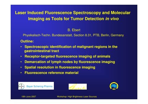
<strong>Laser</strong> <strong>Induced</strong> <strong>Fluorescence</strong> <strong>Spectroscopy</strong> <strong>and</strong> Molecular<br />
Imaging as Tools for Tumor Detection in vivo<br />
B. Ebert<br />
Physikalisch-Techn. Bundesanstalt, Section 8.31, PTB, Berlin, Germany<br />
Outline:<br />
• Spectroscopic identification of malignant regions in the<br />
gastrointestinal tract<br />
• Receptor-targeted fluorescence imaging of animals<br />
• Demarcation of lymph nodes by fluorescence imaging<br />
• Spatial resolution in fluorescence imaging<br />
• <strong>Fluorescence</strong> reference material<br />
Bayer Schering Pharma<br />
2007<br />
18th June 2007<br />
Workshop: High Brightness <strong>Laser</strong> Sources
<strong>Fluorescence</strong> imaging of lymph nodes<br />
Experimental setup<br />
shutter<br />
dichroic<br />
beam splitter<br />
beam dump<br />
505 nm<br />
dichroic<br />
beam splitter<br />
excitation<br />
fiber<br />
OPO<br />
355<br />
nm<br />
colonoscope<br />
THG<br />
SHG<br />
power<br />
supply<br />
electr. delay<br />
HV generator<br />
Nd:YAG<br />
50 Hz<br />
synchronisation<br />
imaging<br />
polychromator<br />
gastrointestinal<br />
tract<br />
intensified<br />
CCD camera<br />
controller<br />
2007<br />
18th June 2007<br />
Workshop: High Brightness <strong>Laser</strong> Sources
Heme biosynthesis<br />
Succinyl-CoA + Glycine<br />
COO -<br />
5 - Aminolevulinic acid (ALA)<br />
CH 2<br />
2 mol ALA<br />
CH 2<br />
C=O<br />
V<br />
M<br />
Porphobilinogen<br />
NH 3<br />
+<br />
M<br />
NH<br />
N<br />
V<br />
4 mol Porphobilinogen<br />
N<br />
HN<br />
Uroporphyrinogen III<br />
Coproporphyrinogen<br />
Protoporphyrinogen IX<br />
Protoporphyrin IX<br />
(PpIX)<br />
CH 2<br />
2007<br />
M<br />
P<br />
P<br />
M<br />
M: CH 3<br />
V: CH=CH 2<br />
P: CH 2<br />
_ CH2<br />
_ COOH<br />
18th June 2007<br />
Workshop: High Brightness <strong>Laser</strong> Sources
<strong>Fluorescence</strong> spectroscopy of tumors<br />
endoscopic view<br />
18th June 2007<br />
Workshop: High Brightness <strong>Laser</strong> Sources<br />
2007
<strong>Fluorescence</strong> spectroscopy of malignant tissue<br />
Normalized fluorescence intensity<br />
1.0<br />
0.8<br />
0.6<br />
0.4<br />
0.2<br />
0.0<br />
18th June 2007<br />
I(599 nm, 0 ns)<br />
0 ns delay<br />
20 ns delay<br />
I(635 nm, 20 ns)<br />
600 700 nm<br />
Wavelength<br />
R= I(635 nm, 20 ns) / I(599 nm, 0ns)<br />
Workshop: High Brightness <strong>Laser</strong> Sources<br />
2007
<strong>Fluorescence</strong> spectroscopy of lymph nodes<br />
Comparison with histology (cumulative frequency)<br />
Normalized cumulative frequency<br />
1.0<br />
0.8<br />
0.6<br />
0.4<br />
0.2<br />
0.0<br />
connective tissue (n = 272)<br />
lymph nodes (n = 434)<br />
involved lymph nodes (n = 90)<br />
1 10<br />
R=I(633 nm, 20 ns) / I(595 nm, 20 ns)<br />
Cancer Research, 2001, 61, 991-999<br />
18th June 2007<br />
Workshop: High Brightness <strong>Laser</strong> Sources<br />
2007
Experimental setup: Gated fluorescence imaging<br />
Gate: 10 ns, 100 Hz Gate:200 ps, 20 MHz<br />
OPO<br />
SHG<br />
THG<br />
Nd:YAG. 100 Hz<br />
Excitation fiber<br />
Delay<br />
generator<br />
ICCD<br />
200 ps<br />
Photocathode<br />
driver<br />
HV pulse<br />
generator<br />
Filter<br />
PC & Delay<br />
card<br />
ICCD-<br />
Camera<br />
Controller<br />
Programmable<br />
delay line<br />
depth<br />
<strong>Laser</strong><br />
<strong>Laser</strong>driver<br />
& Trigger<br />
Filter<br />
18th June 2007<br />
Workshop: High Brightness <strong>Laser</strong> Sources<br />
2007
Receptor targeted NIR- imaging of mouse xenografts<br />
with fluorescent lig<strong>and</strong>s<br />
ITCC- Octreotid<br />
ITCC-(M 2 , M 7 ) Octreotid<br />
In vivo fluorescence<br />
imaging of tumors<br />
SSTR2 Tumor, NIR38:<br />
20nmol /kg body weight<br />
Octreotid<br />
R = dPhe-Cys-Phe-dTrp-Lys-Thr-Cys- Thr<br />
6 h 6 h<br />
nature biotechnology, 2001, • 19, 327 - 331<br />
Bayer Schering Pharma<br />
18th June 2007<br />
Workshop: High Brightness <strong>Laser</strong> Sources<br />
2007
Optical molecular imaging of lymph nodes using a targeted<br />
vascular contrast agent<br />
A<br />
A´<br />
CH CH CH<br />
+<br />
N<br />
3<br />
-<br />
(CH 2 ) 4 SO 3<br />
N<br />
O<br />
(CH 2 ) 4 SO 3<br />
- Na<br />
+<br />
OH<br />
ITCC<br />
MECA<br />
B<br />
B´<br />
Lymph nodes<br />
strong fluorescence in the liver consistent<br />
with the hepatobiliary elimination pathway<br />
Liver<br />
C<br />
C´<br />
Bladder<br />
Bayer Schering Pharma<br />
J Biomed Opt. 2005, 10(4):41205<br />
18th June 2007<br />
Workshop: High Brightness <strong>Laser</strong> Sources<br />
2007
Optical molecular imaging of lymph nodes using a targeted<br />
vascular contrast agent<br />
Contrast: K = (I lymph - I muscle )/I muscle<br />
MECA- 79<br />
0.5<br />
High sensitivity, requiring as little<br />
as 0.25 nmol dye per animal<br />
0.4<br />
Contrast<br />
0.3<br />
0.2<br />
0.1<br />
-<br />
Cyanine dye<br />
Control<br />
conjugate<br />
0.0<br />
0 50 100 150<br />
Time (min)<br />
Bayer Schering Pharma<br />
18th June 2007<br />
Workshop: High Brightness <strong>Laser</strong> Sources<br />
2007
Experimental setup: Gated fluorescence imaging<br />
Gate: 10 ns, 100 Hz Gate:200 ps, 20 MHz<br />
OPO<br />
SHG<br />
THG<br />
Nd:YAG. 100 Hz<br />
Excitation fiber<br />
Delay<br />
generator<br />
ICCD<br />
200 ps<br />
Photocathode<br />
driver<br />
HV pulse<br />
generator<br />
Filter<br />
PC & Delay<br />
card<br />
ICCD-<br />
Camera<br />
Controller<br />
Programmable<br />
delay line<br />
depth<br />
<strong>Laser</strong><br />
<strong>Laser</strong>driver<br />
& Trigger<br />
Filter<br />
18th June 2007<br />
Workshop: High Brightness <strong>Laser</strong> Sources<br />
2007
Depth resolved fluorescence imaging<br />
Fluorescent rod at different depth in a scattering solution µ´s = 14 cm -1<br />
depth: 1 mm<br />
depth: 12 mm<br />
1.7 ns 2.2 ns 3.3 ns<br />
2.7 ns 3.7 ns 4.8 ns<br />
Normalized intensity<br />
1.0<br />
0.8<br />
0.6<br />
0.4<br />
0.2<br />
response<br />
fluorescence<br />
Normalized intensity<br />
1.0<br />
0.8<br />
0.6<br />
0.4<br />
0.2<br />
response<br />
fluorescence<br />
0<br />
0 2 4 6 8 10 12 14 16 18 20<br />
0<br />
0 2 4 6 8 10 12 14 16 18 20<br />
time / ns<br />
time / ns<br />
18th June 2007<br />
Workshop: High Brightness <strong>Laser</strong> Sources<br />
2007
Depth resolved fluorescence imaging<br />
Fluorescent rod at different depth in a scattering solution µ´s = 14 cm-1<br />
Rod<br />
3 mm<br />
x 10 -9<br />
7<br />
Influence of geometry<br />
Depth 5 mm<br />
Depth 10 mm<br />
6<br />
Mean time of flight<br />
5<br />
4<br />
3<br />
0 10 20<br />
x / mm<br />
0 10 20<br />
x / mm<br />
2.2 ns<br />
2.85 ns 4.0 ns<br />
2<br />
1<br />
0 5 10 15 20 25<br />
Depth / mm<br />
18th June 2007<br />
Workshop: High Brightness <strong>Laser</strong> Sources<br />
2007
<strong>Fluorescence</strong> reference material <strong>and</strong> application<br />
<strong>Fluorescence</strong> excitation spectra<br />
Normalized fluorescence intensity<br />
1.0<br />
0.8<br />
0.6<br />
0.4<br />
0.2<br />
0.0<br />
NIR96007 in solution with spheres<br />
NIR96007 in solution<br />
covalently bound to spheres<br />
NIR96007 in solution 1µM<br />
NIR96007 in solution 100 µM<br />
Observation at 790 nm<br />
600 650 700 750<br />
Wavelength (nm)<br />
Bayer Schering Pharma<br />
18th June 2007<br />
Workshop: High Brightness <strong>Laser</strong> Sources<br />
2007
<strong>Fluorescence</strong> intensity in dependence on concentration<br />
of cyanine dye molecules<br />
<strong>Fluorescence</strong> intensity (counts/sec)<br />
4x10 7 NIR960078 in solution<br />
NIR960078 bound to glass spheres<br />
3x10<br />
7<br />
2x10 7<br />
1x10 7<br />
0<br />
0 10 20 30 40<br />
Dyes (µmol)<br />
Bayer Schering Pharma<br />
18th June 2007<br />
Workshop: High Brightness <strong>Laser</strong> Sources<br />
2007
Scattering phantom with NIR96007, 2% on glass spheres<br />
4<br />
3<br />
X (cm)<br />
2<br />
1<br />
Normalized fluorescence intensity<br />
1.0<br />
0.8<br />
0.6<br />
0.4<br />
0.2<br />
0.0<br />
18th June 2007<br />
0 1 2 3 4 5<br />
X (cm)<br />
0<br />
0.0 0.2 0.4 0.6 0.8 1.0<br />
Workshop: High Brightness <strong>Laser</strong> Sources<br />
Normalized fluorescence intensity<br />
Bayer Schering Pharma<br />
2007
Monitoring of inflammation of ankle joints using<br />
cyanine dyes as contrast agents<br />
Regions of interest<br />
Reference<br />
Inflamed region<br />
Inflamed region<br />
Bayer Schering Pharma<br />
18th June 2007<br />
Workshop: High Brightness <strong>Laser</strong> Sources<br />
2007
Increase of fluorescence intensity in the right ankle joint<br />
after i.v. application of a cyanine dye<br />
Normalized fluorescence intensity<br />
1.0<br />
0.8<br />
0.6<br />
0.4<br />
0.2<br />
0.0<br />
measured values<br />
corrected values (normalized<br />
to reference)<br />
0 20 40 60 80 100 120 140 160<br />
J Fluoresc. 2005 15(3):337-62<br />
Time (s)<br />
Bayer Schering Pharma<br />
18th June 2007<br />
Workshop: High Brightness <strong>Laser</strong> Sources<br />
2007
Conclusion<br />
Small malignant regions (dysplasias) in the gastrointestinal<br />
tract can be identified.<br />
Light sources with high pulse energy <strong>and</strong> low repetition rates<br />
are preferable to suppress ambient light<br />
Succesful targeting of endothelial surface-expressed<br />
molecules - l<strong>eu</strong>kocyte homing<br />
Determination of depth of inclusions by time resolved<br />
fluorescence imaging<br />
Fluorescent phantoms are needed, however, to match<br />
optical properties of biological tissue is difficult<br />
18th June 2007<br />
Workshop: High Brightness <strong>Laser</strong> Sources