
Create successful ePaper yourself
Turn your PDF publications into a flip-book with our unique Google optimized e-Paper software.
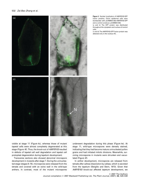
532 Zai-Bao Zhang et al.<br />
(a)<br />
(b)<br />
Figure 3. Nuclear localization of AtMYB103-GFP<br />
fusion proteins. Onion epidermal cells were<br />
bombarded with pCAMBIA1302-AtMYB103-GFP<br />
and a control construct, pCAMBIA1302.<br />
(a and b) The GFP protein was distributed<br />
throughout the cytoplasm and nucleus of control<br />
cells.<br />
(c and d) The AtMYB103-GFP fusion protein was<br />
detected only in the nucleus (N).<br />
(c)<br />
(d)<br />
visible at stage 11 (Figure 4c), whereas those of mutant<br />
tapetal cells were almost completely degenerated at this<br />
stage (Figure 4f). Thus, the knock-out of AtMYB103 resulted<br />
in defects of tapetal cell wall degradation and tapetal cell<br />
protoplast degeneration during tapetum development.<br />
Transverse sections also showed abnormal microspore<br />
development in mutants after stage 7. During the uninucleated<br />
stage (stages 8–10), microspores were released from the<br />
tetrads and covered with an exine wall in the wild-type<br />
anthers. In contrast, most of the mutant microspores<br />
underwent degradation during this phase (Figure 4e). At<br />
stage 11, wild-type microspores were densely stained,<br />
indicating that they had become mature uninucleated pollen<br />
grains and had initiated mitotic divisions. Meanwhile, surviving<br />
microspores in mutants were shrunken and vacuolated<br />
(Figure 4f).<br />
In anther development, microspores are released from<br />
tetrads after callose dissolution by callase, which is secreted<br />
from the tapetum (Steiglitz and Stern, 1973). Given that<br />
AtMYB103 knock-out affected tapetum development, we<br />
ª 2007 The Authors<br />
Journal compilation ª 2007 Blackwell Publishing Ltd, The Plant Journal, (2007), 52, 528–538