
You also want an ePaper? Increase the reach of your titles
YUMPU automatically turns print PDFs into web optimized ePapers that Google loves.
Molecular cloning and functional analysis of AtMYB103 535<br />
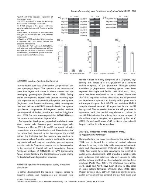
Figure 7. AtMYB103 regulates expression of<br />
downstream genes.<br />
(a) RT-PCR analysis of 12 genes that encode b-<br />
1,3-glucanase in wild type and ms188-1.<br />
(b) RT-PCR analysis of four genes involved in<br />
exine formation in the ms188-1 mutants and in<br />
wild type.<br />
(c) Real-time RT-PCR analysis of A6 expression in<br />
wild-type and mutant (ms188-1 and pst00809)<br />
backgrounds.<br />
(d) Real-time RT-PCR analysis of MS2 expression<br />
in wild-type (Ler), and mutant (ms188 and<br />
pst00809) inflorescences.<br />
(e) Real-time RT-PCR analysis of AtMYB103 in<br />
both wild-type and ms2 backgrounds. WT-inf,<br />
wild-type inflorescences; ms188-1-inf, ms188-1<br />
inflorescences; pst00809-inf, pst00809 inflorescences;<br />
ms2-inf, ms2 inflorescences.<br />
(a)<br />
(c) (d) (e)<br />
(b)<br />
AtMYB103 regulates tapetum development<br />
In Arabidopsis, each lobe of the anther comprises four distinct<br />
sporophytic layers. The tapetum is the innermost of<br />
these four layers and comes in direct contact with the<br />
developing gametophyte (Sanders et al., 1999). During<br />
anther development, tapetal cells become secretory type<br />
cells to provide the nutrition critical for pollen development<br />
(Raghavan, 1989; Stevens and Murray, 1981). In transgenic<br />
lines with reduced AtMYB103 transcript levels, the tapetum<br />
cytoplasmic components disintegrated earlier, without<br />
releasing small oil bodies, plastids and vesicles (Higginson<br />
et al., 2003). Our data also suggested that AtMYB103 knockout<br />
results in early tapetum degeneration.<br />
During anther development, tapetal cell walls break down<br />
when tapetal cells become polar secretary-type cells<br />
(Stevens and Murray, 1981). In ms188, the tapetal cell walls<br />
remain intact late in anther development. Given that most of<br />
the callose had dissolved by the late stage of the ms188<br />
anther, this indicates that the tapetum may continue to<br />
secret some callase from the tapetum, and that the remaining<br />
tapetal cell wall may not completely prevent tapetum<br />
secretary activity. No gene or enzyme has yet been reported<br />
to be involved in tapetal cell wall degradation. Future<br />
functional analysis of AtMYB103, an MYB transcription<br />
factor, should facilitate the identification of genes coding<br />
for tapetal cell wall degradation enzymes.<br />
AtMYB103 regulates A6 transcription during the callose<br />
dissolution<br />
In anther development the tapetum releases callase to<br />
dissolve callose, and microspores are released from<br />
tetrads. Callose is mainly composed of b-1,3-glucan, suggesting<br />
that callase is a b-1,3-glucanase or a complex<br />
mainly composed of b-1,3-glucanase. Although several<br />
candidate b-1,3-glucanase encoding genes have been<br />
reported (Bucciaglia and Smith, 1994; Hird et al., 1993),<br />
none has been confirmed to be a callase. Given that<br />
AtMYB103 regulated callose dissolution, ms188 provided<br />
an experimental approach to identify which gene was a<br />
callase-specific gene. Both RT-PCR and real-time RT-PCR<br />
analysis showed reduced A6 expression in the ms188<br />
background. The expression level of the A6 gene was in<br />
agreement with the partial degradation of callose in<br />
ms188. This indicates that A6 may be a callase or a part of<br />
the callase enzyme complex, as suggested by Hird et al.<br />
(1993). Future identification of A6 knock-out plants should<br />
help to confirm its role as a callase.<br />
AtMYB103 is required for the expression of MS2<br />
to regulate exine formation<br />
Sporopollenin is the major constituent of the exine (Scott,<br />
1994) and is formed by a series of related polymers<br />
derived from long-chain fatty acids, oxygenated aromatic<br />
rings and phenylpropanoids (Piffanelli et al., 1998; Scott,<br />
1994). Four genes have been reported to be involved in<br />
pollen exine development. MS2 encodes a putative fatty<br />
acid reductase that catalyzes fatty acyl groups to fatty<br />
alcohol groups, and that may be involved in sporopollenin<br />
synthesis (Aarts et al., 1997). The remaining three genes,<br />
FLP1, DEX1 and NEF1, have been shown to play roles<br />
in exine pattern formation (Ariizumi et al., 2003, 2004;<br />
Paxson-Sowders et al., 2001). In ms2 male sterile mutants,<br />
pollen development was arrested and no thick exine wall<br />
ª 2007 The Authors<br />
Journal compilation ª 2007 Blackwell Publishing Ltd, The Plant Journal, (2007), 52, 528–538