
You also want an ePaper? Increase the reach of your titles
YUMPU automatically turns print PDFs into web optimized ePapers that Google loves.
Molecular cloning and functional analysis of AtMYB103 533<br />
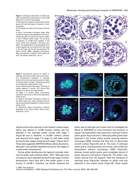
Figure 4. Cytological observation of anther and<br />
pollen development of wild-type (a–c) and ms188<br />
plants (d–f). (a and d) Tetrad stage.<br />
(a) The tapetum cytoplasm was shrunken and the<br />
tapetal cell wall had begun to dissolve in wildtype<br />
plants.<br />
(d) The tapetal cell walls of the mutant remained<br />
intact.<br />
(b and e) Uninucleate microspore stage. Most<br />
ms188 microspores were degraded in the locule.<br />
Tapetal cell walls of mutants were clearly visible.<br />
(c and f) Mitosis pollen stage. The surviving<br />
microspores of ms188 fused together in the<br />
locule, which also displayed a shrunken cytoplasm.<br />
The degeneration of the protoplast of the<br />
mutant tapetal was more severe than that noted<br />
in the wild type, and its cell wall persisted. FB,<br />
fibrous bands; dMSp, degraded microspores;<br />
MSp, microspores; T, tapetum; TCW, tapetal cell<br />
wall; Tds, tetrads. Scale bars = 20 lm.<br />
(a) (b) (c)<br />
(d) (e) (f)<br />
Figure 5. Cytochemical staining for callose in<br />
wild-type and mutant anthers with aniline blue.<br />
(a–c) Fluorescence expression in wild-type<br />
anther with aniline blue staining under UV light.<br />
(a) Anther section at stage 7, showing the tetrad<br />
surrounded with callose (white arrow).<br />
(b and c) Anther section after stage 7, showing no<br />
callose detected in locules. (d–f) Aniline blue<br />
staining on a section of mutant anthers.<br />
(d) Stage 7 of mutant anther, fluorescence<br />
expression was similar to that of wild-type plants<br />
(white arrow).<br />
(e) Stage 9: decreased fluorescence relative to<br />
the tetrad stage was noted, indicating that the<br />
callose was partially degraded in mutant anthers<br />
(white arrow).<br />
(f) Dehiscence stage: fluorescence remained<br />
detectable in the mutant anther (white arrow).<br />
Scale bars = 20 lm.<br />
(a) (b) (c)<br />
(d) (e) (f)<br />
utilized aniline blue staining to test whether callose degradation<br />
was altered in ms188 mutants. Callose was not<br />
detected in the wild-type anther locules after stage 7<br />
(Figure 5b and c). However, in ms188-1 anthers, callose<br />
was observed from stage 7 to stage 12, with fewer noted<br />
during the later stages compared with stage 7 (Figure 5d–f).<br />
These data suggested AtMYB103 affects callose dissolution,<br />
although it was partially degraded during the late stages of<br />
mutant anther development.<br />
During anther development the tapetum provides materials<br />
for pollen wall synthesis (Steer, 1977). Although most<br />
microspores were degraded during the late stage of anther<br />
development, there were still a few pollen grains in the<br />
locules of ms188-1. Therefore, we further observed the<br />
pollen wall of wild-type and mutant lines to investigate the<br />
affects of AtMYB103 on exine formation and structure. A<br />
regular reticulate pattern was observed in wild-type mature<br />
pollen grains (Figure 6a and c). Although pollen grains were<br />
observed in the mutant anther locules, scanning electron<br />
microscopy (SEM) revealed that they had an abnormally<br />
smooth surface (Figure 6b and d). The exine of wild-type<br />
pollen grains, including sexine and nexine, were evident by<br />
transmission electron microscopy (TEM) (Figure 6e),<br />
whereas mutant pollen grains were completely devoid of<br />
sexine (baculum and tectum) (Figure 6f). The pollen coat,<br />
mainly derived from the tapetum, filled the interstices of<br />
wild-type exine (Figure 6e), whereas no pollen coat was<br />
observed in the mutant locule (Figure 6f). These results<br />
ª 2007 The Authors<br />
Journal compilation ª 2007 Blackwell Publishing Ltd, The Plant Journal, (2007), 52, 528–538