Surface-enhanced Raman and fluorescence joint analysis of soil ...
Surface-enhanced Raman and fluorescence joint analysis of soil ...
Surface-enhanced Raman and fluorescence joint analysis of soil ...

Create successful ePaper yourself
Turn your PDF publications into a flip-book with our unique Google optimized e-Paper software.
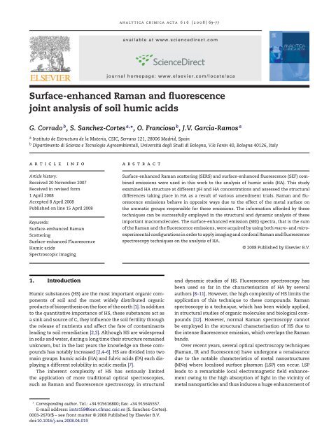
analytica chimica acta 616 (2008) 69–77<br />
available at www.sciencedirect.com<br />
journal homepage: www.elsevier.com/locate/aca<br />
<strong>Surface</strong>-<strong>enhanced</strong> <strong>Raman</strong> <strong>and</strong> <strong>fluorescence</strong><br />
<strong>joint</strong> <strong>analysis</strong> <strong>of</strong> <strong>soil</strong> humic acids<br />
G. Corrado b , S. Sanchez-Cortes a,∗ , O. Francioso b , J.V. Garcia-Ramos a<br />
a Instituto de Estructura de la Materia, CSIC, Serrano 121, 28006 Madrid, Spain<br />
b Dipartimento di Scienze e Tecnologie Agroambientali, Università degli Studi di Bologna, V.le Fanin 40, Bologna 40126, Italy<br />
article<br />
info<br />
abstract<br />
Article history:<br />
Received 20 November 2007<br />
Received in revised form<br />
1 April 2008<br />
Accepted 8 April 2008<br />
Published on line 15 April 2008<br />
Keywords:<br />
<strong>Surface</strong>-emhanced <strong>Raman</strong><br />
Scattering<br />
<strong>Surface</strong>-<strong>enhanced</strong> Fluorescence<br />
Humic acids<br />
Spectroscopic imaging<br />
<strong>Surface</strong>-<strong>enhanced</strong> <strong>Raman</strong> scattering (SERS) <strong>and</strong> surface-<strong>enhanced</strong> <strong>fluorescence</strong> (SEF) combined<br />
emissions were used in this work to the <strong>analysis</strong> <strong>of</strong> humic acids (HA). This study<br />
examined HA structure at different pH <strong>and</strong> HA concentrations <strong>and</strong> assessed the structural<br />
differences taking place in HA as a result <strong>of</strong> various amendment trials. <strong>Raman</strong> <strong>and</strong> <strong>fluorescence</strong><br />
emissions behave in opposite ways due to the effect <strong>of</strong> the metal surface on<br />
the aromatic groups responsible for these emissions. The information afforded by these<br />
techniques can be successfully employed in the structural <strong>and</strong> dynamic <strong>analysis</strong> <strong>of</strong> these<br />
important macromolecules. The surface-<strong>enhanced</strong> emission (SEE) spectra, that is the sum<br />
<strong>of</strong> the <strong>Raman</strong> <strong>and</strong> the <strong>fluorescence</strong> emissions, were acquired by using both macro- <strong>and</strong> microexperimental<br />
configurations in order to apply imaging <strong>and</strong> confocal <strong>Raman</strong> <strong>and</strong> <strong>fluorescence</strong><br />
spectroscopy techniques on the <strong>analysis</strong> <strong>of</strong> HA.<br />
© 2008 Published by Elsevier B.V.<br />
1. Introduction<br />
Humic substances (HS) are the most important organic components<br />
<strong>of</strong> <strong>soil</strong> <strong>and</strong> the most widely distributed organic<br />
products <strong>of</strong> biosynthesis on the face <strong>of</strong> the earth [1]. In addition<br />
to the quantitative importance <strong>of</strong> HS, these substances act as<br />
a sink <strong>and</strong> source <strong>of</strong> C, they influence the <strong>soil</strong> fertility through<br />
the release <strong>of</strong> nutrients <strong>and</strong> affect the fate <strong>of</strong> contaminants<br />
leading to <strong>soil</strong> remediation [2,3]. Although HS are widespread<br />
in <strong>soil</strong>s <strong>and</strong> water, during a long time their structure remained<br />
unknown, but in the last years the knowledge on these compounds<br />
has notably increased [2,4–6]. HS are divided into two<br />
main groups: humic acids (HA) <strong>and</strong> fulvic acids (FA) each displaying<br />
a different solubility in acidic media [7].<br />
The inherent complexity <strong>of</strong> HS has seriously limited<br />
the application <strong>of</strong> more traditional optical spectroscopies,<br />
such as <strong>Raman</strong> <strong>and</strong> <strong>fluorescence</strong> spectroscopy, in structural<br />
<strong>and</strong> dynamic studies <strong>of</strong> HS. Fluorescence spectroscopy has<br />
been used so far in the characterisation <strong>of</strong> HA by several<br />
authors [8–11]. However, the high complexity <strong>of</strong> HS limits the<br />
application <strong>of</strong> this technique to these compounds. <strong>Raman</strong><br />
spectroscopy is a technique, which has been widely applied,<br />
in structural studies <strong>of</strong> organic molecules <strong>and</strong> biological compounds<br />
[12]. However, normal <strong>Raman</strong> spectroscopy cannot<br />
be employed in the structural characterisation <strong>of</strong> HS due to<br />
the intense <strong>fluorescence</strong> emission, which overlaps the <strong>Raman</strong><br />
b<strong>and</strong>s.<br />
Over recent years, several optical spectroscopy techniques<br />
(<strong>Raman</strong>, IR <strong>and</strong> <strong>fluorescence</strong>) have undergone a renaissance<br />
due to the notable characteristics <strong>of</strong> metal nanostructures<br />
(MNs) where localised surface plasmon (LSP) can occur. LSP<br />
leads to a remarkable local electromagnetic field enhancement<br />
owing to the high absorption <strong>of</strong> light in the vicinity <strong>of</strong><br />
metal nanoparticles <strong>and</strong> thus induces a huge enhancement <strong>of</strong><br />
∗ Corresponding author. Tel.: +34 915616800; fax: +34 915645557.<br />
E-mail address: imts158@iem.cfmac.csic.es (S. Sanchez-Cortes).<br />
0003-2670/$ – see front matter © 2008 Published by Elsevier B.V.<br />
doi:10.1016/j.aca.2008.04.019
70 analytica chimica acta 616 (2008) 69–77<br />
spectroscopic signals, mainly in the case <strong>of</strong> emission signals<br />
<strong>of</strong> molecules placed in the proximities <strong>of</strong> the surface [13–15].<br />
<strong>Raman</strong> scattering is greatly <strong>enhanced</strong> as a consequence<br />
<strong>of</strong> LSP. For this reason, the surface-<strong>enhanced</strong> <strong>Raman</strong> scattering<br />
(SERS) technique can be successfully applied to the<br />
study <strong>of</strong> highly fluorescent molecules in water, due to<br />
the <strong>fluorescence</strong> quenching occurring on the metal surface<br />
via a charge-transfer mechanism [16]. This effect<br />
enables <strong>Raman</strong> to be applied to the characterisation <strong>of</strong> HS<br />
[17–20].<br />
SERS spectra can provide important information on the<br />
HS structure in function <strong>of</strong> factors such as the humification<br />
degree [21], extraction <strong>and</strong> purification processes [22], the <strong>soil</strong><br />
amendment trials [23], HS extraction fractions [17,24] <strong>and</strong> pH<br />
[25]. The growing importance <strong>of</strong> SERS in the <strong>analysis</strong> <strong>of</strong> HS<br />
has recently been highlighted in several reviews dealing with<br />
techniques applied to the <strong>analysis</strong> <strong>of</strong> HS [26–28]. SERS has also<br />
been used to examine pesticide–HA interaction [29]. However,<br />
SERS is not a quantitative technique because <strong>of</strong> effects such<br />
as the resonance <strong>Raman</strong> <strong>and</strong> the adsorption on surfaces [30],<br />
although these characteristics can also serve to study structural<br />
<strong>and</strong> dynamic processes undertaken by macromolecules<br />
in the presence <strong>of</strong> surfaces [31].<br />
The surface-<strong>enhanced</strong> <strong>fluorescence</strong> (SEF) technique has<br />
not been employed as much as SERS because <strong>of</strong> chargetransfer<br />
effects, which take place on the surface <strong>and</strong> which,<br />
in turn, lead to an actual quenching <strong>of</strong> the <strong>fluorescence</strong>. SEF<br />
effect is also induced by LPR near a metal surface [32,33].However,<br />
the net effect seems to vary depending on the distance<br />
between the fluorophore <strong>and</strong> the surface, as well as on the<br />
intrinsic quantum yield <strong>of</strong> the fluorophore [34]. In general,<br />
the <strong>fluorescence</strong> is not quenched if the molecule is relatively<br />
far from the surface. The estimated optimal distance for a<br />
good SEF enhancement is achieved above 100 Å [35,36]. At distances<br />
further than this value, SERS <strong>and</strong> SEF signals can be<br />
simultaneously emitted <strong>and</strong> registered for molecular species<br />
placed in the vicinity <strong>of</strong> MNs [36–38]. Since the average size<br />
<strong>of</strong> HA is larger than this value [39], the fluorophores included<br />
in the HA structure could be good c<strong>and</strong>idates to give rise to<br />
intense SEF + SERS <strong>joint</strong> emission spectra, without the intrinsic<br />
quenching observed on a surface. This is possible due to the<br />
fact that the HS cross-sections <strong>of</strong> either <strong>Raman</strong> or <strong>fluorescence</strong><br />
are <strong>of</strong> the same order on MNs, <strong>and</strong> thus, a surface-<strong>enhanced</strong><br />
emission (SEE) is seen for HS adsorbed on these substrates<br />
(Fig. 1, bottom). Furthermore, the SEF emission affords information<br />
on the HA macromolecules adsorbed onto a surface,<br />
Fig. 1 – Top: experimental set-up for SEE measurements. Bottom left: a SEM image <strong>of</strong> typical Ag nanoaggregates. Bottom<br />
right: scheme displaying the adsorption <strong>of</strong> HA micelles <strong>and</strong> the emission <strong>of</strong> <strong>Raman</strong> a <strong>fluorescence</strong> combined process.
analytica chimica acta 616 (2008) 69–77 71<br />
i.e. on the aggregation process undergone by these macromolecules,<br />
which are <strong>of</strong> importance to underst<strong>and</strong> how these<br />
compounds are attached on the inorganic matrices existing<br />
in <strong>soil</strong>s. This fact represented an evident advantage regarding<br />
the normal <strong>fluorescence</strong> spectra obtained in solution.<br />
Spectroscopic imaging <strong>and</strong> micro-confocal techniques<br />
have been developed for both <strong>Raman</strong> <strong>and</strong> <strong>fluorescence</strong> spectroscopy<br />
with promising analytical applications [40], <strong>and</strong><br />
could also be <strong>of</strong> interest in the study <strong>of</strong> HA, as demonstrated by<br />
previous studies [41]. The combination <strong>of</strong> surface-<strong>enhanced</strong><br />
techniques <strong>and</strong> microscopy is also a powerful methodology<br />
which has attracted much attention in the <strong>analysis</strong> <strong>of</strong> biological<br />
systems such as cells or bacteria [42,43] <strong>and</strong> which could<br />
be very helpful in the characterisation <strong>of</strong> HS.<br />
In this work we analysed SEE spectra <strong>of</strong> HA. This work<br />
represents the first time that SEF technique is used in the characterisation<br />
<strong>of</strong> HA. The HA compounds selected for this study<br />
were extracted from a <strong>soil</strong> amended over a period <strong>of</strong> 30 years<br />
under several conditions <strong>and</strong> the corresponding control experiments<br />
in order to evaluate the structural variations induced<br />
by the different treatments. The SEE spectra were acquired by<br />
using both macro- <strong>and</strong> micro-experimental configurations in<br />
order to investigate the abilities <strong>of</strong> the new imaging <strong>and</strong> confocal<br />
<strong>Raman</strong> <strong>and</strong> <strong>fluorescence</strong> spectroscopy techniques on the<br />
<strong>analysis</strong> <strong>of</strong> these HA.<br />
2. Materials <strong>and</strong> methods<br />
The <strong>soil</strong> samples analysed (horizon Ap, 0–40 cm depth) were<br />
taken from plots <strong>of</strong> a 30 plus year field experiment conducted<br />
at the agricultural farm <strong>of</strong> the University <strong>of</strong> Bologna’s Agricultural<br />
Faculty (Cadriano, Italy). This <strong>soil</strong> is classified (Soil Survey<br />
Staff, USDA SCS 1989) as a Typic Udochrept.<br />
The experimental design included plots amended over a<br />
30-year period with cattle manure (CM) <strong>and</strong> crop residues (CR)<br />
constituted by wheat straw or corn stalks (each biomass followed<br />
in succession), with unamended <strong>soil</strong> as control (C). CM<br />
was added to the plots at the following rate: 6 t ha −1 <strong>of</strong> dry matter<br />
after wheat tillage <strong>and</strong> 7.5 t ha −1 <strong>of</strong> dry matter after corn<br />
tillage. All the plots examined, including C, were fertilised with<br />
100 kg N ha −1 yr −1 added in the form <strong>of</strong> ammonium nitrate.<br />
Each treatment (CM, CR <strong>and</strong> C) was conducted in triplicate<br />
using a r<strong>and</strong>omized block design.<br />
Analyses were carried out on <strong>soil</strong> samples taken from:<br />
(i) unamended <strong>soil</strong> in 1972 (C 0 ), <strong>and</strong> (ii) after 30 years (C 30 )<br />
<strong>and</strong> (iii) after a 30-year amendment period with CM <strong>and</strong> CR.<br />
The extraction procedures used to isolate these samples have<br />
already been described in Ref. [23].<br />
to enhance the electromagnetic field on the metal surface. A<br />
typical Ag MNs used to record the SEE spectra is shown in Fig. 1<br />
(bottom).<br />
Samples for SEE measurements were made by adding an<br />
aliquot <strong>of</strong> an aqueous HS solution, prepared by solving 1 mg <strong>of</strong><br />
the corresponding humic fraction in 1 mL <strong>of</strong> tri-distilled water,<br />
to 1 mL <strong>of</strong> the silver colloid until the desired concentration.<br />
The pH <strong>of</strong> the mixture was adjusted by adding 0.1 M NaOH or<br />
HNO 3 . All solutions were prepared with tri-distilled water.<br />
The SERS spectra were recorded by means <strong>of</strong> a Renishaw<br />
<strong>Raman</strong> Microscope System RM2000, using the macroconfiguration<br />
equipped with a notch filter <strong>and</strong> an electrically<br />
refrigerated CCD camera (575× pixels) as indicated in Fig. 1.<br />
The <strong>Raman</strong> <strong>and</strong> <strong>fluorescence</strong> emission spectrum is recorded<br />
by the CCD camera. The 514.5 nm line <strong>of</strong> an Ar + laser was used.<br />
The laser power at the sample was 2.0 mW. Spectral resolution<br />
was 2 cm −1 . Micro-SERS spectra were obtained by using a 100×<br />
objective (NA = 0.9). Confocal micro-SERS <strong>and</strong> <strong>Raman</strong> images<br />
were recorded by using a PRIOR motorised stage coupled to the<br />
microscope with a 1 m step. The SERS spectra were baselined<br />
to withdraw the contribution <strong>of</strong> the <strong>fluorescence</strong> by using the<br />
algorithm provided by the Origin 6.0 Program.<br />
3. Results <strong>and</strong> discussion<br />
3.1. SERS spectra <strong>of</strong> humic acids<br />
Fig. 2 shows the SERS spectra <strong>of</strong> HA at pH 10.0, after correction<br />
<strong>of</strong> the <strong>fluorescence</strong> background. At pH 10.0 the SERS spectra<br />
<strong>of</strong> all HA samples are dominated by two very strong <strong>and</strong> narrow<br />
b<strong>and</strong>s, at 1308 <strong>and</strong> 1611 cm −1 , which can be attributed<br />
to the aromatic part <strong>of</strong> HS <strong>and</strong> in particular to polycyclic aromatic<br />
hydrocarbon (PAH) structures. The nature <strong>of</strong> these PAH<br />
residues is still unknown, but the similarity <strong>of</strong> the SERS pattern<br />
observed at high pH <strong>and</strong> the <strong>Raman</strong> spectra <strong>of</strong>, phenanthrene,<br />
perylene [45] <strong>and</strong> coronene [46] compounds suggests that similar<br />
structures could exist in HA, either from anthropogenic<br />
2.1. SERS spectroscopy<br />
Colloidal silver nanoparticles were prepared by using hydroxylamine<br />
hydrochloride as reducing agent. In a recent work, a<br />
deep study <strong>of</strong> Ag MNs prepared by reduction with hydroxylamine<br />
(AgHx) was accomplished [44]. AgHx was activated by<br />
the addition <strong>of</strong> potassium nitrate, which induced the particle<br />
aggregation to form other larger aggregations. This process<br />
is necessary when using these systems as a substrate for<br />
SERS, since specific morphological requirements are needed<br />
Fig. 2 – SERS spectra <strong>of</strong> the different HA samples<br />
(10 gmL −1 ) employed in this work after correcting the<br />
<strong>fluorescence</strong> background to better show the <strong>Raman</strong><br />
features.
72 analytica chimica acta 616 (2008) 69–77<br />
or natural origins [2,7]. The situation seems to be different<br />
in the case <strong>of</strong> FA, since no PAH b<strong>and</strong>s are observed in these<br />
compounds [24]. The presence <strong>of</strong> PAH in the SERS spectrum<br />
recorded at alkaline pH, suggests that PAH residues might be<br />
mainly localised inside the HA structure, since a HA structure<br />
expansion is expected at high pH due to ionisation <strong>of</strong><br />
carboxylic <strong>and</strong> phenolic groups [47]. Although the macromolecular<br />
expansion is limited by chemical cross-linkages<br />
[48], it seems to be enough to allow a higher exposure <strong>of</strong> the<br />
polyaromatic groups to the metal surface. The b<strong>and</strong>s corresponding<br />
to PAH residues are resonantly <strong>enhanced</strong> as revealed<br />
the appearance <strong>of</strong> the overtone <strong>and</strong> combination b<strong>and</strong>s at<br />
1948, 2577, 2913 <strong>and</strong> 3228 cm −1 in the SERS spectra (Fig. 2).<br />
However, we cannot deduce that PAH groups are quantitatively<br />
predominant in the HA structure, since these groups<br />
undergone a selective enhancement by SERS.<br />
The SERS spectra in Fig. 2 were normalised to the water<br />
b<strong>and</strong> appearing at 3420 cm −1 . As can be seen, the CR <strong>and</strong> CM<br />
samples display a lower SERS intensity in comparison to the<br />
control samples C 0 <strong>and</strong> C 30 . In order to better underst<strong>and</strong> this<br />
behaviour we carried out a more detailed SERS study <strong>of</strong> HA<br />
at different pH values using the C 30 sample (Fig. 3), since this<br />
sample was the one, which displayed the most intense variation.<br />
The b<strong>and</strong>s at 1308 <strong>and</strong> 1611 cm −1 observed at pH 10.0<br />
(Fig. 3a) decreased markedly at pH 5 (Fig. 3d), as a consequence<br />
<strong>of</strong>: (i) an increase <strong>of</strong> intermolecular interactions, due to both<br />
a decrease <strong>of</strong> electric repulsions <strong>and</strong> an increase [47] <strong>of</strong> H-<br />
bonds <strong>and</strong> (ii) coagulation <strong>of</strong> smaller HA micelles into larger<br />
ones [49]. These facts lead to a macromolecular shrinkage.<br />
Below this pH value, a significant enhancement <strong>of</strong> the SERS<br />
intensity was observed (Fig. 3e), which is related to the HA<br />
adsorption tendency increase <strong>of</strong> at low pH as also reported by<br />
several authors [50,51]. The HA adsorption increase is related<br />
to the higher hydrophobic nature <strong>of</strong> HA at low pH caused<br />
by the negative charge decrease upon protonation <strong>of</strong> acidic<br />
HA groups. The increase <strong>of</strong> HA molecules adsorbed on the<br />
metal is further intensified due to the reduction <strong>of</strong> the area<br />
per molecule induced by the molecular shrinkage occurring<br />
at low pH [52].<br />
The SERS spectrum seen at low pH changes with respect to<br />
that observed at high pH (Fig. 3a). The <strong>analysis</strong> <strong>of</strong> the <strong>Raman</strong><br />
broad b<strong>and</strong>s seen at low pH is rather difficult owed to the sum<br />
<strong>of</strong> different components <strong>and</strong> the possible contribution from<br />
carbonaceous materials such as charcoal [53,54], which may<br />
be incorporated in the humic structure <strong>of</strong> that can be formed<br />
due to photodegradation as indicated by other authors [55].<br />
Nevertheless, the different <strong>Raman</strong> pr<strong>of</strong>ile with respect to charcoal<br />
<strong>Raman</strong> spectrum [54], the variation <strong>of</strong> this spectrum with<br />
the pH, as revealed below, <strong>and</strong> the appearance <strong>of</strong> b<strong>and</strong>s which<br />
are attributable to functional groups actually existing in HA,<br />
such as benzoic, polyphenolic <strong>and</strong> aliphatic ones, discharge<br />
the possibility <strong>of</strong> a massive photodegradation <strong>of</strong> the adsorbed<br />
HA as an effect <strong>of</strong> light irradiation.<br />
The dependency <strong>of</strong> SERS intensity on pH is better seen in<br />
the inset figure <strong>of</strong> Fig. 3, which displays a minimum at ca.<br />
5.5. Recently, Terashima et al. [52] have demonstrated that<br />
the apparent pK (pK app ) <strong>of</strong> HA is approximately 5.5 <strong>and</strong>, as<br />
a consequence, at pH 5.5 drastic changes take place in several<br />
physical properties, such as surface tension, surface area<br />
occupied by HA molecules when adsorbed onto a surface<br />
<strong>and</strong> HA hydrophobicity. A similar pH value was reported by<br />
other authors when studying the aggregation tendency <strong>of</strong> HA<br />
[56,57].<br />
It therefore follows that the lower SERS intensity <strong>of</strong> CR <strong>and</strong><br />
CM samples in comparison to the control C 0 <strong>and</strong> C 30 , can<br />
be attributed to the effect <strong>of</strong> amendants. The introduction <strong>of</strong><br />
lignin in the HA structure may increase the stability <strong>of</strong> the<br />
macromolecule due to the higher H-bonds formed between<br />
OH groups afforded by lignin. In the case <strong>of</strong> CM, the enrichment<br />
in fatty acids, due to the treatment with manure, also<br />
lead to a higher stabilisation <strong>of</strong> the macromolecule due to<br />
the formation <strong>of</strong> hydrophobic interactions between aliphatic<br />
chains <strong>of</strong> fatty acid residues.<br />
3.2. <strong>Surface</strong>-<strong>enhanced</strong> emission spectra <strong>of</strong> humic acids<br />
Fig. 3 – SERS spectra <strong>of</strong> C 30 sample (10 gmL −1 ) after<br />
<strong>fluorescence</strong> correction at the following pH: (a) 10.0, (b) 8.0,<br />
(c) 6.0, (d) 5.5 <strong>and</strong> (e) 3.0. Inset: variation <strong>of</strong> the 1609 cm −1<br />
SERS b<strong>and</strong> on changing the pH.<br />
Fig. 4 compares the normal <strong>Raman</strong> spectrum <strong>of</strong> the C 30 sample<br />
(1 gmL −1 ) with SEE spectrum <strong>of</strong> the same sample in the<br />
presence <strong>of</strong> Ag NPs. The emission spectrum <strong>of</strong> the solution<br />
shows the <strong>Raman</strong> b<strong>and</strong>s <strong>of</strong> water at 1640 cm −1 (very weak) <strong>and</strong><br />
3420 cm −1 (strong) <strong>and</strong> a broad <strong>fluorescence</strong> b<strong>and</strong> produced<br />
by HA centered at 630 nm, while no <strong>Raman</strong> b<strong>and</strong> <strong>of</strong> HA are<br />
seen. In contrast, the SEE spectrum <strong>of</strong> HA was characterised<br />
by strong <strong>Raman</strong> b<strong>and</strong>s in the 1200–1650 cm −1 region, <strong>and</strong> by<br />
a <strong>fluorescence</strong> emission with a maximum at 650 nm, which<br />
is threefold intensified in relation to that seen in aqueous<br />
solution. The intensification <strong>of</strong> <strong>Raman</strong> b<strong>and</strong>s corresponding<br />
to the HA <strong>and</strong> the shift observed for the <strong>fluorescence</strong> emission<br />
maximum suggests structural changes in the HA upon<br />
adsorption on the metal. Thus, the SEE spectrum <strong>of</strong> the C 30<br />
sample provided a combined <strong>enhanced</strong> <strong>Raman</strong> <strong>and</strong> <strong>fluorescence</strong><br />
information because the cross-sections corresponding
analytica chimica acta 616 (2008) 69–77 73<br />
Fig. 4 – <strong>Surface</strong>-<strong>enhanced</strong> emission (SEE) spectra <strong>of</strong> C 30<br />
sample (1 gmL −1 ) in absence (a) <strong>and</strong> presence (b) <strong>of</strong> Ag<br />
nanoparticles.<br />
Fig. 6 – Panel (a): variation with pH <strong>of</strong> the surface-<strong>enhanced</strong><br />
<strong>fluorescence</strong> emission (excitation at 514.5 nm <strong>and</strong> emission<br />
at 625 nm). Panel (b): the surface-<strong>enhanced</strong> <strong>Raman</strong><br />
scattering intensity <strong>of</strong> 1611 cm −1 b<strong>and</strong> (excitation at<br />
514.5 nm) measured from the C 30 , CR <strong>and</strong> CM samples at a<br />
HA concentration <strong>of</strong> 10.0 gmL −1 .<br />
to both emissions are <strong>of</strong> the same magnitude order. Although<br />
the total emission measured from adsorbates adsorbed on<br />
metal NPs is also a combined luminescence signal with contributions<br />
from both the molecule <strong>and</strong> the metal background<br />
luminescence [58], we can consider that the latter one is negligible<br />
in relation to the luminescence <strong>of</strong> HA in the SEE spectra.<br />
SEE spectra recorded at three different HA concentrations<br />
are shown in Fig. 5. At high concentration, the <strong>fluorescence</strong><br />
signal predominates over the <strong>Raman</strong> signal, while at lower<br />
concentrations the opposite behaviour is witnessed. This is<br />
probably due to the formation <strong>of</strong> HA multilayers on the Ag<br />
NPs, which leads to higher <strong>fluorescence</strong> intensities as the multilayer<br />
width is increased due to a lower quenching effect by<br />
the metal. The predominance <strong>of</strong> the SERS signal over the SEF<br />
Fig. 5 – <strong>Surface</strong>-<strong>enhanced</strong> emission (SEE) spectra <strong>of</strong> C 30<br />
sample on Ag nanoparticles at the following<br />
concentrations: (a) 10.0 gmL −1 , (b) 1.0 gmL −1 <strong>and</strong> (c)<br />
0.1 gmL −1 .<br />
one (in fact limit <strong>of</strong> detection <strong>of</strong> 1–100 ppb can be obtained,<br />
depending on the case) points out the powerful analytical possibilities<br />
<strong>of</strong> SERS in comparison to the <strong>fluorescence</strong>, in spite <strong>of</strong><br />
the higher sensibility <strong>of</strong> <strong>fluorescence</strong> over the <strong>Raman</strong> effect.<br />
The effect <strong>of</strong> pH on the <strong>Raman</strong> <strong>and</strong> <strong>fluorescence</strong> <strong>of</strong> HA samples<br />
is shown in Fig. 6. The <strong>Raman</strong> intensity changes with the<br />
pH reaching a minimum at pH 5.5 (Fig. 6b). In contrast, the<br />
<strong>analysis</strong> <strong>of</strong> the <strong>fluorescence</strong> emission revealed an opposite<br />
behaviour, i.e. a maximum at precisely pH 5.5 (Fig. 6a).<br />
This contrary behaviour <strong>of</strong> the <strong>fluorescence</strong> <strong>and</strong> SERS signals<br />
<strong>of</strong> HA can be explained on the basis <strong>of</strong> the structural<br />
modifications induced on HA as a consequence <strong>of</strong> the acidic<br />
groups ionisation on increasing the pH. Other authors have<br />
also reported similar variations with pH <strong>of</strong> the HS <strong>fluorescence</strong><br />
emission [8,59]. The HA uncoiling or structural expansion<br />
occurring in the pH range between 8 <strong>and</strong> 10 provoked an opposite<br />
response on the <strong>fluorescence</strong> <strong>and</strong> <strong>Raman</strong> intensities. By<br />
one h<strong>and</strong>, it favours the approach <strong>of</strong> the inner PAH residues<br />
<strong>of</strong> HA to the metal leading to an enhancement <strong>of</strong> the SERS<br />
signal. On the other h<strong>and</strong>, this expansion induces the <strong>fluorescence</strong><br />
quenching due to the higher exposure <strong>of</strong> fluorophores<br />
to the aqueous polar medium <strong>and</strong> the charge-transfer induced<br />
by the closer metal surface.<br />
In contrast, at pH 5.5 the HA is compacted, <strong>and</strong> consequently<br />
the PAHs residues remain inside the HA structure, <strong>and</strong><br />
therefore, far from the metal surface, yielding a weaker SERS<br />
intensity. However, the <strong>fluorescence</strong> signal is <strong>enhanced</strong> due<br />
to the lower <strong>fluorescence</strong> quenching <strong>of</strong> fluorophore groups,<br />
which are therefore protected from the aqueous medium<br />
<strong>and</strong> the metal surface quenching effects. This behaviour<br />
is similar to the <strong>fluorescence</strong> change in proteins due to<br />
folding–unfolding processes observed on changing the pH [60].<br />
At pH below 5.0, the <strong>Raman</strong> intensity is remarkably intensified<br />
due to the higher adsorption tendency <strong>of</strong> HA, as<br />
mentioned above, but the <strong>fluorescence</strong> quenching observed<br />
at lower pH is now attributed to the protonation <strong>of</strong> its acidic
74 analytica chimica acta 616 (2008) 69–77<br />
Fig. 8 – (a) Intensity <strong>of</strong> HA 1611 cm −1 <strong>Raman</strong> b<strong>and</strong><br />
measured for the C 30 sample adsorbed on the Ag<br />
nanoaggregates which were selected <strong>and</strong> labelled with<br />
letters in the top optical micrograph (b). Excitation at<br />
514.5 nm.<br />
ate behaviour between CR <strong>and</strong> C 30 samples due to the presence<br />
<strong>of</strong> a high amount <strong>of</strong> aliphatic chains introduced by the residual<br />
fatty acids <strong>of</strong> manures, which may contribute to a further<br />
stabilisation <strong>of</strong> the HS structure through hydrophobic interactions,<br />
as also corroborated in a previous work [63].<br />
3.3. Micro-SEE spectra <strong>of</strong> HA<br />
Fig. 7 – Spectroscopic images obtained by mapping the<br />
intensity <strong>of</strong> the <strong>Raman</strong> 1611 cm −1 b<strong>and</strong> (excitation at<br />
514.5 nm) (b) <strong>and</strong> the <strong>fluorescence</strong> emission (excitation at<br />
514.5 nm <strong>and</strong> emission at 590 nm) (c) from the C 30 sample<br />
adsorbed on the Ag nanoaggregates shown in the optical<br />
image (a).<br />
groups <strong>and</strong> the lower quantum efficiency <strong>of</strong> protonated groups<br />
[8].<br />
Although this is a general behaviour <strong>of</strong> HA, as we have seen<br />
for many HA types, they also display differences, which can be<br />
attributed to the structural changes induced by amendants,<br />
which the <strong>soil</strong> have received for over 30 years. For instance,<br />
the CR <strong>and</strong> C 30 samples show the minimum <strong>and</strong> maximum<br />
variations <strong>of</strong> <strong>fluorescence</strong> <strong>and</strong> <strong>Raman</strong> signals, respectively.<br />
This can be attributed again to the presence <strong>of</strong> a larger number<br />
<strong>of</strong> phenolic groups in CR sample due to the incorporation<br />
<strong>of</strong> lignin derivatives coming from the residual crops used in<br />
the amendment. Several authors have also detected a higher<br />
sensitivity to pH in HS bearing a higher content in phenolic<br />
hydroxyl groups [8,61,62]. CM sample exhibits an intermedi-<br />
Micro-SEE spectra were obtained from Ag nanoaggregates<br />
immobilised on a glass substrate corresponding to the optical<br />
image shown in Fig. 7a. One <strong>of</strong> the distinct advantages <strong>of</strong><br />
micro-<strong>Raman</strong> instruments is the possibility to record images<br />
<strong>and</strong> maps, which enable the distribution <strong>of</strong> a certain species to<br />
be followed by recording the intensity <strong>of</strong> a characteristic b<strong>and</strong>.<br />
The intense b<strong>and</strong> at 1611 cm −1 was used as reference to study<br />
the SERS intensity obtained from different points in the same<br />
metal aggregate onto which the C 30 was adsorbed. Fig. 7b <strong>and</strong><br />
c shows the images obtained by mapping the intensity <strong>of</strong> the<br />
1611 cm −1 <strong>Raman</strong> b<strong>and</strong> <strong>and</strong> the <strong>fluorescence</strong> at 572 nm (with<br />
excitation at 514.5 nm) in 50 m × 50 m areas <strong>of</strong> the aggregate<br />
shown in the optical image <strong>of</strong> Fig. 7a. The spectroscopic<br />
images indicate that both the <strong>Raman</strong> <strong>and</strong> <strong>fluorescence</strong> signals<br />
are greatly <strong>enhanced</strong> in the vicinity <strong>of</strong> the metal aggregate,<br />
thus highlighting the fact that the emission spectra are actually<br />
produced by surface-<strong>enhanced</strong> <strong>Raman</strong> <strong>and</strong> <strong>fluorescence</strong><br />
effects with a negligible contribution from the bulk.<br />
The maps <strong>of</strong> Fig. 7b <strong>and</strong> c indicate that the emission intensity<br />
is higher in the lower part <strong>of</strong> the nanoaggregate. According<br />
to the electromagnetic mechanism <strong>of</strong> LSP-based surface<strong>enhanced</strong><br />
spectroscopy [13–15], the enhancement depends on<br />
the morphology <strong>of</strong> metallic clusters. In addition, the existence
analytica chimica acta 616 (2008) 69–77 75<br />
observed features was interesting to gain an insight into the<br />
adsorption <strong>of</strong> a macromolecular heterogeneous system such<br />
as HA.<br />
Fig. 8a indicates that the <strong>Raman</strong> intensity depends on the<br />
morphology <strong>of</strong> the aggregate studied. The “d” nanoaggregate<br />
was the most efficient at enhancing the <strong>Raman</strong> intensity.<br />
This can be attributed to the more appropriate morphology<br />
for electromagnetic field enhancement on the surface <strong>of</strong> this<br />
aggregate. In contrast, aggregate a yields a very low <strong>Raman</strong><br />
intensity, even if the size is comparable to that <strong>of</strong> the “d”<br />
aggregate. This study affords interesting information to investigate<br />
the influence <strong>of</strong> the Ag nanoaggregate morphology on<br />
the SERS enhancement.<br />
Fig. 9 shows that the spectral pattern undergoes a slight<br />
variability from point to point, which was not observed in<br />
the corresponding macro-SEE spectra. This effect can be<br />
attributed to the intrinsic structural heterogeneity <strong>of</strong> HA,<br />
which can be adsorbed onto the surface through the different<br />
chemical groups existing in their structure.<br />
It is worth noting that the different features detected in<br />
the micro-SERS spectra obtained at the different points indicated<br />
in Fig. 8a precisely matched the b<strong>and</strong>s resulting from<br />
the curve-fitting <strong>of</strong> the macro-SERS spectrum registered in<br />
macro-conditions for the same sample, as indicated in the<br />
upper spectrum <strong>of</strong> Fig. 9 (see the arrows). This fact clearly<br />
indicates that in the surface-<strong>enhanced</strong> <strong>analysis</strong> <strong>of</strong> macromolecular<br />
systems the spectrum obtained in macro is actually<br />
the sum <strong>of</strong> the different spectra, which can be obtained, in<br />
micro-conditions.<br />
4. Conclusions<br />
Fig. 9 – Bottom: micro-SERS spectra <strong>of</strong> the C 30 sample<br />
(excitation at 514.5 nm) measured at the different Ag<br />
nanoaggregates selected in the optical image shown in<br />
Fig. 8b. Top: curve fitted SERS spectrum recorded for the<br />
same sample in macro-conditions.<br />
<strong>of</strong> areas with large SERS enhancements, so-called hot spots,<br />
has also been reported on metallic NPs. Suitable hot spots are<br />
believed to be formed, for example, at a junction between<br />
two metallic nanoparticles [64,65] <strong>and</strong> are highly localised.<br />
Thus it seems that the local morphology <strong>of</strong> the lower aggregate<br />
<strong>of</strong> Fig. 7a is highly favourable to an enhancement <strong>of</strong> the<br />
electromagnetic field, as deduced from the high SEE signal<br />
observed. This is probably due to the existence there <strong>of</strong> hot<br />
spots.<br />
A r<strong>and</strong>om <strong>Raman</strong> mapping was also performed in a region<br />
where different Ag nanoaggregates having different sizes <strong>and</strong><br />
shapes can be found (Fig. 8b). The micro-SERS spectra recorded<br />
for the points labelled with letters in Fig. 8b are represented<br />
in Fig. 9, while the <strong>Raman</strong> intensity <strong>of</strong> the 1611 cm −1 b<strong>and</strong><br />
is plotted in Fig. 8a for all the examined points. The <strong>analysis</strong><br />
<strong>of</strong> the overall <strong>Raman</strong> intensity <strong>and</strong> relative intensity <strong>of</strong> the<br />
SEE is a promising combined <strong>Raman</strong> plus <strong>fluorescence</strong> technique<br />
to be applied in the structural <strong>and</strong> dynamic <strong>analysis</strong> <strong>of</strong><br />
HA. SEE signal is very sensitive to the HA shrinkage, which<br />
in turn may vary with the pH, the concentration <strong>and</strong> the<br />
structural modifications <strong>of</strong> HA induced by different <strong>soil</strong> treatments.<br />
<strong>Raman</strong> <strong>and</strong> <strong>fluorescence</strong> emissions follow an opposite<br />
behaviour due to the effect <strong>of</strong> the metal surface on the groups<br />
responsible for these emissions. This fact can be related to<br />
the different shrinking state <strong>of</strong> the HA structure, which may<br />
vary with the pH or the amendment trials. At high HA concentration<br />
the SEE spectra are dominated by the <strong>fluorescence</strong><br />
emission. However, at low HA concentrations the SERS signal<br />
predominates over the <strong>fluorescence</strong> <strong>and</strong> a limit <strong>of</strong> detection<br />
as low as 1 ppb can be achieved.<br />
The sensitivity <strong>of</strong> SEE is further increased in microspectroscopy.<br />
A very low detection limit can be achieved in<br />
micro-conditions, with the detection <strong>of</strong> few HA molecules<br />
(probably much lower taking into account that HA molecules<br />
placed in hot spots are those contributing to a highest extent<br />
to the overall SEE signal). The micro-SERS spectra show a high<br />
variability from point to point due to the inherent complexity<br />
<strong>of</strong> HA micelles.<br />
The SEE technique has promising applications in the influence<br />
<strong>of</strong> other factors affecting the HA structure such as: origin,<br />
humification rank, <strong>and</strong> interaction with pollutants. Indeed,<br />
this is a work that we are currently doing.
76 analytica chimica acta 616 (2008) 69–77<br />
Acknowledgements<br />
The authors wish to acknowledge grants from the Dirección<br />
General de Investigación, Ministerio de Educación y Ciencia (Project<br />
FIS2004-00108, FIS2007-63065) <strong>and</strong> Comunidad Autonoma de<br />
Madrid (Project MICROSERES-S-0505/TIC-0191). GC acknowledges<br />
a grant from the University <strong>of</strong> Bologna. This research<br />
was also supported with funds provided by (i) University<br />
<strong>of</strong> Bologna (ex quota 60%, 2006). The authors would like<br />
to thank Pr<strong>of</strong>s. Giovanni Toderi <strong>and</strong> Guido Baldoni <strong>and</strong><br />
Dr. Gianni Giordani for kindly providing the historical <strong>soil</strong><br />
samples.<br />
references<br />
[1] K.H. Tan, Humic Matter in Soil <strong>and</strong> the Environment, Marcel<br />
Dekker, New York, 2003.<br />
[2] F.J. Stevenson, Humus Chemistry: Genesis, Composition,<br />
Reactions, 2nd ed., John Wiley & Sons, New York, 1994.<br />
[3] J. Dec, J.M. Bollag, Soil Sci. 162 (1997) 858.<br />
[4] D.L. Sparks, Chemistry <strong>of</strong> Soil Organic Matter in<br />
Environmental Soil Chemistry, 2nd ed., Academic Press,<br />
Elsevier Science, London, 2003, pp. 75–112.<br />
[5] T.M. Miano, N. Senesi, Sci. Total Environ. 117 (1992) 41.<br />
[6] W. Lindberg, J. Person, S. Wold, Anal. Chem. 55 (1983) 643.<br />
[7] F.J. Stevenson, Humus Chemistry: Genesis, Composition,<br />
Reactions, John Wiley & Sons, New York, 1982.<br />
[8] N. Senesi, Anal. Chim. Acta 232 (1990) 77.<br />
[9] K.M. Spark, R.S. Swift, Sci. Total Environ. 152 (1994) 9.<br />
[10] R. Belfatmi, M. Lamotte, S. Ait-Lyazidi, Ph. Fornier de Violet,<br />
Chemosphere 61 (2005) 761.<br />
[11] M. Fuentes, G. González-Gaitano, J.M. García-Mina, Org.<br />
Geochem. 37 (2006) 1949.<br />
[12] A.T. Tu, <strong>Raman</strong> Spectroscopy in Biology: Principle <strong>and</strong><br />
Applications, John Wiley & Sons, New York, 1982.<br />
[13] M. Moskovits, J. <strong>Raman</strong> Spectrosc. 36 (2005) 485.<br />
[14] R. Aroca, <strong>Surface</strong>-Enhanced Vibrational Spectroscopy, John<br />
Wiley & Sons, Chichester, 2006.<br />
[15] M. Moskovits, Rev. Mod. Phys. 57 (1985) 783.<br />
[16] D. Jancura, S. Sanchez-Cortes, E. Kocisova, P. Miskovsky, A.<br />
Tinti, A. Bertoluzza, Biospectroscopy 1 (1995) 265.<br />
[17] O. Francioso, S. Sanchez-Cortes, V. Tugnoli, C. Ciavatta, L.<br />
Sitti, C. Gessa, Appl. Spectrosc. 50 (1996) 1165.<br />
[18] Y.H. Yang, D.H. Zhang, Spectrosc. Lett. 28 (1996) 1203.<br />
[19] E. Vogel, R. Geszner, M.H.B. Hayes, W. Kiefer, J. Mol. Struct.<br />
482 (1999) 195.<br />
[20] S. Sanchez-Cortes, G. Corrado, O.E. Trubetskaya, O.A.<br />
Trubetskoj, B. Hermosin, C. Saiz-Jimenez, Appl. Spectrosc. 60<br />
(2006) 48.<br />
[21] O. Francioso, S. Sanchez-Cortes, V. Tugnoli, C. Marzadori, C.<br />
Ciavatta, J. Mol. Struct. 565 (2001) 481.<br />
[22] O. Francioso, S. Sanchez-Cortes, D. Casarini, J.V.<br />
Garcia-Ramos, C. Ciavatta, C. Gessa, J. Mol. Struct. 609 (2002)<br />
137.<br />
[23] O. Francioso, S. Sanchez-Cortes, G. Corrado, P. Gioacchini, C.<br />
Ciavatta, Spectrosc. Lett. 38 (2005) 291.<br />
[24] O. Francioso, S. Sanchez-Cortes, C. Ciavatta, V. Tugnoli, C.<br />
Gessa, Appl. Spectrosc. 52 (1998) 270.<br />
[25] S. Sanchez-Cortes, O. Francioso, C. Ciavatta, J.V.<br />
Garcia-Ramos, C. Gessa, J. Colloid Interface Sci. 198 (1998)<br />
308.<br />
[26] T. Visser, Infrared spectroscopy in environmental <strong>analysis</strong>,<br />
in: R.A. Meyers (Ed.), Encyclopedia <strong>of</strong> Analytical Chemistry,<br />
John Wiley & Sons, Chichester, 2000.<br />
[27] S. McDonald, A.G. Bishop, P.D. Prenzler, K. Robards, Anal.<br />
Chim. Acta 527 (2004) 105.<br />
[28] L.A. Lyon, C.D. Keating, A.P. Fox, B.E. Baker, L. He, S.R.<br />
Nicewarner, S.P. Mulvaney, M.J. Natan, Anal. Chem. 70 (1998)<br />
341R.<br />
[29] M.M. Carabba, R.B. Edmons, R.D. Rauh, Anal. Chem. 59 (1987)<br />
2559.<br />
[30] J.A. Creighton, The selection rules for surface-<strong>enhanced</strong><br />
<strong>Raman</strong> spectroscopy, in: R.J.H. Clark, R.E. Hester (Eds.),<br />
Spectroscopy <strong>of</strong> <strong>Surface</strong>s, John Wiley & Sons, Chichester,<br />
1988.<br />
[31] E. Koglin, J.M. Sequaris, <strong>Surface</strong> <strong>enhanced</strong> <strong>Raman</strong> scattering<br />
<strong>of</strong> biomolecules, in: H.U. Borgstedt (Ed.), Topics in Current<br />
Chemistry, Springer-Verlag, Berlin, 1986.<br />
[32] D.A. Weitz, S. Gar<strong>of</strong>f, J.I. Gerstein, A. Nitzan, J. Electron<br />
Spectrosc. Relat. Phenom. 29 (1983) 363.<br />
[33] R. Aroca, G.J. Kovacs, C.A. Jennings, R.O. Loutfy, P.S. Vincett,<br />
Langmuir 4 (1988) 518.<br />
[34] K. Sokolov, G. Chumanov, T.M. Cotton, Anal. Chem. 70 (1998)<br />
3898.<br />
[35] J.R. Lakowicz, Anal. Biochem. 298 (2001) 1.<br />
[36] J.R. Lakowicz, C.D. Geddes, I. Gryczynski, J. Malicka, Z.<br />
Gryczynski, K. Aslan, J. Lukomska, E. Matveeva, J. Zhang, R.<br />
Badugu, J. Huang, J. Fluoresc. 14 (2004) 425.<br />
[37] C. Constantino, J. Duff, R. Aroca, Spectrochim. Acta A 57<br />
(2001) 1249.<br />
[38] C.J.L. Constantino, R.F. Aroca, J. <strong>Raman</strong> Spectrosc. 31 (2000)<br />
887.<br />
[39] M. Kawahigashi, H. Sumida, K. Yamamoto, J. Colloid<br />
Interface Sci. 284 (2005) 463.<br />
[40] P.M. Cooke, Anal. Chem. 68 (1996) 333R.<br />
[41] S.C.B. Myneni, J.T. Brown, G.A. Martinez, W. Meyer-Ilse,<br />
Science 286 (1999) 1335.<br />
[42] S. Lee, S. Kim, J. Choo, S.Y. Shin, Y.H. Lee, H.Y. Choi, S. Ha, K.<br />
Kang, Ch.H. Oh, Anal. Chem. 79 (2007) 916.<br />
[43] C. Eliasson, A. Lorén, J. Engelbrektsson, M. Josefson, J.<br />
Abrahamsson, K. Abrahamsson, Spectrochim. Acta A 61<br />
(2005) 755.<br />
[44] M.V. Cañamares, J.V. García-Ramos, J.D. Gómez-Varga, C.<br />
Domingo, S. Sanchez-Cortes, Langmuir 21 (2005)<br />
8546.<br />
[45] F. Negri, E. di Donato, M. Tommasini, C. Castiglioni, G. Zerbi,<br />
J. Chem. Phys. 120 (2004) 11889.<br />
[46] A. Sadezky, H. Muckenhuber, H. Grothe, R. Niessner, U.<br />
Poschl, Carbon 43 (2005) 1751.<br />
[47] R.S. Swift, Soil Sci. 164 (1999) 790.<br />
[48] E. Tombacz, Soil Sci. 164 (1999) 814.<br />
[49] R. Sutton, G. Sposito, Environ. Sci. Technol. 39 (2005)<br />
9009.<br />
[50] E. Illés, E. Tombácz, Colloid Surf. A 230 (2004) 99.<br />
[51] G. Abate, J.C. Masini, Colloid Surf. A 226 (2003) 25.<br />
[52] M. Terashima, M. Fukushima, S. Tanaka, Colloid Surf. A 247<br />
(2004) 77.<br />
[53] J.O. Skjemstad, P. Clarke, J.A. Taylor, J.M. Oades, S.G. McClure,<br />
Aust. J. Soil Res. 34 (1996) 251.<br />
[54] N. Poirier, S. Derenne, J.-N. Rouzaud, C. Largeau, A. Mariotti,<br />
J. Balesdent, J. Maquet, Org. Geochem. 31 (2000) 813.<br />
[55] H. Darmstadt, D. Pantea, L. Sümmchen, U. Rol<strong>and</strong>, S.<br />
Kaliaguine, C.J. Roy, Anal. Appl. Pyrol. 53 (2000) 1.<br />
[56] E. Balnois, K.J. Wilkinson, J.R. Lead, J. Buffle, Environ. Sci.<br />
Technol. 33 (1999) 3911.<br />
[57] R.L. Revia, G.A. Makharadze, Talanta 48 (1999) 409.<br />
[58] T. Itoh, Y. Kikkawa, V. Biju, M. Ishikawa, A. Ikehata, Y. Ozaki,<br />
J. Phys. Chem. B 110 (2006) 21536.<br />
[59] R.A. Saar, J.H. Weber, Anal. Chem. 52 (1980) 2095.<br />
[60] S.K. Grant, I.C. Deckman, J.S. Culp, M.D. Minnich, I.S. Brooks,<br />
P. Hensley, C. Debouck, T.D. Meek, Biochemistry 31 (1992)<br />
9491.
analytica chimica acta 616 (2008) 69–77 77<br />
[61] S.A. Visser, in: R.F. Christman, Gjessing (Eds.), Aquatic <strong>and</strong><br />
Terrestrial Humic Materials, Ann Arbor Science, Ann Arbor,<br />
1983.<br />
[62] U. Müller-Wegener, Chem. Erde 36 (1977) 352.<br />
[63] O. Francioso, C. Ciavatta, S. Sanchez-Cortes, V. Tugnoli, L.<br />
Sitti, C. Gessa, Soil Sci. 165 (2000) 495.<br />
[64] E.C. Le Ru, P.G. Etchegoin, M. Meyer, J. Chem. Phys. 125 (2006)<br />
204701.<br />
[65] M. Käll, H. Xu, P. Johansson, J. <strong>Raman</strong> Spectrosc. 36 (2005)<br />
510.