English - ZEUS Scientific
English - ZEUS Scientific
English - ZEUS Scientific

You also want an ePaper? Increase the reach of your titles
YUMPU automatically turns print PDFs into web optimized ePapers that Google loves.
TM<br />
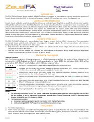
ASA Test System<br />
FA6001<br />
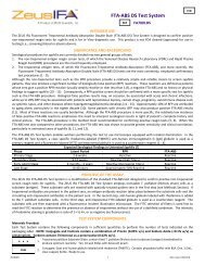
INTENDED USE<br />
The <strong>ZEUS</strong> IFA Anti-Skin Antibody (ASA) Test System is designed for the qualitative and semi-quantitative detection of antibodies<br />
associated with Pemphigus and Bullous Pemphigoid by the indirect fluorescent antibody (IFA) technique, and is for In Vitro<br />
diagnostic use.<br />
SIGNIFICANCE AND BACKGROUND<br />
Most authorities agree that serum antibody studies of all cases of suspected Pemphigus Vulgaris (PV) and Bullous Pemphigoid (BP)<br />
are advisable (1 - 4). Specific antibodies are demonstrable in virtually all active cases, particularly if repeated tests are performed<br />
during different stages of the disease (1). The most frequently recommended substrate for these serum antibody studies is monkey<br />
esophagus (6). Both PV and BP specific antibodies can be detected in a single assay using this substrate. PV antibodies react with<br />
the intercellular substances between the squamous epithelium of monkey esophagus, and BP antibodies react with the basement<br />
membrane of the squamous epithelial cell layer (1, 4, and 6). Antinuclear antibody staining of the squamous epithelial cell nuclei<br />
may occur if serum is obtained from patients with systemic lupus or other connective tissue disorders (7). Distinct intercellular<br />
staining may be detected in cases of pemphigus vegetans, pemphigus foliaceus (including the Brazilian form), and pemphigus<br />
erythematosus (1). Although a single serological test is not sufficient for determining the dosage of drug therapy in PV, a two-fold<br />
drop in titer is an indication of effective control of the disease process. Although some correlation in titer and disease activity is<br />
found in BP, the prognostic value is less than in PV due to fluctuations in titer. Serum studies may be diagnostic of PV or BP;<br />
however, it is frequently advisable to perform direct immunohistochemical studies on biopsies of skin lesions (1, 2). Direct<br />
immunofluorescent studies of skin biopsies may be facilitated with <strong>ZEUS</strong> Tissue Fixative and Wash Solution, prepared according to<br />
Michel (5). This new fixative eliminates the necessity of immediate freezing of the biopsy specimen and allows transport of fresh<br />
tissue biopsies of skin and other tissues up to 5 days at ambient temperatures.<br />
PRINCIPLE OF THE ASSAY<br />
The <strong>ZEUS</strong> IFA ASA Test System is designed to detect the presence of circulating pemphigus and bullous pemphigoid antibodies in<br />
human sera. The assay employs monkey esophagus tissue substrate and goat anti-human immunoglobulin adjusted for optimum<br />
use dilution and free of nonspecific background staining. The reaction occurs in two steps:<br />
1. Step one is the interaction of anti-skin antibodies in patient’s sera with the monkey esophagus.<br />
2. Step two is the interaction of FITC-labeled anti-human immunoglobulin with antibodies attached to the intercellular cement of<br />
the epithelial cells in pemphigus, or the basement membrane zone in bullous pemphigoid in a positive assay (see Assay<br />
Procedure section for details).<br />
The <strong>ZEUS</strong> IFA ASA Test System will detect both PV and BP antibodies. The assay is a particularly useful laboratory aid in the diagnosis<br />
of PV and BP since the majority of untreated patients with active disease will contain anti-skin antibodies in their serum.<br />
TEST SYSTEM COMPONENTS<br />
Materials Provided:<br />
Each Test System contains the following components in sufficient quantities to perform the number of tests indicated on the<br />
packaging label. NOTE: Conjugate and Controls contain a combination of Proclin (0.05% v/v) and Sodium Azide (
NOTES:<br />
1. The following components are not Test System Lot Number dependent and may be used interchangeably with the <strong>ZEUS</strong><br />
IFA Test Systems, as long as the product numbers are identical: SAVe Diluent ® (Product #: FA005CC), Mounting Media<br />
(Product #: FA0009S), and PBS (Product #: 0008S).<br />
2. Test System also contains:<br />
a. Component Label containing lot specific information inside the Test System box.<br />
b. A CD containing all <strong>ZEUS</strong> IFA Product Inserts, providing instructions for use.<br />
PRECAUTIONS<br />
1. For In Vitro diagnostic use.<br />
2. Follow normal precautions exercised in handling laboratory reagents. In case of contact with eyes, rinse immediately with<br />
plenty of water and seek medical advice. Wear suitable protective clothing, gloves, and eye/face protection. Do not breathe<br />
vapor. Dispose of waste observing all local, state, and federal laws.<br />
3. The wells of the Slide do not contain viable organisms. However, consider the Slide potentially bio-hazardous materials and<br />
handle accordingly.<br />
4. The Controls are potentially bio-hazardous materials. Source materials from which these products were derived were found<br />
negative for HIV-1 antigen, HBsAg and for antibodies against HCV and HIV by approved test methods. However, since no test<br />
method can offer complete assurance that infectious agents are absent, these products should be handled at the Bio-safety<br />
Level 2 as recommended for any potentially infectious human serum or blood specimen in the Centers for Disease<br />
Control/National Institutes of Health manual “Biosafety in Microbiological and Biomedical Laboratories”: current edition; and<br />
OSHA’s Standard for Bloodborne Pathogens (20).<br />
5. Adherence to the specified time and temperature of incubations is essential for accurate results. All reagents must be allowed<br />
to reach room temperature (20 - 25°C) before starting the assay. Return unused reagents to their original containers<br />
immediately and follow storage requirements.<br />
6. Improper washing could cause false positive or false negative results. Be sure to minimize the amount of any residual PBS, by<br />
blotting, before adding Conjugate. Do not allow the wells to dry out between incubations.<br />
7. The SAVe Diluent ® , Conjugate, and Controls contain Sodium Azide at a concentration of
5. Staining dish: A large staining dish with a small magnetic mixing set-up provides an ideal mechanism for washing Slides between<br />
incubation steps.<br />
6. Cover slips, 24 x 60mm, thickness No. 1.<br />
7. Distilled or deionized water.<br />
8. Properly equipped fluorescence microscope.<br />
9. 1 Liter Graduated Cylinder.<br />
10. Laboratory timer to monitor incubation steps.<br />
11. Disposal basin and disinfectant (i.e.: 10% household bleach – 0.5% Sodium Hypochlorite).<br />
The following filter systems, or their equivalent, have been found to be satisfactory for routine use with transmitted or incident light<br />
darkfield assemblies:<br />
Transmitted Light<br />
Light Source: Mercury Vapor 200W or 50W<br />
Excitation Filter Barrier Filter Red Suppression Filter<br />
KP490 K510 or K530 BG38<br />
BG12 K510 or K530 BG38<br />
FITC K520 BG38<br />
Light Source: Tungsten – Halogen 100W<br />
KP490 K510 or K530 BG38<br />
Incident Light<br />
Light Source: Mercury Vapor 200, 100, 50 W<br />
Excitation Filter Dichroic Mirror Barrier Filter Red Suppression Filter<br />
KP500 TK510 K510 or K530 BG38<br />
FITC TK510 K530 BG38<br />
Light Source: Tungsten – Halogen 50 and 100 W<br />
KP500 TK510 K510 or K530 BG38<br />
FITC TK510 K530 BG38<br />
SPECIMEN COLLECTION<br />
1. <strong>ZEUS</strong> <strong>Scientific</strong> recommends that the user carry out specimen collection in accordance with CLSI document M29: Protection of<br />
Laboratory Workers from Occupationally Acquired Infectious Diseases. No known test method can offer complete assurance<br />
that human blood samples will not transmit infection. Therefore, all blood derivatives should be considered potentially<br />
infectious.<br />
2. Only freshly drawn and properly refrigerated sera obtained by approved aseptic venipuncture procedures with this assay (8).<br />
No anticoagulants or preservatives should be added. Avoid using hemolyzed, lipemic, or bacterially contaminated sera.<br />
3. Store sample at room temperature for no longer than 8 hours. If testing is not performed within 8 hours, sera may be stored<br />
between 2 - 8°C, for no longer than 48 hours. If delay in testing is anticipated, store test sera at –20°C or lower. Avoid multiple<br />
freeze/thaw cycles which may cause loss of antibody activity and give erroneous results.<br />
STORAGE CONDITIONS<br />
Unopened Test System.<br />
Mounting Media, Conjugate, SAVe Diluent ® , Slides, Positive and Negative Controls.<br />
Rehydrated PBS (Stable for 30 days).<br />
Phosphate-buffered-saline (PBS) Packets.<br />
ASSAY PROCEDURE<br />
1. Remove Slides from refrigerated storage and allow them to warm to room temperature (20 - 25°C). Tear open the protective<br />
envelope and remove Slides. Do not apply pressure to flat sides of protective envelope.<br />
2. Identify each well with the appropriate patient sera and Controls. NOTE: The Controls are intended to be used undiluted.<br />
Prepare a 1:10 dilution (e.g.: 10µL of serum + 90µL of SAVe Diluent ® or PBS) of each patient serum. The SAVe Diluent ® will<br />
undergo a color change confirming that the specimen has been combined with the Diluent.<br />
Dilution Options:<br />
a. Users may titrate the Positive Control to endpoint to serve as a semi-quantitative (1+ Minimally Reactive) Control. In such<br />
cases, the Control should be diluted two-fold in SAVe Diluent ® or PBS. When evaluated by <strong>ZEUS</strong> <strong>Scientific</strong>, an endpoint<br />
dilution is established and printed on the Positive Control vial (± one dilution). It should be noted that due to variations<br />
within the laboratory (equipment, etc.), each laboratory should establish its own expected end-point titer for each lot of<br />
Positive Control.<br />
b. When titrating patient specimens, initial and all subsequent dilutions should be prepared in SAVe Diluent ® or PBS only.<br />
R2030EN 3 (Rev. Date 7/8/2013)
3. Wash Slides for 3 - 5 minutes in PBS.<br />
4. Remove Slides from PBS and blot dry with six-well blotting paper. It is suggested that blotting paper be placed on a flat surface.<br />
Then place substrate Slide in an inverted position over the blotter. Press firmly on back of Slide. Do not allow tissue substrate<br />
to dry throughout the test procedure.<br />
5. With suitable dispenser (listed above), dispense 20µL of each Control and each diluted patient sera in the appropriate wells.<br />
6. Incubate Slides at room temperature (20 - 25°C) for 30 minutes.<br />
7. Gently rinse Slides with PBS. Do not direct a stream of PBS into the test wells.<br />
8. Wash Slides for two, 5 minute intervals, changing PBS between washes.<br />
9. Remove Slides from PBS one at a time. Invert Slide and key wells to holes in blotters provided. Blot Slide by wiping the reverse<br />
side with an absorbent wipe. CAUTION: Position the blotter and Slide on a hard, flat surface. Blotting on paper towels may<br />
destroy the Slide matrix. Do not allow the Slides to dry during the test procedure.<br />
10. Add 20µL of Conjugate to each well.<br />
11. Repeat steps 6 through 9.<br />
12. Apply 3 - 5 drops of Mounting Media to each Slide (between the wells) and coverslip. Examine Slides immediately with an<br />
appropriate fluorescence microscope.<br />
NOTE: If delay in examining Slides is anticipated, seal coverslip with clear nail polish and store in refrigerator. It is recommended<br />
that Slides be examined on the same day as testing.<br />
QUALITY CONTROL<br />
1. Every time the assay is run, the Positive Controls, a Negative Control and a Buffer Control must be included.<br />
2. It is recommended that one read the Positive and Negative Controls before evaluating test results. This will assist in establishing<br />
the references required to interpret the test sample. If Controls do not appear as described, results are invalid.<br />
a. Negative Control - characterized by the absence of fluorescent staining of the squamous epithelial cells, and/or basement<br />
membrane zone.<br />
b. Positive Controls - PV Positive control is characterized by any apple-green fluorescent staining between the squamous<br />
epithelial cells. The BP Positive Control is characterized by specific staining of the basement membrane zone.<br />
3. Additional Controls may be tested according to guidelines or requirements of local, state, and/or federal regulations or<br />
accrediting organizations.<br />
NOTES:<br />
a. The intensity of the observed fluorescence may vary with the microscope and filter system used.<br />
b. Non-specific reagent trapping may exist. It is important to adequately wash slides to eliminate false positive results.<br />
INTERPRETATION OF RESULTS<br />
1. Any apple-green staining of the specific structures noted above on a scale of 1+ to 4+ is considered positive. A 1+ is considered<br />
a weak reaction, and 4+ a strong reaction. All sera positive at a 1:10 dilution should be titered to endpoint dilution. This is<br />
accomplished by making 1:20, 1:40, 1:80, etc., serial dilutions of all positives. The endpoint is the highest dilution that produces<br />
a discernible positive reaction. Specific nuclear staining of the epithelial cell nuclei is considered a positive test for antinuclear<br />
antibodies which may be associated with SLE and other connective tissue diseases.<br />
2. Titers less than 1:10 are considered negative.<br />
3. Positive Test:<br />
a. Specific intercellular staining between the squamous epithelial cells is considered a positive test for pemphigus antibodies.<br />
b. Specific basement membrane zone staining is considered a positive test for bullous pemphigoid.<br />
LIMITATIONS OF THE ASSAY<br />
1. The <strong>ZEUS</strong> IFA ASA Test System is a laboratory aid and by itself is not diagnostic.<br />
2. The results should be interpreted in light of the patient’s clinical condition.<br />
3. No definitive association between the pattern of fluorescence and any specific disease state is intended with this product.<br />
4. No U.S. standard of potency.<br />
REFERENCES<br />
1. Cooperative study. Uses for Immunofluorescence test of skin and sera. Utilization immunofluorescence in the diagnosis of Bullous diseases, Lupus<br />
Erythematosus, and certain other dermatoses. Arch. Dermatol. 111:372-381, 1975.<br />
2. Jablonski S, Chorzelski TP, Beutner EH, et al: Indications for skin and serum immunofluorescent studies in dermatology. In: Beutner EH, Chorzelski TP, Bean S, et<br />
al (Eds): Immunopathology of the Skin. Stroudsburg, PA, Dowden, Hutchinson, and Ross, pp. 1-24, 1973.<br />
3. Chorzelski TP, Jablonski S, Beutner EH: Clinical significance of pemphigus antibodies in: Beutner EH, Chorzelski TP, Bean S, et al (Eds): Immunopathology of the<br />
Skin, Stroudsburg, PA, Dowden, Hutchinson, and Ross. pp. 25-43, 1973.<br />
4. Lever WF: Pemphigus and Pemphigoid. Springfield, IL, Charles C. Thomas, publisher. pp. 3-226, 1965.<br />
5. Michel B, Milner Y, David K: Preservation of Tissue-Fixed immunoglobulins in skin biopsies of patients with lupus erythematosus and bullous diseases:<br />
Preliminary report. J. Invest. Dermatology 59: 449-454, 1973.<br />
6. Anderson P, Hale WL: Immunohistology of human antibodies on monkey esophagus, In: Beutner EH, Chorzelski TP, Bean S, et al (Eds): Immunopathology of the<br />
Skin. Stroudsburg, PA, Dowden, Hutchinson, and Ross: pp. 271-286, 1973.<br />
R2030EN 4 (Rev. Date 7/8/2013)
7. Tan EM, Vaughn JH: antinuclear antibodies: Significance of biochemical specificities, In Beutner EH, Chorzelski TP, Bean S, et al (Eds): Immunopathology of the<br />
Skin. Stroudsburg, PA, Dowden, Hutchinson, and Ross: pp. 367-378, 1973.<br />
8. Procedures for the collection of diagnostic blood specimens by venipuncture - Second Edition: approved Standard (1984). Published by National Committee for<br />
Clinical Laboratory Standards.<br />
9. Lennette DA: Collection and preparation of specimens for virological examination. In: Manual of Clinical Microbiology, 4th ed., EH Lennette, A Balows, WJ<br />
Hausler, and HJ Shadomy (Eds): American Society for Microbiology, Washington, DC. Ch. 61, pp. 687-693, 1985.<br />
10. U.S. Department of Labor, Occupational Safety and Health Administration: Occupational Exposure to Bloodborne Pathogens, Final Rule. Fed. Register 56:64175-<br />
64182, 1991.<br />
<strong>ZEUS</strong> <strong>Scientific</strong>, Inc.<br />
200 Evans Way, Branchburg, New Jersey, 08876, USA<br />
Toll Free (U.S.): 1-800-286-2111<br />
International: +1 908-526-3744<br />
Fax: +1 908-526-2058<br />
<strong>ZEUS</strong> IFA and SAVe Diluent ® are trademarks of <strong>ZEUS</strong> <strong>Scientific</strong>, Inc.<br />
Customer Service E-mail: orders@zeusscientific.com<br />
Technical Support E-mail: support@zeusscientific.com<br />
Website: www.zeusscientific.com<br />
© 2013 <strong>ZEUS</strong> <strong>Scientific</strong>, Inc. All Rights Reserved.<br />
R2030EN 5 (Rev. Date 7/8/2013)