MIC Test Strip MIC Test Strip - Simoco Diagnostics
MIC Test Strip MIC Test Strip - Simoco Diagnostics
MIC Test Strip MIC Test Strip - Simoco Diagnostics
You also want an ePaper? Increase the reach of your titles
YUMPU automatically turns print PDFs into web optimized ePapers that Google loves.
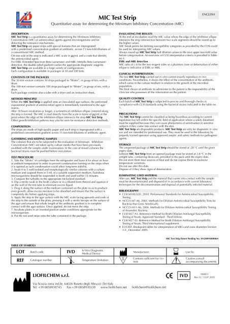
<strong>MIC</strong> <strong>Test</strong> <strong>Strip</strong><br />
Quantitative assay for determining the Minimum Inhibitory Concentration (<strong>MIC</strong>)<br />
ENGLISH<br />
DESCRIPTION<br />
<strong>MIC</strong> <strong>Test</strong> <strong>Strip</strong> is a quantitative assay for determining the Minimum Inhibitory<br />
Concentration (<strong>MIC</strong>) of antimicrobial agents against microorganisms and for<br />
detecting the resistance mechanisms.<br />
<strong>MIC</strong> <strong>Test</strong> <strong>Strip</strong> are paper strips with special features that are impregnated<br />
with a predefined concentration gradient of antibiotic, across 15 two-fold dilutions of<br />
a conventional <strong>MIC</strong> method.<br />
On one side of the strip is indicated a <strong>MIC</strong> scale in μg/mL and a code that identify<br />
the antimicrobial agent.<br />
For ESBL (Extended Spectrum Beta Lactamase) and MBL (Metallo Beta Lactamase)<br />
detection, the double-sided gradient carries the appropriate diagnostic reagents.<br />
<strong>MIC</strong> <strong>Test</strong> <strong>Strip</strong> are available in a large variety of configurations.<br />
Each configuration is available in packages of 30 and 100 tests.<br />
CONTENTS OF THE PACKAGES<br />
The 30-test version contains 30 strips packaged in “blister”, in group of ten, with a<br />
dryer.<br />
The 100-test version contains 100 strips packaged in “blister”, in group of ten, with a<br />
dryer.<br />
Each package contains also a tube with a dryer and an instruction sheet.<br />
METHOD PRINCIPLE<br />
When the <strong>MIC</strong> <strong>Test</strong> <strong>Strip</strong> is applied onto an inoculated agar surface, the preformed<br />
exponential gradient of antimicrobial agent is immediately transferred to the agar<br />
matrix.<br />
After 18 hours incubation or longer, a symmetrical inhibition ellipse centered along<br />
the strip is formed. The <strong>MIC</strong> is read directly from the scale in terms of μg/mL at the<br />
point where the edge of the inhibition ellipse intersects the strip <strong>MIC</strong> <strong>Test</strong> <strong>Strip</strong>.<br />
Other growth/inhibition patterns may also be seen for resistance detection methods.<br />
COMPOSITION<br />
The strips are made of high-quality paper and each strip is impregnated with a<br />
predefined concentration gradient across 15 two-fold dilutions of antibiotic agent.<br />
GATHERING AND KEEPING SAMPLES<br />
The colonies that are to be subjected to the evaluation of Minimum Inhibition<br />
Concentration (<strong>MIC</strong>) are taken up by culture media that have been previously<br />
swabbed with the sample under examination. In the case of mixed colonies the<br />
bacterial strains must be purified before inoculation.<br />
TEST PROCEDURE<br />
1. Take the “blister” of cartridges from the refrigerator and leave it for about an hour<br />
at ambient temperature in order to prevent condensation forming on the strips when<br />
it is opened as such condensation could affect long-term stability.<br />
2. Swab 4 to 5 well isolated and morphologically similar colonies with a culture<br />
medium and suspend them in 5 mL of a suitable suspension medium. Fastidious<br />
microorganisms should be suspended in broth and used within 15 minutes.<br />
3. Compare the turbidity to the appropriate McFarland standard.<br />
4. Dip a sterile swab in the broth culture or in a diluted form thereof and squeeze it<br />
on the wall of the test tube to eliminate excess liquid.<br />
5. Drag it along the surface of the medium contained on the plate so as to produce<br />
even growth; allow excess moisture to be absorbed and ensure that the surface is<br />
completely dry before applying strips.<br />
6. Apply the strip to the agar surface with the <strong>MIC</strong> scale facing upwards and code of<br />
the strip to the outside of the plate, pressing it with a sterile forceps on the surface of<br />
the agar and ensure that whole length of the antibiotic gradient is in complete<br />
contact with the agar surface. Once applied, do not move the strip.<br />
7. Incubate plates in an inverted position under conditions appropriate for the<br />
microorganism.<br />
8. Put the not used strips onto the tube contained in the package.<br />
EVALUATING THE RESULTS<br />
At the end of incubation read the <strong>MIC</strong> value where the edge of the inhibition ellipse<br />
intersects the strip (intersection between two scale segments should be round up to<br />
the higher value).<br />
<strong>MIC</strong> break points for defining susceptibility categories as provided by the CLSI could<br />
be used for interpreting <strong>MIC</strong> values.<br />
Always round up <strong>MIC</strong> <strong>Test</strong> <strong>Strip</strong> half dilution values to the next upper two-fold value<br />
before categorisation. An overview of CLSI interpretative criteria is provided in Table<br />
no.1.<br />
ESBL and MBL detection<br />
<strong>MIC</strong> ratio of ≥ 8 for the two reagent sides or a phantom zone or deformation of either<br />
ellipse is indicative of ESBL or MBL.<br />
CLINICAL INTERPRETATION<br />
The test <strong>MIC</strong> <strong>Test</strong> <strong>Strip</strong> carried out in vitro cannot exactly reproduce in vivo<br />
conditions. Nevertheless, it shows the effect of the concentration of the antibiotic,<br />
which varies in the culture medium in relation to the growth of the microbial<br />
population.<br />
The final choice of antibiotic to administer to the patient is the responsibility of the<br />
clinician who possesses all the information on the patient.<br />
QUALITY CONTROL<br />
Each batch of <strong>MIC</strong> <strong>Test</strong> <strong>Strip</strong> is subjected to precise and thorough checks in<br />
compliance with CLSI standards using the bacterial strains indicated in the table no.<br />
1.<br />
PRECAUTIONS<br />
The <strong>MIC</strong> <strong>Test</strong> <strong>Strip</strong> cannot be classified as being hazardous according to current<br />
legislation but fall within the specific field of application where a safety datasheet<br />
must be supplied because they can cause phenomena of sensitisation in sensitive<br />
subjects if they come into contact with the skin.<br />
<strong>MIC</strong> <strong>Test</strong> <strong>Strip</strong> are disposable products. <strong>MIC</strong> <strong>Test</strong> <strong>Strip</strong> are only for diagnostic in vitro<br />
use and are intended for professional use. They must be used in the laboratory by<br />
properly trained operators using approved aseptic and safety methods for pathogenic<br />
agents.<br />
STORAGE<br />
The unopened package of <strong>MIC</strong> <strong>Test</strong> <strong>Strip</strong> should be stored at -20 °C until the given<br />
expiry date.<br />
Leftover <strong>MIC</strong> <strong>Test</strong> <strong>Strip</strong> from an opened package must be stored at 2-8 °C in the<br />
airtight tube, containing desiccant, provided in the pack until the expiry date.<br />
Do not store them near sources of heat and do not expose them to excessive<br />
temperature variations.<br />
Do not use after this date.<br />
Dispose of if they show signs of deterioration.<br />
ELIMINATING USED MATERIAL<br />
After use, <strong>MIC</strong> <strong>Test</strong> <strong>Strip</strong> and the material that comes into contact with the sample<br />
must be decontaminated and disposed of in accordance with current laboratory<br />
techniques for the decontamination and disposal of potentially infected material.<br />
BIBLIOGRAPHY<br />
• CLSI M100-S20 , 2010. Performance Standards for Antimicrobial Susceptibility<br />
<strong>Test</strong>ing.<br />
• NCCLS M7-A6, 2003. Methods for Dilution Antimicrobial Susceptibility <strong>Test</strong>s for<br />
Bacteria that Grow Aerobically.<br />
• NCCLS M11-A6, 2004. Methods for Dilution Antimicrobial Susceptibility <strong>Test</strong>ing<br />
of Anaerobic Bacteria.<br />
• CLSI M27-A3. Reference Method for Broth Dilution Antifungal Susceptibility<br />
<strong>Test</strong>ing of Yeasts; Approved Standard - Third Edition.<br />
• CLSI M27-S3. Reference Method for Broth Dilution Antifungal Susceptibility<br />
<strong>Test</strong>ing of Yeasts; Third Informational supplement.<br />
• EUCAST. Breakpoint tables for interpretation of <strong>MIC</strong>s and zone diameters Version<br />
1.0 , December 2009.<br />
<strong>MIC</strong> <strong>Test</strong> <strong>Strip</strong> Patent Pending No. AN2009A000064<br />
TABLE OF SYMBOLS<br />
LOT Batch code IVD<br />
In Vitro Diagnostic<br />
Medical Device<br />
REF Catalogue number Temperature limitation<br />
Manufacturer<br />
Contains sufficient for <br />
tests<br />
Use by<br />
Caution,consult<br />
accompanying documents<br />
LIOFILCHEM s.r.l.<br />
Via Scozia zona ind.le, 64026 Roseto degli Abruzzi (Te) Italy<br />
Tel. +39 0858930745 Fax +39 0858930330 www.liofilchem.net liofilchem@liofilchem.net<br />
IVD<br />
F00023<br />
Rev.13 / 13.07.2010


