Soft tissue expansion with self-filling osmotic tissue expanders ...
Soft tissue expansion with self-filling osmotic tissue expanders ...
Soft tissue expansion with self-filling osmotic tissue expanders ...

You also want an ePaper? Increase the reach of your titles
YUMPU automatically turns print PDFs into web optimized ePapers that Google loves.
J Clin Periodontol 2010 doi: 10.1111/j.1600-051X.2010.01630.x<br />
<strong>Soft</strong> <strong>tissue</strong> <strong>expansion</strong> <strong>with</strong><br />
<strong>self</strong>-<strong>filling</strong> <strong>osmotic</strong> <strong>tissue</strong><br />
<strong>expanders</strong> before vertical ridge<br />
augmentation: a proof of<br />
principle study<br />
Doğan Kaner 1 and Anton Friedmann 2<br />
1 Department of Operative Dentistry and<br />
Periodontology, CharitéCentrum 3, Charité –<br />
Universitätsmedizin Berlin, Berlin, Germany;<br />
2 Department of Periodontology, University of<br />
Witten/Herdecke, Witten, Germany.<br />
Kaner D, Friedmann A. <strong>Soft</strong> <strong>tissue</strong> <strong>expansion</strong> <strong>with</strong> <strong>self</strong>-<strong>filling</strong> <strong>osmotic</strong> <strong>tissue</strong> <strong>expanders</strong><br />
before vertical ridge augmentation: a proof of principle study. J Clin Periodontol<br />
2010; doi: 10.1111/j.1600-051X.2010.01624.x.<br />
Abstract<br />
Introduction: Post-surgical graft exposition and loss of grafted bone are a common<br />
complication of vertical bone augmentation. <strong>Soft</strong> <strong>tissue</strong> <strong>expansion</strong> (STE) by<br />
implantation of <strong>osmotic</strong> <strong>self</strong>-<strong>filling</strong> <strong>tissue</strong> <strong>expanders</strong> before reconstructive surgery is an<br />
effective method for generation of soft <strong>tissue</strong>. The aim of this study was to investigate<br />
the feasibility of STE before bone augmentation <strong>with</strong> regard to clinical and histological<br />
outcomes and complications.<br />
Methods: Tissue <strong>expanders</strong> were implanted in patients requiring vertical bone<br />
augmentation. Onlay grafting was carried out after 2 months of STE. Implants were<br />
placed 4–6 months after augmentation. Vertical bone gain was analysed <strong>with</strong> conebeam<br />
computed tomography (CBCT). Bone biopsies were investigated <strong>with</strong> microcomputed<br />
tomography (micro-CT).<br />
Results: Twenty-four sites in 12 patients were treated <strong>with</strong> STE. Complications of<br />
STE were perforation (two sites) and infection (two sites). At augmentation after STE,<br />
primary wound closure was easily achieved and the incidence of graft expositions was<br />
low (4%). At implant placement, high vertical bone gain of 7.5 2.4 mm was found.<br />
Micro-CTs of bone revealed a good ratio of bone volume/<strong>tissue</strong> volume (mean BV/<br />
TV 5 0.1614 0.0582). All implants were osseointegrated.<br />
Conclusions: The combination of STE and subsequent vertical augmentation<br />
provided high gain of well-structured bone for further successful implant therapy and<br />
was accompanied by minimal complications.<br />
Key words: bone graft; dental implants; <strong>tissue</strong><br />
<strong>expansion</strong>; vertical bone augmentation<br />
Accepted for publication 9 September 2010<br />
Placement of dental implants often<br />
requires reconstruction of resorbed<br />
alveolar ridges and a variety of bone<br />
Conflict of interest and source of<br />
funding statement<br />
D. K. lectures for Osmed and A. F.<br />
lectures for Institut Straumann AG.<br />
This study was supported by unrestricted<br />
research grants and donation of clinical materials<br />
by Osmed (Ilmenau, Germany) and<br />
Institut Straumann AG (Basel, Switzerland).<br />
r 2010 John Wiley & Sons A/S<br />
augmentation techniques is used to<br />
improve the horizontal and vertical<br />
dimensions of the implant site. When<br />
applied for lateral ridge augmentation,<br />
autogenous bone block grafts or guided<br />
bone regeneration (GBR) techniques<br />
provide a predictable volume of generated<br />
bone after healing (McAllister &<br />
Haghighat 2007). On the other hand, the<br />
outcome of vertical ridge augmentation<br />
appears less clear. Clinical and histological<br />
data support the feasibility of<br />
vertical augmentation procedures such<br />
as onlay grafting, inlay grafting, distraction<br />
osteogenesis or GBR (Rocchietta<br />
et al. 2008, Esposito et al. 2009). However,<br />
these surgical procedures are considered<br />
highly technique sensitive and<br />
the findings are difficult to extrapolate<br />
(Rocchietta et al. 2008, Esposito et al.<br />
2009). Although the reported incidence<br />
of post-operative complications varies<br />
highly among studies, it is apparent that<br />
soft <strong>tissue</strong> dehiscence and exposure of<br />
bone grafts to the oral cavity are common<br />
complications of vertical ridge<br />
1
2 Kaner & Friedmann<br />
augmentation, compromising the outcome<br />
and leading to partial or complete<br />
loss of the graft in up to 40% of the<br />
cases (Verhoeven et al. 1997, Bahat &<br />
Fontanesi 2001, Chiapasco et al. 2004,<br />
2007, Roccuzzo et al. 2004, 2007,<br />
Proussaefs & Lozada 2005, Barone &<br />
Covani 2007, Merli et al. 2007, Canullo<br />
& Malagnino 2008, Felice et al. 2009,<br />
Urban et al. 2009).<br />
Exposure of grafts is mainly attributed<br />
to difficulties in achieving tension-free<br />
closure of the flap (Lundgren et al.<br />
2008). Generally, the elevation of a flap<br />
disturbs perfusion and causes ischaemia<br />
(McLean et al. 1995). Preservation of<br />
sufficient blood flow is important for<br />
<strong>tissue</strong> survival (Nakayama et al. 1982).<br />
Conversely, reduction of blood supply<br />
and ischaemia-reperfusion injury may<br />
affect the operated <strong>tissue</strong> and may cause<br />
complications such as necrosis of the flap.<br />
The severity of <strong>tissue</strong> damage relates to<br />
the duration and intensity of ischaemia<br />
(Morris et al. 1993, Carroll & Esclamado<br />
2000). Accordingly, a direct relation<br />
between the extent of surgical trauma<br />
and concomitant disturbance of perfusion<br />
has been shown for periodontal surgical<br />
procedures <strong>with</strong> different degree of <strong>tissue</strong><br />
traumatization (Retzepi et al. 2007).<br />
Given that <strong>tissue</strong> mobilization for achieving<br />
tension-free primary wound closure<br />
for vertical augmentation is considerably<br />
more traumatic compared <strong>with</strong> a straightforward<br />
lateral augmentation procedure,<br />
soft <strong>tissue</strong> quality and quantity appear as<br />
key factors for predictable success.<br />
Not<strong>with</strong>standing complications, volume<br />
maintenance during healing is another<br />
major concern, as up to 60% of graft<br />
volume may be resorbed during healing<br />
(McAllister & Haghighat 2007). Again,<br />
compromised vascularization and tension<br />
of the flap caused by soft <strong>tissue</strong> movement<br />
and subsequent limitation of regenerative<br />
space have been considered as causes for<br />
limited outcomes in vertical augmentation<br />
in animals (Rothamel et al. 2009) and in<br />
humans (Lundgren et al. 2008). Thus, it<br />
may be concluded that an antecedent<br />
improvement of soft <strong>tissue</strong> quality and<br />
quantity could enhance the outcome of<br />
vertical bone regeneration.<br />
Generation of soft <strong>tissue</strong> by using<br />
subcutaneous <strong>tissue</strong> <strong>expanders</strong> before<br />
reconstructive procedures is an established<br />
method in plastic surgery. After implantation,<br />
the increase of expander volume<br />
over time causes tension on the surrounding<br />
<strong>tissue</strong>s and results finally in <strong>tissue</strong> gain<br />
(Bennett & Hirt 1993, Bascom & Wax<br />
2002). Osmotic <strong>self</strong>-<strong>filling</strong> <strong>tissue</strong> <strong>expanders</strong><br />
consist of a polymer of methylmethacrylate/vinylpyrrolidone<br />
and expand due<br />
to absorption of body fluids. Presently,<br />
these <strong>expanders</strong> are used for a variety of<br />
indications, such as breast reconstruction,<br />
defect coverage after excisions and preparation<br />
of <strong>tissue</strong> donor sites (Berge et al.<br />
2001, Ronert et al. 2004). Here, we report<br />
for the first time on the application<br />
of <strong>osmotic</strong> <strong>tissue</strong> <strong>expanders</strong> to improve<br />
soft <strong>tissue</strong> before vertical augmentation of<br />
severely resorbed ridges. The aim<br />
of this study was to evaluate the feasibility<br />
of a combined soft <strong>tissue</strong> <strong>expansion</strong><br />
(STE) and vertical augmentation procedure<br />
<strong>with</strong> regard to gain of bone and<br />
complications.<br />
Material and Methods<br />
Patients<br />
Patients were recruited from patients<br />
seeking implant treatment at the Department<br />
of Periodontology, Charité – Universitätsmedizin<br />
Berlin. Resorbed<br />
edentulous or partially edentulous ridges<br />
class C or D (Misch & Judy 1987) and<br />
the need of vertical bone augmentation<br />
of 43 mm before placement of dental<br />
implants were criteria for inclusion.<br />
Exclusion criteria were untreated<br />
periodontal disease; caries; insufficient<br />
oral hygiene; previous radiation therapy;<br />
smoking; systemic disorders potentially<br />
affecting the outcome in implant therapy<br />
(e.g. uncontrolled diabetes mellitus,<br />
haemorrhagic disorders) and medications<br />
putatively affecting implant therapy<br />
(e.g. bisphosphonates). Written<br />
informed consent has been obtained<br />
from each patient. The study protocol<br />
has been approved by the institutional<br />
Ethics Committee of the Charité – Universitätsmedizin<br />
Berlin (EA2/117/07).<br />
Implantation of <strong>tissue</strong> <strong>expanders</strong><br />
Expander type and size (hemisphere<br />
<strong>with</strong> 0.35 ml final volume; round-ended<br />
cylinders <strong>with</strong> 0.24, 0.7, 1.3 or 2.1 ml<br />
final volume; ‘‘Dental Cupola slow’’<br />
and ‘‘Dental cylinder slow’’, Osmed,<br />
Ilmenau, Germany) appropriate for the<br />
edentulous site and <strong>with</strong> a swelling time<br />
of 60 days (Fig. 1a and b) were selected<br />
<strong>with</strong> surgical templates corresponding to<br />
the final expander volume (Fig. 2a and<br />
b). A submucosal pouch was prepared<br />
<strong>with</strong> scalpel and scissors <strong>with</strong>out elevation<br />
of the periosteum (Fig. 2c). The size<br />
of the pouch was controlled <strong>with</strong> the<br />
surgical template (corresponding to<br />
the initial expander volume) that should<br />
easily fit into the pouch (Fig. 2d).<br />
The expander was placed into the pouch<br />
and fixed <strong>with</strong> a bone fixation screw<br />
(Fig. 2e). A meticulous two-layer wound<br />
closure was performed using fine monofilament<br />
sutures. Administration of<br />
antibiotics (amoxicillin 750 mg or clindamycin<br />
600 mg) was started 1 h before<br />
surgery and continued for 7 days. Ibuprofen<br />
(400 mg) was prescribed as<br />
analgesic. Patients were followed up<br />
weekly and were advised to rinse <strong>with</strong><br />
0.2% chlorhexidine for 2 weeks until<br />
suture removal. Abstention from removable<br />
prostheses was required. Fixed<br />
provisional prostheses were adjusted<br />
regularly according to the increasing<br />
soft <strong>tissue</strong> volume. Bone augmentation<br />
was carried out after 6–8 weeks of<br />
<strong>expansion</strong>, when the expander had<br />
reached its final volume (Figs 1a and 2f).<br />
Expander removal and bone<br />
augmentation<br />
Depending on the needed graft volume<br />
and the availability of intra-oral donor<br />
bone, bone grafts were harvested either<br />
Fig. 1. (a) Cylindrical <strong>tissue</strong> expander before and after swelling. (b) Volume increase over<br />
time of a <strong>tissue</strong> expander in vitro (0.9% saline). The final volume (here: 0.7 ml) is reached<br />
after approx. 60 days.<br />
r 2010 John Wiley & Sons A/S
Tissue <strong>expansion</strong> before bone augmentation 3<br />
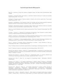
Fig. 2. (a) Resorbed edentulous ridge (class C) requiring vertical bone augmentation of approx. 5 mm. (b) The appropriate expander size is<br />
selected using the surgical template (final expander volume). (c) A supraperiosteal mucosal pouch is prepared using scalpel and scissors. (d)<br />
The preparation is controlled <strong>with</strong> the surgical template (initial expander volume). (e). The <strong>tissue</strong> expander is inserted into the pouch and fixed<br />
<strong>with</strong> a bone fixation screw. (f) After 8 weeks of <strong>tissue</strong> <strong>expansion</strong> (1.3 ml expander), a considerable gain of soft <strong>tissue</strong> can be observed.<br />
r 2010 John Wiley & Sons A/S<br />
from the mandibular ramus or the posterior<br />
ilium.<br />
Bone augmentation <strong>with</strong> ramus grafts<br />
was carried out under local anaesthesia,<br />
under sedation, anti-phlogistic medication<br />
<strong>with</strong> prednisolone and antibiotic<br />
coverage. Ibuprofen 600 mg was prescribed<br />
as analgesic and patients rinsed<br />
<strong>with</strong> 0.2% chlorhexidine for 2 weeks.<br />
At the donor site, a mucoperiostal flap<br />
was reflected distally of the second<br />
molar, exposing the lateral aspect of<br />
the ramus along the external oblique<br />
ridge. Block grafts were prepared <strong>with</strong><br />
a piezoelectric device (Piezosurgery II,<br />
Mectron, Köln, Germany).<br />
After midcrestal incision at the recipient<br />
site, a mucoperiostal flap was<br />
reflected. The expander was removed<br />
and the bone was exposed. Generally,<br />
vertical releasing incisions were<br />
avoided; in case of adjacent teeth present,<br />
the incision was extended into the<br />
gingival sulcus for ease of reflection.<br />
The local bone was perforated and the<br />
block graft was secured <strong>with</strong> screws<br />
(Institut Straumann AG, Basel, Switzerland).<br />
The graft was covered <strong>with</strong> a<br />
granular bone substitute (BioOss, Geistlich,<br />
Wolhusen, Switzerland) and a<br />
collagen membrane (Ossix plus, Colbar,<br />
Hertzelia, Israel). The incision was<br />
closed <strong>with</strong> modified vertical mattress<br />
sutures and single interrupted sutures,<br />
using fine monofilament sutures. Sutures<br />
were removed after 2 weeks.<br />
Bone augmentation <strong>with</strong> grafts from<br />
the posterior ilium was performed under<br />
general anaesthesia and under antibiotic<br />
coverage. The patient was placed in a<br />
prone position and an incision was<br />
placed extending cranially from the<br />
posterior iliac spine. Grafts were harvested<br />
from the external wall of the<br />
posterior iliac crest <strong>with</strong> an oscillating<br />
saw and a chisel.<br />
At the recipient sites, mucoperiostal<br />
flaps were elevated (Fig. 3a). Again,<br />
releasing incisions were avoided. After<br />
removal of the <strong>expanders</strong>, the local<br />
bone was exposed and perforated,<br />
and onlay grafts were fixed <strong>with</strong> screws<br />
(Fig. 3b). Patients were mobilized after<br />
24 h and sutures were removed after 1<br />
week.<br />
The <strong>expanders</strong> were weighed after<br />
removal and a biopsy was taken from<br />
the expanded soft <strong>tissue</strong>.<br />
Implant placement<br />
In patients treated <strong>with</strong> ramus grafts and<br />
GBR, implants (Standard Plus, SLActive<br />
surface, Institut Straumann AG)<br />
were placed 6 months after bone augmentation,<br />
following the standard protocol<br />
for non-submerged healing. In<br />
patients treated <strong>with</strong> iliac grafts,<br />
implants of the same type were placed<br />
4 months after augmentation. Sutures<br />
were removed after 1–2 weeks.<br />
Radiographs<br />
Cone-beam computed tomographies<br />
(CBCT, Galileos, Sirona, Bensheim,<br />
Germany) were taken before implantation<br />
of <strong>tissue</strong> <strong>expanders</strong>, and 4–6 months<br />
after bone grafting, before placement of<br />
dental implants. Digital panoramic<br />
radiographs (Sirona) were made after<br />
bone augmentation and after implant<br />
surgery. The mean vertical bone gain/<br />
surgical site was calculated to the nearest<br />
millimetre by subtraction of bone height<br />
before grafting from bone height before<br />
implantation, using the CBCT software<br />
measurement tool after aligning the
4 Kaner & Friedmann<br />
Fig. 3. (a) The <strong>tissue</strong> expander is explanted in the course of bone augmentation surgery. (b)<br />
After fixation of the bone graft, primary closure of the flap is easily achieved <strong>with</strong>out further<br />
mobilization.<br />
Fig. 4. (a) Cone-beam computed tomographic cross section of a resorbed mandible before<br />
augmentation. Mandibular height: 15.7 mm. (b) Same section of the same patient, 6 months<br />
after augmentation. Mandibular height: 22.6 mm, radiographic bone gain approx. 6.9 mm.<br />
investigation window at reproducible<br />
anatomical landmarks (Fig. 4a and b).<br />
Biopsies<br />
At the time of bone augmentation, biopsies<br />
were taken from the expanded soft<br />
<strong>tissue</strong> surrounding the expander, fixed in<br />
4% formalin, embedded in paraffin,<br />
stained <strong>with</strong> haematoxylin/eosin and<br />
investigated <strong>with</strong> a light microscope.<br />
Bone core biopsies were harvested at<br />
the time of implant placement using a<br />
trephine drill (inner diameter 2.2 mm)<br />
for preparation of the implant site, and<br />
fixed in formalin. The bone biopsies<br />
were investigated <strong>with</strong> micro-computed<br />
tomography (micro-CT), using an<br />
experimental cone-beam micro-CT<br />
scanner <strong>with</strong> a micro-focus tube (voxel<br />
size 20 mm).<br />
Results<br />
Twelve patients (three men, nine<br />
women, mean age 45 years, range 21–<br />
73 years) were included in the study<br />
since November 2007. Treatment was<br />
concluded in October 2009.<br />
Expanders were placed in 24 surgical<br />
sites. Post-operative sequelae were minor<br />
edemata and slight pain; generally,<br />
the treatment was well tolerated.<br />
Healing and <strong>expansion</strong> period were<br />
uneventful in 10 patients. In two<br />
patients, <strong>expanders</strong> perforated through<br />
the mucosa and were removed. Reasons<br />
for perforation were seroma formation<br />
and infection after 4 weeks (one site),<br />
and probably, the use of an oversized<br />
expander type (one site). These sites<br />
were allowed to heal for 6 weeks and<br />
were successfully retreated <strong>with</strong> smaller<br />
<strong>expanders</strong>. In two sites, fistulae developed<br />
after seroma formation shortly<br />
before bone augmentation. These sites<br />
were treated <strong>with</strong> instillation of a tetracycline/cortisol<br />
paste until augmentation<br />
surgery.<br />
Augmentation <strong>with</strong> ramus grafts and<br />
GBR was carried out in 12 sites in nine<br />
patients. In three patients (12 recipient<br />
sites), corticocancellous bone grafts<br />
from the posterior ilium were placed as<br />
onlay grafts onto the resorbed mandible,<br />
or in the maxilla, onlay grafting was<br />
combined <strong>with</strong> a sinus lift procedure<br />
<strong>with</strong> granular bone substitute. In all<br />
cases, wound closure at the recipient<br />
sites was easily achieved <strong>with</strong>out further<br />
mobilization of <strong>tissue</strong> beyond the incision<br />
for the removal of the <strong>tissue</strong> <strong>expanders</strong><br />
(Fig. 3b).<br />
Paraesthesia of the mental region<br />
occurred in one patient after ramus<br />
grafting, but resolved spontaneously<br />
after 4 months. One minor exposition<br />
occurred after vertical augmentation in<br />
the posterior maxilla, but healed spontaneously<br />
after debridement and repeated<br />
application of chlorhexidine gel.<br />
In all other cases and augmented<br />
sites, wound healing was uneventful.<br />
Weighing of the <strong>expanders</strong> after explantation<br />
showed that all <strong>expanders</strong> had<br />
reached their expected final volume<br />
(data not shown). Histological analysis<br />
of the soft <strong>tissue</strong> capsule surrounding<br />
the expander showed dense connective<br />
<strong>tissue</strong> and absence of infiltration (Fig.<br />
6a).<br />
After the designated healing time of 4<br />
and 6 months, respectively, analysis<br />
<strong>with</strong> the CBCT measurement tool<br />
revealed a mean vertical bone gain of<br />
7.5 2.4 mm (range 3–12 mm) before<br />
implant surgery. In all patients, the<br />
desired height of augmentation was<br />
reached and 53 implants (1–19 implants<br />
per patient, length 8–12 mm) could be<br />
placed as intended (Fig. 5a and b) <strong>with</strong>out<br />
additional grafting. In 22 of 24 sites,<br />
the width of keratinized gingiva was<br />
increased <strong>with</strong> free gingival grafts (20<br />
sites) or connective <strong>tissue</strong> grafts (two<br />
sites) simultaneously <strong>with</strong> placement of<br />
the implant (six sites) or after healing<br />
(16 sites). All implants were osseointegrated<br />
and were used for fixed partial<br />
dentures, crowns or bar-retained removable<br />
prostheses.<br />
Eleven bone biopsies were analysed<br />
by micro-CT (Fig. 6b and c; supporting<br />
information Video S1) and a mean BV/<br />
TV (bone volume/<strong>tissue</strong> volume) of<br />
0.1614 0.0582 was found.<br />
Discussion<br />
The use of <strong>tissue</strong> <strong>expanders</strong> before intraoral<br />
bone graft surgery has been occasionally<br />
described in case reports (Lew<br />
et al. 1988, Wittkampf 1989, Bahat &<br />
Handelsman 1991). In these studies,<br />
‘‘classical’’ types of <strong>tissue</strong> <strong>expanders</strong><br />
r 2010 John Wiley & Sons A/S
Tissue <strong>expansion</strong> before bone augmentation 5<br />
Fig. 5. (a) Panoramic radiograph before bone augmentation. Minimal bone height over the<br />
mandibular canal. (b) Panoramic radiograph after implant surgery. Vertical bone gain of<br />
approx. 8 mm.<br />
Fig. 6. (a) Biopsy of the <strong>tissue</strong> capsule surrounding the expander. After 8 weeks of <strong>tissue</strong><br />
<strong>expansion</strong>, a fibre-rich dense connective <strong>tissue</strong> <strong>with</strong>out the presence of inflammatory cells can<br />
be seen. (b and c) Micro-CT of a bone core biopsy taken at implant site preparation 4 months<br />
after grafting from the posterior ilium shows distinct trabecular structures (BV/TV 0.237). A<br />
supplemental movie file shows the full volume of the scan.<br />
were used, i.e. variations of inflatable<br />
silicone balloons. Usually, these <strong>expanders</strong><br />
are filled once a week by injection<br />
of saline into subcutaneous <strong>filling</strong> ports<br />
or percutaneous valve constructions<br />
to the extent until the skin over the<br />
expander appears blanched. However,<br />
decreased <strong>tissue</strong> perfusion and hypoxia<br />
are caused by intra-luminal pressure<br />
spikes that result from the intermittent<br />
<strong>filling</strong> technique (Pietila 1990), and may<br />
lead to <strong>tissue</strong> necrosis and subsequent<br />
perforation of the balloon expander<br />
through skin or mucosa (Wiese 1993).<br />
Generally, percutaneous injections into<br />
subcutaneous ports require local anaesthesia,<br />
while percutaneous valve constructions<br />
that penetrate the skin<br />
increase the risk of infection (Wiese<br />
1993). These disadvantages may<br />
become even more relevant in the oral<br />
environment and may have as yet<br />
impeded the systematic application of<br />
<strong>tissue</strong> <strong>expanders</strong> for ridge augmentation.<br />
r 2010 John Wiley & Sons A/S<br />
In our study, we used for the first time<br />
<strong>osmotic</strong> <strong>tissue</strong> <strong>expanders</strong> before vertical<br />
ridge augmentation. Complications such<br />
as perforation and seroma formation<br />
occurred in four of 24 sites, similar to<br />
the use of <strong>osmotic</strong> <strong>expanders</strong> in other<br />
indications (Ronert et al. 2004). In two<br />
sites, expander implantation was successfully<br />
repeated using a smaller<br />
expander type. Seroma formation in<br />
two sites shortly before bone augmentation<br />
was treated <strong>with</strong> a local antibiotic<br />
and did not interfere <strong>with</strong> bone augmentation<br />
surgery. Osmotic <strong>expanders</strong><br />
increase their size by absorption of<br />
body fluids and the need for external<br />
<strong>filling</strong>s is eliminated, which may explain<br />
the low incidence of infectious complications<br />
during <strong>osmotic</strong> <strong>tissue</strong> <strong>expansion</strong><br />
in our study and in other indications<br />
(Ronert et al. 2004).<br />
Further, this type of expander is<br />
ensheathed <strong>with</strong> a silicone shell; perforations<br />
in the impermeable shell allow<br />
influx of <strong>tissue</strong> fluid, while the rate of<br />
influx over time (and therefore speed of<br />
volume increase) is controlled by the<br />
number of perforations. Unlike balloons,<br />
<strong>osmotic</strong> <strong>expanders</strong> <strong>with</strong> suchlike<br />
silicone shells swell slowly and continuously,<br />
and injection-dependent pressure<br />
peaks are avoided (Anwander et al.<br />
2007). The <strong>expanders</strong> used in our study<br />
reach their final volume after 60 days.<br />
Slow and continuous <strong>expansion</strong> results<br />
in safe and effective generation of soft<br />
<strong>tissue</strong> (Wiese 1993, Wiese et al. 2001),<br />
as experienced during bone augmentation<br />
surgery and confirmed by the<br />
absence of infiltration in soft <strong>tissue</strong><br />
biopsies (Fig. 6a).<br />
The <strong>expanders</strong> were placed in a submucosal<br />
pouch <strong>with</strong>out elevation of the<br />
periosteum. Expansion of the periosteum<br />
is not to be expected, as it is<br />
replaced by fibrous connective <strong>tissue</strong><br />
after subperiosteal implantation of <strong>tissue</strong><br />
<strong>expanders</strong> (Tominaga et al. 1993), while<br />
a new periosteum is formed underneath<br />
the expander. Further, subperiosteal<br />
implantation causes significant resorption<br />
of the underlying bone (Stuehmer et<br />
al. 2009), a finding that was not<br />
observed in our patients. In addition, a<br />
submucosal pouch is easily and quickly<br />
prepared and well tolerated by the<br />
patient; hence, supraperiosteal implantation<br />
appears preferable over subperiosteal<br />
implantation of <strong>osmotic</strong> <strong>tissue</strong><br />
<strong>expanders</strong>.<br />
After <strong>expansion</strong>, major bone augmentation<br />
procedures were carried out. The<br />
quality of expanded <strong>tissue</strong> was excellent<br />
and the space created by <strong>tissue</strong> <strong>expansion</strong><br />
permitted easy primary closure<br />
<strong>with</strong>out the need for additional flap<br />
advancement. Accordingly, the incidence<br />
of post-operative graft expositions<br />
was very low (one in 24 sites,<br />
4%), when compared <strong>with</strong> studies of<br />
vertical bone augmentation <strong>with</strong>out previous<br />
<strong>tissue</strong> <strong>expansion</strong> (mean incidence<br />
of expositions 21.4%, up to 50%,<br />
Table 1) (Verhoeven et al. 1997, Proussaefs<br />
et al. 2002, Roccuzzo et al. 2004,<br />
2007, Chiapasco et al. 2004, 2007,<br />
Proussaefs & Lozada 2005, 2006, Barone<br />
& Covani 2007, Merli et al. 2007,<br />
Canullo & Malagnino 2008, Fontana<br />
et al. 2008, Felice et al. 2009, Urban<br />
et al. 2009).<br />
Before implant surgery, after 4–6<br />
months of healing, standardized CBCT<br />
measurements showed a mean vertical<br />
bone gain of 7.5 2.4 mm. These<br />
results compare favourably <strong>with</strong> the<br />
mean bone gain of 4.13 1.05 mm
6 Kaner & Friedmann<br />
Table 1. Overview of studies reporting on vertical ridge augmentation using variations of guided bone regeneration (GBR) techniques and/or onlay<br />
grafts, illustrating the incidence of post-surgical graft expositions, and radiographic vertical gain of bone 4–6 months after augmentation<br />
References Method of augmentation Vertical bone<br />
gain (mm)<br />
Incidence of<br />
expositions, n (%)<br />
Barone and Covani (2007) Onlay graft Not reported 4/37 (11)<br />
Canullo and Malagnino (2008) GBR 5.3 1.9 1/10 (10)<br />
Chiapasco et al. (2004) GBR 3.87 1.05 3/11 (27.3)<br />
Chiapasco et al. (2007) Onlay graft 5.0 1.07 1/8 (12.5)<br />
Fontana et al. 2008 GBR 4.7 0.48 0/5 (0)<br />
GBR 4.1 0.88 1/5 (20)<br />
Merli et al. (2007) GBR 2.2 1.1 4/11 (25)<br />
GBR 2.5 1.2 5/11 (22)<br />
Proussaefs and Lozada (2005) Onlay graft 4.75 1.29 3/12 (25)<br />
Proussaefs and Lozada (2006) Granular autologous bone and bone substitute, titanium mesh 2.59 0.91 6/18 (33)<br />
Roccuzzo et al. (2004) Granular autologous bone, titanium mesh 4.8 0.9 3/18 (17)<br />
Roccuzzo et al. (2007) Onlay graft 3.4 1.4 6/12 (50)<br />
Onlay graft and titanium mesh 4.8 1.5 4/12 (33.3)<br />
Urban et al. (2009) 4.7 1.67 0/12 (0)<br />
5.1 2.13 1/16 (6.25)<br />
Verhoeven et al. (1997) Onlay graft Not reported 3/13 (23)<br />
Mean 4.13 1.05 45/211 (21.3)<br />
Present investigation Onlay graft/GBR subsequent to soft <strong>tissue</strong> <strong>expansion</strong> 7.5 2.4 1/24 (4)<br />
(range 2.2–5.1 mm) reported in the<br />
aforementioned studies after similar<br />
healing periods (Table 1) and to data<br />
similarly aggregated in a systematic<br />
review (mean vertical bone gain:<br />
4.8 mm, incidence of graft expositions:<br />
18.8%; Jensen & Terheyden 2009).<br />
Bone biopsies were investigated <strong>with</strong><br />
micro-CT (Fig. 6b and c; supporting<br />
information Video S1). Three-dimensional<br />
micro-CT gives a better estimation<br />
of bone regeneration than classical twodimensional<br />
histomorphometry using<br />
histologic sections, because the histologic<br />
processing results in loss of biopsy<br />
material (Muller et al. 1998, Stiller et al.<br />
2009). As microarchitecture reflects bone<br />
quality (Majumdar 2003), an appropriate<br />
ratio of BV/TV and the distinct trabecular<br />
structure found in biopsies of regenerated<br />
bone further illustrate the good<br />
outcome after vertical bone augmentation<br />
subsequent to STE.<br />
In conclusion, our findings demonstrate<br />
the feasibility of <strong>tissue</strong> <strong>expansion</strong><br />
using <strong>osmotic</strong> <strong>expanders</strong> before vertical<br />
bone augmentation. STE was accompanied<br />
by minimal complications and the<br />
incidence of graft expositions after augmentation<br />
surgery was very low. The<br />
combined treatment resulted in comparably<br />
high vertical gain of well-structured<br />
bone and may help to further<br />
improve the outcome and predictability<br />
of implant therapy of patients showing<br />
severe bone resorption.<br />
Acknowledgements<br />
We thank Ms Nannette Richter, DMD,<br />
for dental care of the patients and<br />
Dr Thomas Bauer, MD, DMD (Clinic<br />
for Maxillofacial Surgery, Dessau, Germany)<br />
for his cooperation. Micro-CTs<br />
were kindly provided by Dr. Zully Ritter<br />
and Prof. Dr. Dieter Felsenberg, Center<br />
for Bone and Muscle Research, Charité<br />
– Universitätsmedizin Berlin.<br />
References<br />
Anwander, T., Schneider, M., Gloger, W., Reich, R.<br />
H., Appel, T., Martini, M., Wenghoefer, M., Merkx,<br />
M. & Berge, S. (2007) Investigation of the <strong>expansion</strong><br />
properties of <strong>osmotic</strong> <strong>expanders</strong> <strong>with</strong> and<br />
<strong>with</strong>out silicone shell in animals. Plastic and<br />
Reconstructive Surgery 120, 590–595.<br />
Bahat, O. & Fontanesi, F. V. (2001) Complications of<br />
grafting in the atrophic edentulous or partially<br />
edentulous jaw. The International Journal of Periodontics<br />
& Restorative Dentistry 21, 487–495.<br />
Bahat, O. & Handelsman, M. (1991) Controlled <strong>tissue</strong><br />
<strong>expansion</strong> in reconstructive periodontal surgery.<br />
The International Journal of Periodontics &<br />
Restorative Dentistry 11, 32–47.<br />
Barone, A. & Covani, U. (2007) Maxillary alveolar<br />
ridge reconstruction <strong>with</strong> nonvascularized autogenous<br />
block bone: clinical results. Journal of Oral<br />
and Maxillofacial Surgery 65, 2039–2046.<br />
Bascom, D. A. & Wax, K. A. (2002) Tissue <strong>expansion</strong><br />
in the head and neck: current state of the art.<br />
Current Opinion in Otolaryngology & Head and<br />
Neck Surgery 10, 273–277.<br />
Bennett, R. G. & Hirt, M. (1993) A history of <strong>tissue</strong><br />
<strong>expansion</strong>. Concepts, controversies, and complications.<br />
The Journal of Dermatologic Surgery and<br />
Oncology 19, 1066–1073.<br />
Berge, S. J., Wiese, K. G., von Lindern, J. J., Niederhagen,<br />
B., Appel, T. & Reich, R. H. (2001) Tissue<br />
<strong>expansion</strong> using <strong>osmotic</strong>ally active hydrogel systems<br />
for direct closure of the donor defect of the<br />
radial forearm flap. Plastic and Reconstructive<br />
Surgery 108, 1–5, discussion 6–7.<br />
Canullo, L. & Malagnino, V. A. (2008) Vertical ridge<br />
augmentation around implants by e-PTFE titaniumreinforced<br />
membrane and bovine bone matrix: a 24-<br />
to 54-month study of 10 consecutive cases. The<br />
International Journal of Oral & Maxillofacial<br />
Implants 23, 858–866.<br />
Carroll, W. R. & Esclamado, R. M. (2000) Ischemia/<br />
reperfusion injury in microvascular surgery. Head<br />
& Neck 22, 700–713.<br />
Chiapasco, M., Romeo, E., Casentini, P. & Rimondini,<br />
L. (2004) Alveolar distraction osteogenesis vs.<br />
vertical guided bone regeneration for the correction<br />
of vertically deficient edentulous ridges: a 1-3-year<br />
prospective study on humans. Clinical Oral<br />
Implants Research 15, 82–95.<br />
Chiapasco, M., Zaniboni, M. & Rimondini, L. (2007)<br />
Autogenous onlay bone grafts vs. alveolar distraction<br />
osteogenesis for the correction of vertically<br />
deficient edentulous ridges: a 2-4-year prospective<br />
study on humans. Clinical Oral Implants Research<br />
18, 432–440.<br />
Esposito, M., Grusovin, M. G., Felice, P., Karatzopoulos,<br />
G., Worthington, H. V. & Coulthard, P.<br />
(2009) Interventions for replacing missing teeth:<br />
horizontal and vertical bone augmentation techniques<br />
for dental implant treatment. Cochrane Database<br />
of Systematic Review 7, CD003607.<br />
Felice, P., Marchetti, C., Iezzi, G., Piattelli, A.,<br />
Worthington, H., Pellegrino, G. & Esposito, M.<br />
(2009) Vertical ridge augmentation of the atrophic<br />
posterior mandible <strong>with</strong> interpositional bloc grafts:<br />
bone from the iliac crest vs. bovine anorganic bone.<br />
Clinical and histological results up to one year after<br />
loading from a randomized-controlled clinical trial.<br />
Clinical Oral Implants Research 20, 1386–1393.<br />
Fontana, F., Santoro, F., Maiorana, C., Iezzi, G.,<br />
Piattelli, A. & Simion, M. (2008) Clinical and<br />
histologic evaluation of allogeneic bone matrix<br />
versus autogenous bone chips associated <strong>with</strong> titanium-reinforced<br />
e-PTFE membrane for vertical<br />
ridge augmentation: a prospective pilot study. The<br />
International Journal of Oral & Maxillofacial<br />
Implants 23, 1003–1012.<br />
Jensen, S. S. & Terheyden, H. (2009) Bone augmentation<br />
procedures in localized defects in the alveolar<br />
ridge: clinical results <strong>with</strong> different bone grafts and<br />
bone-substitute materials. The International Journal<br />
of Oral & Maxillofacial Implants 24 (Suppl.),<br />
218–236.<br />
Lew, D., Amos, E. L. & Shroyer, J. V. III (1988) The<br />
use of a subperiosteal <strong>tissue</strong> expander in rib reconstruction<br />
of an atrophic mandible. Journal of Oral<br />
& Maxillofacial Surgery 46, 229–232.<br />
r 2010 John Wiley & Sons A/S
Tissue <strong>expansion</strong> before bone augmentation 7<br />
Lundgren, S., Sjostrom, M., Nystrom, E. & Sennerby,<br />
L. (2008) Strategies in reconstruction of the<br />
atrophic maxilla <strong>with</strong> autogenous bone grafts and<br />
endosseous implants. Periodontology 2000 47,<br />
143–161.<br />
Majumdar, S. (2003) Advances in imaging: impact on<br />
studying craniofacial bone structure. Orthodontics<br />
& Craniofacial Research 6 (Suppl. 1), 48–51.<br />
McAllister, B. S. & Haghighat, K. (2007) Bone<br />
augmentation techniques. Journal of Periodontology<br />
78, 377–396.<br />
McLean, T. N., Smith, B. A., Morrison, E. C., Nasjleti,<br />
C. E. & Caffesse, R. G. (1995) Vascular changes<br />
following mucoperiosteal flap surgery: a fluorescein<br />
angiography study in dogs. Journal of Periodontology<br />
66, 205–210.<br />
Merli, M., Migani, M. & Esposito, M. (2007) Vertical<br />
ridge augmentation <strong>with</strong> autogenous bone grafts:<br />
resorbable barriers supported by ostheosynthesis<br />
plates versus titanium-reinforced barriers. A preliminary<br />
report of a blinded, randomized controlled<br />
clinical trial. The International Journal of Oral &<br />
Maxillofacial Implants 22, 373–382.<br />
Misch, C. E. & Judy, K. W. (1987) Classification of<br />
partially edentulous arches for implant dentistry.<br />
The International Journal of Oral Implantology 4,<br />
7–13.<br />
Morris, S. F., Pang, C. Y., Zhong, A., Boyd, B. &<br />
Forrest, C. R. (1993) Assessment of ischemiainduced<br />
reperfusion injury in the pig latissimus<br />
dorsi myocutaneous flap model. Plastic and Reconstructive<br />
Surgery 92, 1162–1172.<br />
Muller, R., Van Campenhout, H., Van Damme, B.,<br />
Van Der Perre, G., Dequeker, J., Hildebrand, T. &<br />
Ruegsegger, P. (1998) Morphometric analysis of<br />
human bone biopsies: a quantitative structural<br />
comparison of histological sections and microcomputed<br />
tomography. Bone 23, 59–66.<br />
Nakayama, Y., Soeda, S. & Kasai, Y. (1982) The<br />
importance of arterial inflow in the distal side of a<br />
flap: an experimental investigation. Plastic and<br />
Reconstructive Surgery 69, 61–67.<br />
Pietila, J. P. (1990) Tissue <strong>expansion</strong> and skin circulation.<br />
Simultaneous monitoring by laser Doppler<br />
flowmetry and transcutaneous oximetry. Scandinavian<br />
Journal of Plastic and Reconstructive Surgery<br />
and Hand Surgery 24, 135–140.<br />
Proussaefs, P. & Lozada, J. (2005) The use of intraorally<br />
harvested autogenous block grafts for vertical<br />
alveolar ridge augmentation: a human study. The<br />
International Journal of Periodontics & Restorative<br />
Dentistry 25, 351–363.<br />
Proussaefs, P. & Lozada, J. (2006) Use of titanium<br />
mesh for staged localized alveolar ridge augmentation:<br />
clinical and histologic–histomorphometric<br />
evaluation. Journal of Oral Implantology 32, 237–<br />
247.<br />
Proussaefs, P., Lozada, J., Kleinman, A. & Rohrer, M.<br />
D. (2002) The use of ramus autogenous block grafts<br />
Clinical Relevance<br />
Scientific rationale for the study:<br />
Post-surgical graft exposition and<br />
subsequent loss of grafted bone are<br />
common complications of vertical<br />
ridge augmentation and are attributed<br />
to deficient soft <strong>tissue</strong>. We investigated<br />
clinical and histological outcomes<br />
and the feasibility of a<br />
procedure combining vertical ridge<br />
for vertical alveolar ridge augmentation and implant<br />
placement: a pilot study. The International Journal<br />
of Oral & Maxillofacial Implants 17, 238–248.<br />
Retzepi, M., Tonetti, M. & Donos, N. (2007) Comparison<br />
of gingival blood flow during healing of<br />
simplified papilla preservation and modified Widman<br />
flap surgery: a clinical trial using laser Doppler<br />
flowmetry. Journal of Clinical Periodontology 34,<br />
903–911.<br />
Rocchietta, I., Fontana, F. & Simion, M. (2008)<br />
Clinical outcomes of vertical bone augmentation<br />
to enable dental implant placement: a systematic<br />
review. Journal of Clinical Periodontology 35,<br />
203–215.<br />
Roccuzzo, M., Ramieri, G., Bunino, M. & Berrone, S.<br />
(2007) Autogenous bone graft alone or associated<br />
<strong>with</strong> titanium mesh for vertical alveolar ridge<br />
augmentation: a controlled clinical trial. Clinical<br />
Oral Implants Research 18, 286–294.<br />
Roccuzzo, M., Ramieri, G., Spada, M. C., Bianchi, S.<br />
D. & Berrone, S. (2004) Vertical alveolar ridge<br />
augmentation by means of a titanium mesh and<br />
autogenous bone grafts. Clinical Oral Implants<br />
Research 15, 73–81.<br />
Ronert, M. A., Hofheinz, H., Manassa, E., Asgarouladi,<br />
H. & Olbrisch, R. R. (2004) The beginning of a<br />
new era in <strong>tissue</strong> <strong>expansion</strong>: <strong>self</strong>-<strong>filling</strong> <strong>osmotic</strong><br />
<strong>tissue</strong> expander – four-year clinical experience.<br />
Plastic and Reconstructive Surgery 114, 1025–<br />
1031.<br />
Rothamel, D., Schwarz, F., Herten, M., Ferrari, D.,<br />
Mischkowski, R. A., Sager, M. & Becker, J. (2009)<br />
Vertical ridge augmentation using xenogenous bone<br />
blocks: a histomorphometric study in dogs. The<br />
International Journal of Oral & Maxillofacial<br />
Implants 24, 243–250.<br />
Stiller, M., Rack, A., Zabler, S., Goebbels, J., Dalugge,<br />
O., Jonscher, S. & Knabe, C. (2009) Quantification<br />
of bone <strong>tissue</strong> regeneration employing beta-tricalcium<br />
phosphate by three-dimensional non-invasive<br />
synchrotron micro-tomography – a comparative<br />
examination <strong>with</strong> histomorphometry. Bone 44,<br />
619–628.<br />
Stuehmer, C., Rucker, M., Schumann, P., Bormann, K.<br />
H., Harder, Y., Sinikovic, B. & Gellrich, N. C.<br />
(2009) Osseous alterations at the interface of hydrogel<br />
<strong>expanders</strong> and underlying bone. Journal of<br />
Craniomaxillofacial Surgery 37, 258–262.<br />
Tominaga, K., Matsuo, T., Kuga, Y. & Mizuno, A.<br />
(1993) An animal model for subperiosteal <strong>tissue</strong><br />
<strong>expansion</strong>. Journal of Oral & Maxillofacial Surgery<br />
51, 1244–1249.<br />
Urban, I. A., Jovanovic, S. A. & Lozada, J. L. (2009)<br />
Vertical ridge augmentation using guided bone<br />
regeneration (GBR) in three clinical scenarios prior<br />
to implant placement: a retrospective study of 35<br />
patients 12 to 72 months after loading. The International<br />
Journal of Oral & Maxillofacial Implants<br />
24, 502–510.<br />
augmentation and preceding STE<br />
using <strong>self</strong>-<strong>filling</strong> <strong>osmotic</strong> <strong>tissue</strong><br />
<strong>expanders</strong>.<br />
Principal findings: Implantation of<br />
<strong>tissue</strong> <strong>expanders</strong> was accompanied<br />
by minimal complications. After <strong>tissue</strong><br />
<strong>expansion</strong>, primary closure at<br />
vertical augmentation was easily<br />
achieved <strong>with</strong>out further <strong>tissue</strong> mobilization<br />
and the incidence of graft<br />
Verhoeven, J. W., Cune, M. S., Terlou, M., Zoon, M.<br />
A. & de Putter, C. (1997) The combined use of<br />
endosteal implants and iliac crest onlay grafts in the<br />
severely atrophic mandible: a longitudinal study.<br />
The International Journal of Oral & Maxillofacial<br />
Surgery 26, 351–357.<br />
Wiese, K. G. (1993) Osmotically induced <strong>tissue</strong><br />
<strong>expansion</strong> <strong>with</strong> hydrogels: a new dimension in<br />
<strong>tissue</strong> <strong>expansion</strong>? A preliminary report. Journal of<br />
Craniomaxillofacial Surgery 21, 309–313.<br />
Wiese, K. G., Heinemann, D. E., Ostermeier, D. &<br />
Peters, J. H. (2001) Biomaterial properties and<br />
biocompatibility in cell culture of a novel <strong>self</strong>inflating<br />
hydrogel <strong>tissue</strong> expander. Journal of Biomedical<br />
Materials Research 54, 179–188.<br />
Wittkampf, A. R. (1989) Short-term experience <strong>with</strong><br />
the subperiosteal <strong>tissue</strong> expander in reconstruction<br />
of the mandibular alveolar ridge. Journal of Oral &<br />
Maxillofacial Surgery 47, 469–474.<br />
Supporting Information<br />
Additional Supporting Information may<br />
be found in the online version of this<br />
article:<br />
Video S1. Full volume micro-CT scan<br />
of a bone core biopsy taken at implant<br />
site preparation 4 months after grafting<br />
from the posterior ilium (BV/TV 0.237).<br />
Please note: Wiley-Blackwell are not<br />
responsible for the content or functionality<br />
of any supporting materials supplied<br />
by the authors. Any queries (other<br />
than missing material) should be directed<br />
to the corresponding author for the<br />
article.<br />
Address:<br />
Doǧan Kaner<br />
Department of Operative Dentistry and<br />
Periodontology<br />
CharitéCentrum 3<br />
Charité – Universitätsmedizin Berlin<br />
Amannshauser Str. 4–6<br />
14197 Berlin<br />
Germany<br />
E-mail: dogan.kaner@charite.de<br />
expositions was low. At implant placement,<br />
comparatively high vertical<br />
gain of well-structured bone was<br />
found.<br />
Practical implications: The combination<br />
of STE and subsequent vertical<br />
ridge augmentation may be<br />
considered for implant treatment of<br />
patients <strong>with</strong> severely resorbed edentulous<br />
ridges.<br />
r 2010 John Wiley & Sons A/S