Soft tissue expansion with self-filling osmotic tissue expanders ...
Soft tissue expansion with self-filling osmotic tissue expanders ...
Soft tissue expansion with self-filling osmotic tissue expanders ...

You also want an ePaper? Increase the reach of your titles
YUMPU automatically turns print PDFs into web optimized ePapers that Google loves.
2 Kaner & Friedmann<br />
augmentation, compromising the outcome<br />
and leading to partial or complete<br />
loss of the graft in up to 40% of the<br />
cases (Verhoeven et al. 1997, Bahat &<br />
Fontanesi 2001, Chiapasco et al. 2004,<br />
2007, Roccuzzo et al. 2004, 2007,<br />
Proussaefs & Lozada 2005, Barone &<br />
Covani 2007, Merli et al. 2007, Canullo<br />
& Malagnino 2008, Felice et al. 2009,<br />
Urban et al. 2009).<br />
Exposure of grafts is mainly attributed<br />
to difficulties in achieving tension-free<br />
closure of the flap (Lundgren et al.<br />
2008). Generally, the elevation of a flap<br />
disturbs perfusion and causes ischaemia<br />
(McLean et al. 1995). Preservation of<br />
sufficient blood flow is important for<br />
<strong>tissue</strong> survival (Nakayama et al. 1982).<br />
Conversely, reduction of blood supply<br />
and ischaemia-reperfusion injury may<br />
affect the operated <strong>tissue</strong> and may cause<br />
complications such as necrosis of the flap.<br />
The severity of <strong>tissue</strong> damage relates to<br />
the duration and intensity of ischaemia<br />
(Morris et al. 1993, Carroll & Esclamado<br />
2000). Accordingly, a direct relation<br />
between the extent of surgical trauma<br />
and concomitant disturbance of perfusion<br />
has been shown for periodontal surgical<br />
procedures <strong>with</strong> different degree of <strong>tissue</strong><br />
traumatization (Retzepi et al. 2007).<br />
Given that <strong>tissue</strong> mobilization for achieving<br />
tension-free primary wound closure<br />
for vertical augmentation is considerably<br />
more traumatic compared <strong>with</strong> a straightforward<br />
lateral augmentation procedure,<br />
soft <strong>tissue</strong> quality and quantity appear as<br />
key factors for predictable success.<br />
Not<strong>with</strong>standing complications, volume<br />
maintenance during healing is another<br />
major concern, as up to 60% of graft<br />
volume may be resorbed during healing<br />
(McAllister & Haghighat 2007). Again,<br />
compromised vascularization and tension<br />
of the flap caused by soft <strong>tissue</strong> movement<br />
and subsequent limitation of regenerative<br />
space have been considered as causes for<br />
limited outcomes in vertical augmentation<br />
in animals (Rothamel et al. 2009) and in<br />
humans (Lundgren et al. 2008). Thus, it<br />
may be concluded that an antecedent<br />
improvement of soft <strong>tissue</strong> quality and<br />
quantity could enhance the outcome of<br />
vertical bone regeneration.<br />
Generation of soft <strong>tissue</strong> by using<br />
subcutaneous <strong>tissue</strong> <strong>expanders</strong> before<br />
reconstructive procedures is an established<br />
method in plastic surgery. After implantation,<br />
the increase of expander volume<br />
over time causes tension on the surrounding<br />
<strong>tissue</strong>s and results finally in <strong>tissue</strong> gain<br />
(Bennett & Hirt 1993, Bascom & Wax<br />
2002). Osmotic <strong>self</strong>-<strong>filling</strong> <strong>tissue</strong> <strong>expanders</strong><br />
consist of a polymer of methylmethacrylate/vinylpyrrolidone<br />
and expand due<br />
to absorption of body fluids. Presently,<br />
these <strong>expanders</strong> are used for a variety of<br />
indications, such as breast reconstruction,<br />
defect coverage after excisions and preparation<br />
of <strong>tissue</strong> donor sites (Berge et al.<br />
2001, Ronert et al. 2004). Here, we report<br />
for the first time on the application<br />
of <strong>osmotic</strong> <strong>tissue</strong> <strong>expanders</strong> to improve<br />
soft <strong>tissue</strong> before vertical augmentation of<br />
severely resorbed ridges. The aim<br />
of this study was to evaluate the feasibility<br />
of a combined soft <strong>tissue</strong> <strong>expansion</strong><br />
(STE) and vertical augmentation procedure<br />
<strong>with</strong> regard to gain of bone and<br />
complications.<br />
Material and Methods<br />
Patients<br />
Patients were recruited from patients<br />
seeking implant treatment at the Department<br />
of Periodontology, Charité – Universitätsmedizin<br />
Berlin. Resorbed<br />
edentulous or partially edentulous ridges<br />
class C or D (Misch & Judy 1987) and<br />
the need of vertical bone augmentation<br />
of 43 mm before placement of dental<br />
implants were criteria for inclusion.<br />
Exclusion criteria were untreated<br />
periodontal disease; caries; insufficient<br />
oral hygiene; previous radiation therapy;<br />
smoking; systemic disorders potentially<br />
affecting the outcome in implant therapy<br />
(e.g. uncontrolled diabetes mellitus,<br />
haemorrhagic disorders) and medications<br />
putatively affecting implant therapy<br />
(e.g. bisphosphonates). Written<br />
informed consent has been obtained<br />
from each patient. The study protocol<br />
has been approved by the institutional<br />
Ethics Committee of the Charité – Universitätsmedizin<br />
Berlin (EA2/117/07).<br />
Implantation of <strong>tissue</strong> <strong>expanders</strong><br />
Expander type and size (hemisphere<br />
<strong>with</strong> 0.35 ml final volume; round-ended<br />
cylinders <strong>with</strong> 0.24, 0.7, 1.3 or 2.1 ml<br />
final volume; ‘‘Dental Cupola slow’’<br />
and ‘‘Dental cylinder slow’’, Osmed,<br />
Ilmenau, Germany) appropriate for the<br />
edentulous site and <strong>with</strong> a swelling time<br />
of 60 days (Fig. 1a and b) were selected<br />
<strong>with</strong> surgical templates corresponding to<br />
the final expander volume (Fig. 2a and<br />
b). A submucosal pouch was prepared<br />
<strong>with</strong> scalpel and scissors <strong>with</strong>out elevation<br />
of the periosteum (Fig. 2c). The size<br />
of the pouch was controlled <strong>with</strong> the<br />
surgical template (corresponding to<br />
the initial expander volume) that should<br />
easily fit into the pouch (Fig. 2d).<br />
The expander was placed into the pouch<br />
and fixed <strong>with</strong> a bone fixation screw<br />
(Fig. 2e). A meticulous two-layer wound<br />
closure was performed using fine monofilament<br />
sutures. Administration of<br />
antibiotics (amoxicillin 750 mg or clindamycin<br />
600 mg) was started 1 h before<br />
surgery and continued for 7 days. Ibuprofen<br />
(400 mg) was prescribed as<br />
analgesic. Patients were followed up<br />
weekly and were advised to rinse <strong>with</strong><br />
0.2% chlorhexidine for 2 weeks until<br />
suture removal. Abstention from removable<br />
prostheses was required. Fixed<br />
provisional prostheses were adjusted<br />
regularly according to the increasing<br />
soft <strong>tissue</strong> volume. Bone augmentation<br />
was carried out after 6–8 weeks of<br />
<strong>expansion</strong>, when the expander had<br />
reached its final volume (Figs 1a and 2f).<br />
Expander removal and bone<br />
augmentation<br />
Depending on the needed graft volume<br />
and the availability of intra-oral donor<br />
bone, bone grafts were harvested either<br />
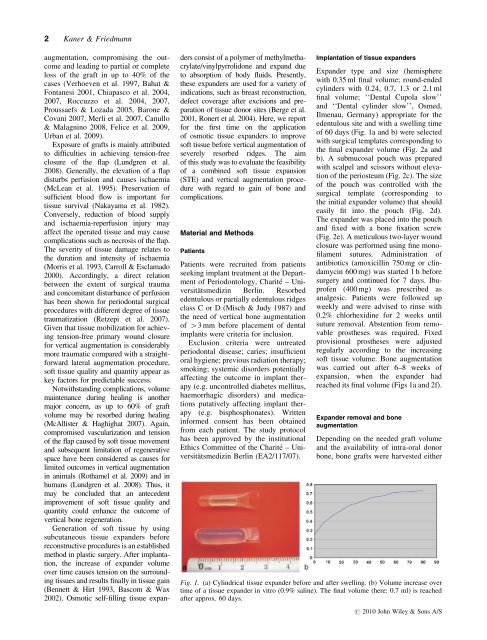
Fig. 1. (a) Cylindrical <strong>tissue</strong> expander before and after swelling. (b) Volume increase over<br />
time of a <strong>tissue</strong> expander in vitro (0.9% saline). The final volume (here: 0.7 ml) is reached<br />
after approx. 60 days.<br />
r 2010 John Wiley & Sons A/S