Soft tissue expansion with self-filling osmotic tissue expanders ...
Soft tissue expansion with self-filling osmotic tissue expanders ...
Soft tissue expansion with self-filling osmotic tissue expanders ...

You also want an ePaper? Increase the reach of your titles
YUMPU automatically turns print PDFs into web optimized ePapers that Google loves.
4 Kaner & Friedmann<br />
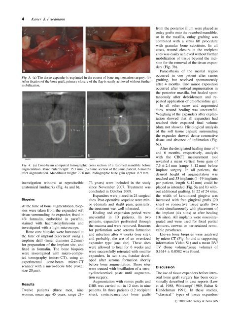
Fig. 3. (a) The <strong>tissue</strong> expander is explanted in the course of bone augmentation surgery. (b)<br />
After fixation of the bone graft, primary closure of the flap is easily achieved <strong>with</strong>out further<br />
mobilization.<br />
Fig. 4. (a) Cone-beam computed tomographic cross section of a resorbed mandible before<br />
augmentation. Mandibular height: 15.7 mm. (b) Same section of the same patient, 6 months<br />
after augmentation. Mandibular height: 22.6 mm, radiographic bone gain approx. 6.9 mm.<br />
investigation window at reproducible<br />
anatomical landmarks (Fig. 4a and b).<br />
Biopsies<br />
At the time of bone augmentation, biopsies<br />
were taken from the expanded soft<br />
<strong>tissue</strong> surrounding the expander, fixed in<br />
4% formalin, embedded in paraffin,<br />
stained <strong>with</strong> haematoxylin/eosin and<br />
investigated <strong>with</strong> a light microscope.<br />
Bone core biopsies were harvested at<br />
the time of implant placement using a<br />
trephine drill (inner diameter 2.2 mm)<br />
for preparation of the implant site, and<br />
fixed in formalin. The bone biopsies<br />
were investigated <strong>with</strong> micro-computed<br />
tomography (micro-CT), using an<br />
experimental cone-beam micro-CT<br />
scanner <strong>with</strong> a micro-focus tube (voxel<br />
size 20 mm).<br />
Results<br />
Twelve patients (three men, nine<br />
women, mean age 45 years, range 21–<br />
73 years) were included in the study<br />
since November 2007. Treatment was<br />
concluded in October 2009.<br />
Expanders were placed in 24 surgical<br />
sites. Post-operative sequelae were minor<br />
edemata and slight pain; generally,<br />
the treatment was well tolerated.<br />
Healing and <strong>expansion</strong> period were<br />
uneventful in 10 patients. In two<br />
patients, <strong>expanders</strong> perforated through<br />
the mucosa and were removed. Reasons<br />
for perforation were seroma formation<br />
and infection after 4 weeks (one site),<br />
and probably, the use of an oversized<br />
expander type (one site). These sites<br />
were allowed to heal for 6 weeks and<br />
were successfully retreated <strong>with</strong> smaller<br />
<strong>expanders</strong>. In two sites, fistulae developed<br />
after seroma formation shortly<br />
before bone augmentation. These sites<br />
were treated <strong>with</strong> instillation of a tetracycline/cortisol<br />
paste until augmentation<br />
surgery.<br />
Augmentation <strong>with</strong> ramus grafts and<br />
GBR was carried out in 12 sites in nine<br />
patients. In three patients (12 recipient<br />
sites), corticocancellous bone grafts<br />
from the posterior ilium were placed as<br />
onlay grafts onto the resorbed mandible,<br />
or in the maxilla, onlay grafting was<br />
combined <strong>with</strong> a sinus lift procedure<br />
<strong>with</strong> granular bone substitute. In all<br />
cases, wound closure at the recipient<br />
sites was easily achieved <strong>with</strong>out further<br />
mobilization of <strong>tissue</strong> beyond the incision<br />
for the removal of the <strong>tissue</strong> <strong>expanders</strong><br />
(Fig. 3b).<br />
Paraesthesia of the mental region<br />
occurred in one patient after ramus<br />
grafting, but resolved spontaneously<br />
after 4 months. One minor exposition<br />
occurred after vertical augmentation in<br />
the posterior maxilla, but healed spontaneously<br />
after debridement and repeated<br />
application of chlorhexidine gel.<br />
In all other cases and augmented<br />
sites, wound healing was uneventful.<br />
Weighing of the <strong>expanders</strong> after explantation<br />
showed that all <strong>expanders</strong> had<br />
reached their expected final volume<br />
(data not shown). Histological analysis<br />
of the soft <strong>tissue</strong> capsule surrounding<br />
the expander showed dense connective<br />
<strong>tissue</strong> and absence of infiltration (Fig.<br />
6a).<br />
After the designated healing time of 4<br />
and 6 months, respectively, analysis<br />
<strong>with</strong> the CBCT measurement tool<br />
revealed a mean vertical bone gain of<br />
7.5 2.4 mm (range 3–12 mm) before<br />
implant surgery. In all patients, the<br />
desired height of augmentation was<br />
reached and 53 implants (1–19 implants<br />
per patient, length 8–12 mm) could be<br />
placed as intended (Fig. 5a and b) <strong>with</strong>out<br />
additional grafting. In 22 of 24 sites,<br />
the width of keratinized gingiva was<br />
increased <strong>with</strong> free gingival grafts (20<br />
sites) or connective <strong>tissue</strong> grafts (two<br />
sites) simultaneously <strong>with</strong> placement of<br />
the implant (six sites) or after healing<br />
(16 sites). All implants were osseointegrated<br />
and were used for fixed partial<br />
dentures, crowns or bar-retained removable<br />
prostheses.<br />
Eleven bone biopsies were analysed<br />
by micro-CT (Fig. 6b and c; supporting<br />
information Video S1) and a mean BV/<br />
TV (bone volume/<strong>tissue</strong> volume) of<br />
0.1614 0.0582 was found.<br />
Discussion<br />
The use of <strong>tissue</strong> <strong>expanders</strong> before intraoral<br />
bone graft surgery has been occasionally<br />
described in case reports (Lew<br />
et al. 1988, Wittkampf 1989, Bahat &<br />
Handelsman 1991). In these studies,<br />
‘‘classical’’ types of <strong>tissue</strong> <strong>expanders</strong><br />
r 2010 John Wiley & Sons A/S