Soft tissue expansion with self-filling osmotic tissue expanders ...
Soft tissue expansion with self-filling osmotic tissue expanders ...
Soft tissue expansion with self-filling osmotic tissue expanders ...

You also want an ePaper? Increase the reach of your titles
YUMPU automatically turns print PDFs into web optimized ePapers that Google loves.
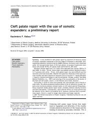
Tissue <strong>expansion</strong> before bone augmentation 3<br />
Fig. 2. (a) Resorbed edentulous ridge (class C) requiring vertical bone augmentation of approx. 5 mm. (b) The appropriate expander size is<br />
selected using the surgical template (final expander volume). (c) A supraperiosteal mucosal pouch is prepared using scalpel and scissors. (d)<br />
The preparation is controlled <strong>with</strong> the surgical template (initial expander volume). (e). The <strong>tissue</strong> expander is inserted into the pouch and fixed<br />
<strong>with</strong> a bone fixation screw. (f) After 8 weeks of <strong>tissue</strong> <strong>expansion</strong> (1.3 ml expander), a considerable gain of soft <strong>tissue</strong> can be observed.<br />
r 2010 John Wiley & Sons A/S<br />
from the mandibular ramus or the posterior<br />
ilium.<br />
Bone augmentation <strong>with</strong> ramus grafts<br />
was carried out under local anaesthesia,<br />
under sedation, anti-phlogistic medication<br />
<strong>with</strong> prednisolone and antibiotic<br />
coverage. Ibuprofen 600 mg was prescribed<br />
as analgesic and patients rinsed<br />
<strong>with</strong> 0.2% chlorhexidine for 2 weeks.<br />
At the donor site, a mucoperiostal flap<br />
was reflected distally of the second<br />
molar, exposing the lateral aspect of<br />
the ramus along the external oblique<br />
ridge. Block grafts were prepared <strong>with</strong><br />
a piezoelectric device (Piezosurgery II,<br />
Mectron, Köln, Germany).<br />
After midcrestal incision at the recipient<br />
site, a mucoperiostal flap was<br />
reflected. The expander was removed<br />
and the bone was exposed. Generally,<br />
vertical releasing incisions were<br />
avoided; in case of adjacent teeth present,<br />
the incision was extended into the<br />
gingival sulcus for ease of reflection.<br />
The local bone was perforated and the<br />
block graft was secured <strong>with</strong> screws<br />
(Institut Straumann AG, Basel, Switzerland).<br />
The graft was covered <strong>with</strong> a<br />
granular bone substitute (BioOss, Geistlich,<br />
Wolhusen, Switzerland) and a<br />
collagen membrane (Ossix plus, Colbar,<br />
Hertzelia, Israel). The incision was<br />
closed <strong>with</strong> modified vertical mattress<br />
sutures and single interrupted sutures,<br />
using fine monofilament sutures. Sutures<br />
were removed after 2 weeks.<br />
Bone augmentation <strong>with</strong> grafts from<br />
the posterior ilium was performed under<br />
general anaesthesia and under antibiotic<br />
coverage. The patient was placed in a<br />
prone position and an incision was<br />
placed extending cranially from the<br />
posterior iliac spine. Grafts were harvested<br />
from the external wall of the<br />
posterior iliac crest <strong>with</strong> an oscillating<br />
saw and a chisel.<br />
At the recipient sites, mucoperiostal<br />
flaps were elevated (Fig. 3a). Again,<br />
releasing incisions were avoided. After<br />
removal of the <strong>expanders</strong>, the local<br />
bone was exposed and perforated,<br />
and onlay grafts were fixed <strong>with</strong> screws<br />
(Fig. 3b). Patients were mobilized after<br />
24 h and sutures were removed after 1<br />
week.<br />
The <strong>expanders</strong> were weighed after<br />
removal and a biopsy was taken from<br />
the expanded soft <strong>tissue</strong>.<br />
Implant placement<br />
In patients treated <strong>with</strong> ramus grafts and<br />
GBR, implants (Standard Plus, SLActive<br />
surface, Institut Straumann AG)<br />
were placed 6 months after bone augmentation,<br />
following the standard protocol<br />
for non-submerged healing. In<br />
patients treated <strong>with</strong> iliac grafts,<br />
implants of the same type were placed<br />
4 months after augmentation. Sutures<br />
were removed after 1–2 weeks.<br />
Radiographs<br />
Cone-beam computed tomographies<br />
(CBCT, Galileos, Sirona, Bensheim,<br />
Germany) were taken before implantation<br />
of <strong>tissue</strong> <strong>expanders</strong>, and 4–6 months<br />
after bone grafting, before placement of<br />
dental implants. Digital panoramic<br />
radiographs (Sirona) were made after<br />
bone augmentation and after implant<br />
surgery. The mean vertical bone gain/<br />
surgical site was calculated to the nearest<br />
millimetre by subtraction of bone height<br />
before grafting from bone height before<br />
implantation, using the CBCT software<br />
measurement tool after aligning the