Clogging of Drainage Catheters: Quantitative and Longitudinal ...
Clogging of Drainage Catheters: Quantitative and Longitudinal ...
Clogging of Drainage Catheters: Quantitative and Longitudinal ...

Create successful ePaper yourself
Turn your PDF publications into a flip-book with our unique Google optimized e-Paper software.
Radiology<br />
grees <strong>of</strong> catheter clogging between different<br />
catheters.<br />
We found a discrepancy between the<br />
curves generated by the fitting Equation<br />
(4) <strong>and</strong> those generated by the Poiseuille<br />
equation (Eq [2]). Furthermore, intracatheter<br />
pressure was found to be proportional<br />
to the square <strong>of</strong> the infusion rate<br />
<strong>and</strong> not to the infusion rate, as indicated<br />
by fitting Equation (4). This discrepancy<br />
might be attributed to technical factors<br />
or to the fact that the system is not modeled<br />
perfectly by the Poiseuille law. The<br />
reasons for these differences remain unknown,<br />
<strong>and</strong> further study is required.<br />
In summary, we propose a method for<br />
in vivo evaluation <strong>of</strong> the effectiveness <strong>of</strong><br />
a drainage catheter, which is based on<br />
the application <strong>of</strong> the Poiseuille law.<br />
With this method, the flow resistance <strong>of</strong><br />
a catheter can be determined quantitatively;<br />
thus, it <strong>of</strong>fers a means <strong>of</strong> quantifying<br />
the degree <strong>of</strong> clogging or the anticlogging<br />
effect <strong>of</strong> drainage catheters. This<br />
method can be used for comparative <strong>and</strong><br />
longitudinal studies concerning the evaluation<br />
<strong>of</strong> drainage procedures or the development<br />
<strong>of</strong> new catheters.<br />
Practical application: By applying the<br />
noninvasive method used in this study,<br />
flow resistance through multiple drainage<br />
catheters can be compared over time<br />
in one animal, which provides quantitative<br />
information about the gradual obstruction<br />
<strong>of</strong> catheters. This information<br />
is critical for the evaluation <strong>of</strong> drainage<br />
procedures or development <strong>of</strong> new catheters<br />
with anticlogging effects, such as<br />
those made with novel biomaterials <strong>and</strong><br />
drug-delivery systems. Furthermore, this<br />
method does not preclude analyses based<br />
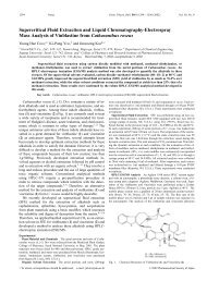
Figure 5. Images show internal surfaces <strong>of</strong> drainage catheters after the catheters were withdrawn.<br />
(a) Stereomicroscopic image shows debris that plugs the catheter side holes <strong>and</strong> lumen,<br />
which were irrigated one time per day. (b) Scanning electron microscopic image shows a catheter<br />
that was irrigated two times per day. Note the irregular surface caused by the debris (D). (Original<br />
magnification, 170.)<br />
on weight or morphologic changes after<br />
the catheter is withdrawn.<br />
Acknowledgments: We thank Hyuk Jae<br />
Choi, Myoung Soo Kim, RT, <strong>and</strong> Hyun Jung<br />
Lee, RT, for their technical assistance in animal<br />
preparation. We are grateful to Myoung Jin<br />
Jang, PhD, for her help with the statistical<br />
analysis.<br />
References<br />
1. Haaga JR, Weinstein AJ. CT-guided percutaneous<br />
aspiration <strong>and</strong> drainage <strong>of</strong> abscesses.<br />
Am J Roentgenol 1980; 135:1187–<br />
1194.<br />
2. vanSonnenberg E, Mueller PR, Ferrucci JT<br />
Jr. Percutaneous drainage <strong>of</strong> 250 abdominal<br />
abscesses <strong>and</strong> fluid collections. I. Results,<br />
failures, <strong>and</strong> complications. Radiology<br />
1984; 151:337–341.<br />
3. Mueller PR, vanSonnenberg E, Ferrucci JT<br />
Jr. Percutaneous drainage <strong>of</strong> 250 abdominal<br />
abscesses <strong>and</strong> fluid collections. II. Current<br />
procedural concepts. Radiology 1984;<br />
151:343–347.<br />
4. Dawson SL, Mueller PR, Ferrucci JT Jr. Mucomyst<br />
for abscesses: a clinical comment.<br />
Radiology 1984; 151:342.<br />
5. Park JK, Kraus FC, Haaga JR. Fluid flow<br />
during percutaneous drainage procedures:<br />
an in vitro study <strong>of</strong> the effects <strong>of</strong> fluid<br />
viscosity, catheter size, <strong>and</strong> adjunctive<br />
urokinase. Am J Roentgenol 1993; 160:165–<br />
169.<br />
6. Han JK. Percutaneous abdominal abscess<br />
drainage. In: Han MC, Park JH, eds. Interventional<br />
radiology. Seoul, Korea: Ilchokak,<br />
1999; 707–714.<br />
7. Pfitzner J. Poiseuille <strong>and</strong> his law. Anaesthesia<br />
1976; 31:273–275.<br />
8. Frazini JB, Finnemore EJ, eds. Fluid mechanics<br />
with engineering applications. Appendix<br />
A. 9th ed. Boston, Mass: McGraw-<br />
Hill, 1997; 764–765.<br />
9. Moon HT, Lee YK, Han JK, Byun Y. A novel<br />
formulation for controlled release <strong>of</strong> heparin-DOCA<br />
conjugate dispersed as nanoparticles<br />
in polyurethane film. Biomaterials<br />
2001; 22:281–289.<br />
838 Radiology June 2003 Lee et al