large - NordiQC
large - NordiQC
large - NordiQC

Create successful ePaper yourself
Turn your PDF publications into a flip-book with our unique Google optimized e-Paper software.
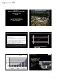
DAKO Satellite Symposium:<br />
CIQC/CAP-ACP Seminar in<br />
Diagnostic Immunohistochemistry<br />
July 08-09, 2010<br />
Immunohistochemical Quality Assurance<br />
and Protocol Optimization<br />
Mogens Vyberg, MD<br />
Assoc professor<br />
Scheme director, <strong>NordiQC</strong><br />
Inst. of Pathology, Aalborg Hospital<br />
Aarhus University Hospital<br />
Denmark<br />
1
Immunohistochemistry: A highly complex analysis<br />
Decalcification<br />
Preparation<br />
Tissue<br />
Type, Dimension,<br />
De-differentiation<br />
Necrosis<br />
Heat cauterisation<br />
Control<br />
Quantification<br />
Reporting<br />
Fixation<br />
Time, Type, Volume<br />
Preanalytic<br />
Postanalytic<br />
Section<br />
Thickness<br />
Storage<br />
Drying<br />
3 choices for 5 variables<br />
in each phase:<br />
4 million different protocols !<br />
Pre-treatment<br />
Manual<br />
Stainer<br />
Visualization<br />
Sensitivity, Specificity<br />
Primary antibody<br />
Clone, Dilution<br />
Buffer, Time, Temp<br />
Analytic<br />
Interpretation<br />
Localization<br />
Positive/Negative - cut-off level<br />
Development<br />
Sensitivity,<br />
Localization
External Quality Assurance: Estrogen Receptor<br />
Correct<br />
False negative<br />
½<br />
Control<br />
Lab. A<br />
Lab. B<br />
3
<strong>NordiQC</strong> EQA: Estrogen Receptor 2003<br />
Sufficient<br />
Insufficient<br />
½<br />
32 / 71 labs = 45% 39 / 71 labs = 55%<br />
4
External Quality Assurance<br />
• Staining quality varies greatly between different<br />
laboratories depending on the individual selection of<br />
methods and the technical expertise<br />
• Internal quality control will often not identify a<br />
poorly calibrated IHC system giving insufficient<br />
staining results<br />
• Standardization of staining methods is not possible<br />
• Standardization of staining results is mandatory<br />
5
Establishment of a Nordic EQA program for IHC<br />
Nordic Immunohistochemical Quality Control<br />
<strong>NordiQC</strong> founded 2003 by Nordic pathologists<br />
• Independent, scientific, not-for-profit organisation<br />
• Institute of pathology<br />
Aalborg Hospital<br />
• General module:<br />
3 annual runs<br />
~16 different markers/tests<br />
• Breast cancer module:<br />
2 annual runs<br />
HER-2 IHC & BRISH, ER/PR<br />
6
Website: www.nordiqc.org<br />
7
<strong>NordiQC</strong> Participants<br />
8
Over-all assessment results<br />
<strong>NordiQC</strong> consensus marks (average 2003-10)<br />
40 runs = 25,000 slides<br />
• Optimal: 36 %<br />
• Good: 33 %<br />
• Borderline:<br />
}<br />
too weak / false neg.: ~ 90 %<br />
31 % {<br />
• Poor:<br />
over-stained / false pos.: ~ 10 %<br />
<strong>NordiQC</strong> regrets any offence<br />
caused to laboratories and companies …<br />
9
<strong>NordiQC</strong> over-all assessment results<br />
Major causes of insufficient stains:<br />
• “Less successful” antibodies 18 %<br />
• Inappropriate antibody dilution 39 %<br />
• Inappropriate epitope retrieval 31 %<br />
• Other inappropriate lab. performance 12 %<br />
• Low sensitivity of detection system<br />
• Inappropriate calibration / home brews<br />
• Endogenous biotin reaction (EBR)<br />
• Section drying-out after HIER<br />
• Technical stainer errors<br />
• . . . .<br />
• Unexplained<br />
10
Results of <strong>NordiQC</strong> tailored recommendations<br />
Lab response to 419 advices (11 markers)<br />
No. Improved %<br />
Positive 268 195 73<br />
Negative 151 21 14<br />
11
<strong>NordiQC</strong> EQA: Estrogen Receptor 2003-11<br />
200<br />
180<br />
160<br />
140<br />
No. participants<br />
120<br />
100<br />
80<br />
67<br />
84<br />
67<br />
72<br />
79 81<br />
74<br />
67<br />
90<br />
60<br />
45<br />
40<br />
20<br />
0<br />
8 10 13 B1 B3 B5 B7 B8 B10 B11<br />
2003 2006 RUN 2009 2011<br />
PASS RATE (%)<br />
No. OF PARTICIPANTS
External IHC Quality Assurance<br />
•Almost 1/3 of all IHC stains produced by <strong>NordiQC</strong><br />
participants are still insufficient !<br />
• New labs<br />
• New antibodies, techniques, platforms<br />
• Increasing demands<br />
•How many IHC stains produced by labs not<br />
participating in an EQA scheme are insufficient ?<br />
•How many scientific publications are based on<br />
insufficient IHC stains ?<br />
•What are the consequences for the patients ?
External Quality Assurance – ER<br />
correct<br />
false negative<br />
“Through the inquiry, the public<br />
learned that between 1997 and<br />
2005 nearly 400 of about 1,000<br />
breast cancer patients received<br />
incorrect test results of the ER<br />
status of their breast tumors.”<br />
Control<br />
14<br />
Craig Allred
External Quality Assurance<br />
“Less successful” antibodies 18 %<br />
• Poor antibodies<br />
• Poor ready-to-use formats<br />
• Less robust antibodies<br />
• Other error-prone antibodies<br />
• Mouse-anti-Golgi (MAG) reaction<br />
• Lot-to-lot variation<br />
• Platform dependent<br />
• Poor cocktail composition<br />
• ….<br />
15
Poor antibodies – CD31 false negative<br />
JC70A<br />
1A10<br />
Optimal (16%)
Poor antibodies – CD31 false negative<br />
JC70A<br />
1A10<br />
Optimal (16%)<br />
Haemangiosarcoma
Poor antibodies – CD31 from 167 labs
Poor antibodies – MLH1 false positive<br />
MLH1 clone ES05<br />
MLH1 clone EPR3894
Poor RTU formats – CD79a<br />
JCB117 optimal JCB117 RTU 760-2630
Less robust Abs – Estrogen receptor alpha
Less robust Abs – Estrogen receptor alpha<br />
Optimal<br />
? Ab/dil./HIER/Viz.<br />
Ductal carcinoma I
Less robust Abs – Estrogen receptor alpha<br />
Optimal<br />
? Ab/dil./HIER/Viz.<br />
Ductal carcinoma II
New Estrogen receptor alpha<br />
rmAb EP1, 1:15 Epitomics<br />
TRS High pH/20M/EnV-FLEX<br />
rmAb SP1, RTU Ventana<br />
CC1M/32M/36˚C/UV<br />
EP1<br />
SP1
Platform dependent antibodies<br />
Antigen Clone XT / Ultra<br />
BSAP Pax5 24<br />
FN<br />
BCL6 PG-B6p<br />
FN<br />
CD4 1F6, 4B12 FN<br />
CD5 4C7 FP<br />
CD56 123.C3<br />
FN<br />
CD79a JCB117 FN<br />
OCT-2 OCT-207 FN<br />
ASMA 1A4<br />
FP<br />
SF-1 NR5A1<br />
FN<br />
SMAD4 BC/B8 FN<br />
Alternative<br />
SP34<br />
GI191E/A8<br />
SP35<br />
SP19<br />
MRQ-42<br />
SP18<br />
MRQ-2<br />
None<br />
None<br />
None<br />
25
Platform dependent antibodies – PAX5
Platform dependent antibodies – PAX5<br />
Hodgkin lymphoma NS<br />
clone SP34<br />
RTU VMS/CM<br />
x200<br />
clone 24<br />
RTU VMS/CM<br />
x200
Inappropriate antibody dilution<br />
28
Inappropriate antibody dilution – CD79a<br />
JCB117 appropriate<br />
JCB117 too dilute<br />
Plasmacytoma<br />
29
Inappropriate antibody dilution – Ig light chains<br />
IgK: Dako pAb A0191<br />
~1:300 ~1:3.000 ~1:30.000
Inappropriate antibody dilution – Ig light chains<br />
239 IgK tests, 12 Abs:<br />
• 12% optimal<br />
Dako pAb A0191:<br />
• 17% optimal<br />
+TRS/Ci 3.000-16.000:<br />
• 29 % optimal<br />
All other Abs:<br />
• 0% optimal<br />
Alternative: FLOW cytometry
Inappropriate epitope retrieval<br />
32
Inappropriate retrieval – CK AE1/AE3<br />
HIER<br />
Proteolysis<br />
AE1 detects CK8 after HIER only<br />
AE1 does not detect CK18<br />
AE3 does not detect CK8/CK18<br />
33
Inappropriate retrieval – CK PAN<br />
Colon AC<br />
Renal CC<br />
HIER<br />
Proteolysis
Inappropriate vendor information<br />
Misleading datasheet: AE1/AE3<br />
2011<br />
35
Confusing vendor information<br />
Correct datasheet:<br />
AE1/AE3<br />
36
Package inserts<br />
RP2/18/22 X16/99<br />
Datasheet changed according<br />
to <strong>NordiQC</strong> recommendation:<br />
mal.melanoma<br />
mal.melanoma
Less sensitive detection systems<br />
38
Melan A, clone A-103 in high expressor<br />
Mal. melanoma<br />
UltraView<br />
OptiView
Melan A, clone A-103 in low expressor<br />
Granulosa cell tumour<br />
UltraView<br />
OptiView
EpCAM<br />
Normal kidney<br />
UltraView, EP4, CC1 OptiView, MOC31, TRS 6
EpCAM<br />
Clear cell renal cell carcinoma<br />
UltraView, EP4, CC1 OptiView, MOC31, TRS 6
Nordic immunohistochemical Quality Control<br />
Conclusion I<br />
• External Quality Assurance (EQA)<br />
• Provides objective evidence of lab proficiency<br />
• Identifies methodological errors<br />
• Provides directions for improvements<br />
• The results of the <strong>NordiQC</strong> work indicate that<br />
• Improvement of IHC is strongly needed<br />
• EQA may have a major impact on lab proficiency<br />
• External quality assurance of staining results through<br />
laboratory proficiency testing is mandatory
Nordic immunohistochemical Quality Control<br />
Conclusion II<br />
• If IHC testing is not done properly, innovation may<br />
not be based on a solid foundation.<br />
• EQA groups must work closely with manufactures in<br />
order to accelerate the companies’ innovative efforts<br />
by early and constructive feedback on new or<br />
candidate new products …<br />
…including improved data sheets<br />
44
Nordic immunohistochemical Quality Control<br />
Thank you<br />
for your<br />
attention !<br />
Aalborg Hospital<br />
45