4th International Myotonic Dystrophy - IDMC.org
4th International Myotonic Dystrophy - IDMC.org
4th International Myotonic Dystrophy - IDMC.org

Create successful ePaper yourself
Turn your PDF publications into a flip-book with our unique Google optimized e-Paper software.
<strong>4th</strong> <strong>International</strong> <strong>Myotonic</strong> <strong>Dystrophy</strong><br />
Consortium Meeting<br />
Welcome to the 4 th <strong>International</strong> <strong>Myotonic</strong> <strong>Dystrophy</strong> Consortium meeting here in<br />
Glasgow. Following on from previous highly successful get-togethers in Kyoto (2001),<br />
Chapel Hill (1999) and Paris (1997), this meeting has a lot to live up to. But, from the<br />
abstracts submitted, it looks like we are in for an exciting few days learning about the<br />
latest breakthroughs in myotonic dystrophy research. Indeed, there were so many<br />
abstracts submitted, that for the first time we haven’t been able to fit everybody in for<br />
an oral presentation and we will be having a poster session as well. In doing so, we have<br />
also reserved a bit more time for discussion at the end of each session and hope that this<br />
period can be used to think about how the various pieces of work fit together. All in all,<br />
this format will give us more time for thinking aloud about what we need to determine<br />
next, and discussing what lies ahead, in this exciting and fast moving field of research.<br />
As at previous meetings though, the timetable allows for some socialising and discussion<br />
in a more relaxed atmosphere as well. This will include the opportunity to sample the<br />
Scottish countryside and one of its most sought after products, uisge beath, ‘the water of<br />
life’ or whisky, as you probably know it. In addition, there will be the chance to try out<br />
your moves on the dance floor at the ceilidh on Friday evening. Whilst on Saturday<br />
evening we will be the guests of the Lord Provost of Glasgow at a Civic Reception he will<br />
be hosting for us at the Kelvingrove Art Gallery and Museum.<br />
It is with great pleasure that I also welcome, not only the scientists and clinicians who<br />
are actively researching in this area, but also the patients and families who will be<br />
attending a joint session of the meeting on Saturday afternoon, as well as the social<br />
events on Friday and Saturday evenings. This session has been made possible by the<br />
simultaneous hosting in Glasgow of the <strong>Myotonic</strong> <strong>Dystrophy</strong> Support Group’s AGM. This is<br />
the first time that all the interested parties have got together at one of the <strong>IDMC</strong><br />
meetings and we hope it will be a big success allowing us all to learn from each others<br />
experiences.<br />
Finally, it just remains for me to say a big thank you to everyone whose hard work has<br />
made this meeting possible, to our generous sponsors and to you all for coming here and<br />
contributing to what I am sure will be another great <strong>IDMC</strong> meeting.<br />
Slàinte mhath!<br />
Darren Monckton<br />
Chair, <strong>IDMC</strong>4<br />
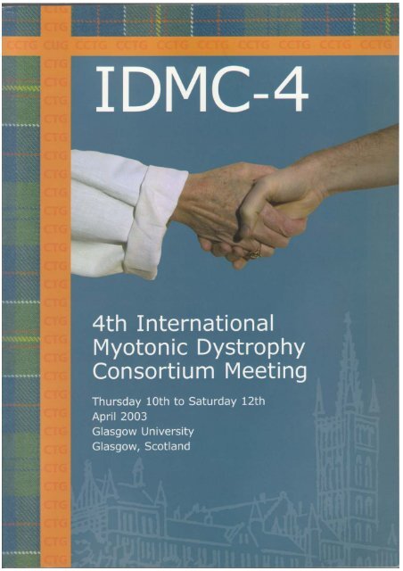
Cover design. The front cover incorporates elements associated with the DNA expansions at the myotonic<br />
dystrophy type 1 and type 2 loci, Glasgow University and Hunter family tartan. The Hunter family provided<br />
collections and donations that made possible the establishment of the Hunterian Museum and Art Gallery within<br />
the University of Glasgow in the late 1700s. It is a striking coincidence that some 200 years later, one of the<br />
Hunter family descendents, Shannon Lord, will be attending <strong>IDMC</strong>4 here at the University of Glasgow to<br />
describe her experiences of living with myotonic dystrophy and the establishment of the Hunter Research Fund.<br />
The shaking hands also depicted on the cover represent not only the simple test often used by clinicians to<br />
detect myotonia, but more importantly symbolises the interactions that we hope to generate between clinicians,<br />
scientists, patients and families here at <strong>IDMC</strong>4. The cover was designed and prepared by Chris Hoke and Scott<br />
Thompson with help from Shannon Lord and Darren Monckton.<br />
Page 1
<strong>IDMC</strong>4 Committees<br />
Local Organising Committee<br />
Alison Wilcox<br />
Department of Medical Genetics<br />
University of Glasgow<br />
Douglas E. Wilcox<br />
Department of Medical Genetics<br />
University of Glasgow<br />
Darren G. Monckton<br />
Institute of Biomedical and Life Sciences<br />
University of Glasgow<br />
UK Committee<br />
Peter Harper<br />
Institute of Medical Genetics<br />
University of Wales College of Medicine<br />
Cardiff<br />
Marion Hamshere<br />
School of Life and Environmental Sciences<br />
University of Nottingham<br />
David Brook<br />
Institute of Genetics<br />
University of Nottingham<br />
David Hilton-Jones<br />
Department of Clinical Neurology<br />
Radcliffe Infirmary<br />
Oxford<br />
<strong>International</strong> Committee<br />
Tetsuo Ashizawa<br />
Department of Neurology<br />
The University of Texas Medical Branch<br />
Galveston, Texas<br />
Martine Devillers<br />
Association Française contre les Myopathies<br />
Département recherche et développement des<br />
thérapeutiques<br />
Évry<br />
Genevieve Gourdon<br />
INSERM UR383<br />
Hôpital Necker Enfants-Malades<br />
Paris<br />
Shannon M. Lord<br />
Hunter Research Fund<br />
Atlanta<br />
Margaret Bowler<br />
<strong>Myotonic</strong> <strong>Dystrophy</strong> Support Group<br />
Nottingham<br />
Daisuke Furutama<br />
First Dept. of Internal Medicine<br />
Osaka Medical College<br />
Osaka<br />
Keith J. Johnson<br />
Pharmacia Corp.<br />
Chicago<br />
Nakaaki Ohsawa<br />
Aino institute for aging research<br />
Ibaraki<br />
Sponsorship<br />
The <strong>org</strong>anising committee is pleased to acknowledge the support of the following<br />
<strong>org</strong>anisations in funding the meeting: GlaxoSmithKline; Pharmacia Corp; The Robertson<br />
Trust; The Genetics Society; The <strong>Myotonic</strong> <strong>Dystrophy</strong> Support Group; The University of<br />
Glasgow; Glasgow City Council; and the Muscular <strong>Dystrophy</strong> Campaign.<br />
The <strong>org</strong>anising committee is also pleased to acknowledge the support of the following<br />
<strong>org</strong>anisations in funding travel costs: The Hunter Research Fund; the Aino Institute for<br />
Aging Research and the Association Française contre les Myopathies.<br />
Page 2
Acknowledgements<br />
The <strong>org</strong>anisers would like to thank all those who have contributed toward the planning<br />
and <strong>org</strong>anisation of <strong>IDMC</strong>4 including: Jane Kelly, Claire McCulloch, Sabrina Sharma,<br />
Bronwyn Syed, Alexis Stevenson, John McAbney, Graham Hamilton and Katherine Allen<br />
from the Institute of Biomedical and Life Sciences, University of Glasgow; Claire Harper<br />
from the University of Glasgow Conference and Visitor Services; Ellen Thompson from<br />
Glasgow University Hospitality Services; Raymond Dixon and Lynne Aikman from The<br />
Print Unit of Glasgow and Strathclyde Universities; Chris Hoke, Scott Thompson and<br />
Susan Greco from Fusion Designworks; Lorna McClelland and Lorna Clarkson from the<br />
Greater Glasgow Tourist Board; Allen Roses from GlaxoSmithKline; Keith Johnson from<br />
Pharmacia Corp; Helen Sang and Jayne Richards from the Genetics Society; James the<br />
janitor for the WILT; The City of Glasgow’s Lord & Lady Provost; Prestige Tours; Alison<br />
Adams from Encore Catering; Neil Drover; RocknReel ceilidh disco band; Harpbeat;<br />
Jenny Versnel from The Muscular <strong>Dystrophy</strong> Campaign; The Grosvenor Hotel; Maragret<br />
Bowler from the <strong>Myotonic</strong> <strong>Dystrophy</strong> Support Group; Anne Theriault and Betty O’Hare<br />
from Yorkhill Hospital. Our sincerest apologies if we have missed anybody out.<br />
Registration<br />
Registration will be in the<br />
foyer of the Western<br />
Infirmary Lecture Theatre<br />
(WILT) from 8.00 am on<br />
Thursday 10 th April. Tea,<br />
coffee and biscuits will be<br />
available. Please arrive in<br />
good time so that we may<br />
start the meeting promptly<br />
at 9.00 am.<br />
How to get to the<br />
Western Infirmary<br />
Lecture Theatre<br />
The Western Infirmary<br />
Lecture Theatre is located in<br />
the rear car cark of the<br />
Western Infirmary Hospital.<br />
Access is from Dumbarton<br />
Road (see maps on following<br />
pages).<br />
By foot. From Dumbarton Road proceed up the hill around the west side of the main<br />
hospital building toward the rear. The WILT is located in the north east corner of the rear<br />
car park.<br />
By taxi. Ask the taxi driver to take you to the rear car park of the Western Infirmary.<br />
By car. Limited car parking for disabled visitors only is available on the Western<br />
Infirmary site. There is no parking within the University grounds. On street car parking is<br />
available in the surrounding area, but the area is very busy and only limited spaces will<br />
be available. You are advised to use public transport or walk.<br />
Page 3
Page 4<br />
The Western Infirmiry Lecture Theather (WILT) and the Kelvingrove Art Gallery are highlighted in black.
Page 5<br />
The Western Infirmary lecture Theatre (WILT), Kelvingrove Art Gallery, Grosvenor Hotel, Novotel Hotel, Lorne Hotel and Holiday Inn are highlighted in black.
Accommodation<br />
The conference hotels are the Kelvin Park Lorne Hotel (923 Sauchiehall Street, Tel: 0141<br />
314 9955) and the Novotel Hotel (181 Pitt Street, Tel: 0141 222 2775). The <strong>Myotonic</strong><br />
<strong>Dystrophy</strong> Support Group are based in the Holiday Inn Glasgow City West (Bothwell<br />
Street, Tel: 0870 400 9032).<br />
If you have any problems concerning your hotel accommodation, please deal with the<br />
hotel concerned directly.<br />
Please note that all attendees should settle their own hotel accounts on checking out.<br />
There may be some limited funds available for support of accommodation costs of<br />
presenting authors (oral and posters), but this will only available after the close of the<br />
meeting.<br />
Local amenities<br />
A good selection of shops, bars and restaurants are located on Byres Road and along<br />
Great Western Road to the north west of the University. The city centre has an extensive<br />
array of shops, including several large shopping centres.<br />
Oral presentations<br />
Each presenter will have a 7 minute slot with an additional 3 minutes for brief questions.<br />
Please ensure you do not overrun. The programme is packed and the session chairmen<br />
will stick rigorously to the schedule. There will be a 30 minute period at the end of each<br />
session for more involved discussion.<br />
If you are using a PowerPoint presentation please ensure that your file is loaded onto one<br />
of the meetings computers before your session begins. Please see Graham Hamilton<br />
(Macintosh) or Douglas Wilcox (PC) to load your file.<br />
If you are using 35mm slides please see John McAbney to obtain a carousel.<br />
Posters<br />
Poster boards will be located in the foyer of the WILT. Please attach your poster to the<br />
appropriately numbered board as soon as possible on arriving at the meeting. Velcro<br />
stickers will be available for this purpose. Please ensure your remove your poster at the<br />
end of the final session on Saturday afternoon.<br />
Cheese and wine poster session<br />
In addition to viewing posters during the tea and coffee and lunch breaks, there will be a<br />
dedicated poster session on the Thursday evening from 5.00 pm until 7.00 pm.<br />
Presenting authors are requested to stand by their posters for the duration of the poster<br />
session. A selection of light nibbles will be served along with wine.<br />
Hospitality<br />
Teas, coffees and lunches throughout the meeting will be served in the foyer of the WILT.<br />
Page 6
Excursion<br />
On Friday afternoon there will be an excursion to<br />
Glengoyne Distillery and Loch Lomond. Two coaches<br />
will depart from outside the WILT at 1.00 pm. Please<br />
make sure you have had your lunch in good time and<br />
are ready to go. The three coaches will visit the venues<br />
in alternate order and will return to the hotels at<br />
around 6.30 pm.<br />
Glengoyne Distillery<br />
Loch Lomond - Scotland’s largest loch<br />
Ceilidh<br />
On Friday evening (11 th April), from 8.00 pm<br />
till midnight, there will be a ceilidh in the<br />
Hilton Glasgow Grosvenor. A ceilidh is a<br />
traditional Scottish social gathering with music<br />
and dancing. The festivities will be lead by the<br />
ceilidh band Rock’n’Reelin.<br />
For refreshments there will be a buffet supper<br />
and a cash bar.<br />
Admission will be by ticket only.<br />
Rock’n’Reelin<br />
“We know from our experience that not<br />
everybody is sure of what to do, therefore we<br />
call every dance and give demonstrations. This<br />
results in a band of highly professional trained<br />
musicians who guarantee a full dance floor and<br />
a memorable night.”<br />
The Hilton Glasgow Grosvenor, Great Western Road.<br />
Tel: 0141 339 8811.<br />
Page 7
Bring and buy sale<br />
The <strong>Myotonic</strong> <strong>Dystrophy</strong> Support Group will be running a ‘bring and buy sale’ during the<br />
joint session on Saturday 12 th April. This is an informal sale to raise money for myotonic<br />
dystrophy research. The MDSG would be delighted if attendees could bring an<br />
inexpensive item to donate to the sale. If the item were representative of the area that<br />
you come from that would be great.<br />
Civic reception and conference dinner<br />
A Civic Reception for the delegates of<br />
<strong>IDMC</strong>4 will be held in Glasgow<br />
Kelvingrove Art Gallery, West Balcony<br />
from 7.00 pm until 8.00 pm on<br />
Saturday 12 th April. This event is<br />
hosted by the Lord Provost of<br />
Glasgow, Mr Alex Mosson.<br />
A Scottish Buffet Dinner will then be<br />
held in the Centre Hall from 8.00 pm<br />
until 11.00 pm. ‘Harpbeat’, a trio of<br />
musicians playing flute, fiddle and<br />
clarsach (Celtic harp) will entertain us<br />
throughout the evening.<br />
The Kelvingrove Art Gallery and Museum is world<br />
renowned for the quality of its international art collection<br />
which includes Impressionists and Italian and Dutch<br />
Renaissance paintings. Without question the Art Gallery<br />
houses one of Scotland’s finest civic art collections.<br />
Delegates will have an opportunity to view part of the<br />
collection following the meal, if they wish.<br />
Admission is by ticket only. Your prebooked<br />
ticket will be in your<br />
registration pack.<br />
Page 8
Programme<br />
Thursday 10 th April AM<br />
8.00 am Registration with tea/coffee<br />
9.00 am Welcome – Meeting chair: Darren G. Monckton<br />
Session 1 – Diagnostics and DNA instability in myotonic dystrophy<br />
Session chair: Genevieve Gourdon<br />
9.10 am 1) Origins of the myotonic dystrophy mutation: worldwide study in the CEPH-Human<br />
Genome Diversity Project<br />
Hidehisa Yamagata* et al.<br />
9.20 am 2) Possible de novo CTG repeat expansion in DM protein kinase gene of a patient with<br />
isolated focal cardiomyopathy<br />
Daisuke Furutama* et al.<br />
9.30 am 3) CTG repeat instability timing during transgenic mice spermatogenesis<br />
Cédric Savouret* et al.<br />
9.40 am 4) Role of mismatch-repair proteins and Fen-1 exonuclease in somatic expansion<br />
behaviour of the (CTG) n -repeat in cellular and animal models for DM1<br />
Marcel R. Nelen, … … and Bé Wieringa*<br />
9.50 am 5) DNA double-strand breaks induce instability of CTG•CAG repeats related to<br />
myotonic dystrophy in an orientation dependent manner in Escherichia coli<br />
Micheal L. Hebert* and Robert D. Wells<br />
10.00 am 6) Could a change in direction of DNA replication make the repeat unstable?<br />
Celia H. Burgoyne* et al.<br />
10.10 am 7) Replication inhibitors modulate instability of an expanded CTG tract at the DM1<br />
disease locus in human cells<br />
Zhi Yang, … … and Christopher E. Pearson*<br />
10.20 am 8) Chemotherapeutically induced deletion of expanded (CAG)•(CTG) repeats<br />
Richard R. Sinden* et al.<br />
10.30 am Tea/coffee break<br />
11.00 am 9) Chemically induced modification of DNA dynamics: towards chemogenetherapy in<br />
the repeat expansion disorders<br />
Mário Gomes-Pereira* et al.<br />
11.10 am 10) High-resolution marker analysis of the region flanking the DM2-(CCTG) n<br />
expansion reveals a single shared haplotype among mutation carriers<br />
L.L. Bachinski* et al.<br />
11.20 pm 11) Molecular genetics of the DM2 CCTG expansion<br />
Laura P.W. Ranum* et al.<br />
11.30 pm 12) Triplet and quadruplet repeat-primed PCR in DM1 and DM2 diagnosis: detection<br />
of unusual mutations<br />
David J. Cockburn* et al.<br />
11.40 pm 13) A new method for routine molecular diagnosis in myotonic dystrophy type 2<br />
using chromogenic in situ hybridization (CISH)<br />
A. Vihola, … … and B. Udd*<br />
11.50 pm Discussion<br />
12.20 pm Lunch<br />
Page 9
Thursday 10 th April PM<br />
Session 2 – Neighbouring gene effects and cell biology in myotonic dystrophy<br />
Session chair: Keith Johnson<br />
2.00 pm 14) Impaired muscle development in congenital myotonic dystrophy<br />
Denis Furling* et al.<br />
2.10 pm 15) Expression of myogenic regulatory factors in myotonic dystrophy myoblasts<br />
Caroline Haineault, … … and Jack Puymirat*<br />
2.20 pm 16) A molecular genetic analysis of muscle differentiation in Drosophila<br />
melanogaster<br />
Michael V. Taylor* et al.<br />
2.30 pm 17) The role of the Drosophila SIX5 homologue, D-Six4, in embryonic muscle<br />
development<br />
Ivan B. N. Clark* et al.<br />
2.40 pm 18) A comparative whole genome approach to study SIX5 function and its<br />
contribution to the DM1 phenotype<br />
Graham M. Hamilton* et al.<br />
2.50 pm 19) DM1 and DM2 transcriptome profiling reveals global gene expression changes<br />
Ralf Krahe* et al.<br />
3.00 pm 20) Six5-target genes and DM1 symptoms<br />
Shigeru Sato* et al.<br />
3.10 pm 21) SIX5 target genes: functional links between SIX5 and DM1 symptoms<br />
Rami A. Jarjour, … … and Keith J. Johnson*<br />
3.20 pm Tea/coffee break<br />
3.40 pm 22) Problems with antibodies<br />
Glenn E Morris* et al.<br />
3.50 pm 23) Characterization of potential DMPK substrates in a cellular blebbing model.<br />
James D. Waring* et al.<br />
4.00 pm 24) Alternative splicing controls DMPK structure, enzymatic activity and subcellular<br />
localization<br />
Derick G. Wansink* et al.<br />
4.10 pm 25) C-terminal targeting-motifs direct DMPK to the endoplasmic reticulum or<br />
mitochondrial outer membrane<br />
René E.M.A. van Herpen* et al.<br />
4.20 pm 26) Towards the understanding of the myotonic dystrophy type 2 pathogenesis<br />
Emanuela Bonifazi* et al.<br />
4.30 pm Discussion<br />
5.00 pm to<br />
7.00 pm<br />
Poster session with cheese and wine<br />
Page 10
Friday 11 th April<br />
Session 3 – Toxic RNA in myotonic dystrophy<br />
Session chair: David Brook<br />
9.00 am 27) Foci formation and the disruption of splicing by CUG-repeat RNA are separable<br />
events<br />
Thai H. Ho* et al.<br />
9.10 am 28) Gene expression profiles of mouse C2C12 cells which stably express human<br />
DMPK with 160 CTG repeat<br />
Noboru Sasagawa* et al.<br />
9.20 am 29) Inhibition of myogenesis in transgenic mice expressing the human DMPK 3’ UTR<br />
C.J. Storbeck* et al.<br />
9.30 am 30) Mouse muscleblind gene knockout models for myotonic dystrophy<br />
Rahul N. Kanadia* et al.<br />
9.40 am 31) Ribonuclear inclusions in the central nervous system in myotonic dystrophy<br />
type 1<br />
Hong Jiang … … and Charles A. Thornton*<br />
9.50 am 32) Analysis of alternative splicing in muscleblind deficient Drosophila<br />
Helena Thorpe* et al.<br />
10.00 am 33) A Drosophila model of myotonic dystrophy<br />
Jonathan M. Houseley* et al.<br />
10.10 am 34) RNA mechanism for myotonic dystrophy 1: elevation of CUGBP1 disorders<br />
myogenesis through the disruption of MEF2A and p21 pathways<br />
Roma Patel … … and Lubov T. Timchenko*<br />
10.20 am Tea/coffee break<br />
10.50 am 35) Aberrant splicing of the ryanodine receptor in myotonic dystrophy<br />
Takashi Kimura* et al.<br />
11.00 am 36) Developmental roles of the muscleblind and CUG-BP homologues in C. elegans<br />
Aidyl-Gonzalez-Serricho* et al.<br />
11.10 am 37) Insights from a C2C12 myoblast model of the effects of the mutant DMPK 3’UTR<br />
RNA on myogenic differentiation<br />
Jeffrey D. Amack … … and Mani S. Mahadevan*<br />
11.20 pm 38) Molecular pathogenesis of the DM2 CCTG Expansion<br />
Laura P.W. Ranum* et al.<br />
11.30 pm Discussion<br />
12.00 pm Lunch<br />
1.00 pm to<br />
6.00 pm<br />
8.00 pm to<br />
11.00 pm<br />
Excursion - Glengoyne Distillery and Loch Lomond<br />
Ceilidh - Grosvenor Hotel<br />
Page 11
Saturday 12 th April AM<br />
Session 4 – Clinical and therapeutic aspects of myotonic dystrophy<br />
Session chair: Peter Harper<br />
9.00 am 39) Somnolence in DM1: cause, assessment and management<br />
David Hilton-Jones* et al.<br />
9.10 am 40) Neuropsychiatric, neuropsychological and neuroimaging studies in myotonic<br />
dystrophy type 1: significance and clinical correlates?<br />
G. Meola* et al.<br />
9.20 am 41) Educational and psychological group observations of children and juveniles with<br />
congenital myotonic dystrophy<br />
Jes Rahbek* et al.<br />
9.30 am 42) A peculiar neurofibrillary degeneration distribution in one DM2 brain: a case<br />
report<br />
Claude-Alain Maurage, Nicolas Sergeant* et al.<br />
9.40 am 43) Testosterone and diurnal rhythmicity of leptin, TNF-alpha, and TNF-II receptor in<br />
insulin-resistant myotonic dystrophy patients<br />
Åsa Johansson* et al.<br />
9.50 am 44) Heart involvement in young myotonic dystrophy type 1 patients<br />
G. Bassez* et al.<br />
10.00 am 45) Steinert’s disease (DM1) with predominant proximal involvement<br />
Frédéric Andreux … … and Bruno Eymard*<br />
10.10 am 46) Clinical and molecular features of myotonic dystrophy type 2<br />
John W. Day* et al.<br />
10.20 am Tea/coffee break<br />
10.50 am 47) Is this myotonic dystrophy type 3?<br />
Robert McWilliam* and Ed Tobias<br />
11.00 am 48) Identification of problems with extended activities of daily living in people with<br />
myotonic dystrophy<br />
Margaret F. Phillips*<br />
11.10 am 49) Parenting in the face of a late-onset genetic disorder: a comparative study of<br />
parents facing either myotonic dystrophy (DM) or Huntington disease (HD)<br />
Claudia Downing*<br />
11.20 pm 50) A patient held “Care Card” to improve the management of myotonic dystrophy<br />
Douglas E Wilcox* et al.<br />
11.30 pm Discussion<br />
12.00 pm Lunch<br />
.<br />
Page 12
Saturday 12 th April PM<br />
Session 5 – All together now – patient, scientist, clinician<br />
and family gathering<br />
Session chair: Darren Monckton<br />
2.00 pm 51) My family has myotonic dystrophy<br />
Shannon Lord<br />
2.20 pm 52) Recent scientific advances in myotonic dystrophy<br />
David Brook<br />
2.40 pm 53) Living with myotonic dystrophy<br />
Margaret Bowler<br />
3.00 pm Tea/coffee<br />
3.40 pm 54) One doctor, two sisters and a professor<br />
Jacqueline Donachie<br />
4.00 pm 55) Towards treatments for myotonic dystrophy<br />
Tetsuo Ashizawa<br />
4.20 pm Panel discussion<br />
7.00 pm to 10.30 pm Civic reception and Scottish buffet – Kelvingrove Art Gallery<br />
Page 13
Abstracts<br />
Oral presentations<br />
Page 14
1) Origins of the myotonic dystrophy mutation:<br />
worldwide study in the CEPH-Human Genome<br />
Diversity Project<br />
Hidehisa Yamagata* 1 , Satoru Akiyama 2 , Yusen Chen 3 , Ikuko<br />
Kondo 1 and Tetsuro Miki 3<br />
1 Department of Medical Genetics, Ehime University School of Medicine.<br />
2 Faculty of Health Science, Ehime University School of Medicine.<br />
3 Department of Geriatric Medicine, Ehime University School of Medicine, Shigenobu-cho,<br />
Onsengun, Ehime 791-0295, Japan.<br />
Although it is believed that a major ancient mutation underlying DM1 has originated from<br />
reservoir pool (CTG19-37), the molecular mechanisms underlying repeat instability are<br />
not well understood. Population-based study will provide a clue for the origins of DM1<br />
mutations and the evolution of modern humans. We previously reported that Japanese<br />
DM1 alleles were always haplotype A and DM1 alleles had not originated from CTG5.<br />
Here we examined the association between CTG repeat length and DM1 locus<br />
polymorphisms in normal individuals from 51 ethnically diverse human populations in 21<br />
countries (CEPH-HGDP).<br />
We determined CTG-repeat number in 1064 CEPH-HGDP individuals using ABI PRISM 310<br />
Gene Scan and examined 8 SNPs at the 20kb DMPK-SIX5 region using PCR-RFLP<br />
methods. As for CTG repeat distributions, the results were similar to those reported by<br />
Tishkoff et al.(1998). Some populations showed trimodal distribution (CTG5, 6-17, 18-<br />
37) and others did not. Frequencies of CTG5, 11-13, >19 were grouped. As expected,<br />
DM1 rare populations (sub-Saharan African, China etc) had no CTG>19 repeats. As for<br />
Amerindians (Karitiana, Maya etc), a major genetic bottleneck from Asia to the Americas<br />
was seen.<br />
The SNPs in promoter region of DMPK showed an evenly distributed pattern when normal<br />
alleles were grouped according to CTG repeat length. Even for CTG>20 alleles there were<br />
different haplotypes according to SIX5 locus SNPs. It suggests that CTG>20 alleles are<br />
also comparatively stable and not all >20 repeat allele will increase its size in successive<br />
generations. Common DM1 founder chromosome region was postulated to be within<br />
about 17kb including CTG repeat. Our data suggest founder chromosomes rather than<br />
predisposing alleles.<br />
Page 15
2) Possible de novo CTG repeat expansion in DM<br />
protein kinase gene of a patient with isolated<br />
focal cardiomyopathy<br />
Daisuke Furutama* 1 , Nobuyuki Negoro 1 , Fumio Terasaki 2 ,<br />
Kuniko Tsuji-Matsuyama 1 , Masaaki Hoshiga 1 , Tadashi Ishihara 1 ,<br />
Toshifumi Tanaka 3 , Takumi Ito 4 , Toshiaki Hanafusa 1 and<br />
Nakaaki Ohsawa 3<br />
1 First Department of Internal Medicine, Osaka Medical College, 2-7 daigaku-cho,<br />
Takatsuki 569-8686, Japan.<br />
2 Third Department of Internal Medicine, Osaka Medical College, 2-7 daigaku-cho,<br />
Takatsuki 569-8686, Japan.<br />
3 Aino institute for aging research, 3-9-25 Ohta, Ibaraki 567-0018, Japan.<br />
4 Deaprtment of Internal Medicine, Seikeikai hospital, 4-2-10 Kohryonaka-machi, Sakai<br />
590-0024, Japan.<br />
It has been believed that triplet repeats of “normal” length are stable and new<br />
pathological expansion could not developed. Here we report an unusual and possible de<br />
novo CTG repeat expansion in myotonic dystrophy protein kinase (DMPK) gene especially<br />
in heart of a patient with “idiopathic” cardiomyopathy.<br />
A case report: A 73 year-old Japanese woman, who had been implanted with a<br />
permanent cardiac pacemaker for complete atrioventricular block, was admitted to our<br />
hospital because of congestive heart failure. She had no family history of myotonic<br />
dystrophy type 1 (DM1). Imaging and pathological studies revealed multiple focal fatty<br />
degeneration of cardiac muscle. She was diagnosed as idiopathic cardiomyopathy.<br />
However, in addition to cardiomyopathy, she had cardiac conduction block, cataracts,<br />
and three episodes of fetal loss of unknown cause, all of which were common symptoms<br />
in DM1. Then, we examined this patient for DM1. Neurological and electrophysiological<br />
studies showed no DM1 signs including myotonia. Although the genomic Southern blot<br />
and conventional PCR analysis showed only normal alleles with 12 CTG repeats in her<br />
peripheral blood mononuclear cells (PBMNC), the small pool PCR (SP-PCR) technique<br />
revealed that there were expanded alleles, with more than 50 CTG repeats, in only a<br />
fraction of cells. Surprisingly, the expanded alleles were more frequently detected in the<br />
biopsied cardiac muscle than in the PBMNC.<br />
Our results suggest de novo CTG repeat expansion in only a fraction of cells and possible<br />
DM1-associated cardiomyopathy in this patient. It could be clinically important to add<br />
DM1 to the list of differential diagnose for “idiopathic” cardiomyopathy, and we believe<br />
that it would stimulate further research about genetic background, i.e. de novo DM1<br />
mutation, in patients with cardiomyopathy, cataract or other isolated DM1 symptoms.<br />
Page 16
3) CTG repeat instability timing during transgenic<br />
mice spermatogenesis<br />
Cédric Savouret* 1 , Claudine Junien 1 and Geneviève Gourdon 1<br />
1 INSERM UR383, Clinique M. Lamy, Hôpital Necker Enfants-Malades<br />
149, rue de Sèvres, 75015 Paris, France.<br />
In order to understand the molecular mechanisms underlying the CTG repeat instability<br />
occurring in DM1, we used our transgenic mice model carrying 45 kb of the human DM1<br />
region with >350 CTG (line DM300-328). As observed in patients, the DM300-328 mice<br />
showed a bias towards expansions (>86%) over generations, with sex- and sizedependence.<br />
Somatic instability towards expansions is also reproduced. By breeding<br />
these mice with DNA repair KO mice to assay the potential role of DNA repair on<br />
instability, we also observed that MSH2-/- mice showed no more expansions, neither<br />
over generations nor in tissues. However, the absence of Msh2 did not stabilize the<br />
repeat, and in sharp contrast with what was observed with other mice models, a strong<br />
bias towards contractions (>90%) was obtained in these mice. This demonstrates that if<br />
expansions require Msh2 to occur, contractions are probably generated through an Msh2-<br />
independant pathway. While no influence of double-strand break repair genes RAD54 and<br />
DNA-PKcs on instability could be noted, our data show that Rad52 may be involved in the<br />
range of expansions.<br />
So far, it was mainly thought that germinal instability in the transmitting parent accounts<br />
for intergenerational instability. However, careful analysis of expansions and contractions<br />
between generations with regards to the parents and kindred MSH2 genotypes<br />
demonstrated that a second instability event takes place just after fertilization. We will<br />
present our current work focusing on the timing of intergenerational instability, especially<br />
during spermatogenesis. We confirmed by single-cell PCR that CTG instability in sperm<br />
increases over lifetime in the DM300-328 mice. In order to see if instability is produced<br />
in spermatozoa or before during spermatogenesis, we sorted the different types of<br />
germinal cells from transgenic mice spermatogenesis by flow cytometry. A double<br />
labelling was used to define precise sorting regions, both against cell DNA content and<br />
against a mitochondrial membrane protein whose repartition and expression change<br />
throughout spermatogenesis. The length of the CTG repeat in each fraction was analyzed<br />
by normal and single-cell PCR. We show that a high level of repeat length mosaicism is<br />
already present in spermatogonia, thus at the very beginning of spermatogenesis, and<br />
preliminary results suggest that the mosaicism in spermatozoa is comparable with that in<br />
spermatogonia. This demonstrates that germinal instability takes place very early during<br />
the spermatogenesis process, in spermatogonia. The same experiments performed with<br />
MSH2-/- males showed that contractions are also produced in spermatogonia.<br />
Page 17
4) Role of mismatch-repair proteins and Fen-1<br />
exonuclease in somatic expansion behaviour of<br />
the (CTG) n -repeat in cellular and animal models<br />
for DM1<br />
Marcel R. Nelen 1 , Walther J.A.A. van den Broek 1 , Derick G.<br />
Wansink 1 , Marga M. Coerwinkel-Driessen 1 , Hein te Riele 2 and<br />
Bé Wieringa* 1<br />
1<br />
Department of Cell Biology, Nijmegen Center for Molecular Life Sciences, University<br />
Medical Center Nijmegen, P.O. Box 9101, 6500 HB Nijmegen, The Netherlands.<br />
2<br />
Division of Molecular Biology, The Netherlands Cancer Institute, Amsterdam, The<br />
Netherlands.<br />
Somatic expansion of the (CTG) n -repeat is thought to be the underlying mechanism<br />
involved in the variability and progression of disease manifestation in myotonic dystrophy<br />
type 1 patients. Both cis- and trans-acting factors may play a role in the mutational<br />
process.<br />
We have produced mice carrying “humanized” myotonic dystrophy protein kinase (DMPK)<br />
alleles with a (CTG) 84 -repeat, inserted at the correct position into the endogenous DM1<br />
locus (Van den Broek et al. (2002) Hum. Mol. Genet. 11:191-198). During breeding over ten<br />
generations the repeat length has now expanded to (CTG) 106 , indicating a marginal<br />
instability during intergenerational segregation. Somatic expansion in these mice occurs<br />
in various tissues, is most conspicuous in kidney, stomach, small intestine and liver, and<br />
prominent after an age of 6 months. We have demonstrated earlier that recognition by<br />
the DNA mismatch repair (MMR) machinery is an important parameter in repeat<br />
instability. Deficiency for the Msh3 MMR-protein has a marked stabilizing effect on the<br />
rate of repeat mutation, whereas deficiency for the Msh6 MMR-protein increases<br />
instability. Cell type remains an important determinant as demonstrated by tissue<br />
fractionation experiments and a high degree of instability in a spontaneously occurring<br />
liver tumour in (CTG) n /Msh6 -/- mice. When genomic amplification of the Dhfr/Msh3 locus<br />
was used to remodel the composition of MMR repair complexes in in vitro cultured<br />
(CTG) 84 -ES cells, we found a conspicuous high degree of repeat instability (now mainly<br />
contractions!). These data support an important role for Msh3 (MutSβ) involvement in<br />
repeat instability.<br />
Finally, (CTG) 84 -ES cells were also used to test the effects of allelic loss (via genetic<br />
inactivation) of the Fen-1 “flap” exonuclease, a candidate protein for being involved in<br />
(CTG) n -repeat expansion via its role in Okazaki fragment processing. Initial tests in<br />
chimaeric animals produced with these cells do not support a clear role for Fen-1 in<br />
repeat instability, but until now effects of complete Fen-1 deficiency could not be tested<br />
due to embryonic lethality.<br />
Taken combined, our results suggest that DNA repair and/or recombination, rather than<br />
replication proper are central processes in DM1 trinucleotide instability.<br />
Page 18
5) DNA double-strand breaks induce instability of<br />
CTG•CAG repeats related to myotonic dystrophy<br />
in an orientation dependent manner in<br />
Escherichia coli<br />
Micheal L. Hebert* 1,2 and Robert D. Wells 1<br />
1 Center for Genome Research, Institute of Biosciences and Technology, Texas A&M<br />
University, Texas Medical Center, 2121 W. Holcombe Blvd. Houston, TX 77030-3303 USA<br />
(E-mail: rwells@ibt.tamu.edu).<br />
2 Member of the Graduate School of Biomedical Sciences at The University of Texas<br />
Health Science Center, Houston, Texas, USA.<br />
This laboratory has investigated the molecular mechanisms of genetic instabilities of<br />
CTG•CAG repeat tracts with respect to myotonic dystrophy. Prior studies revealed the<br />
involvement of replication, repair (methyl-directed mismatch repair, nucleotide excision<br />
repair, and DNA polymerase III proofreading), and recombination in this process. The<br />
role of non-B DNA secondary conformations (slipped structures) in the slippage of the<br />
complimentary strands resulting in primer realignment has been implicated in most<br />
models of instability.<br />
We have recently investigated the influence of double-strand breaks (DSBs) in the<br />
CTG•CAG repeat sequence on its genetic instabilities (expansions and deletions).<br />
Plasmids containing 43 or 70 CTG•CAG repeats were linearised at a unique site in vitro<br />
near the centre of the repeats and transformed into parental, RecA dependent<br />
homologous recombination deficient, or RecBC exonuclease deficient Escherichia coli.<br />
DSBs increased deletion of the repeating sequence. The repeat was further destabilized<br />
in the absence of RecA or RecBC. The orientation of the insert relative to the<br />
unidirectional ColE1 origin of replication affected the amount of instability generated<br />
during the repair of the DSB. When the CTG strand was the template for lagging strand<br />
synthesis, instability was increased, most markedly in the recA - strain. Experiments with<br />
the (CTG•CAG) 70 insert showed more instability than studies with the TRS insert<br />
containing 43 repeats. Hence, ColE1 origin-dependent replication was involved during the<br />
repair of the DSB. We hypothesize an important role of DNA secondary structures in the<br />
replication-repair unpaired intermediates.<br />
Page 19
6) Could a change in direction of DNA replication<br />
make the repeat unstable?<br />
Celia H. Burgoyne* 1,2 , Andrew J. Gibb 1 , John A.L. Armour 2 and<br />
Marion G. Hamshere 1<br />
1 School of Life and Environmental Sciences, University of Nottingham, University Park,<br />
Nottingham, NG7 2RD, UK.<br />
2 Institute of Genetics, Queen’s Medical Centre, Nottingham, NG7 2UH, UK.<br />
The length of the CTG repeat expansion is observed to increase in successive generations<br />
and also to vary between different tissues within an individual. There is a general positive<br />
correlation between the length of the repeat tract and disease severity. However, the<br />
factors responsible for germline instability and somatic instability are largely unknown.<br />
This study aimed to characterise the timing and direction of DNA replication around the<br />
DMPK locus in DM1 and control cell lines, to establish why repeat expansions become<br />
unstable initially and why they are more unstable in some tissues. This would test the<br />
hypothesis that repeat expansion causes a slowing or block to DNA replication in the<br />
normal direction, leading to a switch in the direction of replication, which is in turn<br />
responsible for accelerated repeat length instability.<br />
<strong>Myotonic</strong> and control lymphoblastoid cells have been synchronised for passage through<br />
S-phase by arrest at the G1/S border and subsequent release, and their position in S-<br />
phase confirmed by flow cytometry. DNA has been extracted at time points throughout<br />
replication, for relative copy number analysis using Multiplex Amplifiable Probe<br />
Hybridisation (MAPH). This technique uses a set of identically linkered probes for<br />
genomic loci to assay copy number after hybridisation to genomic DNA, with the signal<br />
from any one probe being proportional to the copy number of its target site in the<br />
sample. Comparison of probes for the DMPK region, with control probes for known early<br />
and late replicating loci, will enable the timing and direction of replication to be<br />
determined.<br />
Page 20
7) Replication inhibitors modulate instability of<br />
an expanded CTG tract at the DM1 disease locus<br />
in human cells<br />
Zhi Yang 1 , Rachel Lau 1 , Julien L. Marcadier 2 , David Chitayat 3<br />
and Christopher E. Pearson* 1,2<br />
1 Program of Genetics & Genomic Biology, The Hospital for Sick Children, 555 University<br />
Avenue, Elm Wing 11-135, Toronto, Ontario M5G 1X8, Canada.<br />
2 Department of Molecular & Medical Genetics, University of Toronto, Ontario, Canada.<br />
3 University Health Network, Toronto, Ontario, Canada.<br />
Gene-specific (CTG)•(CAG) repeat expansion is associated with at least 12 human<br />
diseases, including myotonic dystrophy (DM1). Most of our understanding of trinucleotide<br />
instability is from non-human models which have presented mixed results, supporting<br />
replication errors or processes independent of cell division. Nevertheless, the mechanism<br />
occurring at any of the disease loci in patient cells is poorly understood. Using DM1<br />
patient-derived fibroblasts, we have shown that spontaneous expansion of the diseased<br />
(CTG)216 allele occurred in proliferating but not quiescent cells. Furthermore, cells were<br />
treated with agents known to alter DNA synthesis but not to directly damage DNA.<br />
Inhibiting replication initiation with mimosine had no effect upon instability. Inhibiting<br />
both leading and lagging strand synthesis with aphidicolin, or blocking only lagging<br />
strand synthesis with emetine significantly enhanced CTG expansions. Strikingly, only the<br />
expanded DM1 allele was altered, leaving the normal allele, (CTG)12 and other repeat<br />
loci unaffected. Standard and small-pool PCR revealed that inhibitors enhanced 3-fold the<br />
magnitude of short expansions in most cells, while 11-25% of cells experienced gains of<br />
122-170 repeats. Our results support a role for the perturbation of replication fork<br />
dynamics in DM1 CTG expansions within patient fibroblasts. This is the first report that<br />
repeat length alterations specific to a disease allele can be modulated by exogenously<br />
added compounds. The identification of compounds that specifically alter repeat<br />
instability at a disease locus provides insight for research aimed at developing<br />
pharmacological agents that would intervene with the mutation process to slow<br />
pathogenesis in man.<br />
Page 21
8) Chemotherapeutically induced deletion of<br />
expanded (CAG)•(CTG) repeats<br />
Richard R. Sinden* 1 , Vera I. Hashem 1 , Malgorzata J. Pytlos 1 ,<br />
Kuniko Tsuji 2 , Merhdad Khajavi 2 , Xi Lin 3 and Tetsuo Ashizawa 3<br />
1 Center for Genome Research, Institute of Biosciences and Technology, Texas A&M<br />
University System Health Sciences Center, 2121 West Holcombe Blvd., Houston, Texas<br />
77030-3303, USA.<br />
2 Department of Neurology, Baylor College of Medicine, One Baylor Place, and VAMC,<br />
Houston, Texas 77030, USA.<br />
3 Department of Neurology, The University of Texas Medical Branch, 301 University Blvd.<br />
JSA9.128, Galveston, Texas 77555-053, USA.<br />
The number of neurodegenerative disorders associated with the expansion of DNA<br />
repeats, currently about eighteen, continues to increase as additional diseases caused by<br />
this novel type of mutation are identified. Typically, as is the case for myotonic<br />
dystrophy, expanded repeats are biased toward further expansion upon intergenerational<br />
transmission, and disease symptoms show an earlier age of onset and greater severity as<br />
the length of the triplet repeat tract increases. Most diseases exhibit progressive<br />
neurological and/or muscular degeneration that can lead to total disability and death. At<br />
present, no treatment exists correcting or preventing the genetic basis of any repeat<br />
disease. Given that the severity of these diseases is related to repeat tract length,<br />
reducing repeat lengths might delay the onset and reduce disease severity.<br />
We have tested the hypothesis that the introduction of damage into DNA, which results<br />
in subsequent repair events, can lead to an increased rate of repeat deletion. Applying a<br />
sensitive genetic assay in Escherichia coli we have demonstrated that certain DNA<br />
damaging agents, including EMS, ENU, UV light, and anticancer agents mitomycin C,<br />
cisplatin, and X-rays increase the rate of deletion of (CTG)•(CAG) repeats in a length and<br />
orientation dependent fashion. In addition, oxidative damage to DNA also increases the<br />
deletion rate of repeats. These results suggest that a chemotherapeutic approach to the<br />
reduction in triplet repeat length may provide one possible rationale to slow, stop, or<br />
reverse the progression of these diseases.<br />
We have tested this rationale for repeat deletion on human lymphoblast DM1 cell lines<br />
containing expanded triplet repeats by treating cells with some of these and other<br />
agents. Following treatment and growth, in general, the continued expansion observed in<br />
DM1 cells in culture appears to be prevented and in many cases large deletions in the<br />
repeats are observed. These results suggest that a chemotherapeutic approach to the<br />
reduction in triplet repeat length may provide one possible rationale to slow, stop, or<br />
reverse the progression of CTG-associated diseases.<br />
Page 22
9) Chemically induced modification of DNA<br />
dynamics: towards chemogenetherapy in the<br />
repeat expansion disorders<br />
Mário Gomes-Pereira*, Sanam Mustafa, John P. McAbney and<br />
Darren G. Monckton<br />
Institute of Biomedical and Life Sciences, Anderson College Complex, University of<br />
Glasgow, 56 Dumbarton Road, Glasgow G11 6NU, UK.<br />
<strong>Myotonic</strong> dystrophy type 1 (DM1) is an autosomal dominant disease caused by the<br />
expansion of a CTG repeat in the 3’ untranslated region of the myotonic dystrophy<br />
protein kinase (DMPK) gene. Similarly to other trinucleotide repeat conditions, such as<br />
Huntington disease, fragile X syndrome and Friedreich ataxia, expanded DM1 alleles<br />
exhibit several distinctive features associated with DNA instability. In particular, somatic<br />
mosaicism of repeat length is often prominent and tends to be age-dependent, tissuespecific<br />
and expansion-biased. These properties most likely contribute towards the tissue<br />
specificity and progressive nature of the disease symptoms.<br />
Despite the common features associated with the DNA dynamics, pathology in triplet<br />
repeat disorders is mediated by a variety of complex and unrelated molecular routes.<br />
Thus, therapies aimed at the downstream effects of the repeat expansion will be largely<br />
disease-specific. On the other hand, therapies targeted directly at the repeat mutation<br />
may have general utility in these disorders. Specifically, strategies that result in<br />
suppression of somatic repeat expansion would be expected to be of therapeutic benefit,<br />
whilst reversion of the expanded mutant repeat to within the range observed in the<br />
general population would be predicted to be curative.<br />
To this end we have determined that the dynamics of expanded triplet repeats may be<br />
modified by chemical treatments in a tissue culture model of unstable DNA. Cell lines<br />
established from transgenic animals, carrying a randomly integrated CAG•CTG repeat<br />
derived from the human DM1 locus, have been treated with a variety of chemicals and<br />
repeat size variability assessed by sensitive small pool PCR techniques. Reagents that<br />
result in both acceleration and deceleration of the rate of expansion have been identified,<br />
establishing that drug-induced modification of DNA dynamics, chemogenetherapy,<br />
presents a possible new route to treatment in these disorders.<br />
Page 23
10) High-resolution marker analysis of the region<br />
flanking the DM2-(CCTG) n expansion reveals a<br />
single shared haplotype among mutation carriers<br />
L.L. Bachinski* 1 , B. Udd 2 , G. Meola 3 , G. Bassez 4 , B. Eymard 5 , C.<br />
Thornton 6 , R.T. Moxley 6 , P. Harper 7 , M. Rogers 7 , J. Gamez 8 , L.<br />
Martorell 9 , C. Navarro 10 , A. Bottani 11 , A. Kohler 12 , F.A. Wright 13<br />
and R. Krahe 1<br />
1 Cancer Genetics, Univ. of Texas M.D. Anderson Cancer Center, Houston, TX, USA.<br />
2 Neurology, Vaasa Central Hospital, Finland. 3 Neurology, San Donato Hospital, Milan,<br />
Italy. 4 Pathology, Henri Mondor Univ. Hospital, Créteil and 5 Myology Inst., Salpêtrière<br />
Hospital, Paris, France. 6 Neurology, Univ. of Rochester Medical Center, NY, USA. 7 Inst.<br />
Medical Genetics, Univ. Hospital of Wales, Cardiff, Wales. 8 Neurology, General Hospital<br />
Vall d'Hebron Univ. and 9 Genetics, Hospital San Pau, Barcelona and 9 Anatomical<br />
Pathology, Meixoeiro Hospital, Vigo, Spain. 10 Medical Genetics and 11 Neurology, Geneva<br />
Univ. Hospital, Switzerland. Biostatistics, 12 Univ. of North Carolina, Chapel Hill, NC, USA.<br />
Expansion of the (CCTG) n repeat in ZNF9 causes PROMM/DM2. To understand the<br />
evolution of this mutation, we determined if a unique haplotype segregated with the<br />
expansion. We sampled 14 unrelated Caucasian families from Finland (3), Italy (3), the<br />
USA (3), France (2), Spain (1), Switzerland (1) and the UK (1) and genotyped 47<br />
affected individuals and 16 normal individuals along with 22 index cases from the same<br />
populations. As controls we included 108 members from 11 families and 10 unrelated<br />
individuals from matching ethnic backgrounds. We genotyped 27 microsatellite markers<br />
over ~11 Mb flanking the DM2 locus. Evidence of extensive allele sharing was seen in the<br />
Finnish families (Northern haplotype) over ~1.8 Mb centromeric and ~200 kb telomeric<br />
to the expansion. A subregion of this haplotype was shared with 1 USA and 1 UK family.<br />
Four families (1 Swiss, 1 French, 2 Italian) shared a different haplotype over ~600 kb<br />
(Southern haplotype). The other 4 families shared alleles with either haplotype only 200<br />
kb telomeric to the expansion. These data suggested a single or a few founding<br />
mutations. To refine the haplotype(s) flanking the mutation, BAC clones containing ZNF9<br />
were identified, their sequences assembled and divided into four bins based on overlap.<br />
We identified and interrogated 22 SNPs, present in our BAC contig and verified by STScontent<br />
mapping. Using GeneHunter we characterized 13 independent phase-known<br />
chromosomes containing the DM2 expansion and 137 phase-known normal<br />
chromosomes. Of the 22 SNPs, only 6 showed variation. We observed a total of 6<br />
different normal haplotypes. The most common was present in 126 of 137 (91.9%). All<br />
13 independent DM2 chromosomes were invariant for all markers over ~114 kb<br />
surrounding the mutation and shared a single haplotype identical to the most common<br />
normal haplotype. This is reminiscent of the situation in DM1. It is noteworthy that for<br />
most of the SNPs studied, little variation was observed over a distance of ~200 kb<br />
surrounding ZNF9, suggesting a highly conserved haplotype block. Taken together, these<br />
data suggest a single founding mutation in Caucasian DM2 patients<br />
Page 24
11) Molecular genetics of the DM2 CCTG<br />
expansion<br />
Laura P.W. Ranum* 1 , Melinda Moseley 1 , Christina Liquori 1 ,<br />
Joline Dalton 1 , Danielle Jin 1 , Kenneth Ricker 2 and John W. Day 1<br />
1 Institute of Human Genetics, University of Minnesota, USA; 2 Dept of Neurology,<br />
University of Wurzburg, Germany.<br />
We recently demonstrated that myotonic dystrophy type 2 is caused by a CCTG<br />
expansion in intron 1 of the zinc finger protein 9 (ZNF9) gene. Using a panel of 74 DM2<br />
families, we have identified two conserved haplotypes spanning at least 425 kb.<br />
Additional markers near the DM2 repeat could not distinguish if the two haplotypes are<br />
truly distinct or if they converge close to the mutation. In either case, our data indicate<br />
that a founder haplotype(s) is responsible for the predominant Northern European<br />
ancestry of DM2. The DM2 repeat tract contains the complex repeat motif (TG)n(TCTG)n<br />
(CCTG)n. The CCTG portion of the repeat tract is interrupted on normal alleles and<br />
expands on affected alleles. To gain insight into possible functions of the repeat tract we<br />
looked for evolutionary conservation. Variations of the repeat motif and flanking<br />
sequences within intron 1 are conserved between human, chimp, gorilla, mouse and rat<br />
suggesting a conserved function.<br />
Page 25
12) Triplet and quadruplet repeat-primed PCR in<br />
DM1 and DM2 diagnosis: detection of unusual<br />
mutations<br />
David J. Cockburn* 1 , Lampros A. Mavrogiannis 1 , Kimberley J.<br />
Flintoff 1 , and Graham R. Taylor 1<br />
1 DNA Laboratory, St James’s University Hospital, Leeds, LS9 7TF, UK.<br />
We have introduced triplet repeat primed-PCR (TP-PCR) into our testing protocol for<br />
DM1, and developed an analogous test, quadruplet repeat primed-PCR (QP-PCR) for the<br />
detection of CCTG expansion mutations in DM2 (PROMM). These assays use one primer<br />
which anneals to unique sequence adjacent to the repeat motif; a second one anneals<br />
within the (CTG)n or (CCTG)n motif itself; and a third stabilises the amplification of a 3<br />
or 4bp ladder, characteristic of an expansion mutation.<br />
Since introducing these tests in the diagnostic setting, we have analysed 107 DM1 and<br />
34 DM2 referrals. Among these samples, 42 DM1 and 5 DM2 patients showed typical<br />
expansions. The tests have proved to be powerful and rapid alternatives to Southern blot<br />
analysis, enhancing our diagnostic service.<br />
The power of the TP-PCR and QP-PCR assays has been demonstrated further by the<br />
identification of three atypical expansions in this period. And we have investigated these<br />
unusual cases further by exploiting the flexibility of the assays, employing modifications<br />
to the basic design.<br />
The first atypical case is a DM1 expansion in a 50 year-old patient where TP-PCR<br />
revealed a discontinuous pattern indicative of regular interruptions. Further studies by<br />
Southern blot analysis, a modified TP-PCR and partial sequencing showed that the<br />
expansion is at least 1.5kb long, and contains a CCG triplet every 8-11 repeats, at least<br />
on the proximal side. To our knowledge, this is the first punctuated DM1 expansion to be<br />
described.<br />
The second case is a 30 year old man, clinically affected and from a known DM1 family.<br />
He showed a normal pattern on TP-PCR, even though another affected family member<br />
showed the 3bp ladder typical for an expansion. Southern blot analysis revealed two<br />
abnormally sized bands, indicative of a de novo rearrangement. A modified TP-PCR<br />
designed specifically for the distal side of the (CTG)n repeat, demonstrated that an<br />
expansion is present, thereby suggesting that the rearrangement disrupts only the<br />
proximal side. To our knowledge, this is the first described example of a rearrangement<br />
associated with a DM1 CTG expansion.<br />
The third case is a referral for DM2 investigations where an unusually faint 4bp ladder<br />
was detected by the standard QP-PCR assay. A modified assay was designed, and results<br />
revealed that an expansion is present, but that it does not consist of a pure (CCTG)n<br />
repeat. This is currently being investigated further.<br />
In conclusion, TP-PCR and QP-PCR are rapid and reliable alternatives to Southern blot<br />
analysis for detection of expansion mutations. They may reveal additional features<br />
associated with the expansions, and may be adapted readily for further investigations.<br />
These methods have enabled us to detect and elucidate the first cases of interruptions<br />
and rearrangement associated with DM1 and DM2 expansions.<br />
Page 26
13) A new method for routine molecular<br />
diagnosis in myotonic dystrophy type 2 using<br />
chromogenic in situ hybridization (CISH)<br />
A. Vihola 1 , H. Haapasalo 2 , H. Kalimo 3 , A. Paetau 4 , R. Krahe 5<br />
and B. Udd* 1<br />
1 Department of Neurology, Vaasa Central Hospital, Finland. 2 Department of Pathology,<br />
Tampere University Hospital, Finland. 3 Department of Pathology, Turku University<br />
Hospital, Finland. 4 Department of Pathology, University of Helsinki, Finland. 5 Cancer<br />
Genetics, Univ. of Texas M.D. Anderson Cancer Center, Houston, TX, USA.<br />
The final diagnosis of myotonic dystrophy type 2 (DM2) is based on molecular genetic<br />
verification of the underlying (CCTG)n expansion mutation on chromosome 3q21. Clinical<br />
diagnosis prior to molecular genetic confirmation is a challenge for the neurologist. We<br />
have previously shown that DM2 displays preferential fiber type 2 atrophy in contrast to<br />
fiber type 1 atrophy in DM1, and that nuclear clump fibers occur at very early stages of<br />
the disease in biopsies of proximal muscles. The accumulating RNA product of the<br />
expansion mutation is possible to detect as ribonuclear inclusions by fluorescent in situ<br />
hybridization (FISH). We have used the muscle biopsies of 8 DM2 mutation verified<br />
patients and of DM1 and healthy controls to determine if the mutation is possible to<br />
detect also by using a chromogenic in situ hybridization (CISH) technique which is more<br />
convenient for routine diagnosis in the pathology departments. We applied both<br />
antisense (CAGG)8 and sense (CCTG)8 oligonucleotides for hybridization and a polymeric<br />
commercial amplification kit for detection of the chromogenic label. Both direct DM2<br />
mutation detection as a single dot per nucleus on the leading DNA strand when<br />
hybridizing with the sense oligos, and the RNA accumulation as ribonuclear inclusions<br />
using the antisense oligos were easily identified in all 8 DM2 patient samples. The<br />
method worked also on old paraffin embedded muscle tissue. No chromogenic signal was<br />
found in the control samples. This CISH method is an extension of the the routine<br />
histopathological test panel used for diagnostic work-up of muscle biopsy samples and<br />
could be used as the molecular genetic verification of the mutation and the diagnosis in<br />
DM2 patients.<br />
Page 27
14) Impaired muscle development in congenital<br />
myotonic dystrophy<br />
Denis Furling* 1 , Le Thanh Lam 2 , Glenn E. Morris 2 , Anna Buj-<br />
Bello 3 , Jean-Louis Mandel 3 and Gillian Buttler-Browne 1<br />
1 UMR 7000, Faculté de Médecine Pitié-Salpêtrière, Université Paris VI, CNRS, 105 bld de<br />
l’Hôpital, 75013, Paris, France.<br />
2<br />
MRIC Biochemistry Group, North East Wales Institute, Mold Road, Wrexham, LL11 2AW,<br />
UK.<br />
3<br />
Institut de Génétique et de Biologie Moléculaire et Cellulaire, CNRS/INSERM/ULP, 1 rue<br />
Laurent Fries, B.P.163, 67404 Illkirch Cedex, C.U. de Strasbourg, France.<br />
Impairment in skeletal muscle development represents one of the main features in the<br />
congenital form of DM1 (CDM). A delay in the maturation of the skeletal muscle occurs in<br />
CDM foetuses with large CTG repeats (1000 and more). Moreover muscles from the CDM<br />
foetuses are frequently atrophic and there is a reduction in the number of satellite cells<br />
suggesting that there may be a defect in satellite cells with large CTG expansions.<br />
To follow the maturation of normal and CDM foetal muscles, we analysed the pattern of<br />
expression of myosin heavy chain (MyHC) isoforms in muscles from foetuses aged<br />
between 16- and 35 weeks of development. We showed a persistence of embryonic and<br />
foetal MyHC and a dramatic reduction in the amount of slow MyHC at the end of<br />
gestation in CDM foetuses carrying large CTG expansions (1800 to 3700). The abnormal<br />
muscle development in CDM foetuses is characterised by a delay in maturation and an<br />
almost total absence of the secondary generation of slow muscle fibres. In parallel, we<br />
have measured the level of DMPK protein during muscle development. The level of DMPK<br />
increases dramatically between 9 and 16 weeks of development and remains high<br />
throughout the remaining gestation period. The amount of DMPK protein in CDM skeletal<br />
muscle was reduced to 57% of control levels. Since the DMPK protein was found in both<br />
fast and slow fibres, it’s seems to be unlikely that the reduced DMPK protein level is<br />
involved in the delayed slow fibre maturation in CDM. In addition, we have shown that<br />
abnormal splicing of specific mRNAs occurs in CDM fœtal muscles. We showed that the<br />
splicing of the insulin receptor mRNA is altered and we also demonstrated a novel<br />
aberrant alternative splicing in the myotubularin-related gene 1 (MTMR1) mRNA. In fact<br />
this MTMR1 transcript is muscle-specific and is specifically induced during myogenesis.<br />
We found a striking reduction in the level of this muscle-specific isoform and the<br />
appearance of an abnormal MTMR1 transcript in differentiated CDM muscle cells and<br />
muscle tissue. These results suggest that MTMR1 could play a role in muscle formation<br />
and could be implicated in the abnormal CDM muscle phenotype.<br />
Page 28
15) Expression of myogenic regulatory factors in<br />
myotonic dystrophy myoblasts<br />
Caroline Haineault 1 , Daniel Beaulieu 1 , Denis Furling 2 and<br />
Jack Puymirat* 1<br />
1 Unit of Human Genetics, CHU Laval Research center, 2705 Blvd Laurier, Sainte-Foy,<br />
Quebec, G1V4G2, Canada. 2 UMR CNRS 7000, 105 Blvd de l’Hôpital, Paris 75013, France.<br />
One characteristic feature of the congenital form of myotonic dystrophy (CDM1) is a<br />
delay of skeletal muscle development. Little is known about the mechanisms by which<br />
the DM1 mutation causes the delay in muscle differentiation. The data available suggest<br />
that the delay in muscle differentiation may be caused either by decreased levels in<br />
myogenin or myoD or p21 expression.<br />
To clarify the potential role of these factors in DM1 muscle pathogenesis, we examined<br />
the expression of myogenin, myoD and p21 in normal and six DM1 human myoblasts<br />
with different expansions (from 750 to 3700) by Western blot analysis and we correlate<br />
the levels of these factors with the ability of DM1 myoblasts to fuse.<br />
We found an excellent correlation between the loss of the ability of DM1 myoblasts to<br />
fuse and the length of the expansion in 5 of the 6 DM1 myoblast cell line tested. However<br />
one DM1 myoblast cell line with 2800 repeats differentiate as normal myoblasts.<br />
Myogenin is expressed by DM1 cells which differentiate and its expression is completely<br />
abolished in DM myoblasts which have lost their ability to fuse (DM1 myoblasts with<br />
more than 3,000 repeats). In contrast p21 and myoD mRNAs are expressed by DM1 cells<br />
and their levels are not correlated with the lose of the ability of DM1 myoblasts to fuse.<br />
Conclusion: 0ur data demonstrate that the lose of the ability of DM1 myoblasts to fuse is<br />
correlated with decreased levels in myogenin but not in myoD and p21 gene expression.<br />
Page 29
16) A molecular genetic analysis of muscle<br />
differentiation in Drosophila melanogaster<br />
Michael V. Taylor*, David Liotta and Han Jun<br />
Cardiff School of Biosciences, Cardiff University, Park Place, Cardiff CF10 3TL, UK.<br />
We study muscle development in the fruit fly, Drosophila melanogaster. Our work is<br />
likely to be significant for human muscle biology. This is suggested both by the finding<br />
that 60% of Drosophila genes have human counterparts and by the general conservation<br />
of molecular mechanisms of muscle differentiation between Drosophila and other species.<br />
Moreover, 77% of human “disease genes” match sequences in the Drosophila genome.<br />
This includes genes implicated in myotonic dystrophy.<br />
A great deal is known about the patterning mechanisms that establish the basic animal<br />
body plan and then govern its subsequent subdivision. However, there is a major gap in<br />
our knowledge of what happens next. How do these populations of cells then differentiate<br />
to produce functional tissues, like muscle?<br />
This process is complex involving many different genes, only some of which have been<br />
characterised. Progress therefore requires first identification and then analysis of the<br />
remaining genes. We therefore undertook a systematic molecular screen using<br />
subtractive hybridisation. From this we isolated cDNA clones for a population of genes<br />
that are specifically expressed during muscle differentiation. This cDNA population is a<br />
valuable resource for analysing unexplored features of muscle development.<br />
Our current work centres on the analysis of a small group of genes from this screen<br />
together with an ongoing study of the function of Dmef2, which is a pivotal regulator of<br />
muscle differentiation. Initial studies on the function of two of these genes indicate that<br />
they encode novel nuclear proteins / transcriptional regulators and that they have roles<br />
in muscle differentiation. One may direct muscle migration and/or attachment. The<br />
second inhibits muscle differentiation and interacts genetically with Dmef2. When overexpressed<br />
in the developing myogenic mesoderm it results in almost no myosin positive<br />
muscle fibres being produced. Our results suggest that it functions in a molecular switch<br />
that regulates muscle differentiation.<br />
Page 30
17) The role of the Drosophila SIX5 homologue,<br />
D-Six4, in embryonic muscle development<br />
Ivan B. N. Clark* 1 , David J. Finnegan 1 and Andrew Jarman 2<br />
1 Institute of Cell and Molecular Biology, Darwin Building, University of Edinburgh, Kings<br />
Buildings, Mayfield Road, Edinburgh EH93JR, U.K.<br />
2 Wellcome Centre for Cell Biology, Michael Swann Building, University of Edinburgh,<br />
Kings Buildings, Mayfield Road, Edinburgh EH93JR, U.K.<br />
D-Six4 is the closest Drosophila melanogaster homologue of SIX5, the homeodomain<br />
gene located downstream of DMPK at the human type 1 myotonic dystrophy (DM1) locus.<br />
Expansion of the DMPK CTG(n) repeat results in reduced SIX5 expression, which is likely<br />
to be a contributing factor in DM1 pathology. Drosophila embryos homozygous for EMSinduced<br />
alleles of D-Six4 show defects in gonad and muscle development. Wild-type<br />
larvae show a stereotyped pattern of 30 muscles per hemisegment, each muscle being<br />
formed from the fusion of a single founder cell with a number of fusion competent<br />
myoblasts. Founder cells express a distinct set of transcription factors, encoded by the<br />
muscle identity genes, which determine the eventual size, shape, position and<br />
innervation of the muscle. In D-Six4 mutant embryos, large numbers of unfused cells<br />
indicate a defect in myoblast fusion. Numerous muscles are absent, misshapen or<br />
mislocalised. In contrast to other mutations affecting myoblast fusion, we observe that in<br />
D-Six4 mutants certain groups of muscles are consistently affected more severely than<br />
others. The muscles of the pharynx are always absent in embryos homozygous for the<br />
most severe allele, probably a functional null. In contrast we observe no visible defects in<br />
embryonic cardiac and visceral muscles. Somatic muscles are affected to varying<br />
degrees, the laterally located muscles being consistently more abnormal than more<br />
dorsal and more ventral muscles. We suggest that D-Six4 may regulate expression of<br />
some of the muscle identity genes, leading to defects in the specification of particular<br />
founder lineages. We are testing this hypothesis by visualising specific founders and their<br />
precursors in D-Six4 mutant embryos.<br />
Page 31
18) A comparative whole genome approach to<br />
study SIX5 function and its contribution to the<br />
DM1 phenotype<br />
Graham M. Hamilton* 1 , David J. Finnegan 2 , Andrew P. Jarman 2 ,<br />
Darren G. Monckton 1 and Keith J. Johnson 3<br />
1 Division of Molecular Genetics, University of Glasgow, Glasgow G11 6NU, UK.<br />
2 University of Edinburgh, Institute of Cell and Molecular Biology, Edinburgh EH9 3JR, UK.<br />
3 Pharmacia Corp, 4901 Searle Parkway, Skokie, Illinois, 60077, USA.<br />
<strong>Myotonic</strong> dystrophy type 1 (DM1) is characterised by a number of pathologies including<br />
muscle defects and testicular atrophy. The mutation, a CTG triplet repeat expansion, is<br />
within the 3’ untranslated region of a protein kinase gene DMPK and the promoter region<br />
of a homeobox gene, SIX5. The mechanism by which the mutation causes the disease<br />
phenotype has yet to be elucidated, but the SIX5 transcript has been shown to be<br />
reduced in DM patients. SIX5 is a member of the Six family of genes, named after the<br />
first member found in Drosophila melanogaster, sine oculis. We have shown that D-Six4<br />
is the closest Drosophila homologue of SIX5 and have characterised the wild type<br />
expression pattern. Mutations in D-Six4 have revealed its role in mesoderm, gonad, head<br />
and central nervous system patterning in the developing embryo. Moreover, adult males<br />
with a defective D-Six4 gene exhibit testicular atrophy. The defects in D-Six4 mutant<br />
flies suggest that human SIX5 should be strongly considered as responsible for the<br />
muscle wasting and testicular atrophy of adult phenotypes in DM1 and the distinctive<br />
phenotype of infants with the congenital form of DM1.<br />
We are in the process of studying the changes in RNA transcription levels between these<br />
mutant fly lines and wild type flies by microarray analysis. This will identify the genes<br />
that are directly and indirectly controlled by D-Six4. Preliminary results indicate that a<br />
range of genes are differentially expressed in the Drosophila mutants. The majority of<br />
the differentially expressed genes were of unknown function (>60%), the remaining<br />
known genes had a variety of functions including DNA/RNA binding activities,<br />
transcription factors, protease activity, protease inhibition and cytochrome p450. When<br />
these data from the transcription profile have been analysed and confirmed by RT-PCR it<br />
will be compared with results obtained from similar analyses carried out with DM1 patient<br />
tissue. We envisage that this type of approach will greatly enhance our knowledge of the<br />
disease pathology.<br />
Page 32
19) DM1 and DM2 transcriptome profiling reveals<br />
global gene expression changes<br />
R. Krahe* 1,2 , S. Colella 1,2 , M. Sirito 1 , T. Ashizawa 3 , G. Bassez 4 ,<br />
B. Eymard 5 , B. Udd 6 , F.A. Wright 2,7 and K. Virtaneva 2<br />
1 Cancer Genetics, Univ. of Texas M. D. Anderson Cancer Center, Houston, 2 Human<br />
Cancer Genetics, CCC, Ohio State Univ., Columbus, OH, and 3 Neurology, Univ. of Texas<br />
Medical Branch, Galveston, TX, USA. 4 Pathology, Henri Mondor Univ. Hospital, Créteil and<br />
5 Myology Inst., Salpêtrière Hospital, Paris, France. 6 Neurology, Vaasa Central Hospital,<br />
Finland. 7 Univ. of North Carolina, Chapel Hill, NC, USA.<br />
The myotonic dystrophies (DM) are now collectively recognized as a clinically and<br />
genetically heterogeneous group of neuromuscular disorders, characterized by autosomal<br />
dominant inheritance, muscular dystrophy, myotonia, and multi-system involvement. For<br />
DM1, the variable phenotype and anticipation have been linked to a (CTG)n repeat<br />
expansion in DMPK in 19q13.3. For DM2, the variable phenotype has been linked to a<br />
(CCTG)n repeat expansion in ZNF9 in 3q21.3. In both DM1 and DM2, expression of the<br />
mutant alleles containing (CUG)n and (CCUG)n expansions, respectively, leads to the<br />
formation and accumulation of mutant RNA transcripts in ribonuclear inclusions. For DM1,<br />
transcription of the mutant repeat and accumulation in ribonuclear inclusions appears to<br />
be both necessary and sufficient to cause disease (RNA gain-of-function model).<br />
Ribonuclear inclusions have been shown to bind various trans-acting factors involved in<br />
splicing, resulting – directly or indirectly – in the aberrant splicing of at least five gene<br />
products (CTNT, IR, MAPT, CLCN1 and MTMR1). Given the similar pathophysiological<br />
manifestations of DM1 and DM2, we hypothesized that they share a common pathological<br />
pathway(s). To test these hypotheses, we used gene expression profiling with<br />
microarrays. We globally compared expression in skeletal muscle biopsies of normal,<br />
DM1 and DM2 individuals. Comparison of expression profiles of a pool of 10 normal<br />
skeletal muscle biopsies with DM1 (6) and DM2 (4) biopsies showed considerable overlap<br />
in the genes down-regulated among DM1 and DM2 patients and dysregulation of several<br />
functional gene categories, including muscle, calcium channel, DNA repair, ribosomal<br />
proteins, RNA binding and proteasome. Numerous skeletal muscle-specific genes (e.g.,<br />
myosin heavy chain, β-tropomyosin, desmin, several troponins and skeletal muscle<br />
sodium channel 1) were specifically affected. Similar changes were seen in vitro with<br />
primary myoblast cultures established from skeletal muscle of two DM1 patients for<br />
either total RNA or nuclear and cytoplasmic fractions separated. Expression profiling of<br />
nuclear and cytoplasmic fractions of DM1 myoblasts that displayed an inability to<br />
differentiate into multinucleated myotubes identified sets of genes down-regulated in<br />
both fractions (e.g., β-tropomyosin) and down-regulated in only the cytoplasmic fraction<br />
(e.g., troponin I, the slow-twitch/type-I fibre skeletal muscle isoform; myomesin2, a<br />
titin-associated protein). Taken together, these data suggest a global trans-effect of the<br />
transcribed expansion on the DM1 transcriptome. As the DM1 and DM2 cellular<br />
pathologies show considerable overlap, expression profiling will provide new and valuable<br />
insights into the pathology of DM.<br />
Page 33
20) Six5-target genes and DM1 symptoms<br />
Shigeru Sato* 1 , Mika Takiguchi 1 , Miwa Nakamura 1 , Sita Reddy 2 ,<br />
Nobuhiro Ibaraki 3 and Kiyoshi Kawakami 1<br />
1 Department of Biology, Center for Molecular Medicine, Jichi Medical School,<br />
Minamikawachi, Tochigi, Japan.<br />
2 Institute for Genetic Medicine, Keck School of Medicine, University of Southern<br />
California, Los Angeles, California, USA.<br />
3 Institute Department of Ophthalmology, Chiba Hokusoh Hospital, Nippon Medical School,<br />
Inba, Chiba, Japan.<br />
The CTG repeat expansion located in the 3’ untranslated region of DMPK causes<br />
suppression of the expression of SIX5, the neighbouring homeobox gene. SIX5 is<br />
expressed tissues affected by DM1 and mice deficient for Six5 display several DM1-<br />
related defects such as cataracts and hypogonadism. To elucidate the mechanisms by<br />
which SIX5 haploinsufficiency contributes to the development of DM1 symptoms, we<br />
have previously established an experimental system to identify downstream target genes<br />
whose expression is regulated by SIX5. In this study, we sought to identify SIX5-target<br />
genes in the lens to define how SIX5 deficiency causes cataracts. Overexpression of<br />
constitutively active Six5 (VP16-Six5) in cultured human lens epithelial cells upregulated<br />
(>2-fold) the expression of 227 genes out of 12,814 genes examined. Importantly, the<br />
following genes were identified as potential targets: 1) 5 genes strongly implicated in<br />
cataractogenesis and 2) >10 genes encoding proteins important for the maintenance of<br />
lens transparency (ion-channels and transporters). We are currently investigating the<br />
expression pattern of these genes in Six5-deficient animals.<br />
Page 34
21) SIX5 target genes: functional links between<br />
SIX5 and DM1 symptoms<br />
Rami A. Jarjour 1 , Gurman Pall 2 , Sarah E. Harris 1 , Catherine L.<br />
Winchester 1 , and Keith J. Johnson* 1<br />
1 Institute of Biomedical and Life Sciences, University of Glasgow, Anderson College<br />
Complex, 56 Dumbarton Road, Glasgow G11 6NU, UK.<br />
2 Division of Cancer Sciences and Molecular Pathology, Department of Pathology, Western<br />
Infirmary, University of Glasgow, Glasgow G11 6NT, UK.<br />
We report the identification of the DNA binding site of SIX5, using random<br />
oligonucleotide selection and bacterially expressed recombinant GST-SIX+HD (a GST<br />
fusion protein of the SIX- and homeo- domains of SIX5). Using bioinformatic screens of<br />
the human genome we have identified gene sequences containing the SIX5 binding site<br />
which indicate likely candidates for regulation by SIX5. Gel shift assays showed that<br />
recombinant GST-SIX+HD binds to these sequences which were identified in the<br />
following genes: dopamine receptor gene (DRD5), calcium-activated potassium channel<br />
gene (SK3), myogenin (MYOG), DOCK180 and the insulin receptor gene. In addition, we<br />
have shown that GST-SIX+HD binds to intron 1 of ZNF9. These data suggest a link in the<br />
molecular pathology of DM1 and DM2 and might explain some of the overlap in<br />
phenotypes.<br />
Page 35
22) Problems with antibodies<br />
Glenn E Morris*, Ian Holt, Le Thanh Lam, Pham Nguyen Y Chan<br />
and Nguyen thi Man<br />
Biochemistry Group, North East Wales Institute, Mold Road, Wrexham, LL11 2AW, UK.<br />
Although useful information about the subcellular behaviour of proteins can be obtained<br />
by transfection of cultured cells with corresponding cDNAs, specific antibodies are<br />
essential to understand the behaviour of endogenous protein in normal and pathological<br />
states. This presentation will summarize the experience of our laboratory in producing<br />
antibodies against human DMPK, Six5 and, more recently, CUG-binding proteins.<br />
The main problem with DMPK was cross-reaction with other proteins, including proteins<br />
more abundant than DMPK itself. This was partly due to other cyclic-AMP dependent<br />
kinases, such as MRCK, with significant sequence homology with DMPK. It was partly<br />
solved by avoiding the catalytic domain as immunogen, but only finally solved by the use<br />
of a panel of monoclonal antibodies (mAbs) against several different DMPK epitopes. A<br />
second problem that DMPK may share with other globular enzymes is the protein<br />
conformation requirement for antibody binding; thus, mAbs that bind denatured protein<br />
may not bind native protein and vice-versa. This problem can make immunolocalisation<br />
difficult.<br />
Six5, a nuclear transcription factor, is also potentially subject to cross-reaction because<br />
its homeodomain and Six domain sequences encoded by exon A are closely homologous<br />
to similar sequences in five other proteins in the Six family. However, we have produced<br />
panels of mAbs against exon A and exon B and have shown by epitope mapping that<br />
they all recognise sequences outside the shared domains. Six5, however, is much less<br />
abundant than DMPK and it is difficult to immunolocalise endogenous Six 5 in tissues<br />
(although it is easily detected on western blots). Another problem with nuclear proteins is<br />
epitope masking where epitopes are rendered inaccessible to antibody by the association<br />
of other proteins and nucleic acids with the antigen. This problem is especially significant<br />
where antigen mobility is limited.<br />
It seems likely that similar problems will be encountered with CUG-binding proteins.<br />
Thus, CUGBP-1 is a member of a large family of related proteins (Brunol) with high<br />
sequence homology in their C-terminal domains and three separate genes for human<br />
muscleblind have been identified (MBLL, MBNL and MBXL). Unlike Six5, they are found in<br />
the cytoplasm as well as in the nucleus. Preliminary antibody studies will be presented.<br />
Supported by the MDC and AFM.<br />
Page 36
23) Characterization of potential DMPK substrates<br />
in a cellular blebbing model<br />
James D. Waring* 1 , Tina Subissatti 1 , David Lefebvre 1 and<br />
Robert G. Korneluk 1<br />
1 Solange Gauthier Karsh Laboratory, Research Institute, Children's Hospital of Eastern<br />
Ontario, 401 Smyth Rd., Ottawa, K1H 8L1, Canada.<br />
Current studies have reached a consensus revolving around the importance of the<br />
metabolism of mutant transcripts in the pathology of myotonic dystrophy types I and II.<br />
However, a depletion of DMPK due to retarded processing of mutant transcripts has been<br />
proposed to contribute to certain symptoms of DM1 in a tissue-specific manner, such as<br />
cardiac conduction defects. Little is currently known regarding the role of DMPK in the<br />
metabolism or structure of affected tissues. Elucidation of its activities would allow us to<br />
determine the effects its loss would have upon key sites of expression, such as cardiac<br />
intercalated discs.<br />
DMPK is most closely related to a number of kinases that are regulated by the Rho<br />
subfamily of p21 GTPases. These kinases modulate cytoskeletal structure during a<br />
diverse range of processes, primarily by the stimulation of actomyosin contractility<br />
through the phosphorylation of the myosin regulatory light chain (MRLC). For example,<br />
the related rho-kinase (ROK) both directly phosphorylates MRLC and indirectly stimulates<br />
this through the phosphorylation and consequent inhibition of myosin phosphatase.<br />
Furthermore, DMPK has recently been shown to strongly stimulate cytoplasmic blebbing<br />
(in the absence of cell death) in cultured cells, an activity also believed to depend upon<br />
MRLC phosphorylation, implying that DMPK shares this substrate specificity. We have<br />
tested this in a cellular model of blebbing and found that MRLC phosphorylation at<br />
relevant sites was unaltered, despite that fact that the majority of the cells were<br />
dramatically affected. We have also tested two other substrates of ROK involved in the<br />
regulation of cytoskeletal structure, the ezrin-radixin-moesin family and cofilin. Again, we<br />
found these to be unaffected. These surprising results suggest that DMPK may have a<br />
mode of action distinct from its related kinases, perhaps consistent with its divergent C-<br />
terminal structure. As cultured cells which express DMPK also "round up" concomitant<br />
with blebbing, indicative of detachment from the substratum, we are currently examining<br />
the possibility that DMPK modulates focal adhesion formation.<br />
Page 37
24) Alternative splicing controls DMPK structure,<br />
enzymatic activity and subcellular localization<br />
Derick G. Wansink* 1 , René E.M.A. van Herpen 1 , Marga M.<br />
Coerwinkel-Driessen 1 , Brian A. Hemmings 2 and Bé Wieringa 1<br />
1 Department of Cell Biology, Nijmegen Center for Molecular Life Sciences, University<br />
Medical Center Nijmegen, P.O. Box 9101, 6500 HB Nijmegen, The Netherlands.<br />
2 Friedrich Miescher Institute, Maulbeerstrasse 66, CH-4058 Basel, Switzerland.<br />
Transcripts of the DMPK gene are subject to constitutive and regulated (cell-type<br />
specific) modes of alternative splicing, which are conserved between man and mouse. A<br />
down-regulation of DMPK protein levels, in addition to an imbalance in the DMPK splice<br />
isoform profile, is thought to play a role in aspects of the pathophysiology of DM1 that<br />
cannot be easily explained with dominant-negative interference by expanded mRNA, like<br />
heart arrythmia, CNS features like hypersomnia, or congenital DM1. We have<br />
investigated how structural subdomains determine biochemical and cell biological<br />
properties of DMPK splice isoforms.<br />
Mouse DMPK cDNAs encoding the six major splice isoforms, including several mutants,<br />
were cloned and individually expressed in COS-1 cells. A newly developed kinase assay<br />
(using individual peptides from a peptide library as substrate) revealed that all DMPK<br />
isoforms are Lys/Arg-directed kinases, which is in line with the fact that DMPK is a<br />
member of the Rho kinase/PKB family. Individual DMPK isoforms displayed comparable<br />
transphosphorylation activity and sequence preference for peptide substrates. However,<br />
DMPK autophosphorylation and phosphorylation of MYPT1 (myosin phosphatase targeting<br />
subunit), as putative in vivo target of DMPK, were dependent on presence or absence of<br />
an alternatively spliced VSGGG-motif and the nature of the C-terminus (3 different<br />
variants). In-gel effects of the VSGGG-motif on the migration behaviour of full-length<br />
kinase provide evidence for a model in which this motif mediates 3-D-conformational<br />
changes in DMPK isoforms. Finally, the three different C-termini endow DMPK with the<br />
ability to bind exclusively to either ER or mitochondria, or adopt a cytosolic location (see<br />
for more detail Abstract van Herpen et al.).<br />
We hypothesize that DMPK splice isoforms have cell-type and location-dependent<br />
substrate specificities with a role in cytoarchitectural-<strong>org</strong>anellar dynamics or ion<br />
homeostasis.<br />
Page 38
25) C-terminal targeting-motifs direct DMPK to<br />
the endoplasmic reticulum or mitochondrial outer<br />
membrane<br />
René E.M.A. van Herpen*, Mietske J.P. Wijers, Jack A.M.<br />
Fransen, Derick G. Wansink and Bé Wieringa<br />
Department of Cell Biology, Nijmegen Center for Molecular Life Sciences, University<br />
Medical Center Nijmegen, P.O. Box 9101, 6500 HB Nijmegen, The Netherlands.<br />
The genetic mutation causing myotonic dystrophy type 1 is a (CTG)n-trinucleotide repeat<br />
expansion in the 3’-UTR of the myotonic dystrophy protein kinase (DMPK) gene. Cell-type<br />
specific alternative splicing of DMPK transcripts results in six DMPK splice isoforms with<br />
different C-termini, in man as well as in mouse. DMPK isoforms can have a hydrophobic<br />
tail (Tail 1, isoforms A/B), a less hydrophobic tail (Tail 2, isoforms C/D) or a truncated<br />
tail (Tail 3, isoforms E/F). We have investigated the influence of DMPK C-termini on<br />
subcellular localization and show that different C-termini direct DMPK to specific<br />
subcellular locations.<br />
Confocal laser scanning microscopy and immuno-electron microscopy (EM) studies were<br />
performed on Neuro2A cells transfected with cDNAs encoding single DMPK splice<br />
isoforms. Using <strong>org</strong>anellar markers, colocalization of Tail 1-containing isoforms was found<br />
with the endoplasmic reticulum (ER) and the nuclear envelope. Presence of Tail 2<br />
resulted in a mitochondrial localization. Immuno-EM analysis showed that Tail 2 directs<br />
DMPK to the mitochondrial outer membrane. DMPK isoforms containing Tail 3 exhibited a<br />
cytosolic localization. Using DMPK deletion constructs and YFP fusion proteins we could<br />
demonstrate that the ultimate 47 amino acids of Tail 1 form the minimal region required<br />
for targeting to the endomembrane system. Likewise, the final 45 amino acids of Tail 2<br />
conferred mitochondrial targeting properties to DMPK, but here correct localization was<br />
dependent on presence of the internal alpha-helical coiled-coil domain. The mitochondrial<br />
localization was found to be saturable, suggesting a receptor-type interaction. The<br />
relationship of DMPK targeting motifs with motifs in other known ER-mitochondrial<br />
resident proteins will be discussed.<br />
We hypothesize that the partitioning behaviour of DMPK isoforms may specify their role<br />
in regulation of ion-homeostasis or ER-mitochondrial dynamics.<br />
Page 39
26) Towards the understanding of the myotonic<br />
dystrophy type 2 pathogenesis<br />
Emanuela Bonifazi* 1 , Annalisa Botta 1 , Laura Vallo 1 , Luciano<br />
Merlini 2 , Giovanni Meola 3 , Claudio Castellan 4 , Stefano Previtali 5 ,<br />
Roberto Massa 1 , Fabrizio Loreni 1 , Giuseppe Novelli 1<br />
1 Departments of Biopathology, Biology and Neuroscience, Tor Vergata University of<br />
Rome, Via Montpellier 1 – 00133 Rome, Italy.<br />
2 Neuromuscular Unit, Istituti Ortopedici Rizzoli, Via Pupilli 1 - 40136 Bologna, Italy.<br />
3 Department of Neurology, University of Milan, San Donato Hospital, Via Morandi 30,<br />
20097 Milan, Italy.<br />
4 Clinical Genetics Service, Bolzano General Hospital, Corso Italia 13/M, Italienalee 39100<br />
Bolzano, Italy.<br />
5 Department of Neurosciences and DIBIT, S. Raffaele Scientific Institute, Via Olgettina<br />
58, 20132 Milan, Italy.<br />
<strong>Myotonic</strong> dystrophy type 2 (DM2, MIM #602668) is a dominant inherited disorder with<br />
multisystemic clinical features stickling similar to adult-onset myotonic dystrophy type 1<br />
(DM1, MIM #160900 ). The mutation responsible for DM2 is an untranslated CCTG<br />
expansion in intron 1 of the ZNF9 gene on chromosome 3q21.3. This gene encodes for a<br />
small, highly conserved, protein of 19kDa whose biological function and role in the<br />
disease pathogenesis are still unclear.<br />
As part of a multidisciplinary project focused on the understanding of the DM2 molecular<br />
basis, we developed a rapid genetic test for DM2 mutation which combines a long-PCR<br />
and Southern blot protocol to reveal expanded ZNF9 alleles. This protocol allowed us to<br />
detect expanded alleles up 15 Kb and to reveal somatic heterogeneity. Using this<br />
protocol we analyzed 20 DM1-negative patients and identified the DM2 mutation in 12 of<br />
them. This result confirm the existence of at least another locus involved in DM<br />
phenotype. To analyze the subcellular localization and the expression of the ZNF9<br />
protein, we have performed immunofluorescence (IF) experiments on human and rat<br />
skeletal muscle. In longitudinally sectioned myofibres of both species, IF reactivity for<br />
ZNF9 showed a regular transverse banding pattern throughout the fibre width. The<br />
transverse bands width were similar to sarcomeric I-bands, and showed in some<br />
instances a beaded appearance. In double IF experiments observed by confocal<br />
microscopy, ZNF9 and the sarcoplasmic reticulum (SR) ca/Mg ATPase (SERCA1) localized<br />
to the same transverse elements, but the two signals did not show a superimposition in<br />
merged images. These data indicate that in skeletal myofibres, ZNF9 localized to I-band<br />
associated elements, other than the SR terminal cisternae. Such a distribution does not<br />
match exactly to the one observed for DMPK, the protein product of the DM1 gene,<br />
therefore suggesting different functions for these proteins.<br />
Work supported by grant form the Italian Ministry of Health and Ministry of Education,<br />
University and Research (fondi - FIRB 2002).<br />
Page 40
27) Foci formation and the disruption of splicing<br />
by CUG-repeat RNA are separable events<br />
Thai H. Ho* 1,2 , Michael Poulos 3 , Maurice Swanson 3 and<br />
Thomas A. Cooper 1,2<br />
1 Department of Pathology, Baylor College of Medicine, One Baylor Plaza, Houston, TX<br />
77030, USA.<br />
2 Department of Molecular and Cellular Biology, Baylor College of Medicine, One Baylor<br />
Plaza, Houston, TX 77030, USA.<br />
3 Department of Molecular Genetics and Microbiology, Centers for Gene Therapy and<br />
Mammalian Genetics, University of Florida College of Medicine, Gainesville, FL 32610,<br />
USA.<br />
The RNA gain of function hypothesis proposes that the expression of expanded repeats<br />
leads to a disruption of normal splicing of a subset of pre-mRNAs. Two proteins, CUG<br />
binding protein (CUG-BP) and muscleblind (MBNL1), are thought to be involved in the<br />
pathogenesis of DM1. MBNL1 colocalises with the expanded repeats in the nuclear foci.<br />
CUG-BP binds short CUG motifs and regulates splicing of three pre-mRNAs known to be<br />
mis-regulated in DM1 striated muscle. The splicing pattern observed in DM1 patients can<br />
be reproduced in normal cells by expression of CTG repeats or CUG-BP. Mutation of CUG-<br />
BP binding sites block the effects of repeats or CUG-BP. Interestingly, CUG-BP is not<br />
detected in the nuclear foci containing the expanded CUG repeats. Thus, there seems to<br />
be a paradox between MBNL and CUG-BP: CUG-BP is functionally linked with the<br />
aberrant regulation of splicing but not physically associated with foci while MBNL is<br />
physically linked with the foci but its function is unknown.<br />
Using a transient transfection assay, we have studied the relationship between the<br />
formation of nuclear foci and the disruption of normal splicing regulation of target genes.<br />
Two pre-mRNAs that are affected by the CUG repeats in DM1 striated muscle are cardiac<br />
troponin T (cTNT) and the insulin receptor (IR). Coexpression of a DMPK transgene<br />
containing 960 CUG repeats with a cTNT or IR minigene in COS cells reproduces the<br />
splicing pattern observed for both genes in DM1 (increased exon inclusion for cTNT and<br />
increased exon skipping for IR). The identical minigene containing 960 CAG repeats has<br />
little to no effect on cTNT or IR splicing as does a DMPK minigene with no repeats. In situ<br />
hybridization with Cy3-labeled peptide nucleic acid (PNA) probes demonstrates that both<br />
CUG and CAG repeats form RNA foci.<br />
Since the expanded repeats in DM1 patients colocalise with MBNL1, 960 CUG or 960 CAG<br />
repeats were cotransfected with a GFP-MBNL1 fusion protein. Unexpectedly, both CUG<br />
and CAG repeats recruit GFP-MBNL1 into foci. Furthermore, GFP-MBNL2 and GFP-MBNL3<br />
are also recruited to CUG and CAG foci. Similar amounts of RNA and foci formation were<br />
formed by CUG and CAG repeats. The same relationship between foci formation and<br />
altered splicing regulation was observed in C2C12 myoblasts and HeLa cells, suggesting<br />
that the effect of CUG repeats and foci formation is not cell line specific.<br />
In summary, the formation and number of foci does not correlate with the aberrant<br />
regulation of alternative splicing of two known DM1 targets (cTNT and IR). The nuclear<br />
foci formed by CUG and CAG repeats can recruit GFP-MBNL1, but only CUG repeats<br />
reproduce the splicing pattern observed in DM1.<br />
Page 41
28) Gene expression profiles of mouse C2C12<br />
cells which stably express human DMPK with 160<br />
CTG repeat<br />
Noboru Sasagawa*, Yuya Takeshita, Yoshihiro Kino and Shoichi<br />
Ishiura<br />
Department of Life Sciences, Graduate school of Arts and Sciences, University of Tokyo,<br />
Tokyo 153-8902, Japan.<br />
The molecular basis of <strong>Myotonic</strong> <strong>Dystrophy</strong> (DM) is explained for the expansion of CTG<br />
triplet repeat located in the 3'-untranslated region (3'-UTR) of DM protein kinase (DMPK).<br />
Our stable cell lines (mouse C2C12) expressing human DMPK with 160 CTG triplet repeat<br />
shows several phenotypical change; slow differentiation, higher sensitivity to an oxidative<br />
stress, and so on. To investigate the gene expression profile of this cell, we performed<br />
Northern analysis by using several genes as probes. We observed that the expression<br />
level of EXP and MBLL mRNA increased in the cell with 160 CTG repeat, compared to the<br />
cell with 5 CTG repeat.<br />
On the other hand, our DNA microarray analysis revealed that many genes are differently<br />
expressed in the C2C12 cells with DMPK/long CTG repeat. Especially, we confirmed that<br />
gene expression of the secretory carrier membrane protein 3 was increased, while<br />
endosulfine alpha was decreased by Northern analysis.<br />
Moreover, we further examined promoter regions of genes which showed marked<br />
expression levels by DNA microarray. The gene group having low expression level had an<br />
AP-1 region with a significant percentage. On the other hand, SRF, ICSbf and myosinspecific<br />
sequence were mainly observed in the high expression gene group. These results<br />
may favour an "RNA gain of function" pathway caused by the expansion of CTG triplet<br />
repeat.<br />
Page 42
29) Inhibition of myogenesis in transgenic mice<br />
expressing the human DMPK 3’ UTR<br />
C.J. Storbeck* 1,2 , S. Drmanic 1,2 , K. Daniel 1 , F. Jirik 3 , D. Parry 4 ,<br />
J.D. Waring 1 , N. Ahmed 1 , L.A. Sabourin 4,5 , J-E. Ikeda 1,2 ,6 and<br />
R.G. Korneluk 1,2<br />
1 Solange Gauthier-Karsh Molecular Genetics Laboratory, Children’s Hospital of Eastern<br />
Ontario Research Institute, 401 Smyth Rd., Ottawa K1H 8M5, Canada.<br />
2 Department of Biochemistry, Microbiology and Immunology<br />
3 Centre for Molecular Medicine and Therapeutics, University of British Columbia.<br />
4 Department of Cellular and Molecular Medecine, University of Ottawa.<br />
5 Ottawa Hospital Research Institute, Neuroscience Program.<br />
6 NeuroGenes Project, <strong>International</strong> Cooperative Research Project/Japan Science and<br />
Technology Corporation, and Molecular Genetics, University of Ottawa, 401 Smyth Road,<br />
Ottawa, ON, K1H 8L1, Canada.<br />
<strong>Myotonic</strong> <strong>Dystrophy</strong> (DM) is a multisystemic disorder caused by expanding CTG or CCTG<br />
repeat sequences within the DMPK or ZNF9 genes respectively. DM results in delayed<br />
muscle development, muscle weakness and wasting, cardiac conduction abnormalities,<br />
cognitive defects and eye abnormalities such as cataracts. One theory explaining the<br />
molecular basis of DM is a dominant gain of function of these repeat sequences within<br />
their respective mRNA molecules. Recent evidence from cell culture and mouse models<br />
suggests that the CUG repeats alone, although required, may not be sufficient for the<br />
entire phenotypic spectrum of the disease and that additional DMPK 3’ UTR sequence<br />
may be required for muscle wasting in particular. Here we report that expression of the<br />
DMPK 3’ UTR utilizing endogenous regulatory sequences with a wild type complement of<br />
CUG repeats (11) or mutant number of CUG repeats (91) can result in abberant and<br />
delayed muscle development in vivo. In addition, adult transgenic animals with 91 CUG<br />
repeats display muscle atrophy at 3 months of age. When myoblasts from both 11 and<br />
91 CUG repeat transgenic mice are cultured, both display reduced fusion potential with a<br />
greater reduction observed in the 91 CUG repeat myoblast cultures. Taken together<br />
these data indicate that expression of wild type numbers of CUG repeat sequences in<br />
conjunction with the DMPK 3’ UTR is sufficient to interfere with muscle development in<br />
vivo and that inclusion of additional repeats abrogates the condition.<br />
Page 43
30) Mouse muscleblind gene knockout models for<br />
myotonic dystrophy<br />
Rahul N. Kanadia* 1 , Patana Teng-umnuay 1 , Yuan Yuan 1 , Myrna<br />
G. Stenberg 1 , Charles A. Thornton 2 and Maurice S. Swanson 1<br />
1 Department of Molecular Genetics and Microbiology, Powell Gene Therapy Center,<br />
University of Florida College of Medicine, Gainesville, Florida 32610, USA.<br />
2 Department of Neurology, University of Rochester School of Medicine, Rochester Ney<br />
York, USA.<br />
We have proposed a toxic RNA model for the myotonic dystrophies in which transcription<br />
of mutant DMPK and ZNF9 genes results in the synthesis of dsCUG and dsCCUG RNAs<br />
that are sequestered by members of the human muscleblind-like (MBNL) protein family.<br />
Interactions between mutant DMPK and ZNF9 gene transcripts and the muscleblind<br />
proteins results in the formation of aberrant ribonucleoprotein complexes in the nucleus<br />
and the corresponding loss of normal MBNL protein functions.<br />
To test this model, we compared the expression of mouse Mbnl and Dmpk genes both in<br />
adult tissues and during embryonic development and subsequently generated mouse<br />
Mbnl knockout lines to examine the physiological effects resulting from specific loss of<br />
Mbnl proteins. The mouse genome contains three Mbnl genes (Mbnl1, Mbnl2/Mbll,<br />
Mbnl3/Mbxl/Mblx/Chcr) related to the Drosophila mindmelt/muscleblind (mm/mbl) gene.<br />
Based on our expression analysis, we selected Mbnl1 for our initial knockout studies. We<br />
will report preliminary behavioural, biochemical and physiological characterization of<br />
Mbnl1 knockout mice and describe the relevance of our findings to the toxic RNA model.<br />
Page 44
31) Ribonuclear inclusions in the central nervous<br />
system in myotonic dystrophy type 1<br />
Hong Jiang 1 , Ami Mankodi 1 , Richard T. Moxley 1 , Maurice S.<br />
Swanson 2 and Charles A. Thornton* 1<br />
1 Department of Neurology, University of Rochester, Box 673, 601 Elmwood Avenue,<br />
Rochester, New York 14642, USA.<br />
2 Department of Molecular Genetics & Microbiology, University of Florida College of<br />
Medicine, 1600 SW Archer Road, Gainesville, Florida 32610, USA.<br />
Manifestations of DM in skeletal muscle result from toxicity of RNA produced from the<br />
mutant gene. These repeat expansion RNAs accumulate in the myonucleus, forming<br />
ribonuclear inclusions (RIs). Do RIs also form in the central nervous system? We used<br />
fluorescence in situ hybridization and immunofluorescence to examine postmortem brain<br />
tissue obtained from patients with DM1 (n=7) or controls (Huntington disease, n=2;<br />
Alzheimer disease, n=2; no neurologic disease, n=4). RIs were not observed in any<br />
region of the CNS in controls. By contrast, RIs were found in many neuronal populations<br />
in every patient with DM1. In cerebral cortex, RIs were present in >85% of neurons and<br />
were distributed throughout all cortical layers and in all lobes. RIs were also abundant in<br />
hippocampus, thalamus, substantia nigra, and brainstem tegmentum. In the cerebellum,<br />
RIs were observed only in Purkinje cells and were smaller and less intensely fluorescent<br />
than in cerebral cortex. Small RIs were also present in oligodendrocytes of the<br />
subcortical and deep hemispheric white matter. Of note, RIs in cerebral cortical neurons<br />
were larger, more numerous, and more intensely fluorescent than in skeletal muscle<br />
from the same patient. Muscleblind proteins were redistributed into RIs in neurons.<br />
Analyses of other RNA binding proteins are in progress and will be presented. These<br />
studies show that mutant DMPK mRNA is expressed in neurons and could be a factor in<br />
producing the CNS manifestations of DM1.<br />
Page 45
32) Analysis of alternative splicing in muscleblind<br />
deficient Drosophila<br />
Helena Thorpe*, Laura Machuca-Tzili, Majid Fardaei, Thelma<br />
Robinson and J. David Brook<br />
1 Institute of Genetics, University of Nottingham, Nottingham, UK.<br />
The human homologues of the drosophila gene Muscleblind have been implicated in the<br />
pathogenesis of myotonic dystrophy due to the fact that the proteins they encode colocalise<br />
with expanded repeats in DM cell lines. It has also been seen that mutant flies<br />
lacking muscleblind show disruption of their z-band <strong>org</strong>anisation, a feature also seen in<br />
<strong>Myotonic</strong> <strong>Dystrophy</strong> patients.<br />
There is an increasing amount of evidence to suggest that disrupted alternative splicing<br />
of certain genes may be a key mechanism underlying the pathophysiology of <strong>Myotonic</strong><br />
Dystophy. Aberrant splicing has been reported in several genes including cTNT, Insulin<br />
Receptor, Tau and more recently ClC-1 chloride channel protein.<br />
It has been suggested that the sequestration of muscleblind proteins may represent a<br />
bystander effect, not related to DM pathophysiology. In order to examine the link<br />
between muscleblind sequestration in DM and the observed alternative splicing defects<br />
we have examined alternative splicing in mutant flies lacking muscleblind. In particular<br />
our interest has focussed on genes encoding proteins involved in the z-band structure,<br />
concentrating initially on alpha-actinin.<br />
Several muscleblind knockout flies are available, the majority are lethal at embryonic<br />
stage 17, or hatching. The genomic <strong>org</strong>anisation of drosophila alpha-actinin was<br />
established from database searches and alternatively spliced forms identified. PCR<br />
primers were designed to amplify the different splice-forms. RT-PCR was performed on<br />
muscleblind (-/-) and muscleblind (+/+) control flies and the products analysed by<br />
restriction digest and sequencing. Our initial data supports the theory that loss of<br />
muscleblind leads to an alteration in the splicing pattern of alpha-actinin.<br />
Page 46
33) A Drosophila model of myotonic dystrophy<br />
Jonathan M. Houseley*, Judith Soloway, Jane Evans, Sarah<br />
Mole, Graham J.R. Brock, Kevin M.C. O’Dell and Darren G.<br />
Monckton<br />
Institute of Biomedical and Life Sciences, Level 5 Anderson College, 56 Dumbarton Road,<br />
Glasgow G11 6NU, UK.<br />
<strong>Myotonic</strong> dystrophy is a relatively common genetic disease, but its mode of action has<br />
until recently remained elusive. It is caused by an expansion of CTG repeats in the 3'<br />
untranslated region of the DMPK gene; this is unusual since the mutation does not<br />
directly effect the structure or function of a protein. Therefore, the mutation must act<br />
through a DNA or an RNA based mechanism (or both), and the prospect of a mutation<br />
asserting its effects through RNA is unusual to say the least. Nevertheless, it has recently<br />
been demonstrated that transgenic mice expressing expanded CTG repeats in an<br />
unrelated gene's mRNA exhibit symptoms resembling those of the human disease, albeit<br />
to a lesser extent. Just as in patients, mutant mRNA forms aggregates within muscle cell<br />
nuclei and muscle structure is perturbed. It is also known that in human cells, expression<br />
of mRNA containing expanded CUG repeats is sufficient to cause certain other mRNAs to<br />
be mis-spliced, presumably accounting for the observed physiological effects. To model<br />
myotonic dystrophy in Drosophila, we have established transgenic lines expressing<br />
expanded CTG repeats in the 3’ untranslated region of a GFP marker. Surprisingly, flies<br />
expressing the expanded repeats show no adverse phenotype, but the expanded repeat<br />
RNA does form nuclear foci, although not in all cells in which it is expressed. We have<br />
also noticed that a significant proportion of the expanded repeat RNA contains less<br />
repeats than the DNA, although it is not known whether this effect occurs in humans.<br />
Page 47
34) RNA mechanism for myotonic dystrophy 1:<br />
elevation of CUGBP1 disorders myogenesis<br />
through the disruption of MEF2A and p21<br />
pathways<br />
Roma Patel 1 , Polina Iakova 2 , Zong-Jin Cai 1 , Nikolai A.<br />
Timchenko 2 and Lubov T. Timchenko* 1,3<br />
1 Department of Medicine, 2 Department of Pathology and Huffington Center on Aging,<br />
3 Molecular Physiology and Biophysics, Baylor College of Medicine, One Baylor Plaza,<br />
Houston, TX 77030, USA.<br />
<strong>Myotonic</strong> <strong>Dystrophy</strong> 1 (DM1) is caused by expansion of CTG trinucleotide repeats within<br />
the 3’ UTR of DMPK gene. Accumulation of long RNA CUG repeats in DM1 patients<br />
increases levels of a CUG binding protein, CUGBP1. To examine the role of the unprogrammed<br />
elevation of CUGBP1 in DM1 pathology, we generated transgenic mice that<br />
overexpress CUGBP1 mainly in skeletal muscle. Analyses of CUGBP1 transgenic mice<br />
showed that genetically un-programmed elevation of CUGBP1 in mice disrupts<br />
myogenesis. The severity of muscle disruptions correlates with the levels of CUGBP1<br />
elevation. Mutant mice with the levels of CUGBP1 comparable to patients with congenital<br />
DM1 show significant delay in muscle development. CUGBP1 transgenic mice with the<br />
levels of CUGBP1 similar to adult form of DM1 develop muscular dystrophy. Examination<br />
of key regulators of muscle differentiation showed that protein levels of MEF2A and p21<br />
are misregulated in skeletal muscle of CUGBP1 transgenic mice and in DM1 patients.<br />
These data suggest that the “un-programmed” elevation of CUGBP1 increases p21 and<br />
MEF2A levels at wrong time of development leading to muscle deficiency.<br />
Investigation of molecular mechanisms by which CUGBP1 increases expression of p21<br />
and MEF2A showed that CUGBP1 interacts with MEF2A and p21 mRNAs and increases<br />
translation of these proteins. In cell culture models, expression of CUGBP1 is induced<br />
during myoblast differentiation, which leads to the up-regulation of p21 and MEF2. In<br />
DM1 cells with elevated levels of RNA CUG repeats, protein levels of CUGBP1 and<br />
intracellular localization of CUGBP1 are altered. These alterations cause a reduction of<br />
protein levels of p21, MEF2A and myogenin. Taken together, data from transgenic<br />
animals and from tissue culture models support the hypothesis that the elevation of<br />
CUGBP1 is, at least in part, responsible for skeletal muscle pathology in DM1 patients.<br />
Page 48
35) Aberrant splicing of the ryanodine receptor in<br />
myotonic dystrophy<br />
Takashi Kimura*, Masanori P. Takahashi, Harutoshi Fujimura<br />
and Saburo Sakoda<br />
Department of Clinical Neuroscience (Neurology), Graduate School of Medicine, Osaka<br />
University, 2-2 Yamadaoka, Suita, Osaka 565-0871, Japan.<br />
It was reported that mRNAs for chloride channel, cardiac troponin T, insulin receptor and<br />
myotubularin-related 1 were aberrantly spliced in muscles from myotonic dystrophy<br />
(DM). In all cases, the developmentally regulated splice switch that involves a choice<br />
between two or more alternative isoforms is skewed, resulting in preferential expression<br />
of the isoform that is usually expressed in immature or non-skeletal muscle tissues. We<br />
previously reported the induction of the mRNA of small-conductance Ca 2+ -actvated K +<br />
(SK3) channel that is usually expressed in immature fibres. Taken together, we postulate<br />
that there is a maturation-related abnormality in DM that explains the abnormal splicing<br />
and transcription. Based on this hypothesis, we investigated the splicing of two candidate<br />
mRNAs, which are developmentally regulated.<br />
We used 28 muscle specimens; 10 from DM1, 5 from ALS, 5 from PM, 2 from limb-girdle<br />
muscular dystrophy and 6 from normal control. Myotubes cultured from 2 muscle<br />
specimens; 1 from DM and 1 from normal control, were also used. We examined splicing<br />
pattern of insulin receptor, ryanodine receptor of skeletal muscle type (RyR1) and β-<br />
dystroglycan, using RT-PCR. The total amount of RyR3 mRNA, which is expressed in<br />
immature muscles, were also quantified.<br />
We found a significant increase of an alternatively spliced isoform of RyR1. The<br />
alternative splicing results in the insertion of 5 amino acids at modulatory region of the<br />
receptor and the isoform is normally expressed in undifferentiated muscles. The other<br />
splicing isoform of RyR1 was not significantly altered. The splicing of β-dystroglycan and<br />
the total amount of mRNA for RyR1 and RyR3 did not differ significantly.<br />
The increase of an immature isoform of RyR1, supports our hypothesis of maturationrelated<br />
abnormality in DM. Furthermore, RyR1 channel is a major calcium release<br />
pathway from sarcoplasmic reticulum and regulate contraction of skeletal muscle. It is<br />
possible that the aberrant isoform may be responsible for muscular degeneration of DM,<br />
although functional studies for this isoform are needed,<br />
Page 49
36) Developmental roles of the muscleblind and<br />
CUG-BP homologues in C. elegans<br />
Aidyl-Gonzalez-Serricho*, Laurence Kedes and Sita Reddy<br />
Institute for Genetic Medicine, Room 240, University of Southern California<br />
Keck School of Medicine, 2250 Alcazar Street, Los Angeles, CA<br />
Loss of function of MBNL, MBLL and MBXL, the Muscleblind family of RNA-binding<br />
proteins and increased activity of CUG-BP, the prototype of the CELF family of RNAbinding<br />
proteins, have been hypothesized to contribute to DM pathophysiology. In this<br />
study we have used the nematode, C. elegans, to study the developmental roles of these<br />
proteins.<br />
A gene showing 58% homology with the Muscleblind family of proteins is located on the<br />
Caenorhabditis elegans’ cosmid K02H8. K02H8 is found on the long arm of the X<br />
chromosome in C. elegans’. We silenced K02H8.1 by designing siRNAs directed to the<br />
second exon of K02H8.1 whose amino acid sequence is homologous to the first Zinc<br />
knuckle in MBNL and MBLL: TLEVCR. We also directed silencing to the 1 st exon of<br />
K02H8.1 that does not show homology to any of muscleblinds, in order to confirm single<br />
gene silencing with the same reproducible “null” phenotype compared to the siRNA of the<br />
second exon of K02H8.1. We injected 1 µg of K02H8.1 siRNA into gonads of wild type<br />
adult hermaphrodites. The next generation was viewed at the plate level for any unusual<br />
phenotypes and then viewed in more detail under Nomarski. At the plate level, we saw<br />
that at least 15-20% worms in which KO2H8.1, were silenced were lethargic and small.<br />
Under closer inspection, we viewed young larvae undergoing necrosis of the head<br />
neurons and ventral/dorsal nerve cord but the motor movements were normal until the<br />
neurons were totally destroyed at about larval stage 4. Embryonic arrests were also<br />
seen. Currently, we are making [K02H8.1::gfp] constructs by fusing a 4 kb genomic<br />
fragment upstream of the first exon of K02H8.1 to sequences encoding a green<br />
fluorescent protein (GFP) in order to determine its expression pattern of KO2H8.1. We<br />
are also injecting K02H8.1 siRNA into different mutant background labeled with GFP to<br />
decipher when and where during development KO2H8 is required.<br />
etr-1 encodes a muscle-specific ELAV-type RNA-binding protein, whose homologues<br />
include Drosophila ELAV and human CUG-BP . etr-1 function is needed for embryonic<br />
muscle development, suggesting that etr-1's homology to CUG-BP (implicated in<br />
myotonic dystrophy) echoes conserved roles in development (Milne et al., 1999). We<br />
have replicated the gene silencing experiments done by Milne et al. resulting in muscle<br />
defects and embryonic lethality. In complimentary studies we have over-expressed etr-1<br />
by injecting etr-1 coda in to a wild type background. Over-expression of etr-1 results in<br />
failure to hatch. Those worms that did hatch did not have a posterior and demonstrated<br />
extra cuticle in around the head. The data obtained demonstrate that the Muscleblind<br />
and CUG-BP proteins control important developmental pathways and misregulation of<br />
these proteins could contribute to the development of one or more features of DM<br />
pathophysiology.<br />
Page 50
37) Insights from a C2C12 myoblast model of the<br />
effects of the mutant DMPK 3’UTR RNA on<br />
myogenic differentiation<br />
Jeffrey D. Amack 1 , Shannon Reagan 2 and Mani S. Mahadevan* 3<br />
1 Huntsman Cancer Institute, University of Utah, 2000 Circle of Hope, Salt Lake City, UT<br />
84112, USA.<br />
2 Laboratory of Genetics, University of Wisconsin-Madison, Madison, WI 53706, USA.<br />
3 Department of Pathology, University of Virginia, PO Box 800904, Charlottesville, VA<br />
22908, USA.<br />
Over the past several years, we have established a cell culture model to study the effects<br />
of the DMPK 3’UTR using C2C12 mouse myoblasts. To establish this model, the human<br />
DMPK 3’UTR sequence containing either a pathogenic (CUG) 200 repeat tract or a wild-type<br />
(CUG) 5 tract was fused to a reporter gene (lacZ or GFP) and expressed in C2C12 cells.<br />
C2C12 are muscle precursor cells originally isolated from injured adult mouse muscle<br />
that have proven an excellent system to study myogenic differentiation. When cultured<br />
appropriately, the C2C12 cells, exit the cell cycle and form multinucleated myotubes.<br />
Using this model, we have shown that transcripts containing the mutant DMPK 3’UTR<br />
aggregate into nuclear foci and also block myogenic differentiation. This differentiation<br />
defect may represent muscle development abnormalities found in congenital DM patients<br />
and/or point to defects in muscle regeneration, which could contribute to muscle wasting<br />
in adult patients.<br />
Using our cell culture model, we have previously shown that the programmed upregulation<br />
of myogenin and p21 is severely blunted in cells expressing the mutant DMPK<br />
3’UTR, suggesting defects in the early steps of myogenic differentiation. MyoD, a<br />
transcription factor required for myoblast differentiation during muscle regeneration, is<br />
thought to be required at these early stages. We now show that MyoD protein levels are<br />
compromised in cells that express the mutant DMPK 3’UTR transcripts and that this<br />
reduction correlates with blunted E-box (MyoD binding site) mediated gene expression at<br />
time points critical for initiating differentiation. We show that this effect occurs at a posttranscriptional<br />
stage. Importantly, restoring MyoD levels rescues this defect. We<br />
conclude that mutant DMPK 3’UTR transcripts disrupt myoblast differentiation by<br />
reducing MyoD levels below a threshold required to activate the differentiation program<br />
and suggest that this may be relevant to muscle defects in congenital DM and muscle<br />
regeneration in adult DM.<br />
Page 51
38) Molecular pathogenesis of the DM2 CCTG<br />
expansion<br />
Laura P.W. Ranum* 1 , Melinda Moseley 1 , Christina Liquori 1 ,<br />
Joline Dalton 1 , Danielle Jin 1 , Kenneth Ricker 2 and John W. Day 1<br />
1 Institute of Human Genetics, University of Minnesota, USA.<br />
2 Dept of Neurology, University of Wurzburg, Germany.<br />
The DM2 expansion is larger than previously reported microsatellite expansions with<br />
CCTGs that can exceed 44 kb (mean ~20 kb). Similar to DM1, RNA containing the repeat<br />
accumulates as nuclear foci. Because haploinsufficiency has been suggested to play a<br />
role in DM1, we performed a series of experiments to determine if the DM2 expansion<br />
interferes with normal ZNF9 transcription and to define what portion of the ZNF9<br />
transcript is present in the nuclear foci. Consistent with normal splicing, RNA in situ<br />
hybridization in affected muscle and fibroblasts indicate that transcripts containing exon<br />
1 (5’ of the expansion) or exon 5 (3’ of the expansion) localize to the cytoplasm but do<br />
not co-localize with the CCUG-containing nuclear foci. Probes to sites distributed<br />
throughout intron 1 detect transcripts as small punctate signals that are evenly dispersed<br />
throughout the nucleus, but that do not co-localize to the much larger nuclear CCUG<br />
containing foci. The effects of the CCTG expansion on ZNF9 expression were<br />
independently evaluated using monoallelic mouse-human cell lines containing either a<br />
normal or an expanded (~1000 repeats) human DM2 allele. RT-PCR between exons 2<br />
and 1 amplified spliced human transcripts from cells containing either the normal or the<br />
expanded allele and Northern analysis showed comparable levels of human ZNF9 mRNA<br />
with no evidence of unprocessed transcripts. Similarly, equal transcript levels were<br />
detected by Northern analysis in diploid fibroblasts from normal and affected individuals.<br />
Consistent with a gain rather than a loss of function model, our results demonstrate that<br />
the DM2 expansion does not prevent transcription or splicing of ZNF9, the repeatcontaining<br />
nuclear foci do not contain intron 1 or other portions of the transcript, and the<br />
DM2 expansion does not reduce allele specific or overall ZNF9 mRNA levels.<br />
Page 52
39) Somnolence in DM1: cause, assessment and<br />
management<br />
David Hilton-Jones* 1 , Maxwell Damian 2 and Giovanni Meola 1<br />
1 Department of Clinical Neurology, Radcliffe Infirmary, Oxford, UK.<br />
2 Department of Neurology, Leicester Royal Infirmary, UK.<br />
3 Department of Neurology, University of Milan, Italy.<br />
Somnolence (or excessive-daytime sleepiness) is an extremely common, but under<br />
recognised, symptom in DM 1. It can be a major cause of morbidity for the patient, and a<br />
source of considerable irritation for other family members. It may be multifactorial in<br />
origin. Recent research has indicated that it is potentially treatable.<br />
Cause Evidence will be presented to suggest that it is mainly central in origin, relating to<br />
underlying cerebral dysfunction. In some patients, there may be a contribution from<br />
sleep fragmentation associated with sleep apnoea.<br />
Assessment Although the clinical history of somnolence may be clear it is helpful to<br />
have objective and quantitative measures of daytime sleepiness. This is important for<br />
clinical trials, but is also useful in everyday practice to help assess response to<br />
intervention. The Epworth Sleepiness Scale is widely used, in large part because of its<br />
simplicity. Objective assessments of sleepiness include the multiple sleep latency test<br />
(MSLT), the maintenance of wakefulness test (MWT), and modifications of the MWT.<br />
Recent studies in DM 1 have suggested that each of these standard measures have<br />
limitations when applied to DM.<br />
It is important to exclude sleep apnoea. Formal overnight sleep studies in hospital are<br />
expensive and inconvenient for the patient. More limited overnight oximetry, using a<br />
home oximeter, is satisfactory to exclude major abnormality.<br />
Management If troublesome daytime somnolence is identified, then further assessment<br />
is indicated. Routine “sleep hygiene” advice should be given. Sleep apnoea should be<br />
excluded. Then, it is appropriate for a trial of modafinil.<br />
Since Damian’s et al (2001) open study of modafinil, which suggested substantial<br />
benefit, two double-blind studies have been reported. The findings of the Oxford study<br />
(Talbot et al 2003) will be presented and compared with the North American (MacDonald<br />
et al 2002) study. The findings of the French study are awaited.<br />
Page 53
40) Neuropsychiatric, neuropsychological and<br />
neuroimaging studies in myotonic dystrophy<br />
type 1: significance and clinical correlates?<br />
G. Meola* 1 , V. Sansone 1 , C. Dragoni 2 , S. Scarone 2 , E.<br />
Cattaneo 3 , S. Cappa 3 , S. Gandossini 1 and B. Cotticelli 4<br />
1 Dept. Neurology, Univ. Milan, Ist. Policlinico San Donato,; 2 Dept Neuropsychiatry, Osp.<br />
S. Paolo, Milan; 3 Dept. Neuropsychology, Univ. Vita e Salute, Osp. San Raffaele, Milan;<br />
4 Dept. Neuroradiology, Ist. Policlinico San Donato, Milan, Italy.<br />
Background. In a previous study we demonstrated that patients with DM1 had a<br />
selective visual-spatial impairment on neuropsychological tests which correlated with<br />
hypoperfusion in frontal and temporal poles determined by PET scans more than with<br />
white matter hyperintense lesions observed on brain MRI.<br />
Aims: On the basis of our previous PET studies the aims of our study are: (i) to further<br />
investigate into frontal lobe function to determine whether there is a typical cognitive or<br />
behavioural trait in DM1; (ii) To correlate neuropsychological test results with brain MRI<br />
findings; (iii)To explore the social consequences of this brain involvement.<br />
Methods: Twenty-one patients with moderately severe DM-1 (500-700 CTGn)(age<br />
range: 25-62. mean age 44.1 ± 9.5; 8 women and 13 men) from 11 unrelated families<br />
underwent neuropsychiatric interviews, neuropsychological tests selective for frontal lobe<br />
function, brain CT or MRI scans. Results were compared to 20 age, sex and educationmatched<br />
controls (age range: 25-62. mean age 44.3 ± 8.7; 9 women and 11 men).<br />
Results: (i) Neuropsychiatric interviews demonstrated an avoidant trait personality<br />
disorder in DM1 compared to controls (p < 0.05). (ii) Neuropsychological tests: DM1<br />
patients had significantly lower scores on tests of frontal lobe function (Tower of London<br />
Test, Coloured Word Stroop Test, Wisconsin Card Sorting Test) compared to controls (p<br />
< 0.001). (iii) Neuroimaging demonstrated: mild to moderate general cerebral atrophy<br />
and temporal cortical atrophy in 9 of 21 patients. Small non-confluent, diffuse white<br />
matter hyperintense lesions were present in 5 of 21 patients while in 3 patients these<br />
lesions were greater than 2 cm in some areas. In only 1, 57 year-old woman, the lesions<br />
were confluent and greater than 2 cm.<br />
Conclusions: (i) The low scores on those tests selective for frontal lobe function suggest<br />
a frontal disexecutive syndrome is present in these patients. (ii) All patients show an<br />
avoidant personality trait disorder. (iii) There seems to be no correlation between the<br />
neuropsychological test scores and the degree of cerebral atrophy and white matter<br />
hyperintense lesions. Cognitive and behavioural limitations associated with this condition<br />
adversely affect the patients in their educational and employment levels independently of<br />
the degree of muscle weakness or myotonia. In general there was a lower educational<br />
level compared to the general population. Eighteen of 21 patients were reluctant to make<br />
new friends, carry out new activities or take personal risks. Fourteen of 21 patients were<br />
employees with no responsibility or decision-making positions. The remainder were<br />
unemployed and showed no concern over being unemployed.<br />
Significance: Awareness that the brain is involved in DM1 is helpful in understanding<br />
why some patients with myotonic dystrophy may not function as well as would be<br />
expected from their physical disabilities. In addition, knowledge that the avoidant trait<br />
disorder is a feature of DM1, is helpful in planning medical visits and examinations on a<br />
regular basis, because these patients are reluctant to seek medical attention. Timely<br />
intervention on potentially fatal cardiac arrhythmias or respiratory problems has obvious<br />
therapeutic and prognostic implications.<br />
Page 54
41) Educational and psychological group<br />
observations of children and juveniles with<br />
congenital myotonic dystrophy<br />
Jes Rahbek* 1 , Isabelle Schwartzbach 1 and Bettina Paulsen 1<br />
1 Institute of Neuromuscular Diseases, Kongsvang Allé 23, 8000 Århus C, Denmark.<br />
There is a lack of systematic knowledge of the behavioural features in children and<br />
juveniles with congenital myotonic dystrophy. Generally the patient group continues to<br />
be regarded as mentally retarded without any specific studies have been carried out to<br />
describe in details the mental traits. Consequently nurseries and schools treat the group<br />
according to this undifferentiated idea of retardation.<br />
We have identified 16 patients diagnosed with congenital myotonic dystrophy and aged<br />
3-18 years. The patients took part in three courses <strong>org</strong>anized and guided by a team from<br />
the Institute of Neuromuscular Diseases composed of a doctor, an educationalist and a<br />
psychologist. The main purpose was to observe behavioural features and assess possible<br />
unmet needs of the patient group. Courses lasted 10, 3 and 3 days respectively.<br />
The patients seem to have a substantial demand for physical contact with other people.<br />
At the same time they require their own personal “space”. They have difficulties in<br />
adjusting to shifting situations, and generally they are not able to interpret expressions<br />
of other people. They lack initiative as well as capabilities to plan and to judge a situation<br />
when needed. They perceive concretely and seem to learn best by use of the body. Most<br />
participants expressed a wish to learn more about the rules of being together.<br />
We have reason to believe that these patients clearly constitute their own entity in a<br />
behavioural perspective. Studies, preferably multi centre studies, are needed to define<br />
feasible and appropriate educational, psychological and medicosocial support and<br />
interventions.<br />
Page 55
42) A peculiar neurofibrillary degeneration<br />
distribution in one DM2 brain: a case report<br />
Claude-Alain Maurage 1 , Nicolas Sergeant* 1 , Luc Buée 1 ,<br />
Geneviève Gourdon, Hannu Kalimo 2 , Bjarne Udd 2 and André<br />
Delacourte 1<br />
1 Department of Cerebral Ageing and Neurodegeneration, INSERM U422, place de Verdun,<br />
59045 Lille, France<br />
2 Department of Neurology, Vaasa Central Hospital, FIN-65130 Vaasa, Finland<br />
Neurofibrillary degeneration (NFD) is a neuropathological hallmark of more than 22<br />
neurological disorders, referred to as Tauopathies. NFD is characterised by the<br />
intraneuronal accumulation of pathological fibrils made of abnormally modified tau<br />
proteins. In the human brain, tau proteins consist of six isoforms generated by<br />
alternative splicing. We have previously shown that NFD was present in the brain tissue<br />
from DM1 affected individuals and that a reduced number of tau isoforms was expressed,<br />
thus suggesting that tau pathology occurs in DM1. Herein, we report the presence of tau<br />
pathology in one genetically confirmed DM2 affected patient.<br />
The brain was obtained at autopsy of a 71 year-old man, who had never been affected by<br />
cognitive decline or Parkinsonism. Formalin-fixed brain tissue was investigated for NFD<br />
by the mean of Gallyas silver staining and immunohistochemistry. Tau pathology,<br />
consisting of neurofibrillary tangles and neuropil threads, was observed in the neocortex<br />
(polymodal), in the allocortex (hippocampus), and in the midbrain (periaqueducal grey<br />
substance, occulomotor nuclei, locus niger, and locus ceruleus). Nor pigment neither<br />
neuron loss of both nuclei were observed. Intriguingly, tau-immunoreactive neurites<br />
were observed in the anterior horn of the spinal cord. Multiple neuropathological markers<br />
were analysed to ascertain that the tau pathology was not overlapping with other<br />
neurodegenerative disorders. Nor amyloid deposition, as in Alzheimer’s disease, neither<br />
alpha-synuclein, the main component of Lewy bodies and Lewy neurites, were detected.<br />
Cystatin labelling was normal. In contrast, ubiquitin-positive, intranuclear inclusions,<br />
reminiscent of Marinesco bodies, were observed in many pigmented neurons in the locus<br />
niger.<br />
In conclusion, tau pathology is also observed in DM2. The topographic distribution of the<br />
tau pathology closely resembles that of the fives DM1 individuals previously examined.<br />
Neuropathological examination strongly suggests that this tau pathology does not result<br />
from cerebral ageing or other neurological causative factors, as Alzheimer’s disease,<br />
Parkinson disease or Lewy body dementia. Further analyses would be necessary to<br />
determine whether the tau isoform expression is affected similarly to that of DM1<br />
individuals. Nevertheless, our results suggest that tau pathology and neurodegeneration<br />
occur in DM2 patients.<br />
Page 56
43) Testosterone and diurnal rhythmicity of<br />
leptin, TNF-alpha, and TNF-II receptor in insulinresistant<br />
myotonic dystrophy patients<br />
Åsa Johansson* 1,2 , Bo AhrÈn 3 , HÂkan Forsberg 4 and Tommy<br />
Olsson 2<br />
1 Department of Public Health and Clinical Medicine, Ume University Hospital, UmeÂ,<br />
Sweden.<br />
2 Department of Medical Biosciences, Ume University, UmeÂ, Sweden.<br />
3 Department of Medicine, Lund University, Lund, Sweden.<br />
4 Department of Internal Medicine, Sunderby Hospital, LuleÂ, Sweden.<br />
The metabolic syndrome consists of a cluster of factors including insulin resistance,<br />
impaired glucose tolerance, hypertension, central obesity, and hypertriglyceridemia.<br />
Insulin resistance is also a cardinal feature of DM1. Like other patients with the metabolic<br />
syndrome, DM1 patients have an increased body fat mass and increased circulating<br />
triglyceride levels. However, glucose intolerance is unusual and blood pressure is not<br />
increased. We have previously shown disturbed cortisol regulation, impaired fibrinolysis<br />
and altered secretion of cytokines. A baseline hyperleptinemia has been shown but so far<br />
the 24h leptin profile has not been established.<br />
Leptin is mainly produced by adipose tissue and circulating levels are closely related to<br />
fat mass. Leptin secretion has been suggested to be modified by e g TNF and<br />
testosterone. TNF has also been attributed an important position in the pathogenesis of<br />
insulin resistance. Testosterone may contribute to the distinct gender differences in leptin<br />
secretion. Importantly, testosterone levels are markedly decreased in DM1 males. We<br />
investigated the leptin and TNF systems in relation to testosterone in insulin resistant<br />
DM1 subjects.<br />
Forty-two DM1 subjects (20 women and 22 men) and 50 controls participated. Body<br />
composition was measured and circulating levels of leptin, TNF-alpha, TNFR-II, insulin,<br />
testosterone and lipids were measured in fasting morning samples. Nine patients and<br />
controls participated in diurnal sampling. The number of CTG repeats was analysed.<br />
Basal as well as median 24 h levels of leptin and TNFR-II were significantly increased in<br />
DM1 patients, independent of body fat mass. This was associated with higher insulin and<br />
lower testosterone levels in DM1 patients. The genetic defect was related to leptin and<br />
TNFR-II levels.<br />
Hyperleptinemia in DM1 is clearly linked to the concomitant hypogonadism. The genetic<br />
defect may directly or indirectly contribute to increased leptin levels. Increased exposure<br />
of cytokines may contribute to insulin resistance and other hormonal disturbances in<br />
DM1.<br />
Page 57
44) Heart involvement in young myotonic<br />
dystrophy type 1 patients<br />
G. Bassez* 1,4 , A. Lazarus 2 , I. Desguerre 3 , P. Laforêt 4 , HM.<br />
Bécane 2 , J. Varin 2 , B. Eymard 4 and D. Duboc 2<br />
1 Myology Institute, Salpêtrière Hospital, Paris, France.<br />
2 Department of Cardiology, Cochin University Hospital, Paris, France.<br />
3 Paediatric Neurology, Necker University hospital, Paris, France.<br />
4 Nerve-Muscle Group, Henri Mondor University Hospital, Créteil, France.<br />
Cardiac involvement is a well-known crucial prognostic factor in adults DM1 patients and<br />
the occurrence of cardiac symptoms in young affected individuals is usually considered as<br />
uncommon. However, there is little available data concerning cardiac findings for children<br />
and teenagers. It is also unclear whether severe cardiac signs may be encountered as an<br />
isolated inaugural symptom in DM1 adolescents, or constantly combined with other<br />
significant clinical DM1 signs. Cardiac evaluation in young DM1 patients (
45) Steinert’s disease (DM1) with predominant<br />
proximal involvement<br />
Frédéric Andreux 1 , Guillaume Bassez 1 , Pascal Laforêt 1 , Hélène<br />
Radvanyi 2 , Marc Jean-Pierre 3 and Bruno Eymard* 1<br />
1 Myology Institute, Salpêtrière Hospital, Paris France.<br />
2 Departement of Molecular Genetics, Ambroise Paré Hospital, Boulogne, France.<br />
3 Departement of Molecular Genetics Cassini, Cochin Hospital, Paris, France.<br />
<strong>Myotonic</strong> <strong>Dystrophy</strong> type 1 (DM1), Steinert’s disease, typically involves the distal<br />
muscles of the extremities. The proximal musculature is only later affected. Scapulae,<br />
shoulder girdle, and pectoral muscle are frequently spared. On the contrary, a prominent<br />
and early proximal involvement characterizes the proximal myotonic myopathy<br />
(PROMM), the main clinical phenotype related to the DM2 mutation. We report Steinert’s<br />
disease affected patient with a predominant involvement of proximal musculature<br />
mimicking PROMM and even facio scapulo humeral dystrophy. Six patients (4 men, 2<br />
women) had a genetically confirmed DM1 mutation (expansion of CTG triplets ranging<br />
from 466 to 1470). At first examination, all patients showed an prominent proximal<br />
upper limb weakness with a severe and progressive limitation of arm’s elevation and<br />
abduction. The muscle weakness was initially asymmetric for 4 patients mimicking FSH.<br />
Five patients had scapular winging which was unilateral in one. In one patient scapular<br />
winging was only observed after 6 years of follow-up. All patients had facial and axial<br />
involvement. Clinical myotonia was marked in 3 patients, mild in 2 patients and absent in<br />
one. Mean age at onset was 28 years (10-51). First signs were myotonia (3/6),<br />
dysarthria and mental retardation (1), scapular winging since childhood (1), swallowing<br />
impairment (1). In Conclusion we describe here six observations of DM1 affected patients<br />
with unusual proximal musculature involvement. Thus, prominent proximal involvement<br />
in a myotonic dystrophy, if it is usually suggestive of PROMM, may also be found in<br />
molecularly proven Steinert’s disease.<br />
Page 59
46) Clinical and molecular features of myotonic<br />
dystrophy type 2<br />
John W. Day* 1,3 , Kenneth Ricker 4 , Joline C. Dalton 2,3 , Jennifer<br />
Jacobson 2,3 , Katherine Dick 2,3 , Laura Rassmusen 2,3 and Laura P.<br />
W. Ranum 2,3<br />
Department of Neurology 1 , Department of Genetics, Cell Biology and Development 2 , and<br />
the Institute of Human Genetics 3 , University of Minnesota, Minneapolis, MN, USA.<br />
Department of Neurology 4 , University of Wurzburg, Germany.<br />
To better define the pathophysiology of myotonic dystrophy we have studied families<br />
with the characteristic multisystemic features but without the DM1 expansion on<br />
chromosome 19. We have compared clinical, histological and molecular features of<br />
genetically diagnosed subjects with DM2, which we recently reported is caused by a<br />
CCTG repeat expansion in intron 1 of ZNF9.<br />
Due to the repeat tract’s unprecedented size (mean ~5000 CCTGs) and somatic<br />
instability, expansions were detectable by Southern analysis in only 80% of known<br />
carriers. We developed a repeat assay that increased the molecular detection rate to<br />
99%. Among all genetically diagnosed adults, 90% had electrical myotonia, 82%<br />
weakness, 61% cataracts, 23% diabetes and 3% cardiomyopathy. The majority of<br />
tested subjects had insulin resistance, hypogammaglobulinemia and testicular failure.<br />
Only 30% of the positive samples had single sizeable expansions by Southern analysis;<br />
70% showed multiple bands or smears. Among individuals with single expansions,<br />
repeat size did not correlate with age of disease onset, age of onset of weakness or age<br />
at initial cataract extraction, but did correlate with age when blood was drawn. In<br />
parent child pairs in which both had single sizeable bands on Southern analysis, the<br />
children had a repeat size that on average was 17kb (-4250 CCTGs) less than their<br />
parents. Offspring had earlier age of onset than parents (mean=-13 years, n=79), but<br />
lack of correlation between repeat length and disease severity. as well as the apparent<br />
intergenerational shortening of repeat length, prevented clear interpretation of this<br />
observation. All muscle biopsies had ribonuclear inclusions, even those rare cases with<br />
normal histology. We have no definite examples of DM3; some families have the<br />
cardinal features of myotonic dystrophy without either DM1 or DM2 expansions, but all<br />
features may not truly co-segregate in affected individuals.<br />
DM2 is present in a large number of families of Northern European ancestry. Clinically,<br />
DM2 has the multisystemic features of adult-onset DM1, but no congenital form has been<br />
identified. .The clinical features common to DM1 and DM2 are presumably caused by<br />
CUG and CCUG expansions in RNA. Additional investigations are needed to determine<br />
whether the clinical differences of DM1 and DM2 are also caused by deleterious effects of<br />
the repeat expansions in RNA, or whether additional pathogenic mechanisms are<br />
involved.<br />
Page 60
47) Is this myotonic dystrophy type 3?<br />
Robert McWilliam* 1 and Ed Tobias 2<br />
1 Fraser of Allander Neurosciences Unit, Royal Hospital for Sick Children, Yorkhill, Glasgow<br />
G3 8SJ, UK.<br />
2 Duncan Guthrie Institute of Clinical Genetics, Royal Hospital for Sick Children, Yorkhill,<br />
Glasgow G3 8SJ, UK.<br />
We describe a four generation pedigree in whom at least seven (possibly ten) members<br />
presented with congenital cataract. These individuals also express a constellation of<br />
additional features, highly suggestive of myotonic dystrophy. Dominant inheritance<br />
appears likely and there may be a degree of anticipation. Molecular genetic studies have<br />
failed to demonstrate a mutation in or linkage to either the DM1 or DM2 gene.<br />
The proband, a 12 year old boy, was referred to the Neurogenetic Clinic following his<br />
second cataract operation. He also has bilateral ptosis, mild learning disability and<br />
chronic constipation. Both his mother and maternal aunt had congenital cataracts and<br />
have myopathic facies with mild distal weakness and myotonia. Both have experienced<br />
delayed recovery from anaesthesia and the aunt has recurrent tachy – arrhythmias. The<br />
mother has longstanding faecal incontinence. Family members from earlier generations<br />
who have had congenital cataract also have a myopathic facial appearance but no other<br />
symptoms.<br />
Page 61
48) Identification of problems with extended<br />
activities of daily living in people with myotonic<br />
dystrophy<br />
Margaret F. Phillips*<br />
University of Nottingham Rehabilitation Research Unit, Derby City Hospital, Uttoxeter<br />
Road, Derby, DE22 3NE, UK.<br />
People with myotonic dystrophy have difficulty with several extended activities of daily<br />
living (EADL) and so may benefit from interventions directed at problem areas. This was<br />
a retrospective audit of the use of a specific EADL questionnaire in patients attending a<br />
muscle clinic in a department of rehabilitation medicine. The questionnaire was a self<br />
report instrument which is undergoing validation, and is derived from the Nottingham<br />
Extended Activities of Daily Living Index (NEADL). It covers 48 aspects of EADL, each<br />
being graded from ‘alone easily’ through ‘alone with difficulty’ and ‘ with help’ to ‘not at<br />
all’, and scored 3 to 0. Patients were asked to complete it whilst in the waiting room at<br />
the time of their appointment. 21 of 23 patients so requested completed the<br />
questionnaire. Median age was 36, range 17 to 67. Total score ranged from 42 to 144,<br />
median 126. There was a ceiling effect (5 patients with maximum score). There were<br />
only 2 items which no patients had difficulty with (‘open the front door to visitors’ and<br />
‘read a newspaper or book’), and these items might have been difficult in a more<br />
disabled population. Five items were found difficult by over 50% of the group: ‘walking<br />
over uneven ground, ‘doing the housework’, ‘using items e.g. scissors, screwdrivers’,<br />
managing the garden’, and ‘driving a car’. A further 22 items were found to be difficult by<br />
25 – 50% of the group. 7 patients reported falls within the last few weeks.<br />
Therefore this tool does recognise difficulties with EADL in myotonic dystrophy. Some of<br />
these problems could be recognised from scales already in common use in DM, such as<br />
those in the Cardiff protocol, but others would not be. In addition to research use it could<br />
be used as a means of identifying areas where intervention may benefit patients. A<br />
formal prospective study of reliability and validity would assist in modifying the tool for<br />
this population. A related question concerning the scale would be regarding the<br />
sensitivity of a self report tool in DM. In addition this tool, by its nature, will only<br />
recognise problems that are apparent at the level of activity limitation (disability) and<br />
further questions regarding participation restriction (handicap) may also be of use in both<br />
research and for clinical use.<br />
Page 62
49) Parenting in the face of a late-onset genetic<br />
disorder: a comparative study of parents facing<br />
either myotonic dystrophy (DM) or Huntington<br />
disease (HD)<br />
Claudia Downing*<br />
Centre for Family Research, University of Cambridge, Free School Lane, Cambridge CB2<br />
3RF, UK.<br />
Late-onset disorders typically manifest in mid-life making it likely that people will have<br />
had had to make decisions about childbearing and know of their risk (whether or not they<br />
have had genetic tests) before developing any overt symptoms. Thus, in addition to<br />
concerns about whether any child will inherit the disorder, they face uncertainty about<br />
whether the at-risk parent will be able to sustain a parenting role.<br />
This study deploys a model of responsibility generated in my doctoral research (Downing,<br />
2002) to first, compare ethical dilemmas associated with having genetic testing, becoming a<br />
parent and parenting across two genetic disorders, DM and HD. Both disorders affect men<br />
and women making it possible to second, compare ethical issues across conditions around<br />
fathering and mothering in recognition that parenting is experienced differently by men and<br />
women. Since the introduction in 1987 of predictive testing for HD and in 1992 for MMD<br />
an increasing number of men and women are now living with the certain knowledge of<br />
their gene positive status but they still represent a small proportion of the total at-risk<br />
population. The third aim of the study is to focus specifically on the ethical dilemmas that<br />
arise in connection with mothering and fathering when risks for these two conditions are<br />
transformed into certainties. This is being achieved by comparing accounts from those<br />
having and not having predictive testing.<br />
Participants are being recruited to the qualitative in-depth interview study through<br />
support groups and clinical genetics departments. By theoretically sampling from initial<br />
respondents it become possible to select for interview within each disorder group<br />
mothers and fathers who are either mildly affected, have tested positive, or are still atrisk<br />
and their partners, who are not themselves at-risk. The study also has sought to<br />
include those parenting under different conditions, such as couples, single parents, and<br />
step-parents. Parenting concerns are also likely to vary according to the age of the<br />
dependent child or children. Participants are also therefore being selected to ensure that<br />
within each group some will have only young children (under six years of age) and others<br />
will have older children (up to sixteen years, or still dependent in that they are affected<br />
by juvenile forms of the disorders).<br />
Interviews with parents are identifying the nature of ethical concerns that genetic<br />
information raises for them in their interactions with each other, their children, and<br />
professionals such as lawyers, social workers, community health workers and genetic<br />
counsellors. Some preliminary findings from the first set of interviews will be presented.<br />
Findings from this study will eventually be disseminated to both families and professionals. It<br />
is envisaged that the study will inform counselling being offered to those who are considering<br />
testing and identify ongoing counselling and support needs of parents who have tested<br />
positive for a late-onset genetic disorder.<br />
Downing, C (2002) Reproductive decision-making in families at risk for Huntington's<br />
disease: perceptions of responsibility. Unpublished PhD thesis. Centre for Family<br />
Research, University of Cambridge, Cambridge.<br />
Page 63
50) A patient held “Care Card” to improve the<br />
management of myotonic dystrophy<br />
Douglas E Wilcox* 1 , Alison Wilcox 1 , Robert McWilliam 2 , Richard<br />
Petty 3 and Kevin Kelly 4<br />
1 Department of Medical Genetics, University of Glasgow, Yorkhill Hospitals, Glasgow G3<br />
8SJ, UK.<br />
2 Fraser of Allander Neurosciences Unit, Royal Hospital for Sick Children, Yorkhill NHS<br />
Trust, Glasgow G3 8SJ, UK.<br />
3 Institute of Neurological Sciences, Southern General Hospital, 1345 Govan Road,<br />
Glasgow G51 4TF, UK.<br />
4 Aberdeen Genetics Service, Aberdeen Royal Hospitals NHS Trust, Aberdeen, AB25 2ZN<br />
Although myotonic dystrophy (DM) is a multi-system disorder, many UK patients do not<br />
have a physician who takes an overall view of their management. Care may be<br />
uncoordinated and delivered by staff who are not expert in DM. Management pathways<br />
for DM have been developed but are available only in muscle centres. At the Scottish<br />
Muscle Network meeting in 2000, the <strong>Myotonic</strong> <strong>Dystrophy</strong> Support Group (MDSG)<br />
challenged us: "What are you going to do about our members who do not attend special<br />
clinics?" The MDSG supported a meeting of 33 UK specialists. The result was the patient<br />
held “Care Card” (www.gla.ac.uk/muscle/dm.htm). Printed on A4 it folds to fit a small<br />
plastic wallet. The Care Card has a plain language list of ways DM may affect health and<br />
a summary about main risks. There are also personal and specialist contact details. The<br />
reverse lists specialists that can help and has a diary for clinic attendances and<br />
recommended screening procedures. There is also a web address for an anaesthetic<br />
protocol. Patients are encouraged to carry the Care Card and show it at clinics. We have<br />
been monitoring its introduction in a number of ways. Patients seem to like them and<br />
worldwide, over 4,000 have now been downloaded from our website. It is also available<br />
on a number of other websites. 126 professionals have contacted us, all have been<br />
positive; all found the Care Card helpful, often suggesting improvements and requesting<br />
copies for their patients or for local versions. A parallel study has found that in Scotland<br />
DM patients feel better informed than other muscular dystrophy patients they also feel<br />
more involved in their own management. We are auditing the effect of the card in a<br />
group of 360 patients and their affected relatives who have been identified from clinical<br />
and DNA diagnostic records. We are comparing a number of outcomes in card users and<br />
non-users, such as the time since the last ECG.<br />
Page 64
51) My family has myotonic dystrophy<br />
Shannon Lord<br />
The Hunter Research Fund, Atlanta, Ge<strong>org</strong>ia, USA.<br />
As the daughter of a career Naval officer, Shannon lived in several places in the<br />
continental United States and Hawaii before her father retired in Mobile, Alabama when<br />
she was fourteen. As a child, life’s challenges came relatively easy for her in academics,<br />
athletics and socialization. She and her four siblings adapted easily to each move,<br />
though they always required lots of sleep and struggled to wake up in the morning.<br />
After graduating from college with a Bachelor of Arts in English, she worked, traveled,<br />
and lived in France and Switzerland before getting married in 1971. Since then she has<br />
worked as a teacher, artist, writer, and fundraiser for The Hunter Fund for myotonic<br />
dystrophy research, but most of all as a parent advocate for her two sons with DM. As is<br />
so often the case, she learned that she, herself, has DM only after her older son was<br />
diagnosed thirteen years ago. “Basically,” she says, “my sons, with childhood-onset,<br />
have been my career, and a very challenging one at that.” Alas, many of their life<br />
challenges --- unlike her own --- have come with enormous struggle and difficulty.<br />
Shannon has lived in Atlanta, Ge<strong>org</strong>ia for over thirty years with her architect husband,<br />
Larry, whose design interest and specialty is medical research laboratories.<br />
She will present her family’s varied responses to their genetic disorder: one son’s<br />
response with acceptance, compliance, and contentment; the other’s with anger,<br />
defiance, and self-loathing; her husband’s with excessive work and community<br />
involvement; and her own with initial depression followed by acceptance that she could<br />
not “fix” her children and herself. She ultimately developed a compelling need to “do”<br />
something to support research efforts through fundraising, writing, and cheering from<br />
the sidelines.<br />
For her sons with childhood onset, the most challenging symptoms to date have been the<br />
cognitive and behavioral aspects for which there are few conclusive studies and<br />
treatments. As a family, they fly by the seat of their pants, through trial and error with<br />
medication, “tough love,”and behavior modification, with limited results, especially with<br />
her younger son.<br />
She endorses the shaking hands (on the cover of the abstracts), symbolizing physicians,<br />
scientists, and patients working together to find a treatment for this sad and debilitating<br />
disease.<br />
Page 65
52) Recent scientific advances in myotonic<br />
dystrophy<br />
David Brook<br />
Institute of Genetics, University of Nottingham, Queen's Medical Centre, Nottingham NG7<br />
2UH, UK.<br />
David Brook obtained his BSc degree in Genetics and Cell Biology from the University of<br />
Manchester and his PhD from the University of Edinburgh. He worked as a postdoc in<br />
Peter Harper's Institute for Medical Genetics in Cardiff for six years, and subsequently at<br />
MIT for three years. In 1992, he was appointed to the post of Senior Lecturer at the<br />
University of Nottingham and was promoted to Professor in 1995. He became Head of<br />
the Institute of Genetics at Nottingham in 2000.<br />
Recent years have seen many significant advances in our understanding of the molecular<br />
basis of myotonic dystrophy including the factors that regulate the unusual genetics of<br />
the disorder and give rise to the symptoms observed. David will discuss recent progress,<br />
including a summary of the new data presented earlier in the meeting.<br />
Page 66
53) Living with myotonic dystrophy<br />
Margaret Anne Bowler (nee Jones)<br />
The <strong>Myotonic</strong> <strong>Dystrophy</strong> Support Group, Nottingham, UK.<br />
Margaret was born 1 st February 1937 in Nottingham, UK. Her father was a brick maker<br />
and her mother a dress maker (those skills not passed on to Margaret). She has one<br />
brother. She trained as a State Registered Nurse and State Certified Midwife at the<br />
Nottingham City Hospital and furthered her career by being a Community Midwife in her<br />
home area for 40 years. She is an active member of the Parish Church, having been<br />
Church Warden for several years, and Guide captain of the church guide company for<br />
40years, and was a Guide Commissioner prior to retirement.<br />
She has four children, her daughter is a teacher, her eldest son is a Consultant<br />
Anaesthetist (Cardiff), her second son a director of his building firm. Her third son has<br />
myotonic dystrophy, the only sibling to be affected.<br />
Her introduction to myotonic dystrophy was the diagnosis of husband and son in 1985.<br />
Her son was 12 years old and her husband 42 years old. Her husband's cataracts had<br />
been removed several years before. His occupation was a cartographer and he worked at<br />
Nottingham University. Her son had everyday living difficulties, including clumsiness,<br />
soiling and difficulty in writing. She had been blamed for his emotional state (soiling).<br />
Nine years passed until the diagnosis was made, the fight for survival mentally, had been<br />
a great strain on mother and child. Immediate physical relief was felt by mother on the<br />
announcement of a diagnosis, not her fault after all!<br />
One year later she visited Prof. Peter Harper in Cardiff with her son and husband. The<br />
visit gave confidence, someone else knew about myotonic dystrophy.<br />
She will try to share with you experiences that people have shared with her since she<br />
started the <strong>Myotonic</strong> <strong>Dystrophy</strong> Support Group in 1989. For a few years prior to this<br />
contacts had been made with other families by advertising in the press and radio. This<br />
did help the isolation experienced with the family having a rare condition.<br />
2003 will be the <strong>Myotonic</strong> <strong>Dystrophy</strong> Support Groups' 14 th Annual Conference, and the<br />
Charity is honoured at being asked to join the <strong>IDMC</strong>4 meeting in Glasgow.<br />
MDSG is affiliated to the Muscular <strong>Dystrophy</strong> Campaign, Long term Medical Alliance,<br />
Genetic Interest Group, National Council for Voluntary Organisations, Scottish Muscle<br />
Centre and Self Help Nottingham.<br />
Margaret has Spoken to over 1000 families via the telephone.<br />
The <strong>Myotonic</strong> <strong>Dystrophy</strong> Support Group has 1300 on the mailing list, including 22<br />
different countries. There are 14 Trustees, 7 Medical Advisors and 23 Contact Families.<br />
MDSG is a Registered Charity.<br />
She has been given 20 minutes to share with you a little of 'Living with myotonic<br />
dystrophy'. She can only give you a taster.... you have to live with myotonic dystrophy to<br />
know how and whom it affects.<br />
Page 67
54) One doctor, two sisters and a professor<br />
Jacqueline Donachie<br />
Artist, Glasgow, UK.<br />
Jacqueline Donachie was born in 1969 in Glasgow, Scotland. She studied in the<br />
Environmental Art Department of Glasgow School of Art, where she gained a BA (Hons)<br />
in Fine Art in 1991, and later won a Fulbright Award to study on the Masters Programme<br />
at Hunter College in New York (1995-96), before finally completing her MFA at Edinburgh<br />
College of Art in 1998. Since 1991 she has been exhibiting widely, both nationally and<br />
internationally, and has recently completed solo exhibitions in France, Denmark and<br />
Sweden. In 2001 she was the Henry Moore Sculpture Fellow at Spike Island in Bristol,<br />
and in 2002 was a guest of IASPIS, the <strong>International</strong> Artists’ Studio Programme in<br />
Stockholm, Sweden. She continues to live and work in Glasgow, where she lives with her<br />
husband and 2 sons<br />
Alongside her work for exhibitions and public commissions Donachie has also published a<br />
series of artists’ books, begun in 1994 with Part Edit, produced to coincide with her first<br />
solo exhibition at Tramway in Glasgow. She is currently working on the 6th book in the<br />
series, to be published later this year. The fifth book in the series, DM, was produced as<br />
the result of a collaboration between herself and Dr Darren Monckton and Professor Keith<br />
Johnson of the Division of Molecular Genetics at the University of Glasgow. The<br />
collaboration was instigated by Donachie following the diagnosis of myotonic dystrophy in<br />
her own family, first discovered on the birth of her sister Susan’s second child who was<br />
born congenitally affected in May 1999. Since then the gene has been traced to Susan<br />
and her other son, Fraser, as well as her father, brother and several cousins in the<br />
family. In 2001 the partnership successfully applied for a research award from the Sciart<br />
Consortium, an initiative of the Wellcome Trust set up to encourage collaborations<br />
between scientists and artists. This award enabled Donachie to spend several months<br />
resident within the Division looking at how the department operated, and funded a visit<br />
by the trio, accompanied by Donachie’s sister Susan, to the Montreal and Saguenay<br />
regions of Quebec, Canada to look at the exceptionally high founder effect in the area. It<br />
was this extremely moving journey that is described in part in DM, and that will provide<br />
the basis for her presentation to <strong>IDMC</strong>4.<br />
Relevant Facts<br />
DM<br />
Published by the University of Glasgow<br />
ISBN 0 85261 764 X<br />
Funded by a Research Award from the Sciart Consortium,<br />
with additional support from COPUS, the UK Science Communication Partnership.<br />
The Sciart Consortium no longer exists, but has been replaced by a new sciart award<br />
scheme, funded directly by the Wellcome Trust. The £1 million pound scheme will award<br />
grants for a series of projects over the next two years to further support and encourage<br />
innovative arts projects investigating biomedical science and its cultural and social<br />
contexts. Jacqueline Donachie and Dr. Darren Monckton hope to apply to this new<br />
scheme in 2003 for a major production award, to enable them to continue their<br />
collaboration.<br />
Page 68
55) Towards treatments for myotonic dystrophy<br />
Tetsuo Ashizawa<br />
Department of Neurology, The University of Texas Medical Branch, Galveston, Texas,<br />
USA.<br />
Dr. Ashizawa was born in Tokyo, Japan on September 11 th , 1948. In 1973, he received<br />
his medical degree from Keio University School of Medicine in Tokyo, Japan. He had his<br />
internship at Allegheny General Hospital in Pittsburgh, Pennsylvania and graduated from<br />
neurology residency at Baylor College of Medicine in 1978. After one year of a<br />
postdoctoral clinical fellowship in neuromuscular disease and a two years research<br />
fellowship in neurochemistry, he joined the faculty at Baylor in 1981. He became a full<br />
professor at Baylor in 1997, and about a year ago he moved to The University of Texas<br />
Medical Branch at Galveston, Texas, where he is Professor and Chairman of the<br />
Neurology Department. Dr Ashizawa will discuss current treatments and possible future<br />
therapies of myotonic dystrophy.<br />
Current drug treatments of myotonic dystrophy type 1 (DM1) are aimed to alleviate<br />
symptoms such as myotonia, daytime sleepiness, psychological symptoms, and some of<br />
the cardiac complications. Surgical treatments are for cataracts, gall stone, and jaw and<br />
teeth problems. Pacemaker implantation often saves lives in patients who have severe<br />
cardiac conduction block. When anesthesia must be used, specific precautions are<br />
necessary to minimize risks for breathing complications. Although these “symptomatic”<br />
treatments are important, no treatments are available to improve the disease itself by<br />
intervening in the mechanism of the disease. Researchers are trying to develop these<br />
treatments by two approaches: one is discovery based and the other is hypothesis<br />
based. The former starts with a discovery of a potentially effective treatment. The<br />
efficacy of the treatment must be confirmed by rigorous clinical trials, and the<br />
mechanism of action is then explored. Drugs such as DHEA-S, myotrophin, and CoQ10<br />
may fall into this category. The latter is based on the understanding of the disease<br />
mechanism. In DM1, there may be multiple mechanisms accounting for different sets of<br />
symptoms. Researchers are working on each mechanism to develop innovative<br />
treatments including those that shorten the CTG repeats in DNA and CUG repeats in RNA,<br />
and ones that correct the consequences of the repeat expansion mutation.<br />
Page 69
Abstracts<br />
Poster presentations<br />
Page 70
P1) Clinical and molecular analysis of myotonic<br />
dystrophy in Uruguay<br />
Claudia Braida* 1 , Diana Yorio 2 , Dolores García-Arocena 1 , José<br />
Tort 1 , Mercedes Rodríguez 1 , Diana Peláez 1,2 , María M.<br />
Rodríguez 1 , John P. McAbney 3 , Darren G. Monckton 3 and Mario<br />
Medici 2<br />
1 Departamento de Genética, Facultad de Medicina, Universidad de la República, Av. Gral<br />
Flores 2125, Montevideo 11800, Uruguay.<br />
2 Sección Afecciones Neuromusculares, Instituto de Neurología, Facultad de Medicina,<br />
Universidad de la República, Hospital de Clínicas 2ºpiso, Av. Italia, Montevideo 11600,<br />
Uruguay.<br />
3 Institute of Biomedical and Life Sciences, Anderson College Complex, University of<br />
Glasgow, 56 Dumbarton Road, Glasgow G11 6NU, UK.<br />
<strong>Myotonic</strong> dystrophy type I (DM1) is the most common muscular dystrophy of adult life,<br />
with a world-wide incidence of 1 in 8.000 adults. This autosomal dominant multisystemic<br />
disorder is clinically variable. Due to the variability of this disease a confirmation of the<br />
clinical diagnosis was desirable.<br />
The Institute of Neurology and The Genetics Department, Faculty of Medicine, started in<br />
1998 the first molecular study of myotonic dystrophy in Uruguay. Twelve unrelated<br />
patients and 20 at risk relatives were clinically examined and the CTG repeat length<br />
determined. Patients showed a wide variation in the range and severity of muscular and<br />
systemic signs, with a high prevalence of thyroid abnormalities. The clinical diagnosis<br />
was confirmed by molecular analyses in 31/32 cases. Southern blot analysis showed<br />
large expansions ranging from 130 to 3610 CTG repeats in 17 patients, while PCR<br />
detected 4 carriers of small expansions in the low end of disease range, one being carrier<br />
of a premutation. SP-PCR was done in the carrier of a premutation allele of 45 repeats to<br />
investigate the risk of DM1 to his offspring. A expansion bias was detected predicting<br />
that individuals inheriting the expanded allele would be at high risk of developing the late<br />
onset form of the disease. A patient with clinical diagnosis of myotonic dystrophy but<br />
negative for DM1 mutation was analysed by PCR for DM2 locus, and resulted negative for<br />
this mutation.<br />
The molecular diagnosis by PCR and Southern blot was optimised and an area of<br />
research for this pathology in our country was started. Among the patients studied so far<br />
no significant correlation between the number of CTG repeats and the clinical<br />
manifestations was found. However, a tendency could be observed within some families.<br />
The lack of correlation was probably due to the small number of patients examined and<br />
the confounding effects of age dependent somatic mosaicism on measured allele length.<br />
Page 71
P2) Molecular and genetic studies of the DM1<br />
mutation in Costa Rica<br />
Fernando Morales* 1,2 , Patricia Cuenca 1,2 , Roberto Brian 3 ,<br />
Mauricio Sittenfeld 4 and Gerardo del Valle 5<br />
1 Instituto de Investigaciones en Salud y 2 Escuela de Biología, Programa de<br />
Neurociencias, Universidad de Costa Rica, San José, Costa Rica, América Central.<br />
3 Servicio de Neurología, Hospital Nacional de Niños, Universidad de Costa Rica, San José,<br />
Costa Rica, América Central.<br />
4 Servicio de Neurología, Hospital San Juan de Dios, Universidad de Costa Rica, San José,<br />
Costa Rica, América Central.<br />
5 Neuro-Lab, Clínica Neurológica, Curridabat, Costa Rica, América Central.<br />
In order to improve the clinical management and genetic counselling offered to patients<br />
and their relatives, we established the molecular diagnosis of DM1 in Costa Rica, studied<br />
the intergenerational behaviour of the CTG repeat and the transmission ratio of the DM1<br />
allele. The clinical diagnosis of DM1 was confirmed through molecular diagnosis using<br />
Southern blot and PCR. We studied 126 patients from 30 different families. The mutation<br />
was present in 21 of 30 families, the other families were expansion negative and affected<br />
by other myotonic conditions. The size of the mutation presents a positive and a negative<br />
correlation with the severity of the symptoms and with the age of onset respectively.<br />
Transmission of the DM1 mutation is sex and size-dependent among the Costa Rican<br />
patients as worldwide described. The DM1 allele was transmitted in 49%, so that there is<br />
not preferential transmission of the DM1 allele, however, it is preferentially transmitted<br />
in the firsts generations from carriers males. The more offspring a male with a few CTG<br />
repeats has, the more affected offspring he will have. According to this and other results,<br />
the transmission ratio of the DM1 allele could depend on the number of offspring, the sex<br />
of the initial transmitting parent, its place in the generation, the fertility and mechanism<br />
of instability between sexes, the size of the mutation of the transmitting parent and most<br />
likely, some inherent and unknown events occurring during gametogenesis which are<br />
probably different between sexes. Molecular diagnosis must be used as a tool for a<br />
proper clinical classification of the patients. The adequate clinical approach, since there is<br />
no treatment so far, should include, besides a multidisciplinary clinical management,<br />
prevention through genetic counselling based on the exact molecular diagnosis of the<br />
carriers.<br />
Page 72
P3) <strong>Myotonic</strong> dystrophy type 2: preliminary<br />
results in the Spanish population<br />
Loreto Martorell*, Ivón Cuscó and Montserrat Baiget<br />
Genetics Unit. Hospital de la Santa Creu I Sant Pau. Avda. Pare Claret 167. 08025<br />
Barcelona, Spain.<br />
The aim of this study is the analysis of the CCTG unstable locus in the ZNF9 gene which<br />
causes myotonic dystrophy type 2 (DM2).<br />
Since the beginning in 1988, of myotonic dystrophy studies in our genetics unit until<br />
now, we have analysed the CTG repeat expansion causing DM1 in 1200 families<br />
suspicious of having Steinert disease. In 290 of these families the DM1 molecular<br />
analysis was negative. We have reviewed the clinical data and in 105 families the<br />
diagnosis agreed with a myotonic dystrophy syndrome. Nine of these families have a<br />
PROMM diagnosis.<br />
We started conducting the PCR analysis of the DM2 locus in the families with negative<br />
molecular diagnosis of DM1 ( at this point 38 families) and in the nine families with<br />
PROMM diagnosis.<br />
The PCR has confirmed the DM2 diagnosis in five of the nine families with clinical<br />
diagnosis of PROMM, whereas only one of the families gave a negative result. The<br />
analysis for the three remaining families is still in course.<br />
We have cloned and sequenced some alleles to analyse the complex tract and to<br />
determine the normal range for the DM2.<br />
Our preliminary data in the study of DM2 in that population will be presented.<br />
Page 73
P4) How common is the DM2 mutation as the<br />
cause of clinical myotonic dystrophy?<br />
Moira MacDonald 1 , David Moore 1 , Elena Mavraki 2 , Laura<br />
Ranum 3 , Siv Fokstuen 4 , Mark Rogers 1 , Peter S Harper* 1<br />
1 Institute of Medical Genetics, University Hospital of Wales, Heath Park, Cardiff CF14<br />
4XW, UK.<br />
2 Molecular Genetics Laboratory, Department of Pathology, Ninewells Hospital, Dundee<br />
DD1 9SY, UK.<br />
3 MMC 206, 515 Delaware St. S.E. Department of Genetics, Cell Biology and<br />
Development, Institute of Human Genetics, University of Minnesota, Minneapolis, MN<br />
55455, USA.<br />
4 Division of Medical Genetics, Centre Medical Universitaire, 1 Rue Michel-Servet, 1211<br />
Geneva 4, Switzerland.<br />
<strong>Myotonic</strong> <strong>Dystrophy</strong> is usually the result of a CTG repeat expansion in the 3’ untranslated<br />
region of the DMPK gene on chromosome 19q13.3 (DM1). Recently, a second mutation<br />
(DM2), a CCTG expansion in intron 1 of the ZNF9 gene on chromosome 3q21, has been<br />
found to be responsible for some cases of myotonic dystrophy, as well as for the allied<br />
clinical disorder Proximal <strong>Myotonic</strong> Myopathy (PROMM). We have previously found only<br />
one family with the DM2 mutation in a research series of 95 myotonic dystrophy families<br />
In a series of 205 diagnostic referrals to the Cardiff laboratory between 1992 and June<br />
2000, 101(49.3%) were positive for the DM1 expansion. Of the 104, DM1 negative<br />
patients, 40, unrelated, adult patients, with no family history of the DM1 mutation,<br />
referred from either Neurologists or Geneticists, were analysed using a combination of<br />
direct PCR and QP-PCR for the CCTG expansion at the 3q21 locus. Three patients were<br />
positive, suggesting a frequency of around 1.5% for the DM2 mutation in all cases of<br />
clinically suspected myotonic dystrophy in Wales. All three cases with the DM2 mutation<br />
showed clinically atypical features, suggesting that analysis of the DM2 mutation in UK<br />
patients will be of greatest value in suspected cases of myotonic dystrophy that are<br />
atypical or where the DM1 mutation has proved normal.<br />
Page 74
P5) Quantification of the degree of mutation<br />
length variability in Huntington’s disease<br />
patients: buccal cell analysis<br />
Nicola J. Veitch*, John P. McAbney, Peggy Ennis, The<br />
Venezuelan Collaborative Huntington’s Disease Research Group,<br />
Peggy F. Shelbourne and Darren G. Monckton<br />
Institute of Biomedical and Life Sciences, University of Glasgow, Glasgow G11 6NU, UK.<br />
A number of neurodegenerative disorders have been associated with the expansion of<br />
DNA triplet repeats, including Huntington's disease, myotonic dystrophy, spinocerebellar<br />
ataxia and fragile X syndrome. Huntington’s disease (HD) is associated with an unstable<br />
trinucleotide CAG repeat expansion within exon 1 of the HD gene. Germline repeat<br />
instability at the HD locus leads to mutation sizes in HD patients that vary between 37-<br />
86 repeat units in most cases. Although the trinucleotide repeat length is inversely<br />
correlated to the age at onset of HD in patients, there is still a great deal of variation<br />
between individuals. Interestingly, very large increases in mutation length have been<br />
observed in the striatum of HD patients with some cells carrying more than 1000 CAG<br />
repeats. This pattern of mutation length variability suggests that somatic mosaicism of<br />
the repeat length may contribute to the tissue-specificity and progressive nature of HD<br />
pathogenesis. Genetic modifiers of mutation length instability may therefore modify the<br />
age at onset of the disease.<br />
The aim of the project is to quantify mutation length variability in buccal cells and<br />
determine if repeat length and age at sampling are modifiers of somatic instability.<br />
Buccal cells from 770 individuals were obtained from a Venezuelan community with a<br />
high prevalence of HD. Genotyping was performed by sizing fluorescent PCR products on<br />
a denaturing polyacrylamide gel. The mutation profiles of the samples were obtained by<br />
determining the CAG repeat length on individual mutant alleles using small pool and<br />
single molecule PCR assays. The resulting information will help evaluate whether buccal<br />
cell tissue can be used as a surrogate tissue for brain in mutation instability studies as<br />
well as clarifying the relationship between repeat instability and the age of onset of<br />
disease symptoms. This study will also inform future studies aimed at identifying genetic<br />
modifiers of somatic instability in triplet repeat diseases.<br />
Page 75
P6) Investigating cis-acting factors affecting the<br />
stability of trinucleotide repeats<br />
Helen E. James*, Graham J.R. Brock and Darren G. Monckton<br />
Institute of Biomedical and Life Sciences, University of Glasgow, Anderson College<br />
Complex, 56 Dumbarton Rd, Glasgow G11 6NU, UK<br />
Large expansions of trinucleotide repeats have been identified as causing a growing<br />
number of different human diseases, including myotonic dystrophy type 1 (DM1) and<br />
Huntington's disease (HD). The repeats are genetically unstable both in somatic and<br />
germline cells, with longer repeats associated with more severe forms of disease.<br />
Levels of somatic mosaicism vary between different tissue types - in DM1 patients the<br />
repeat is found to be longer in muscle cells than circulating lymphocytes, while Hdh<br />
knock-in mice show high levels of somatic instability in the striatum, the major affected<br />
tissue in HD. Differences in stability are also seen between different loci in humans, for<br />
example the spinocerebellar ataxia type 3 (SCA3) locus is relatively stable, the DM1<br />
locus far less so.<br />
The mechanism for the expansion of these repeats is not understood, but is affected by<br />
both cis and trans-acting factors. The Dmt mouse lines contain a transgene derived from<br />
the human DM1 repeat and its flanking sequence. The stability of the repeat varies<br />
between lines and tissue types, being most unstable in Dmt-D and very stable in Dmt-E.<br />
Currently we are investigating the insertion site of the transgene in Dmt-D, using<br />
bioinformatic and molecular biology approaches, in order to identify its exact location<br />
within the genome. Further research will compare the different lines, and attempt to<br />
identify the cis-acting factors which affect the stability of the repeat at different locations<br />
in the genome.<br />
Page 76
P7) The effect of altering the ratio of MutSα to<br />
MutSβ on trinucleotide repeat instability in vitro<br />
Joanna S. Bell*, Mário Gomes-Pereira, John P. McAbney and<br />
Darren G. Monckton<br />
Institute of Biomedical and Life Sciences, University of Glasgow, Glasgow G11 6NU, UK.<br />
The expansion of simple sequence repeats has been identified as the cause of a growing<br />
number of hereditary human diseases, including myotonic dystrophy type 1 (DM1).<br />
Disease-associated alleles, in which the repeat has expanded above a threshold number<br />
of repeats, are extremely unstable in both the germline and soma, and show a heavy<br />
bias towards further expansion. While germline instability provides a molecular basis for<br />
the phenomenon of ‘anticipation’, the multi-systemic nature of these progressive<br />
disorders may be explained by the age-dependent, tissue-specific somatic instability that<br />
is observed. In Dmt-D transgenic mice, which carry a single copy of an expanded<br />
CAG•CTG repeat, the dynamic nature of the somatic instability observed in humans is<br />
reconstituted. In vitro cell cultures from a variety of tissues isolated from these mice<br />
have been established, with cultured kidney cells showing the greatest levels of<br />
instability at this locus.<br />
Currently, little is known about the mutational mechanisms underlying trinucleotide<br />
repeat instability, or the factors governing which tissues are affected. However, recent<br />
studies have suggested the involvement of the mismatch repair (MMR) system, as the<br />
stability of expanded trinucleotide repeats is enhanced in mice deficient for Msh2, Msh3<br />
or Pms2, indicating that these proteins facilitate repeat expansion events. Conversely,<br />
mice deficient for Msh6 show a significant increase in repeat instability, implying that the<br />
presence of this component constrains mutation. MSH2 interacts with either MSH6 or<br />
MSH3 to form the heterodimers MutSα and MutSβ respectively. In mammals, the ratio of<br />
MutSα to MutSβ is approximately 6:1, however, treatment of mammalian cell cultures<br />
with the folate antagonist methotrexate has been found to alter this ratio dramatically in<br />
favour of MutSβ. This is due to co-amplification of the MSH3 gene alongside the DFHR<br />
gene, the product of which confers resistance to methotrexate. In order to further<br />
investigate the roles of MutSα and MutSβ in trinucleotide repeat instability, several<br />
subclones derived from a Dmt-D kidney cell line will be treated with methotrexate to<br />
amplify the Msh3 gene. The levels of repeat instability will be assayed over both time and<br />
number of population doublings and compared to control cultures to determine the effect<br />
of altering the ratio of Msh3 to Msh6.<br />
Page 77
P8) Cis-acting modifiers of regional repeat<br />
mosaicism in the brains of SCA7 transgenic mice<br />
John P. McAbney* 1 , Randell T. Libby 2 , Ying-Hui Fu 3 , Refugio A.<br />
Martinez 2 , R. Lau 4 , David D. Einum 5 , K. Nichol 4 , Carol B. Ware 6 ,<br />
Louis J. Ptacek 5,7 , Christopher E. Pearson 4 , Albert La Spada 2,8,9<br />
and Darren G. Monckton 1<br />
1 Institute for Biomedical and Life Sciences, University of Glasgow, Glasgow, UK.<br />
2 Departments of Laboratory Medicine, 6 Comparative Medicine, 8 Medicine (Division of<br />
Medical Genetics) & 9 Neurology, University of Washington Medical Center, Seattle, WA,<br />
USA.<br />
3 Department of Human Genetics, University of California (San Francisco), San Francisco,<br />
CA, USA.<br />
4 Department of Genetics, Hospital for Sick Children, Toronto, Ontario, Canada.<br />
5 Department of Human Genetics & 7 Howard Hughes Medical Institute, University of Utah,<br />
Salt Lake City, UT, USA.<br />
Spinocerebellar ataxia type 7 (SCA7) is an autosomal dominant cerebellar ataxia caused<br />
by a CAG repeat expansion in the ataxin-7 gene. In humans the disease is characterised<br />
by high levels of intergenerational repeat instability. Indeed, the SCA7 repeat is the most<br />
unstable of those associated with polyglutamine tract expansions.<br />
In order to analyse the molecular basis of the repeat instability, Libby et al., generated<br />
lines of transgenic mice with either a SCA7 cDNA construct or a 13.5kb genomic<br />
fragment, both inserts containing 92 CAG repeats. This study concluded that genomic<br />
context drives CAG repeat instability, as those mice carrying the genomic fragment<br />
displayed marked intergenerational repeat instability, while mice with the cDNA construct<br />
showed very little. An analysis of somatic mosaicism revealed that this too was similarly<br />
modified by flanking sequences.<br />
We have gone onto investigate age dependence and regional variation in somatic<br />
mosaicism in the brains of these animals. Our findings concluded that, like previous<br />
studies on these mice, that the transgenic line containing the 13.5kb genomic fragment<br />
displays marked instability, whilst the line carrying the cDNA construct remains relatively<br />
stable. Nonetheless, low level instability is detected in older cDNA construct mice.<br />
Mosaicism is similarly age dependent in the genomic fragment mice, with very large<br />
expansion observed in older animals. Significant variation also exists between different<br />
brain regions with a noticeably higher rate of expansion observed in the cerebellum than<br />
in other transgenic models. However, the precise patterns of regional mosaicism differ<br />
between lines. Thus, although the human genomic fragment use is capable of driving<br />
high levels of instability, the precise pattern appears to be further modified by the<br />
integration site, confirming a role for high order sequence effects.<br />
Page 78
P9) Investigating somatic instability in a<br />
transgenic mouse model of Huntington’s disease<br />
Meera Swami*, Darren G. Monckton and Peggy F. Shelbourne<br />
Division of Molecular Genetics, Institute of Biomedical and Life Sciences, University of<br />
Glasgow, Glasgow, United Kingdom.<br />
Like many of the disease-associated trinucleotide repeats, the myotonic dystrophy (DM)<br />
type 1 CTG repeat undergoes germline and somatic instability. The CTG repeat displays<br />
an age dependent, expansion biased and tissue specific pattern of somatic instability. It<br />
is believed that somatic instability in many of the trinucleotide disorders may contribute<br />
to the tissue specificity and progressive nature of these diseases. In the case of DM, the<br />
primary affected tissue, muscle, has previously been shown to contain repeats bearing<br />
large expansions when compared with blood from DM patients. High levels of repeat<br />
length variation in skeletal muscle, kidney and liver have also been described in a<br />
transgenic mouse model carrying an expanded CTG repeat in the 3' UTR of the DMPK<br />
gene.<br />
The patterns of somatic instability in a transgenic mouse model of Huntington’s disease<br />
(HD) were investigated in this study. HD is a progressive neurodegenerative disorder<br />
caused by the expansion of a (CAG)n repeat in the first exon of the HD gene over ~36<br />
repeats. Expression of the expanded allele leads to the expansion of a polyglutamine<br />
stretch in the product of the HD gene, huntingtin. The HD CAG repeat also undergoes<br />
tissue specific somatic instability, but the striatum and cortex are the tissues that display<br />
the greatest levels of instability. The largest expansions appear to be confined to the<br />
regions most susceptible to HD pathology, therefore it has been suggested that there<br />
may be a link between somatic instability and HD neuropathology.<br />
The R6/2 line used in this study carries a mutant form of the first exon of the HD gene<br />
containing an expanded CAG tract of ~230 repeats. The R6/2 mice display some<br />
pathological features reminiscent of HD, but appear to suffer a particularly severe<br />
disease phenotype when compared with knock-in HD models. The patterns of cell loss in<br />
these mice also do not appear to correlate well with the patterns of selective pathology<br />
observed in human tissue. These observations may be related to the longer<br />
polyglutamine tract present in the R6/2 mice compared with those found in HD patients.<br />
Small pool PCR (SP-PCR) analyses were used to assess the behaviour of the HD repeats<br />
in various tissues from 19 week old R6/2 mice. The HD repeat was found to be unstable<br />
in a wide range of peripheral and CNS tissues. There are notable differences between the<br />
tissue-specific mutation profiles but aspects of these profiles differ from those obtained<br />
from knock-in HD models and HD patients. In addition, very high levels of instability<br />
were observed in these mice, which is somewhat surprising given their age.<br />
Page 79
P10) Differential fibre type myonuclei<br />
internalisation in DM1 and DM2<br />
G. Bassez* 1 , R. Krahe 2 , H. Radvanyi 3 , T. Maisonobe 4 , B.<br />
Eymard 4 and R.K. Gherardi 1<br />
1 Nerve-Muscle Group, Henri Mondor University Hospital, Créteil, France.<br />
2 Cancer Genetics, Univ. of Texas M.D. Anderson Cancer Center, Houston, TX, USA.<br />
3 Department of Molecular Biology, A. Paré University Hospital, Boulogne, France.<br />
4 Department of Neuropathology, Salpêtrière Hospital, Paris, France.<br />
5 Myology Institute, Salpêtrière Hospital, Paris, France.<br />
Light microscopic myopathological findings in both type 1 (DM1) and type 2 (DM2)<br />
myotonic dystrophies share common alterations, including increased fibre size variation<br />
and numerous internally placed myonuclei sometimes forming nuclear chains or nuclear<br />
clumps in highly atrophic fibres. Ringed fibres and subsarcolemmal masses are mainly<br />
encountered in DM1. Consequently, PROMM/DM2 muscle biopsy features have been<br />
reported to be similar to those of DM1 with no specific findings. Recently, we together<br />
with our collaborators demonstrated a preferential type 2 fibre atrophy in PROMM/DM2,<br />
the smallest fibres being mostly identified by the fast MHC isoform immunohistochemical<br />
analysis, and the absence of type 1 fibre atrophy compared to DM1. Both disorders are<br />
characterized by ribonuclear inclusions, but the comparative distribution of internal nuclei<br />
in DM1 and DM2 human muscles has not yet been evaluated. Deltoid muscle biopsies<br />
from 6 age-matched patients with mutation confirmed DM1 (n=3) or DM2 (n=3) have<br />
been studied by morphometric analysis after fast MHC/emerin double immunolabelling in<br />
order to quantify the number of internally placed myonuclei in type 1 and type 2 muscle<br />
fibres, respectively. The overall proportion of muscle fibres with internal nuclei was<br />
29.3% in DM1 and 36% in DM2 muscle sections, regardless of the fibre type. In DM1,<br />
the mean percentage of internal myonuclei was 49.6 +/- 13.6 in type 1 fibres and 9.6<br />
+/- 3.1 in type 2 fibres. Inversely, the results were 17.3 +/- 15.0 in type 1 fibres and<br />
60.7 +/- 32.7 in type 2 fibres in PROMM/DM2 cases. In each sample the difference in the<br />
fibre type distribution of central nuclei was significant (p
P11) The human lens expresses calcium activated<br />
SK channels linking cataract with myotonic<br />
dystrophy<br />
Jeremy D. Rhodes*, David J. Collison and Ge<strong>org</strong>e Duncan<br />
School of Biological Sciences, University of East Anglia, Norwich, UK.<br />
Apamin-sensitive, calcium-activated SK potassium channels have been implicated in<br />
myotonic dystrophy (MD) and schizophrenia, and both conditions carry an increased risk<br />
of cataract. In MD a reduction in anterior epithelial cell density has been observed.<br />
Therefore, the presence and functional activity of SK channels were investigated in the<br />
human lens.<br />
The expression of all 3 members of the SK channel family was investigated by TaqMan<br />
RT-PCR using human primers for SK1, SK2 and SK3. The functional activity was<br />
investigated following G-protein and tyrosine kinase receptor activation using<br />
electrophysiological and calcium-imaging techniques. Lens voltage was monitored by<br />
inserting a single electrode into the intact human lens, perifused with artificial aqueous<br />
humour, and changes in intracellular calcium were recorded simultaneously after fura-2<br />
incorporation. Lens epithelial cell growth was quantified by imaging cell cover of the<br />
posterior capsule in an in vitro capsular bag model.<br />
Expression of all 3 SK family members was detected in both anterior and equatorial lens<br />
epithelial cells. Application of either G-protein (e.g. ATP, 100 μM) or tyrosine kinase<br />
(EGF, 10 ng/ml) receptor agonists induce a rapid hyperpolarisation of lens voltage which<br />
was accompanied by a parallel increase in intracellular calcium. Application of the calcium<br />
ionophore ionomycin also induced a rapid hyperpolarisation. All of the hyperpolarising<br />
responses were abolished by apamin (10 nM) and trifluoperazine (100 μM). The<br />
hyperpolarising responses were accentuated by the SK channel activator 1-EBIO. The<br />
growth of epithelial cells on the posterior capsule was inhibited by apamin.<br />
This study shows that SK channels are an integral part of the G-protein and tyrosine<br />
kinase calcium signalling mechanisms in the human lens and inhibiting their activation<br />
leads to a decline in cell function. This helps to explain why a change in their activity,<br />
whether by disrupted gene expression (e.g. in MD) or by drug intervention (e.g. in<br />
schizophrenia), can lead to cataract.<br />
Page 81
P12) Biochemical properties of native forms of<br />
DMPK<br />
S. Salvatori* 1 , S. Reddy 2 , J. Nagy 3 , S. Furlan 4 , M.A. Pagano 5 , O.<br />
Marin O. 5 and F. Meggio 5<br />
1 Department of Biomedical Sciences, University of Padova, Viale G. Colombo 3, 35121-<br />
Padova, Italy.<br />
2 Institute for Genetic Medicine, University of Southern California, School of Medicine, Los<br />
Angeles, California 90033, USA.<br />
3 Department of Physiology, Faculty of Medicine, University of Manitoba, 730 William<br />
Avenue, Winnipeg, Manitoba R3E 3J7, Canada.<br />
4 C.N.R. Unit for Muscle Biology and Physiopathology c/o University of Padova, Viale G.<br />
Colombo 3, 35121-Padova, Italy.<br />
5 Department of Biochemistry, University of Padova, Viale G. Colombo 3, 35121-Padova,<br />
Italy.<br />
Most of data referring to physico-chemical and biochemical properties of DMPK, including<br />
its catalytic activity arise from studies carried out on human recombinant proteins. We<br />
show here some preliminary results on the native forms of DMPK.<br />
As a starting point, we have raised several peptide-specific anti-DMPK antibodies directed<br />
against different domains of the protein. We found that CD1 and TA1, which are directed<br />
against epitopes located within the catalytic site and the C-terminal domain, respectively,<br />
were immunoreactive against two proteins of 85 kDa and 72 kDa which are lacking in<br />
homozygous DMPK-deficient mice. Similar results were obtained with commercially<br />
available anti-DMPK antibodies (Zymed Laboratories inc., CA). Another antibody (CC1) is<br />
directed against an epitope of the coiled-coil region, not completely shared between<br />
human and mouse sequences. The reactivity of CC1 against the two proteins in mice<br />
over-expressing DMPK has been previously demonstrated. In rat striated muscles, we<br />
found that CD1, CC1 and TA1 reacted with the same proteins demonstrating that they<br />
are bona fide genuine forms of native DMPK.<br />
By using these anti-DMPK antibodies in Western blot analysis, we found that both 85 kDa<br />
and 72 kDa proteins were easily released from muscle after homogenization. However, a<br />
significant amount of the 85 kDa was more firmly bound to the membranes, from where<br />
it was removed after alkali treatment. Taking advantage of these solubility properties, we<br />
have obtained by conventional chromatographic procedures a fraction enriched in DMPK.<br />
The high-speed supernatant and the chromatographic fraction have been used for<br />
binding and activity assays. The results suggest that: a) a soluble low molecular weight<br />
component may be bound in vitro from partially purified DMPK, b) a big molecular<br />
complex of about 250 kDa is present in the high-speed supernatant, as shown by non<br />
denaturing electrophoresis, c) a thiol-inducible basophilic activity seems to be maintained<br />
after the first purification step toward specific peptides.<br />
Page 82
P13) Role of Wilms Tumor 1 (WT1) protein in<br />
myotonic dystrophy<br />
Ana M. Tari* 1 and Tetsuo Ashizawa 2<br />
1 Department of Bioimmunotherapy, The University of Texas M.D. Anderson Cancer<br />
Center, Houston, Texas, USA.<br />
2 Department of Neurology, University of Texas Medical Branch, Galveston, Texas, USA.<br />
Previously we established clonal lymphoblastoid cell lines (LBCLs) prepared from DM1<br />
patients to study the somatic instability of the DM1 CTG repeat. We showed that larger<br />
CTG repeat expansion had a growth advantage over those with smaller expansions in<br />
culture. We attributed this growth advantage to a decrease in the expression of the cell<br />
cycle inhibitor, p21 WAF1 . However, the mechanisms by which p21 WAF1 expression is<br />
decreased in DM1 cells with large CTG repeats are unknown.<br />
The Wilms Tumor 1 (WT1) gene encodes a zinc-finger transcription factor that has high<br />
affinity towards CG- and TC-rich sequences. WT1 regulates the transcription of numerous<br />
genes that are vital for the regulation of cell growth, apoptosis, and differentiation,<br />
including p21 WAF1 . Furthermore, the expression of WT1 protein is essential for the<br />
development of the heart, male and female gonads, and retina, which are <strong>org</strong>ans that<br />
are often affected by the disease in DM1 patients. We hypothesize that a decrease in the<br />
expression/function of WT1 protein is part of the disease-causing mechanism in DM1. We<br />
used the 313 LBCL cell lines that have different CTG repeat sizes to investigate the role<br />
of WT1 protein on the expression of p21 WAF1 and the regulation of cell growth and<br />
apoptosis in DM1 cells.<br />
Western blot was used to study WT1 protein expression in DM1 LBCLs with 1150-CTGs<br />
(313-1), 770-CTGs (313-15), and 550-CTGs (313-2). Similar levels of WT1 protein<br />
expression were found in LBCLs with 770-CTGs and 550-CTGs. However, a 2-fold<br />
decrease in WT1 protein expression was found in LBCLs containing the 1150-CTGs, thus<br />
correlating decreased WT1 protein level with decreased p21 WAF1 level. To determine<br />
whether WT1 protein has a role in regulating the expression of p21 WAF1 protein, antisense<br />
oligodeoxynucleotides (oligos) specific for the translation initiation site of the WT1 mRNA<br />
were used to downregulate WT1 expression. We found that downregulation of WT1<br />
protein expression led to a decrease in p21 WAF1 protein level. When LBCLs with 1150-<br />
CTGs (313-1) were incubated with WT1 antisense oligos for 3 days, an increase in cell<br />
growth was observed. However, when the incubation of 313-1 cells with WT1 antisense<br />
oligos was prolonged to 6 days, cell growth was inhibited and cells appear to be dying.<br />
These data suggest that WT1 plays an important role in regulating the cell growth and/or<br />
cell death of DM1 cells, possibly by regulating p21 WAF1 expression.<br />
Page 83
P14) Identification and characterisation of<br />
CG3800, the Drosophila homologue of ZNF9, the<br />
gene involved in myotonic dystrophy type 2<br />
Donna Rix* 1 , Graham M. Hamilton 1 , Li Feng 2 , David J.<br />
Finnegan 2 , Andrew P. Jarman 2 , Keith J. Johnson 1 and Darren G.<br />
Monckton 1<br />
1 Institute of Biomedical and Life Sciences, University of Glasgow, Anderson College<br />
Complex, 56 Dumbarton Road, Glasgow, G11 6NU, UK.<br />
2 Wellcome Trust Centre for Cell Biology, School of Biological Sciences, University of<br />
Edinburgh, Kings Buildings, Edinburgh, EH9 3JR, UK.<br />
ZNF9 is the only known human member of the Cellular Nucleic Acid Binding Protein<br />
(CNBP) family; distinguished by their seven closely linked CCHC zinc knuckle motifs<br />
similar to those seen in retroviral nucleic acid binding proteins. CNBP has a putative role<br />
in regulating β-myosin heavy chain and c-myc, as well as translational control of L4<br />
ribosomal proteins. Expansion of a CCTG repeat element in intron one of ZNF9 has been<br />
shown to cause myotonic dystrophy type 2 (DM2), a late onset disorder characterised by<br />
myotonia, myopathy, cataracts, gonadal atrophy and cardiac conduction defects. In order<br />
to gain insight into ZNF9 function, we are characterising its homologue in the genetically<br />
tractable model <strong>org</strong>anism, Drosophila melanogaster. Bioinformatic analysis of the<br />
Drosophila genome identified the as yet uncharacterised CG3800 as the most likely<br />
homologue of ZNF9; and indicated the gene is contained in one exon and comprises of<br />
only six CCHC zinc knuckles. BLAST searches identified putative homologues in mosquito,<br />
Anopheles gambiae, and silkworm, Bombyx mori, that also have six CCHC motifs. This<br />
indicates a rearrangement of CNBP between insects and vertebrates. RT-PCR using RNA<br />
from various stages of Drosophila development indicate CG3800 is expressed at all<br />
stages in the life cycle, except early embryo. In-situ hybridisation of whole mount<br />
embryos show that CG3800 expression is ubiquitous; at low level in most tissues but<br />
much higher in brain, gut and segment muscle precursors from development stage 11.<br />
Additional studies will look at CG3800 mutant phenotypes to attempt to clarify its<br />
function.<br />
Page 84
P15) A soluble factor produced by myotonic<br />
dystrophy myoblasts inhibits myogenic<br />
differentiation<br />
Jack Puymirat* 1 , Edith Belanger 1 , Caroline Haineault 1 , Daniel<br />
Beaulieu 1 , Denis Furling 2 and Gilles Doucet 1<br />
1 Unit of Human Genetics, CHU Laval Research center, 2705 Blvd Laurier, Sainte-Foy,<br />
Quebec, G1V4G2, Canada. 2 UMR CNRS 7000, 105 Blvd de l’Hôpital, Paris 75013, France.<br />
One characteristic of myotonic dystrophy (DM1) is the presence of a congenital form<br />
(CDM1) which differs from the adult form in that the characteristic features of the<br />
skeletal muscle is a delay of development. To date little is known about the mechanisms<br />
by which the expansion causes the delay of CDM1 skeletal muscle development.<br />
Using primary DM1 human myoblast cultures with different expansions (from 750 to<br />
3700), we showed that the ability of DM1 myoblasts to fuse is inversely proportional to<br />
the length of the expansion. We also showed that conditioned media by DM1 myoblasts<br />
block the fusion of normal human myoblasts and this effect was directly correlated with<br />
the loss of the ability of DM1 myoblast to fuse. These data suggest that the loss of DM1<br />
myoblasts to fuse may be caused by a soluble factor secreted in the culture medium. Our<br />
efforts were then toward the identification of this soluble factor produced by DM1<br />
myoblasts. The first potential candidate tested was BMP-4 since it was shown to inhibit<br />
terminal differentiation of myoblastic progenitor cells. We showed that recombinant BMP-<br />
4 blocks the fusion of normal myoblasts in a dose-dependent manner. The levels of BMP-<br />
4 RNAs were undetectable or very low in normal myoblasts and in DM1 myoblasts with<br />
750 repeats and, its expression increased by 8-fold in DM1 myoblasts with 1200 repeats.<br />
However, the levels of BMP-4 was not found to be increased in 3 other DM1 myoblasts<br />
which have lost their ability to fuse. These data indicate that there is not a clear<br />
correlation between BMP-4 mRNA in DM1 cells and their ability to fuse. The BMP-4<br />
protein is regulated by the binding of its antagonist, Noggin and the balance between<br />
noggin and BMP-4 proteins is essential for BMP-4 activity. We measured the levels of<br />
Noggin mRNA and we observed that Noggin expression is increased only in DM1<br />
myoblasts which have retained their ability to fuse. Furthermore, the ability of DM1<br />
myoblasts to fuse is directly correlated with the levels of Noggin mRNAs in all of DM1<br />
myoblast cell lines. Finally, we have shown that treatment of DM1 myoblasts with<br />
exogenous noggin is capable to force DM1 myoblasts to fuse.<br />
The DM1 mutation induced a misregulation of Noggin and BMP-4 signalling pathway, both<br />
proteins being involved in the differentiation of myoblasts. Our data also suggest that<br />
another soluble factor produced by DM1 myoblasts is involved in the fusion defect of<br />
myoblasts with large expansions.<br />
Page 85
P16) Changes in extractability of DMPK during<br />
muscle development<br />
Le Thanh Lam*, Nguyen thi Man and Glenn E Morris<br />
Biochemistry Group, North East Wales Institute, Mold Road, Wrexham LL11 2AW, UK.<br />
Expression of DMPK (myotonic dystrophy protein kinase) is important for DM1 because<br />
the expanded CUG repeats in this disease are carried by DMPK transcripts in the nucleus.<br />
Reduction of DMPK expression levels is a potential approach to therapy. DMPK is<br />
expressed mainly in skeletal and cardiac muscle. Related proteins that are more widely<br />
expressed include MRCK and an unidentified cross-reacting protein (CRP), both of which<br />
are recognised by the anti-DMPK mAb, MANDM1 (Lam, Pham, Nguyen thi Man and<br />
Morris, Hum Mol Genet, 9 (2000) 2167-2173).<br />
DMPK increases during human myoblast differentiation in culture and even more<br />
dramatically between 9 and 16 days of human foetal development (Furling, Lam,<br />
Agbulut, Butler-Browne and Morris, Amer. J. Pathol. March 2003). Simple biochemical<br />
fractionation can distinguish between soluble cytosolic proteins, proteins associated with<br />
membranes (Triton X-100 extractable) and insoluble matrix proteins that include many<br />
nuclear and myofibrillar proteins.<br />
In differentiating myotubes in culture, most DMPK was easily extractable in low-salt<br />
buffer, consistent with a mainly cytosolic protein. In adult skeletal muscle, neither DMPK<br />
nor MRCK were extracted by low-salt buffers, although CRP did appear in the cytosolic<br />
fraction. DMPK remained insoluble when pellets were extracted sequentially with Triton<br />
X-100 (to extract membranes), up to 2M NaCl (to dissolve myofibres) or buffers up to pH<br />
12 (to dissociate complexes). In a sample of 24-week foetal human muscle, CRP was<br />
cytosolic, but both DMPK and MRCK were non-extractable with low-salt buffers, with or<br />
without Triton X-100.<br />
The results suggest that a change in the subcellular localisation of DMPK occurs during<br />
later stages of human muscle development.<br />
Supported by the Muscular <strong>Dystrophy</strong> Campaign.<br />
Page 86
P17) The cellular localisation of DMPK<br />
Ian Holt*, Le Thanh Lam and Glenn E Morris<br />
Biochemistry Group, North East Wales Institute, Mold Road, Wrexham LL11 2AW, UK.<br />
DM1 and DM2 are caused by dominant negative effects of expanded CUG or CCUG<br />
repeats in the nucleus. Expression of DMPK (myotonic dystrophy protein kinase) remains<br />
vitally important for DM1, however, since the expanded CUG repeats are carried by<br />
DMPK transcripts in the nucleus. Reduction of DMPK expression levels is a potential<br />
approach to therapy.<br />
We have investigated the cellular localisation of DMPK in transfected COS-7 cells. Three<br />
DMPK cDNA fragments were cloned into the mammalian expression plasmid, pcDNA4.<br />
The cloned sequences contained exons 2 to 15 (full-length, except for exon 1), exons 2<br />
to 12 (“catalytic + coil domains”) and exons 9 to 15 (“coil + tail domains”).<br />
After transfection (48h), cells were fixed with 50:50 acetone/methanol and expressed<br />
protein was detected with anti-XPress tag antibody. Transfected “full-length” DMPK<br />
appeared to form aggregates in the cytoplasm of the COS cells, possibly an artefact of<br />
over-expression. Transfection with either “kinase + coil” or with the “coil + tail”<br />
constructs produced a generally even and intense expression staining of the cytoplasm<br />
with less intense staining in the nuclear compartment. Aggregation of over-expressed<br />
“full length” DMPK appears to require both the tail and catalytic domains.<br />
To examine the same transfected cells using monoclonal antibodies (mAbs) against the<br />
catalytic or coil domains of DMPK, the cells were fixed with formalin and permeabilised<br />
with Triton X-100. As before, cytoplasmic aggregates were observed using anti-DMPK<br />
mAbs on cells transfected with “full length” DMPK. However, use of anti-DMPK mAbs on<br />
cells transfected with either kinase + coil or with coil + tail domains showed a stronger<br />
nuclear localisation than the anti-XPress antibody in acetone/methanol fixed cells. The<br />
results suggest that some DMPK is localised in the nucleus, although the apparent<br />
distribution depends on the method of fixation<br />
To determine whether the distribution of transfected DMPK reflected the endogenous<br />
distribution, cultured human foetal muscle cells were formalin-fixed and incubated with<br />
anti-DMPK mAbs. DMPK was observed in both cytosolic and nuclear compartments.<br />
Supported by the MDC and, more recently, by AFM.<br />
Page 87
P18) Glucose tolerance and insulinemia in<br />
transgenic mice carrying expanded CTG repeats<br />
Céline Guiraud* 1 , Edith Brisson 1 , Grégoire Morisse 1 , Claudine<br />
Junien 1 and Geneviève Gourdon 1<br />
1 Inserm U383. Clinique M. Lamy, Hôpital Necker Enfants-Malades, 149 rue de Sèvres,<br />
75015 Paris, France.<br />
Glucose intolerance, associated with an hyperinsulinemia in response to a glucose load. is<br />
frequently observed in myotonic dystrophy type 1 and type 2 patients. Although the<br />
insulin resistance mechanisms remained unclear until recently, several data<br />
demonstrated alterations in the insulin receptor (IR) function. A lot of common<br />
symptoms between DM1 and DM2 are probably resulting from a trans-dominant gain of<br />
function of RNA containing CUG or CCUG expansions, leading to splicing defects of other<br />
RNA. The switch from the normally predominant IR isoform to the lower-signalling<br />
isoform, observed in DM patients, could explain glucose intolerance and decreased insulin<br />
sensitivity.<br />
In order to verify if these abnormalities could be modelled in transgenic mice, we<br />
investigated glucose tolerance and insulinemia in transgenic mice carrying the human<br />
DMPK gene with >350 CTG repeat. We previously showed that the human DMPK<br />
transgene was expressed in mice with a similar expression pattern than in human<br />
although at a lower level. As in DM1 patients, expanded DMPK RNA are retained in the<br />
nucleus and homozygous mice showed myotonia, myopathy and alteration of the tau<br />
isoforms in brain, in concordance with the trans-dominant effect of the DM1 mutation.<br />
We performed glucose-tolerance tests in mice carrying >350 CTG repeats or normal<br />
repeats of 20 CTG and in non transgenic mice. Mice were tested after a 16 hour fast.<br />
Glucose and insulin levels in blood were measured before and after intraperitoneal<br />
injection of glucose. Results and abnormalities observed in the transgenic mice will be<br />
presented and discussed.<br />
Page 88
P19) An in vitro model of myotonic dystrophy<br />
Christine Haworth* and Darren G. Monckton<br />
Institute of Biomedical and Life Sciences, University of Glasgow, Anderson College, 56<br />
Dumbarton Road, Glasgow G11 6NU, UK.<br />
Molecularly DM1 is associated with the expansion of a CTG repeat in the 3’ UTR of the DM<br />
protein kinase (DMPK) gene. How this expansion leads to the DM1 phenotype is still<br />
unclear but evidence is gathering for a gain of function model in which the repeat<br />
expansion within the DMPK RNA renders it toxic to downstream cellular processes.<br />
Indeed, in DM patient cells foci of aggregated expanded DMPK RNA are present which are<br />
not seen in control samples. These foci co-localise with CUG binding proteins MBNL;<br />
MBLL and MBXL, but have not been associated with a phenotype.<br />
Although it doesn’t co-localise with foci, CUG binding protein (CUGBP1) -a regulator of<br />
RNA processing, is known to have increased activity and levels in DM1 patient cells. This<br />
alteration can also be induced in vitro by expression of an expanded repeat and<br />
furthermore leads to aberrant splicing directly related to the phenotype: so far the<br />
chloride channel (myotonia), insulin receptor (diabetes), tau protein (dementia) and<br />
cardiac troponin T (reduced cardiac function) genes have been identified.<br />
We believe that major aspects of DM pathology, in a variety of affected tissues are<br />
triggered by toxic RNA foci and CUG-BP mediated mis-splicing. To test this theory we are<br />
creating a mouse model using the cre-lox system to achieve conditional expression of an<br />
expanded CUG array. Expression can be spatial or temporal. Here we can delineate each<br />
facet of the complicated multisystemic phenotype in vivo. Temporal control will allow the<br />
study of the earliest molecular events —which are likely to be the most critical in DM<br />
pathogenesis.<br />
Cre is a site-specific recombinase which recognises a DNA sequence termed loxP, and<br />
excises DNA situated between two of these sites. In the transgene, we have an EGFP<br />
reporter gene with the CUG repeat array in the 3’UTR. It is only expressed after Cre<br />
mediated excision of a ‘stuffer’ fragment flanked by lox P sites. The ‘stuffer’ region of<br />
DNA contains an SV40 polyadenylation to eliminate downstream gene expression. Our<br />
analyses have revealed that in the presence of the stuffer fragment no reporter gene<br />
expression can be detected. After the mouse model has been generated, expression of<br />
the CUG array is instigated by genetic breeding with a Cre-expressing mouse. It is the<br />
Cre mouse which defines tissue specificity and temporal expression. The original<br />
transgenic line may be bred with many different cre-expressing lines. Control may be<br />
achieved temporally using cre mice with inducible promoters such as the Mx1 promoter.<br />
This promoter is silent in healthy mice, but can be induced to high levels of transcription<br />
by administration of interferon alpha, interferon beta, or synthetic double-stranded RNA.<br />
To date the constructs have been tested in HeLa and Cos7 cells by co-transfection of a<br />
cre-expressing plasmid. The 250 repeat construct forms foci, whereas the control with 5<br />
repeats does not. We have generated the control mouse and pronuclear injections are<br />
ongoing with the 250 repeat.<br />
Page 89
P20) Three proteins, MBNL, MBLL and MBXL, colocalize<br />
in vivo with nuclear foci of expandedrepeat<br />
transcripts in DM1 and DM2 cells<br />
Majid Fardaei* 1 , Mark T. Rogers 2 , Helena M. Thorpe 1 , Kenneth<br />
Larkin 1 , Marion G. Hamshere 3 , Peter S. Harper 2 and J. David<br />
Brook 1<br />
1 Institute of Genetics, University of Nottingham, Queen’s Medical Centre, Nottingham<br />
NG7 2UH, UK.<br />
2 Institute of Medical Genetics, University of Wales College of Medicine, Heath Park,<br />
Cardiff CF4 4XN, UK.<br />
3 School of Life and Environmental Sciences, University of Nottingham, Biological Sciences<br />
Building, University Park, Nottingham NG7 2RD, UK.<br />
<strong>Myotonic</strong> dystrophy is a complex neuromuscular disorder associated with DNA expansion<br />
mutations in two different genes. In DM1 a CTG repeat in the 3'-untranslated region of<br />
DMPK is expanded, whereas in DM2 an intronic CCTG expansion occurs in the gene ZNF9.<br />
Transcripts containing expanded repeats form foci in the nuclei of DM1 and DM2 cells.<br />
Recent work using antibodies has shown that proteins related to Drosophila muscleblind<br />
co-localize with repeat foci in DM1 and DM2 cells. We show that rather than there being a<br />
single human muscleblind gene producing multiple proteins through alternative splicing,<br />
there are in fact three different muscleblind genes, MBNL, MBLL and MBXL, which map to<br />
chromosomes 3, 13 and X, respectively, and which show extensive alternative splicing.<br />
Two of the genes, MBNL and MBLL, are expressed in many adult tissues whereas MBXL is<br />
expressed predominantly in the placenta. Green fluorescent protein-tagged versions of<br />
MBNL, MBLL and MBXL co-localize with nuclear foci in DM1 and DM2 cells, suggesting<br />
that all three proteins may play a role in DM pathophysiology.<br />
Page 90
P21) Cis-effect and cytotoxicity of a reporter gene<br />
containing an expanded CTG repeat in a stable<br />
transfectant of PC12 neuronal cell<br />
Hirokazu Furuya*, Nobue Shin-noh, Koji Ikezoe, Yasumasa<br />
Ohyagi and Jun-ichi Kira<br />
Department of Neurology, Neurological Institute, Graduate School of Medical Sciences,<br />
Kyushu University 60, Fukuoka 812-8582, Japan.<br />
Objective: The expansion of an unstable CTG triplet repeats in the 3'-untranslated region<br />
of the DM protein kinase mRNA is known to be responsible for myotonic dystrophy type<br />
1(DM1). To understand the pathogenesis of DM1 in detail, we have established stable<br />
transfectant of PC12 cell (a rat neuronal cell line) and detected the cytotoxicity of<br />
expanded repeats containing LUC mRNA (Ex-mRNA).<br />
Materials and Methods: A stable cell culture system expressing the reporter gene<br />
luciferase (LUC) alone or fused to the 250 CTG repeats (CTG250) were established using<br />
pCAGGSneo vector and PC12 cell. These cells were cultured with or without nerve growth<br />
factor (NGF) in serum-free medium as an oxidative stress model. Following several days<br />
of culture, cell cytotoxicity was detected by an LDH cytotoxicity detection kit (Takara,<br />
Co.), counting the number of cells and a LUC assay after neuronal differentiation. Cell<br />
apoptosis and cytoskeletal gene expression was confirmed by detection of the activated<br />
form of caspase-3 or several neuronal marker proteins by Western blotting.<br />
Results: The number of CTG250 cells was significantly decreased comparing with LUC<br />
after replating those cells maintained in serum-free media with NGF. We probed for the<br />
active form of caspase-3 by Western blotting indicating that the cytotoxicity of Ex-mRNA<br />
causes apoptosis. Western blotting also showed low expression ß-tubulin in CTG250.<br />
Discussion: PC12 cells show frequent cell division and the nuclear membrane disappears<br />
at intervals until NGF is added to the culture medium. Once NGF is added to the medium,<br />
the cells start differentiation that results in formation of a permanent nuclear membrane.<br />
In the undifferentiated condition, there are no differences in cell number and cytotoxicity<br />
between CTG250 and LUC, while in differentiated condition, decreased cell number, LDH<br />
activity and LUC activity were seen in the CTG250 cell line, indicating that Ex-mRNA is<br />
toxic to the neuronal cell and causes apoptosis. Our data also shows that the CTG-repeat<br />
expanded construct has a negative trans-effect on expression of other cytoskeletal<br />
genes. In addition to a cis-effect, a trans-effect was also observed even in neuronal<br />
cultured cells. We assume that the Ex-mRNA facilitate neurodegeneration in the CNS in<br />
DM, resulting in tauopathy.<br />
Page 91
P22) Comparative analysis of RNA repeat-binding<br />
proteins<br />
Yoshihiro Kino* 1 , Yoko Oma 1 , Daisuke Mori 1 , Yuya Takeshita 1 ,<br />
Noboru Sasagawa 1 and Shoichi Ishiura 1<br />
1 Department of Life Sciences, Graduate School of Arts and Sciences, University of Tokyo,<br />
3-8-1 Komaba, Meguro-ku, Tokyo 153-8902, Japan.<br />
Recent studies have suggested that RNA-binding proteins are involved in the<br />
pathogenesis of myotonic dystrophy with the expanded CUG/CCUG repeat. First, we<br />
analysed the RNA-binding specificity of three such proteins, CUG-BP, EXP/MBNL, PKR.<br />
For this purpose, we used yeast three-hybrid system in which an interaction in vivo<br />
between repeat RNA and protein can be detected by the activity of reporter gene.<br />
CUG-BP was shown to interact strongly with sequences containing UG/GU dinucleotide<br />
repeat. However, its binding to CUG and CCUG was not reproduced in this system.<br />
Although abnormal splicing events caused by altered expression of CUG-BP have been<br />
suggested, CUG-BP does not appear to be a direct target of expanded repeats. PKR,<br />
known as a double-stranded RNA-binding protein, also showed little affinity to these<br />
repeats. In contrast, EXP distinctly interacted with both CUG and CCUG repeats,<br />
suggesting that this protein is directly involved in the repeat expansion in the<br />
pathogenesis of both DM1 and DM2.<br />
Deletion analysis of EXP indicated the splice variation in the affinity to the RNA repeat,<br />
reflecting the composition of the zinc finger motifs. From the results of yeast threehybrid<br />
analysis, we deduced the target sequence of EXP. Based on this sequence, we<br />
listed several candidate targets of EXP by database search. Identification of the target<br />
genes of EXP is important because if the molecular function of EXP is disturbed by the<br />
expanded repeat, the innate target of EXP might also be affected and this would lead to<br />
the pathogenesis of myotonic dystrophy. We are now trying to detect the binding of EXP<br />
to one of the candidate genes by in vitro experiments.<br />
Page 92
P23) RNA-based gene therapies for myotonic<br />
dystrophy type 1<br />
Marc-André Langlois* 1 , Nan Sook Lee 2 , John J. Rossi 2 and Jack<br />
Puymirat 1<br />
1 Department of Human Genetics, Laval University Medical Research Centre, 2705 Laurier<br />
blvd, Quebec City, G1V 4G2, Canada.<br />
2 Division of Molecular Biology, Graduate School of Biological Sciences, Beckman Research<br />
Institute of the City of Hope, Duarte, CA 91010, USA.<br />
The main obstacle for developing a successful gene therapy approach for myotonic<br />
dystrophy is specific targeting of the mutant DMPK transcripts sequestered in the<br />
nucleus. Many of the molecular and cellular disruptions seen in the disease are believed<br />
to be related to the presence of the large CUG expansion on the RNA. Here we assess<br />
whether different RNA-based gene therapies such as antisense RNAs and ribozymes<br />
could prove effective in DM1.<br />
In order to specifically target mutant DMPK transcripts, we developed a vector system<br />
employing a modified tRNA-met promoter capable of producing high levels of DMPKspecific<br />
hammerhead ribozymes in the nucleus. Accessible ribozyme target sites were<br />
identified in 3’UTR of the DMPK mRNA thus allowing targeting of most DMPK splicing<br />
isoforms. Ribozymes transcribed with this expression system are retained in the cell<br />
nucleus because of mutations blocking RNA maturation and nuclear export. Cleavage of<br />
DMPK transcripts by the ribozymes generates a 5’-hydroxyl group rendering the RNA<br />
suitable for degradation. Here we report 50% and 63% reductions of endogenous wild<br />
type and mutant DMPK transcripts respectively. Destruction of DMPK mRNA correlates<br />
with a reduction in nuclear foci number and intensity and also with partial restoration of<br />
type B insulin receptor mRNA expression in DM1 myoblasts with 750 CTG repeats. Our<br />
results thus indicate that destruction of mutant DMPK mRNA is possible and may prove<br />
necessary to restore normal cell functions in DM1 myoblasts.<br />
Page 93
P24) An RNA gain of function for CUG-repeats<br />
disrupts regulation of alternative splicing<br />
Thomas A. Cooper* 1, 2 , Rajesh Savkur 1 and Gopal Singh 1<br />
1 Department of Pathology, Baylor College of Medicine, One Baylor Plaza, Houston, TX<br />
77030, USA.<br />
2 Department of Molecular and Cellular Biology, Baylor College of Medicine, One Baylor<br />
Plaza, Houston, TX 77030, USA.<br />
A major component of DM pathogenesis involves disruption of nuclear RNA processing<br />
due to expression of RNA transcripts from the expanded allele. We have previously<br />
identified three genes which undergo aberrant regulation of alternative splicing in DM<br />
striated muscle: cardiac troponin T (cTNT), insulin receptor (IR), and the muscle-specific<br />
chloride channel (ClC-1). Aberrant splicing regulation results in expression of natural<br />
mRNAs at inappropriate times or in inappropriate tissues. Mis-regulated alternative<br />
splicing of IR and ClC-1 is linked to insulin resistance and myotonia in DM1, respectively.<br />
The mis-regulated pre-mRNAs are natural targets of CUG-BP, a known splicing regulator<br />
and one of a family of six related CELF proteins. The splicing patterns for all three genes<br />
reflect increased CUG-BP activity consistent with increased levels of CUG-BP detected in<br />
the relevant DM tissues. It is unclear what fraction of alternative splicing is regulated by<br />
CELF proteins and, by extension, what fraction of pre-mRNA alternative splicing is<br />
disrupted in DM. We have analyzed additional alternative splicing events in tissues from<br />
unaffected individuals and DM1 patients and have found additional genes whose splicing<br />
regulation is affected (including tau and FXR1) and genes not affected (including amyloid<br />
precursor protein and clathrin light chain). Splicing of the clathrin light alternative exon is<br />
not significantly affected by overexpression of CUG-BP consistent with the absence of<br />
aberrant regulation in DM1 tissue. We plan to expand this analysis using technologies to<br />
assay large numbers of alternative splicing events.<br />
Page 94
P25) The myotonic dystrophy mutation sensitizes<br />
myoblasts to apoptosis<br />
C.J. Storbeck 1,2 , J.D. Waring 1 , K. Daniel 1 , A. Mirizai 1 , R.<br />
Carriere 1 , N. Ahmed 1 , H. McLeod 1 , L.A. Sabourin 3,4 J-E.<br />
Ikeda 1,2,5 and R.G. Korneluk* 1,2<br />
1 Solange Gauthier Karsh Molecular Genetics Laboratory, Children's Hospital of Eastern<br />
Ontario Research Institute, 401 Smyth Rd., Ottawa, Ontario K1H 8L1, Canada.<br />
2 Department of Biochemistry, Microbiology and Immunology, University of Ottawa;<br />
3 Department of Cellular and Molecular Medicine, University of Ottawa; 4 Ottawa Hospital<br />
Research Institute, Neuroscience Program; 5 NeuroGenes Project, <strong>International</strong><br />
Cooperative Research Project/Japan Science and Technology Corporation, University of<br />
Ottawa, 401 Smyth Road, Ottawa, Ontario K1H 8M5, Canada.<br />
The pathogenic mechanism for DM is unknown, however, mounting evidence suggests a<br />
dominant gain of function conferred by CUG repeat expansions upon mutant transcripts.<br />
We have used stable C2C12 mouse myoblast lines to model the effects of this gain-offunction<br />
in muscle. A GFP reporter gene was expressed alone or was fused to the DMPK<br />
3’ UTR containing either 11 or 99 CTG repeats. We found that transgene expression<br />
levels were approximately ten fold higher in the control and 11 CTG repeat cell lines<br />
compared with the 99 CTG repeat lines, suggesting a negative selection upon clones<br />
expressing high levels of longer repeats. Consistent with this idea, 99 repeat lines were<br />
more sensitive to staurosporine-induced apoptosis than cell lines with 11 CTG repeats.<br />
Similarly, DM patient amniocytes and myoblasts were more susceptible to staurosporineinduced<br />
apoptosis than control cells. Furthermore, DM myoblasts also showed higher<br />
than normal levels of the apoptosis that is normally associated with myogenic<br />
differentiation, suggesting again a heightened sensitivity to apoptosis, which is triggered<br />
by cell cycle withdrawal.<br />
One candidate mediator of these effects is the double stranded RNA-activated protein<br />
kinase (PKR), which induces apoptosis and determines myoblast cell fate upon<br />
differentiation. PKR was activated preferentially by the DMPK 3’ UTR containing 99 CTG<br />
repeats compared with that containing 11 CTG repeats or by a control UTR. PKR protein<br />
levels were elevated in myoblasts expressing the DMPK 3’ UTR with 11 or 99 CTG repeats<br />
but not in cells expressing a control UTR. Taken together, these data demonstrate that a<br />
possible pathogenic mechanism for DM is a lowered apoptotic threshold in part through<br />
activation of PKR, resulting in delayed myogenesis and muscle loss.<br />
Page 95
P26) Development and analyses of mice<br />
expressing expanded CUG repeat tracts in the<br />
heart<br />
Partha S. Sarkar, Agnes Banfalvi, Charles Berul and Sita<br />
Reddy*<br />
Children’s Hospital, Boston, USA.<br />
Institute for Genetic Medicine, USC Keck School of Medicine, 2250 Alcazar Street, Los<br />
Angeles, CA 90033, USA.<br />
DM1 is a multi-system disorder characterized by cardiac disease. Heart disease in DM1<br />
manifests primarily as conduction disorders with cardiac hypertrophy and dilated<br />
cardiomyopathy being observed at lower frequency. We have previously shown that<br />
decreased levels of Dmpk result in cardiac conduction defects. To test if expression of<br />
expanded (CUG)n repeats in heart result in cardiac defects, we have developed novel<br />
strains of mice expressing 0.7 and 1.2 kb CTG tracts in cardiac muscle. In these<br />
experiments expanded CTG tracts were cloned in the 3’ UTR of ß-galactosidase gene<br />
driven by myosin heavy chain promoter. Structure-function analyses of these mice will<br />
be presented.<br />
Page 96
P27) Expression of (CUG) n RNA in skeletal muscle<br />
lead to decreased tetanic force development and<br />
mitochondrial abnormalities<br />
Partha S. Sarkar*, Agnes Banfalvi, Jyoti Nautiyal, Kirkwood E.<br />
Personius and Sita Reddy<br />
State University at Buffalo, New York, Institute for genetic Medicine, USC Keck School of<br />
Medicine, 2250 Alcazar Street, Los Angeles, CA 90033<br />
<strong>Myotonic</strong> dystrophy type1 (DM1) is an autosomal dominant multi-system disorder<br />
associated with a CTG repeat expansion located in the 3’ untranslated region of DMPK<br />
and upstream of a homeo-domain encoding gene, SIX5. Skeletal muscle is prominently<br />
affected in DM1. DM1 skeletal muscle defects include myotonia, muscle weakness and<br />
wasting. Ultra-structural abnormalities in mitochondria, the Z line and in the<br />
sarcoplasmic reticulum are features observed in DM1 muscles. Previous studies by<br />
Mankodi et al (Science, 289,1769, 2000) have shown that expression of an -actin<br />
cassette containing a 0.7 kb tract of CUG repeats in skeletal muscle leads to myotonia,<br />
muscle fibre loss and the formation of central nuclei. To test if expression of various<br />
lengths and dosage of (CUG)n repeats in muscle result in other muscle abnormalities, we<br />
have developed mice expressing various lengths of CUG repeats in skeletal muscle. In<br />
these experiments expanded CTG tracts were cloned in the 3’ UTR of -galactosidase<br />
gene under the transcriptional control of the human -actin promoter. Mice expressing<br />
CUG RNA show as myotonia, as previously reported by Mankodi et al. Significantly, we<br />
demonstrate a 30-40% reduction of muscle tetanic force generation. Electron<br />
microscopic analysis show swollen cristae and mitochondrial loss. The details of the<br />
various analyses will be presented.<br />
Page 97
P28) Assessing neurological disability in myotonic<br />
dystrophy<br />
Abhijit Chaudhuri* 1 , Barrie Condon 2 , Donald Hadley 3 , Walter<br />
Watson 4 , Alison Wilcox 5 and Douglas Wilcox 6<br />
1 Clinical Neurosciences, University of Glasgow; 2 Institute of Neurological Sciences,<br />
Glasgow; 3 Neuroradiology, University of Glasgow; 4 South Glasgow University Hospitals<br />
NHS Trust; 5 Scottish Muscle Network, Yorkhill Hospital, Glasgow; 6 Duncan Guthrie<br />
Institute, University of Glasgow.<br />
<strong>Myotonic</strong> dystrophy (MD) is an inherited multi-systemic disease. Although myotonia,<br />
muscle weakness and wasting are characteristic features of the disease, neurological<br />
disability in MD is often due to somnolence, fatigue and related behavioural changes.<br />
Unlike other progressive neurological disorders, there is no agreed assessment scale in<br />
MD to monitor disease progression and disability. No specific treatment is yet available to<br />
modify the natural history of neurological disease in MD.<br />
An ongoing prospective pilot study in Glasgow is currently assessing a cohort of<br />
genetically confirmed MD patients longitudinally in order to identify appropriate markers<br />
for disease severity and neurological disability in MD. Recruited patients have periodic<br />
assessments for 2 years. At the predetermined assessment points during this period, the<br />
patients fill in previously validated self-completed questionnaires for daily activities,<br />
range of symptoms and quality of life in addition to their physical examination and<br />
annual electrocardiogram. Proton magnetic resonance spectroscopy scans of brain are<br />
also carried out in the regions of interest that are known to correlate with the severity of<br />
MD gene expression. Muscle mass in the arms and legs are measured by using dual X-<br />
ray energy absorptiometry to objectively identify limb muscle wasting over time.<br />
Cumulative longitudinal data in these patients are expected to provide an opportunity to<br />
develop and validate the first unified MD disease severity rating scale. This scale will be<br />
essential not only for disease prognosis but also to test the efficacy of specific<br />
therapeutic interventions in limiting progression of neurological disability.<br />
Page 98
P29) The relationship between myotonic<br />
dystrophy type 1 and tumours<br />
Masanobu Kinoshita *1 , Tetsuo Komori 2 , Toshiyuki Ohtake 2 ,<br />
Akihiko Matsuda 1 , Misato Takahama 1 , Manabu Ohnuki 1 , Naoki<br />
Hayashi 3 , Satoru Takeda 3 , Ryuichi Osanai 4 , Tetsuya Mitarai 1<br />
and Kazuhiko<br />
1 Fourth Department of Internal Medicine, and 3 Department of Gynecology, Saitama<br />
Medical Center, Saitama Medical School, Saitama, Japan.<br />
2 Department of Neurology, Tokyo Metropolitan Neurological Hospital, Tokyo, Japan.<br />
4 Department of Otolaryngology, Kashiwa Kousei General Hospital, Chiba, Japan.<br />
5 Internal Medicine, Ueno Hospital, Tokyo, Japan<br />
<strong>Myotonic</strong> dystrophy type 1 (DM 1) is an autosomal dominant disorder characterized by a<br />
wide variety of clinical features. The mutation responsible for DM1 is an unstable<br />
expansion of a CTG triplet repeat ((CTG) n ) in the 3’ untranslated region of a gene<br />
encoding DM protein kinase (DMPK). Genetically, it has been speculated that the DMPK<br />
gene with normal (CTG) n length may act as a tumor suppressor. Clinically, it has been<br />
often reported that a variety of benign and malignant tumours are associated with DM1.<br />
We found a woman’s patient with DM 1 associated with an ovarian cancer and a man’s<br />
patient with DM 1 accompanied by laryngeal and renal cell carcinomas previously. We<br />
therefore investigated whether tumours that accompany DM1 are incidental or diseaserelated.<br />
The subjects included 55 patients with DM1, 27 men and 28 women (mean age<br />
46.2±10.9). We examined whether they were affected with tumours, using medical<br />
history, cervical and abdominal echography, and abdominal CT. Southern blot analysis<br />
was done as described previously to determine the (CTG) n lengths. Briefly, the genomic<br />
DNAs extracted from the leukocytes were digested with the restriction enzymes of Eco R<br />
I, then hybridized with a 32 P-labeled cDNA25 probe<br />
Abnormal expanded alleles were exhibited in all patients. Those allelic sizes ranged from<br />
9.9 to 18.0 kb. Ovarian cyst, myoma uteri, and endometriosis were present in 21 (77.8<br />
%), 12 (44.4 %), and 3 (11.1 %) of 27 women’s patients, respectively. Adenomatous<br />
goiter in the thyroid gland and parotid gland tumor were found in 22 (7.3 %), 3 (11.1 %)<br />
of 55 patients, respectively. In addition, an ovarian cancer was found in one women’s<br />
patient, and laryngeal and renal cell carcinomas in one man’s patient. Abnormal<br />
expanded allelic sizes in the leukocytes obtained from the former patient and the latter<br />
were 15.4 and 9.9 kb, respectively<br />
Ovarian cyst and myoma uteri might be associated with DM 1 because the frequency of<br />
each tumor was relatively great. However, it was unclear the relationship between the<br />
(CTG) n length and tumours.<br />
Page 99
P30) Database for myotonic dystrophies (DM1<br />
and DM2): general principles and description<br />
B. Eymard* and the members of INSERM/AFM clinical research<br />
group network<br />
Myology Institute, Salpêtrière Hospital, Paris, France.<br />
An international database dedicated to myotonic dystrophies is a very useful tool for<br />
promoting clinical research. The main objective of such a database is to collect the<br />
clinical and paraclinical data from a large group of type 1 and type 2 DM patients in order<br />
to develop clinical research in the field of myotonic dystrophies. We present here the<br />
main elements of the database, which include two complementary sections. The first part<br />
is an inclusion document collecting the following basic information: identification,<br />
elements for diagnosis, clinical history, clinical evaluation of neuromuscular and systemic<br />
signs, professional and social consequences of the disease. The second part is an annual<br />
follow-up section reporting the recent clinical events and an actualised clinical evaluation.<br />
The clinical research objectives derived from the database will be: 1) to compare the<br />
main features of both diseases in patients from different countries. In particular, the<br />
study will compare the DM1 genetically homogenous Quebec population to the<br />
genetically heterogeneous European patients 2) to identify severity factors (weakness,<br />
cardiac and respiratory involvement, swallowing disturbances, intellectual impairment)<br />
and to study their potential interrelations; 3) to study mortality and morbidity; 4) to<br />
precise genotype/phenotype correlations; 5) to compare DM1 and DM2 features; and 6)<br />
to select patients for future therapeutic trials.<br />
Page 100
Contact details for attendees<br />
Ruben Artero<br />
Genetics<br />
University of Valencia<br />
Doctor Moliner, 50<br />
Burjasot<br />
46100<br />
Spain<br />
Tel: 34963543005<br />
Fax: 34963543029<br />
ruben.artero@uv.es<br />
Tetsuo Ashizawa<br />
Neurology<br />
University of Texas Medical<br />
Branch<br />
301 University Blvd. JSA9.128<br />
Galveston, TX<br />
77555-0539<br />
USA<br />
Tel: 409-772-2466<br />
Fax: 409-772-2390<br />
teashiza@utmb.edu<br />
Linda Bachinski<br />
Cancer Genetics<br />
M.D. Anderson Cancer Centre<br />
1515 Holcombe Blvd<br />
Box 11<br />
Houston, TX<br />
77030<br />
USA<br />
Tel: 713-745-4540<br />
Fax: 713-745-7403<br />
lbachins@mdanderson.<strong>org</strong><br />
Monserrat Baiget<br />
Genetics Unit<br />
Hospital Sant Pau<br />
Pare Claret 167<br />
Barcelona<br />
08025<br />
Spain<br />
Tel: 34-93-2919194<br />
Fax: 34-93-2919494<br />
mbaiget@hsp.santpau.es<br />
Guillaume Bassez<br />
Pathology<br />
Creteil University, hospital<br />
Av De Lattre de Tassigny<br />
Creteil<br />
94000<br />
France<br />
Tel: +33 1 49 81 27 32<br />
Fax: +33 1 49 81 27 33<br />
bassez@creteil.inserm.fr<br />
Silvia Begni<br />
Genetics Unit<br />
IRCCS Centro S. Giovanni di<br />
Dio - FBF<br />
via Pilastroni, 4<br />
Brescia<br />
25123<br />
Italy<br />
Tel: 39.030.3501599<br />
Fax: 39.030.3533513<br />
silviabegni@hotmail.com<br />
Joanna S Bell<br />
Division of Molecular Genetics<br />
University of Glasgow<br />
56 Dumbarton Road<br />
Glasgow<br />
G11 6NU<br />
Scotland<br />
Tel: 0141-330-6220<br />
Fax: 0141-330-4878<br />
J.Bell@bio.gla.ac.uk<br />
Emanuela Bonifazi<br />
Biopathology<br />
Tor Vergata University of Rome<br />
Via Montpellier, 1<br />
Rome<br />
00133<br />
Italy<br />
Tel: +39 067 259 6079<br />
Fax: +39 062 042 7313<br />
emanuelabonifazi@yahoo.it<br />
Claudia Braida<br />
Genetics<br />
Fucultad de Medicina<br />
Av. Gral Flores 2125<br />
Montevideo<br />
11880<br />
Uruguay<br />
Tel: (5982) 9249562<br />
Fax: (5982) 924563<br />
cbraida@fmed.edu.uy<br />
David Brook<br />
Institute of Genetics<br />
University of Nottingham<br />
Queen's Medical Centre<br />
Nottingham<br />
NG7 2UH<br />
UK<br />
Tel: 0115-8493217<br />
Fax: 0115-9709906<br />
david.brook@nottingham.ac.uk<br />
Celia<br />
Burgoyne<br />
School of Life and<br />
Environmental Sciences<br />
University of Nottingham<br />
University Park<br />
Nottingham<br />
NG7 2RD<br />
UK<br />
Tel: 0115 9518502<br />
Fax: 0115 9513251<br />
ceila.burgoyne@nottingham.ac<br />
.uk<br />
Abhijit Chauduri<br />
Neurology<br />
University of Glasgow<br />
Institute of Neurological<br />
Sciences<br />
1345 Govan Road<br />
Glasgow<br />
G51 4TF<br />
UK<br />
Tel: 0141 201 2492<br />
Fax: 0141 201 2993<br />
ac54p@udcf.gla.ac.uk<br />
Page 101<br />
Ivan Clark<br />
Institute of Cell and Molecular<br />
Biology<br />
University of Edinburgh<br />
Kings Buildings<br />
Mayfield Road<br />
Edinburgh<br />
EH9 3JR<br />
Scotland<br />
Tel: 0131-650-5380<br />
Fax:<br />
ivan.clark@ed.ac.uk<br />
David Cockburn<br />
DNA Laboratory<br />
St James Hospital<br />
Beckett St<br />
Leeds<br />
LS9 7TF<br />
UK<br />
Tel: 0113 206 6058<br />
Fax: 0113 246 7090<br />
david.cockburn@leedsth.nhs.u<br />
k<br />
Tom Cooper<br />
Pathology<br />
Bayor College of Medicine<br />
1 Baylor Plaza<br />
Houston<br />
77030<br />
USA<br />
Tel: 713-798-3141<br />
Fax: 713-798-5838<br />
tcooper@bcm.tmc.edu<br />
Patricia Cuenca<br />
Human Genetics<br />
Instituto de Investigaciones en<br />
Salud<br />
Universidad de Cota Rica<br />
San Jose<br />
2060<br />
Costa Rica<br />
Tel: (506)2073047<br />
Fax: (506)2075130<br />
pcuenca@cariari.ucr.ac.cr<br />
John W. Day<br />
Neurology and Human<br />
Genetics<br />
University of Minnesota, MMC<br />
206<br />
420 Delaware Street SE<br />
Minneapolis, MN<br />
55455-0392<br />
USA<br />
Tel: 612-625-6180<br />
Fax: 612-626-2600<br />
johnday@umn.edu<br />
Jacqueline Donachie<br />
2 Clayton Terrace<br />
Glasgow<br />
G31 2JA<br />
Scotland<br />
Tel: 0141.550.3903<br />
Fax:
jacquelinedonachie@hotmail.co<br />
m<br />
Claudia Downing<br />
Center for Family Research<br />
University of Cambridge<br />
Free School Lane<br />
Cambridge<br />
CB2 3RF<br />
England<br />
Tel: 01223 334510<br />
Fax: 01223 330574<br />
cd10008@cam.ac.uk<br />
Bruno Eymard<br />
Myology<br />
Salpetriere Hospital<br />
Bd de I'hopital<br />
Paris<br />
75651<br />
France<br />
Tel: 33 1 42163775<br />
Fax: 33 1 42163775<br />
anne-marie.maronne@psl.aphop-paris.fr<br />
Majid Fardaei<br />
Genetics<br />
Genetics<br />
QMC<br />
Nottingham University<br />
Nottingham<br />
NG7 2UH<br />
UK<br />
Tel: 0115 9249924<br />
Fax: 0115 9709906<br />
MRXMF@nottingham.ac.uk<br />
Jane Fenton-May<br />
Medical Genetics<br />
UHW<br />
Heath Park<br />
Cardiff<br />
CF5 6AF<br />
UK<br />
Tel: 2920593221<br />
Fax:<br />
jfm@fenton-may.oirg<br />
David Finnegan<br />
Institute of Cell and Molecular<br />
Biology<br />
University of Edinburgh<br />
Kings Buildings<br />
Mayfield Road<br />
Edinburgh<br />
EH9 3JR<br />
UK<br />
Tel: 0131 447 6956<br />
Fax: 0131 650 8650<br />
david.finnegan@ed.ac.uk<br />
Denis Furling<br />
UMR 7000 CNRS<br />
Cytoskeleton/Development<br />
105, bld de l'hospital<br />
Paris<br />
75013<br />
France<br />
Tel: (33) 1 40 77 96 91<br />
Fax: (33) 1 53 60 08 02<br />
furling@ext.jussieu.fr<br />
Daisuke Furutama<br />
The First Department of<br />
Internal Medicine<br />
Osaka Medical College<br />
2-7 Daigaku-cho<br />
Takatsuki<br />
569-8686<br />
Japan<br />
Tel: 81-72-683-1221<br />
Fax: 81-72-683-1801<br />
in1003@poh.osaka-med.ac.jp<br />
Hirokazu Furuya<br />
Department of Neurology<br />
Neurological Institute, Kyushu<br />
University<br />
60 Maidashi 3-1-1<br />
Kyushu University<br />
Fukuoka<br />
812-8581<br />
Japan<br />
Tel: +81 92 642 5340, 5344<br />
Fax: +81 92 642 5352<br />
furuya@neuro.med.kyushuu.ac.jp<br />
Maguire Giselle<br />
School of Biological Sciences<br />
University of Auckland<br />
Moonfleet<br />
St Andrews Road<br />
Wenvoe Cardiff<br />
CF5 6AF<br />
UK<br />
Tel: 7771603811<br />
g.maguire@auckland.ac.nz<br />
Novelli Giuseppe<br />
Biopathology<br />
Tor Vegata University of Rome<br />
Via Montpellier, 1<br />
Rome<br />
00133<br />
Italy<br />
Tel: +39 067 259 6080<br />
Fax: +39 062 042 7313<br />
novelli@med.uniroma2.it<br />
Mario Gomes-Pereira<br />
Division of Molecular Genetics<br />
IBLS<br />
Anderson College<br />
56 Dumbarton Road<br />
Glasgow<br />
G11 6NU<br />
Scotland<br />
Tel: 0141-330-6220<br />
Fax: 0141-330-4878<br />
mpereira@bio.gla.ac.uk<br />
Aidyl Gonzalez-Serricchio<br />
Keck School of Medicine:<br />
Institute for Genetic Medicine<br />
University of Southern<br />
California<br />
2250 Alcazar Street<br />
Los Angeles<br />
90033<br />
USA<br />
Tel: 323-442-2496<br />
Fax: 323-442-2764<br />
asg@hsc.usc.edu<br />
Genevieve Gourdon<br />
InsermU383<br />
Hopital Necker<br />
149 rue de Sevres<br />
Paris<br />
75015<br />
France<br />
Tel: 33144494523<br />
Fax: 33147833206<br />
gourdon@necker.fr<br />
Celine Guiraud<br />
75<br />
Inserm u383<br />
Hopital necker clinique Maurice<br />
Lamy<br />
149 Rue de Sevres<br />
Paris<br />
75015<br />
France<br />
Tel: 144494523<br />
Fax: 147833206<br />
guiraud@necker.fr<br />
Graham Hamilton<br />
Division of Molecular Genetics<br />
University of Glasgow<br />
Anderson College<br />
56 Dumbarton Road<br />
Glasgow<br />
G11 6NU<br />
Scotland<br />
Tel: 44 141 330 6220/9<br />
Fax: 44 141 330 6220/9<br />
Graham.Hamilton@bio.gla.ac.u<br />
k<br />
Marion Hamshere<br />
SLES<br />
University of Nottingham<br />
University Park<br />
Nottingham<br />
NG7 2UH<br />
UK<br />
Tel: 0115 951 3220<br />
Fax: 0115 951 3251<br />
marion.hamshere@nottingham<br />
.ac.uk<br />
Peter Harper<br />
Institute of Medical Genetics<br />
University of Wales College of<br />
Medicine<br />
Heath Park<br />
Cardiff<br />
CF14 4XN<br />
UK<br />
Tel: 029 20744057<br />
Fax: 029 20747603<br />
HarperPS@cardiff.ac.uk<br />
Christine Haworth<br />
Division of Molecular Genetics<br />
University of Glasgow<br />
56 Dumbarton Road<br />
Glasgow<br />
G11 6NU<br />
Scotland<br />
Tel: 0141-330-6220<br />
Fax: 0141-330-4878<br />
C.Haworth@bio.gla.ac.uk<br />
Page 102
Micheal Hebert<br />
Center for Genome Research<br />
Institute of Biosciences and<br />
Technology<br />
2121 W. Holcombe Blvd<br />
803A<br />
Houston<br />
77030-3303<br />
USA<br />
Tel: 713-677-7686<br />
Fax: 713-677-7689<br />
mhebert@ibt.tamu.edu<br />
David Hilton-Jones<br />
Clinical Neurology<br />
Radcliffe Infirmary<br />
Woodstock Road<br />
Oxford<br />
OX2 6HE<br />
UK<br />
Tel: 01865-224891<br />
Fax: 01865-790493<br />
david.hiltonjones@clneuro.ox.ac.uk<br />
Thai Ho<br />
Pathology<br />
Bayor College of Medicine<br />
1 Baylor Plaza<br />
Houston<br />
77054<br />
USA<br />
Tel: 713-798-5021<br />
Fax:<br />
th692353@bcm.tmc.edu<br />
Ian Holt<br />
Biochemistry<br />
North East Wales Institute<br />
Mold Road<br />
Wrexham<br />
LL11 2AW<br />
UK<br />
Tel: 01978 293366<br />
Fax: 01978 290008<br />
holti@newi.ac.uk<br />
Jonathan Mark Houseley<br />
IBLS<br />
Anderson College<br />
56 Dumbarton Road<br />
Lanarkshire<br />
Glasgow<br />
G11 6NU<br />
UK<br />
Tel: 0141 330 6220<br />
Fax:<br />
9902981h@student.gla.ac.uk<br />
Shoichi Ishiura<br />
Life Sciences<br />
Graduate School of Arts and<br />
Sciences, The University of<br />
Tokyo<br />
3-8-1 Komaba<br />
Meguro-ku<br />
Tokyo<br />
153-8902<br />
Japan<br />
Tel: +81-3-5454-6739<br />
Fax: +81-3-5454-6739<br />
cishiura@mail.ecc.utokyo.ac.jp<br />
Takumi Ito<br />
Internal Medicine<br />
Seikeikai Hospital<br />
4-157-2 Higashionodacho<br />
Amagasaki<br />
Japan<br />
Tel: 81-6-6491-9354<br />
Fax: 81-6-6492-2566<br />
in1017@poh.osaka-med.ac.jp<br />
Helen James<br />
Division of Molecular Genetics<br />
University of Glasgow<br />
Anderson College<br />
56 Dumbarton Road<br />
Glasgow<br />
G11 6NU<br />
Scotland<br />
Tel: 0141-330-6220<br />
Fax:<br />
0105568j@student.gla.ac.uk<br />
Rami Jarjour<br />
Molecular Genetics<br />
University of Glasgow<br />
56 Dumbarton Road<br />
Anderson College<br />
Glasgow<br />
G11 6NU<br />
UK<br />
Tel: 7780633307<br />
Fax: 0141 357 0556<br />
9807856j@student.gla.ac.uk<br />
Åsa Johansson<br />
Dept of Anaesthesiology<br />
Karlskoga Lasarett<br />
Karlskoga<br />
691 91<br />
Sweden<br />
Tel: 46.586.10101<br />
Fax:<br />
Asa.Johansson@medicin.umu.s<br />
e<br />
Keith Johnson<br />
Discovery Research<br />
Pharmacia Corporation<br />
4901 Searle Parkway<br />
Skokie<br />
IL 60077<br />
USA<br />
Tel: 8479827055<br />
Fax: 8479827609<br />
keith.j.johnson@pharmacia.co<br />
m<br />
Rahul Kanadia<br />
Molecular Genetics and<br />
Microbiology<br />
University of Florida<br />
JHMHC Box 100266<br />
Gainesville, Florida<br />
32610-0266<br />
USA<br />
Tel: 352-392-9783<br />
Fax: 352-392-5914<br />
rkanadia@ufl.edu<br />
Kevin Kelly<br />
Medical Genetics, Medical<br />
School<br />
Grampian University Hospitals<br />
Trust<br />
Foresthill<br />
Aberdeen<br />
AB25 2ZD<br />
Scotland<br />
Tel: 01224 552120<br />
Fax: 01224 559390<br />
k.f.kelly@abdn.ac.uk<br />
Takashi Kimura<br />
Clinical Neuroscience<br />
(Neurology)<br />
Graduate School of Medicine,<br />
Osaka University<br />
2-2Yamadaoka<br />
Suita<br />
565-0871<br />
Japan<br />
Tel: +81-6-6879-3571<br />
Fax: +81-6-6879-3579<br />
kimuratk@neurol.med.osakau.ac.jp<br />
Yoshihiro Kino<br />
Life Sciences<br />
University of Tokyo<br />
3-8-1 Komaba<br />
Meguro-ku<br />
Tokyo<br />
153-8902<br />
Japan<br />
Tel: 81-3-5454-6759<br />
Fax: 81-3-5454-6739<br />
cc27708@mail.ecc.utokyo.ac.jp<br />
Masanobu Kinoshita<br />
Fourth Department of Internal<br />
Medicine<br />
Saitama Medical Centre,<br />
Saitama Medical School<br />
1981 Kamoda-Tsujido<br />
Kawagoe-shi, Saitama<br />
350-8550<br />
Japan<br />
Tel: 81-492-28-3604<br />
Fax: 81-492-26-8451<br />
mkino@saitama-med.ac.jp<br />
Robert Korneluk<br />
Research<br />
Children's Hospital of Eastern<br />
Ontario<br />
401 Smyth Rd<br />
Ottawa<br />
K1H 8LI<br />
Canada<br />
Tel: 613-523-3281<br />
Fax: 613-523-4833<br />
bob@mgcheo.med.uottawa.ca<br />
Page 103
Ralf Krahe<br />
Cancer Genetics, Unit 11<br />
University of Texas M.D.<br />
Anderson Cancer Centre<br />
1515 Holcombe Blvd<br />
Houston<br />
Texas 77030<br />
USA<br />
Tel: 713-792-2071<br />
Fax: 713-792-8382<br />
RalfKrahe@mdanderson.<strong>org</strong><br />
Le Lam<br />
Biochemistry<br />
North East Wales Institute<br />
Mold Road<br />
Wrexham<br />
LL11 2AW<br />
UK<br />
Tel: 01978 293366<br />
Fax: 01978 290008<br />
lamlt@newi.ac.uk<br />
Marc-Andre Langlois<br />
Human Genetics<br />
Laval University Medical<br />
Research Centre<br />
CHUQ Pavillion CHUL RC-9300<br />
2705 Laurier Blvd<br />
Quebec<br />
G1V 4G2<br />
Canada<br />
Tel: 1+(418)654-2186<br />
Fax: 1+(418)654-2207<br />
marcandre.langlois@crchul.ulaval.c<br />
a<br />
Shannon Lord<br />
Patient<br />
801 Mt.Paran Road NW<br />
Atlanta, Ge<strong>org</strong>ia<br />
30327<br />
USA<br />
Tel: (404) 252 5451<br />
Fax: (404) 252 8356<br />
smlord@mindspring.com<br />
R Lyons<br />
General Practice<br />
Chorley Health Centre<br />
Collison Avenue<br />
Chorley<br />
PR7 2TH<br />
UK<br />
Tel: 01257 265080<br />
Fax:<br />
LehmusLyons@aol.com<br />
Laura Machuca Tzili<br />
Institute of Genetics<br />
Queen's Medical Centre<br />
Nottingham<br />
University of Nottingham<br />
Derby Road<br />
Nottingham<br />
NG7 2UH<br />
UK<br />
Tel: 0115 9249924 ext 43919<br />
Fax: 0115 9709906<br />
mrxlem@nottingham.ac.uk<br />
Mani. S. Mahadevan<br />
Pathology<br />
University of Virginia<br />
MR5 Bldg. Rm 3330<br />
415 Lane Rd, PO Box 800904<br />
Charlottesville, VA<br />
22908<br />
USA<br />
Tel: 434-243-4816<br />
Fax: 434-924-1545<br />
mahadevan@virginia.edu<br />
Devillers Martine<br />
Direction Recherche &<br />
Therapeutiques<br />
AFM<br />
1, rue de l'<strong>International</strong><br />
BP59<br />
EVRY<br />
9100<br />
France<br />
Tel: 01 69 13 22 24<br />
Fax: 01 69 13 22 22<br />
mdevillers@afm.genethon.fr<br />
Loreto Martorell<br />
Genetics Unit<br />
Hospital Sant Pau<br />
Pare Claret 167<br />
Barcelona<br />
08025<br />
Spain<br />
Tel: 34-93-2919361<br />
Fax: 34-93-2919494<br />
mmartorells@hsp.santpau.es<br />
John P. McAbney<br />
Division of Molecular Genetics<br />
University of Glasgow<br />
56 Dumbarton Road<br />
Glasgow<br />
G11 6NU<br />
Scotland<br />
Tel: 0141-330-6220<br />
Fax: 0141-330-4878<br />
jpma1g@udcf.gla.ac.uk<br />
Giovanni Meola<br />
Neurology<br />
University of Milan<br />
Istituto Policlinico San Donato<br />
Via Morandi, 30<br />
San Donato<br />
20097<br />
Italy<br />
Tel: 39-02-5277-4480<br />
Fax: 39-02-5274717<br />
giovanni.meola@unimi.it<br />
Darren Monckton<br />
Institute of Biomedical and Life<br />
Science<br />
University of Glasgow<br />
Anderson College<br />
56 Dumbarton Road<br />
Glasgow<br />
G11 6NU<br />
Scotland<br />
Tel: 44 141 330 6213<br />
Fax: 44 141 330 6871<br />
D.Monckton@bio.gla.ac.uk<br />
Fernando Morales<br />
Universidad de Costa Rica,<br />
Seccion de Genetica Humana<br />
Instituto de Investigaciones en<br />
Salud<br />
San Jose<br />
2060<br />
Costa Rica<br />
Tel: (506) 2073047<br />
Fax: (506) 2075130<br />
famorale@cariari.ucr.ac.cr<br />
Glen Morris<br />
Biochemistry<br />
North Wales East Institute<br />
Mold Road<br />
Wrexham<br />
LL11 2AW<br />
Wales<br />
Tel: 01978 293214<br />
Fax: 01978 290008<br />
morrisge@newi.ac.uk<br />
Kevin O'Dell<br />
Division of Molecular Genetics<br />
University of Glasgow<br />
Anderson College<br />
56 Dumbarton Road<br />
Glasgow<br />
G11 6NU<br />
Scotland<br />
Tel: 44 141 330 6220/9<br />
Fax: 44 141 330 6220/9<br />
K.Odell@bio.gla.ac.uk<br />
Nakaaki Ohsawa<br />
Chairman<br />
Aino Insitute for Aging<br />
Research<br />
3-9-25 Ohta<br />
Ibaraki<br />
567-0018<br />
Japan<br />
Tel: 81-72-621-3745<br />
Fax: 81-72-621-3745<br />
ohsawa@aiar.aino.or.jp<br />
Keiko Ohsawa<br />
Secretary<br />
Aino Insitute for Aging<br />
Research<br />
3-9-25 Ohta<br />
Ibaraki<br />
567-0018<br />
Japan<br />
Tel: 81-72-621-3745<br />
Fax: 81-72-621-3745<br />
ohsawa@aiar.aino.or.jp<br />
Robert Osborne<br />
School of Life and<br />
Environmental Sciences<br />
University of Nottingham<br />
University Park<br />
Nottingham<br />
NG7 2RD<br />
UK<br />
Tel: 0115 9515151_18502<br />
sbxrjo@nottingham.ac.uk<br />
Page 104
Gurman Pall<br />
Pathology<br />
University of Glasgow<br />
Glasgow<br />
G12 8QQ<br />
UK<br />
Tel: 0141 211 1797<br />
g.pall@clinmed.gla.ac.uk<br />
Christopher Pearson<br />
Genetics & Genomic Biology<br />
The Hospital for Sick Children<br />
555 University Avenue<br />
Elim Wing, Lab 11-135<br />
Toronto<br />
M5G 1X8<br />
Canada<br />
Tel: 416-813-8256<br />
Fax: 416-813-4931<br />
cepearson@genet.sickkids.on.c<br />
a<br />
Margaret Phillips<br />
University of Nottingham<br />
Rehabilitation Research Unit<br />
Derby City Hospital<br />
Uttoxeter Road<br />
Derby<br />
DE22 3NE<br />
UK<br />
Tel: 01332 625599<br />
Fax: 01332 625681<br />
Margaret.Phillips@nottingham.<br />
ac.uk<br />
Jack Puymirat<br />
Human Genetics<br />
CHU Laval Research Centre<br />
2705 Blvd Laurier<br />
Sanite-Foy, Quebec<br />
G1V4G2<br />
Canada<br />
Tel: 418-654 2186<br />
Fax: 418-654 2207<br />
jack.puymirat@crchul.ulaval.ca<br />
Jess Rahbek<br />
Institut for Muskelsvind<br />
Kongsvangs Alle 23<br />
Arhus C<br />
8000<br />
Denmark<br />
Tel: 4589482222<br />
Fax: 4589482238<br />
jera@muskelsvindfonden.dk<br />
Laura Ranum<br />
Genetics, Cell Biology and<br />
Development: Institute of<br />
Human Genetics<br />
University of Minnesota, MMC<br />
206<br />
420 Delaware Street SE<br />
Minneapolis, MN<br />
55455<br />
USA<br />
Tel: 612-624-0901<br />
Fax: 612-626-2600<br />
ranum001@umn.edu<br />
Sita Reddy<br />
Biochemistry and Molecular<br />
Biology<br />
Institute for Genetic Medicine<br />
2250 Alcazar Street<br />
USC - Keck School of Medicine<br />
Los Angeles<br />
CA 90033<br />
USA<br />
Tel: 323-442-2457<br />
Fax: 323-442-2764<br />
sitaredd@usc.edu<br />
Jeremy Rhodes<br />
School of Biological Sciences<br />
University of East Anglia<br />
Norwich<br />
NR4 7TJ<br />
UK<br />
Tel: 01603 592252<br />
Fax: 01603 592250<br />
j.rhodes@uea.ac.uk<br />
Donna Rix<br />
Division of Molecular Genetics<br />
University of Glasgow<br />
Anderson College<br />
56 Dumbarton Road<br />
Glasgow<br />
G11 6NU<br />
Scotland<br />
Tel: 44 141 330 6220/9<br />
Fax: 44 141 330 6220/9<br />
0102959r@student.gla.ac.uk<br />
Sergio Salvatori<br />
Scienze Biomediche<br />
Sperimental<br />
University<br />
Viale G.Colombo 3<br />
Padova<br />
I-35121<br />
Italy<br />
Tel: 39 0498276041<br />
Fax: 39 0498276040<br />
patgen08@bio.unipd.it<br />
Partha Sarkar<br />
Institute for Genetic Medicine<br />
University of Southern<br />
California<br />
2250 Alcazar Street<br />
CSA 210<br />
Los Angeles<br />
90033<br />
USA<br />
Tel: 323-442-3914<br />
Fax: 323-442-2764<br />
psarkar@usc.edu<br />
Noboru Sasagawa<br />
Life Sciences<br />
University of Tokyo<br />
Komaba<br />
Tokyo<br />
153-8902<br />
Japan<br />
Tel: +81-3-5454-6977<br />
Fax: +81-3-5454-6739<br />
csasa@mail.ecc.u-tokyo.ac.jp<br />
Shigeru Sato<br />
Division of Biology<br />
Centre for Molecular Medicine<br />
Yakushiji<br />
Minamikawachi<br />
Tochigi<br />
329-0498<br />
Japan<br />
Tel: 81-285-58-7312<br />
Fax: 81-285-44-5476<br />
ssato@jichi.ac.jp<br />
Cedric Savouret<br />
Hopital Necker Enfants-<br />
Malades<br />
Inserm ur383<br />
149 rue de Sevres<br />
Paris<br />
75015<br />
France<br />
Tel: 0144-494-523<br />
Fax: 0147-833-206<br />
savouret@necker.fr<br />
Isabelle Schwarzbach<br />
Institut for Muskelsvind<br />
Kongsvangs Alle 23<br />
Arhus C<br />
8000<br />
Denmark<br />
Tel: +45 8948 2222/+45 2265<br />
2432<br />
Fax: +45 8948 2238<br />
isch@muskelsvindfonden.dk<br />
Nicolas Sergeant<br />
U422<br />
INSERM<br />
1, place de Verdun<br />
Lille<br />
59045<br />
France<br />
Tel: 33320622072<br />
Fax: 33320622079<br />
sergeant@lille.inserm.fr<br />
Richard Sinden<br />
Center for Genome Research<br />
Institute of Biosciences and<br />
Technology<br />
2121 West. Holcombe Blvd<br />
Houston<br />
77030-3303<br />
Texas<br />
Tel: 713 677 7664<br />
Fax: 713677 7689<br />
RSinden@ibt.tamu.edu<br />
David Stevenson<br />
Division of Molecular Genetics<br />
University of Glasgow<br />
56 Dumbarton Road<br />
Glasgow<br />
G11 6NU<br />
UK<br />
Tel: 0141 330 6220<br />
Fax: 0141 330 4878<br />
ds28n@udcf.gla.ac.uk<br />
Page 105
Chris Storbeck<br />
Cellular and Molecular Medicine<br />
Neuroscience Institute,<br />
University of Ottawa<br />
451 Smyth Rd<br />
Ottawa<br />
K1H 8M5<br />
Canada<br />
Tel: (613) 562-5800<br />
Fax: (613)562-5403<br />
cstorbec@uottawa.ca<br />
Meera Swami<br />
Institute of Biomedical and Life<br />
Science<br />
University of Glasgow<br />
Anderson College<br />
56 Dumbarton Road<br />
Glasgow<br />
G11 6NU<br />
Scotland<br />
Tel: 44 141 330 6213<br />
Fax: 44 141 330 6871<br />
0105569s@student.gla.ac.uk<br />
Maurice Swanson<br />
Molecular Genetics and<br />
Microbiology<br />
University of Florida College of<br />
Medicine<br />
1600 SW Archer Road<br />
Gainesville, Florida<br />
USA<br />
Tel: 352.392.31082<br />
Fax: 352.392.3133<br />
mswanson@ufl.edu<br />
Masanori Takahashi<br />
Neurology<br />
Graduate School of Medicine,<br />
Osaka University<br />
2-2 Yamadaoka<br />
Suita<br />
Osaka<br />
565-0871<br />
Japan<br />
Tel: +81-6-6879-3571<br />
Fax: +81-6-6879-3579<br />
mtakahas@neurol.med.osakau.ac.jp<br />
Toshifumi Tanaka<br />
Neurology<br />
Aino Insitute for Aging<br />
Research<br />
3-9-25 Ohta<br />
Ibaraki<br />
567-0018<br />
Japan<br />
Tel: 81-72-621-3745<br />
Fax: 81-72-621-3745<br />
ttanaka@aiar.aino.or.jp<br />
Ana Tari<br />
Bioimmunotherapy<br />
UT MD Anderson Cancer<br />
Centre<br />
1515 Holcombe Blvd, Unit 422<br />
Houston<br />
TX 77030<br />
USA<br />
Tel: 1-713-794-4856<br />
Fax: 1-713-745-4975<br />
atari@mdanderson.<strong>org</strong><br />
Michael Taylor<br />
Cardiff School of Biosciences<br />
Cardiff University<br />
Park Place<br />
Cardiff<br />
CF10 3TL<br />
UK<br />
Tel: 029 2087 5881<br />
Fax: 029 2087 4305<br />
TaylorMV@cf.ac.uk<br />
Charles Thornton<br />
Neurology<br />
University of Rochester Medical<br />
Centre<br />
601 Elmwood Avenue<br />
Box 673<br />
Rochester, New York<br />
146428673<br />
USA<br />
Tel: (585)273-2542<br />
Fax: (585)273-1255<br />
charles_thornton@urmc.roches<br />
ter.edu<br />
Helena Thorpe<br />
Institute of Genetics<br />
Queen's Medical Centre<br />
Nottingham<br />
University of Nottingham<br />
Derby Road<br />
Nottingham<br />
NG7 2UH<br />
UK<br />
Tel: 0115 9249924 ext 43919<br />
Fax: 0115 9709906<br />
helena.thorpe@nottingham.ac.<br />
uk<br />
Lubov Timchenko<br />
Medicine/ CVS<br />
Baylor College of Medicine<br />
1 Baylor Plaza<br />
Houston<br />
77030<br />
USA<br />
Tel: (713) 798-6911<br />
Fax: (713) 798-3142<br />
lubovt@bcm.tmc.edu<br />
Bjarne Udd<br />
Neurology<br />
Vasa Central Hospital<br />
VASA<br />
65130<br />
Finland<br />
Tel: +358-6-3232885<br />
Fax: +358-6-3232888<br />
Bjarne.Udd@vshp.fi<br />
Laura Vallo<br />
Biopathology<br />
Tor Vegata University of Rome<br />
Via Montpellier, 1<br />
Rome<br />
00133<br />
Italy<br />
Tel: +39 067 259 6079<br />
Fax: +39 062 042 7313<br />
lauravallo@yahoo.it<br />
Rene van Herpen<br />
Cell Biology (code 163)<br />
NCMLS, UMC Nijmegen<br />
P O Box 9101<br />
Nijmegen<br />
6500 HB<br />
The Netherlands<br />
Tel: 31.24.3613664<br />
Fax: 34.24.3615317<br />
r.vanherpen@ncmls.kun.nl<br />
Nicola Veitch<br />
Molecular Genetics<br />
University of Glasgow<br />
Level 5 Anderson College<br />
56 Dumbarton Road<br />
Glasgow<br />
G11 6NU<br />
Scotland<br />
Tel: 44 141 330 6220/9<br />
Fax: 44 141 330 6220/9<br />
nicv@molgen.gla.ac.uk<br />
Rick Wansink<br />
Cell Biology (code 163)<br />
NCMLS, UMC Nijmegen<br />
P O Box 9101<br />
Nijmegen<br />
6500 HB<br />
The Netherlands<br />
Tel: 31.24.3613664<br />
Fax: 34.24.3615317<br />
r.wansink@ncmls.kun.nl<br />
James Waring<br />
Research<br />
Children's Hospital of Eastern<br />
Ontario<br />
401 Smyth Rd<br />
Ottawa<br />
K1H 8LI<br />
Canada<br />
Tel: 613-738-3927<br />
Fax: 613-738-4833<br />
jamie@mgcheo.med.uottawa.c<br />
a<br />
Be (Berend) Wieringa<br />
Cell Biology (code 163)<br />
NCMLS, UMC Nijmegen<br />
P O Box 9101<br />
Nijmegen<br />
6500 HB<br />
The Netherlands<br />
Tel: 31.24.3614329/3614287<br />
Fax: 31.24.3615317<br />
b.wieringa@ncmls.kun.nl<br />
Alison Wilcox<br />
Medical Genetics<br />
Duncan Guthrie Institute<br />
Yorkhill NHS Trust<br />
Dalnair Street<br />
Glasgow<br />
G3 8SJ<br />
UK<br />
Tel: 0141 201 0365<br />
Fax: 0141 357 4277<br />
a.wilcox@clinmed.gla.ac.uk<br />
Page 106
Douglas Wilcox<br />
Medical Genetics<br />
Duncan Guthrie Institute<br />
Yorkhill NHS Trust<br />
Dalnair Street<br />
Glasgow<br />
G3 8SJ<br />
UK<br />
Tel: 0141 201 0365<br />
Fax: 0141 357 4277<br />
d.e.wilcox@clinmed.gla.ac.uk<br />
Hidehisa Yamagata<br />
Medical Genetics<br />
Ehime University<br />
Shigenobu-cho<br />
Onsengun<br />
Ehime<br />
791-0295<br />
Japan<br />
Tel: +81-89-960-5278<br />
Fax: +81-89-960-5279<br />
hideyama@m.ehime-u.ac.jp<br />
Helena Willen<br />
Nordic School of Public Health<br />
Box 12133<br />
Goteb<strong>org</strong><br />
SE-40242<br />
Sweden<br />
Tel: +46 31 693961<br />
Fax: +46 31 691777<br />
helena@nhv.se<br />
Page 107
Notes<br />
Page 108
Page 109
<strong>IDMC</strong>5 will be in Quebec, Canada in 2005.<br />
See you all then!<br />
Page 110