
You also want an ePaper? Increase the reach of your titles
YUMPU automatically turns print PDFs into web optimized ePapers that Google loves.
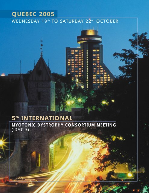
QUEBEC 2005<br />
WEDNESDAY 19 th TO SATURDAY 22 nd OCTOBER<br />
5 th INTERNATIONAL<br />
MYOTONIC DYSTROPHY CONSORTIUM MEETING<br />
(<strong>IDMC</strong>-5)
A MESSAGE FROM THE CHAIRS OF THE 5 th <strong>IDMC</strong>-5 MEETING<br />
Dear Friends,<br />
It is with great pleasure that we welcome you to the magnificent city of Quebec for the 5 th<br />
International Myotonic Dystrophy Consortium meeting.<br />
Every effort was made to include, in almost all sessions, presentations of potential interest to<br />
investigators, physicians and patients, without neglecting social issues and overall the global care of<br />
our patients. As for the previous meeting in Glasgow, there were so many abstracts submitted that we<br />
were not able to fit everybody in for oral presentations. We will be having a poster session as well.<br />
There will be numerous innovations in the course of this congress, including a special lecture on the<br />
historical aspect of <strong>myotonic</strong> <strong>dystrophy</strong> in Quebec, the participation of patients in all sessions, an<br />
interactive session between investigators, physicians and patients as well as between patients coming<br />
from different countries. A videoconference with <strong>myotonic</strong> <strong>dystrophy</strong> support group from Los Angeles<br />
will be also <strong>org</strong>anized. For the first time more than 80 patients and families will be present coming<br />
from France, Belgium, Greece, Italy, the UK, Canada and the USA. We hope that this meeting will be<br />
a great success allowing us all to learn from investigators and patients’ experiences.<br />
We would like to thank all those who actively participated in the <strong>org</strong>anization of this meeting, as well<br />
as our generous sponsors who made this congress possible.<br />
While in Quebec city, we hope that you take some time to enjoy the ambiance of the most European<br />
city in North America and that you get the opportunity to walk through the old part of town with your<br />
friends and colleagues. As for the previous meeting, the timetable allows for some socializing and<br />
discussion in a more relaxed atmosphere as well. This includes a visit to the First Nations village, a<br />
tour along the New France and St-Lawrence River road. On Saturday evening, you are invited to an<br />
exclusive private evening at the « Chapelle du Petit Séminaire ».<br />
Yours sincerely<br />
Jack Puymirat<br />
Chair, <strong>IDMC</strong>-5<br />
Christopher Pearson<br />
Co-chair <strong>IDMC</strong>-5
<strong>IDMC</strong>-5 COMMITTEES<br />
Local Organising Committee<br />
Christopher Pearson Cynthia Gagnon Jean Pierre Bouchard<br />
Genetics & Genomic Biology University of Laval Department of Neurology<br />
Hospital for Sick Children Quebec Hospital Enfant-Jésus<br />
Toronto<br />
Quebec<br />
Co-Chair <strong>IDMC</strong>-5<br />
Pascale Rousseau<br />
MDA. Canada<br />
Montreal<br />
Jack Puymirat<br />
CHU Laval Research Center<br />
Quebec<br />
Chair <strong>IDMC</strong>-5<br />
International Committee<br />
Tetsuo Ashizawa Martine Devillers Daisuke Furutama<br />
Department of Neurology Association Française contre First Department of Internal<br />
The University of Texas Medical les Myopathies Medicine<br />
Branch Departement recherche et Osake Medical College<br />
Galveton, Texas, USA des Thérapeutiques, Evry, France Osaka, Japan<br />
Nakaaki Ohsawa Shannon M. Lord Margaret Bowler<br />
Aino Institute for aging Research Hunter Research Fund Myotonic Dystrophy Support Group<br />
Ibaraki, Osaka, Japan Atlanta, USA Nottingham, UK<br />
Claude Boulier<br />
French Myotonic Dystrophy<br />
Support Group<br />
Evry, France<br />
Honorary Presidents of the <strong>IDMC</strong>-5 meeting<br />
Mr Bernard Barataud Ms Margaret Whal Ms Wyn Chivers<br />
Vice-President of the AFM Medical and Science Editor From National Executive Director of<br />
Director of the Genethon, MDA.USA MDA.Canada<br />
Evry, France<br />
SPONSORS<br />
The <strong>org</strong>anising committee is pleased to acknowledge the support of the following <strong>org</strong>anisations in funding the meeting :<br />
The AFM, MDA.USA, MDA.Canada, the Quebec Network of Applied Genetic Medicine (RMGA), Quebec Dystrophy support<br />
group, Institutes of Genetics and of Neurosciences and Mental Health of the Canadian Institutes of Health Research, Quebec<br />
minister of Health, Air Canada, Roche, Avis, Fisher Scientific, Qiagen, Sarstedt, Aetherna-Zentaris, CHU laval Research<br />
Center.<br />
The <strong>org</strong>anising committee is also pleased to acknowledge the support of the following <strong>org</strong>anisations in funding travel<br />
costs : The Association Française contre les Myopathies; the Hunter Research Fund; the Aino Institute for Aging Research.
GENERAL CONGRESS INFORMATION<br />
REGISTRATION<br />
The registration Area is located in the hall of Room Place Montcalm of lower level at the hotel Loews-<br />
Concorde from 5 h 00 to 7 h 00 pm on Wednesday October 19 th .<br />
ORAL PRESENTATIONS<br />
Each speaker will have a 7 minute period with an additionnal 3 minute for one or two questions. There<br />
will be a 30 minute period at the end of each session for discussion. Please ensure you do not overrun.<br />
The programme is packed and the session chairmen will stick rigorously to the schedule.<br />
Please note that all presentations should be in Power Point and will be projected exclusively via a<br />
centralised server. Therefore, we remind all speakers that they must go to the Speakers Ready Room<br />
with their presentation at least one hour before the session starts.<br />
POSTERS<br />
Posters boards will be located in room Suzor-Côté at third level of the hotel. Please attach your poster<br />
to the appropriated numbered board as soon as possible on arriving at the meeting. Please ensure you<br />
remove your poster at the end of the final session on Saturday afternoon.<br />
Please not that poster presenters are required to be present during the poster session on Saturday 22,<br />
12h-14h and during the poster discussion on Saturday 22, 14h-15h30. During discussion of poster, we<br />
ask you to present one slide (on Powerpoint) which summarises your work.<br />
LUNCHES<br />
Lunches will be served from Thursday to Saturday in the Hotel. Lunch times are listed on the timetable<br />
overview.<br />
Thursday : Restaurant Astral<br />
Elevator Top floor<br />
Friday :<br />
Buffet<br />
Hall third level<br />
Saturday : Buffet<br />
Hall third level<br />
Please note that teas, coffees and juices throughout the meeting will be served during break session.<br />
4 <strong>IDMC</strong>-5
LANGUAGE<br />
The official language of the 5 th <strong>IDMC</strong>-5 meeting is English. For the interactive session with physicians,<br />
investigators and patients, a simultaneous translation will be available to allow patients who do not<br />
speak English to participate to the session.<br />
SOCIAL EVENTS<br />
Opening Ceremony & Welcome Reception<br />
The opening Ceremony will take place on Wednesday October 19 th at 19 h 00 in room Place Montcalm,<br />
lower level. The ceremony will open with welcome addresses from Honorary presidents. This will be<br />
followed by a lecture on the history of <strong>myotonic</strong> <strong>dystrophy</strong> in Quebec.<br />
The Opening Ceremony<br />
will be followed by the<br />
Welcome Reception.<br />
Lunch at Astral restaurant<br />
at Loews Le Concorde<br />
A special lunch will be <strong>org</strong>anised<br />
at the Astral Restaurant on<br />
Thurday<br />
20 October, at 12 h 00. This is the<br />
crowning star of the hotel :<br />
540 feet up in the air, this<br />
revolving restaurant serves up<br />
13 miles of stunning<br />
360-degree views<br />
Visit at First Nations village<br />
A very special evening will be <strong>org</strong>anised at the First Nation village on Thursday, 20 October at 18 h 45.<br />
This visit will allow you to familiarize with songs, dance, medicine, culture, arts and crafts of the<br />
Indian Huron-Wendat Nation.<br />
Admission by ticket only.<br />
Your pre-booked<br />
will be in your<br />
registration pack.<br />
Buses will be available<br />
at front-door.<br />
QUEBEC 2005 5
Tour<br />
On Friday afternoon there will be an<br />
excursion to Quebec and its environ.<br />
Following the New France, St-Lawrence<br />
River road you will visit Montmorency Falls,<br />
Cap-Tourmante, a truly peaceful haven for<br />
snow geeses and the museum of cupper<br />
Albert Gilles<br />
Admission by ticket only. Your pre-booked<br />
will be in your registration pack.<br />
Admission by ticket only.<br />
Your pre-booked<br />
will be in your<br />
registration pack.<br />
Buses will be available<br />
at front-door.<br />
6 <strong>IDMC</strong>-5
Closing ceremony<br />
Gala Dinner on Saturday 22 October at 20 h at the<br />
Chapel of l’Amerique française Museum. There will<br />
be a musical entertainment by Masks & Bergamasques<br />
compagny. Fantastic singers, skilled<br />
pianist and <strong>org</strong>an virtuoso perform in a show that<br />
people will remember.<br />
Admission by ticket only.<br />
Your pre-booked<br />
will be in your<br />
registration pack.<br />
(Shuttle service)<br />
The Chapel is about<br />
10 minutes of step<br />
from the Hotel.<br />
QUEBEC 2005 7
PLAN<br />
8 <strong>IDMC</strong>-5
Scientific Programme Wednesday October 19 th<br />
<strong>IDMC</strong>-5<br />
17 h - 19 h : Registration<br />
Room :<br />
Place Montcalm<br />
Lower Level<br />
19 h - 19 h 30 : Opening by Honorary Presidents<br />
- Wyn Chivers, National Executive Director of MDA.Canada<br />
- Bernard Barataud, Vice-president AFM<br />
- Margaret Wahl, Medical and Science Editor from MDA.USA<br />
19 h 30 - 20 h : Special lecture<br />
A Historical Overview on DM1 in Northeastern Quebec<br />
Dr Jean-Pierre L. Bouchard, MD, FRCPC<br />
Department of Neurological Sciences<br />
CHAUQ-Hospital Enfant-Jesus<br />
Quebec, Canada<br />
20 h : Welcome Cocktail<br />
QUEBEC 2005 9
Scientific Programme Thursday October 20 th AM<br />
Room :<br />
Borduas<br />
<strong>IDMC</strong>-5<br />
Third Floor<br />
SESSION 1 : DNA INSTABILITY, DIAGNOSIS AND MODEL OF MYOTONIC DYSTROPHY<br />
Session chairs : G. Gourdon and C. Pearson<br />
8 h 30<br />
1) Cellular factors that determine (CTG)n hypermutability in DM1<br />
Wieringa, Be et al.<br />
8 h 40<br />
2) The behavior of triplet repeats in human embryonic stem cells<br />
De Temmerman N. et al.<br />
8 h 50<br />
3) The expanded CTG/CAG repeat in <strong>myotonic</strong> <strong>dystrophy</strong> type 1 continues to expand in<br />
non-dividing cells<br />
Monckton D.G. et al.<br />
9 h<br />
4) A role for DNA replication and DNA repair in CTG instability at the DM1 locus<br />
Cleary J.D. et al.<br />
9 h 10<br />
5) Prenatal diagnosis in <strong>myotonic</strong> <strong>dystrophy</strong> type 1 (DM1) : the Spanish experience<br />
Martorell L. et al.<br />
9 h 20<br />
6) Intergenerational contraction of the CTG repeats in large families with <strong>myotonic</strong><br />
<strong>dystrophy</strong>.<br />
Puymirat J. et al.<br />
9 h 30<br />
7) Assessment of in situ hybridization for molecular diagnosis of <strong>myotonic</strong> <strong>dystrophy</strong> type 2<br />
(DM2) : pattern of tissue detection and new phenotype<br />
Bassez G. et al.<br />
9 h 40<br />
8) Identification of a novel locus for <strong>myotonic</strong> <strong>dystrophy</strong> in human chromosome 16<br />
Krahe R. et al.<br />
9 h 50<br />
9) Identification of a candidate gene for a multisystem <strong>myotonic</strong> disorder with<br />
frontotemporal dementia linked to 15q21-24 chromosome (DM3)<br />
Bonifazi E. et al.<br />
10 h -10 h 20<br />
Tea/coffee break<br />
10 <strong>IDMC</strong>-5
Room :<br />
Borduas<br />
Third Floor<br />
10 h 20<br />
10) Long DM1 and DM2 repeat sequences induce gross rearrangements in flanking DNA<br />
sequences<br />
Wojciechowska M. et al.<br />
10 h 30<br />
11) Small-pool PCR study of the mutated alleles in sperm of <strong>myotonic</strong> <strong>dystrophy</strong> 1 patients<br />
Cobo A.M. et al.<br />
10 h 40<br />
12) Somatic mosaicism and improved genotype-phenotype correlations in <strong>myotonic</strong> <strong>dystrophy</strong><br />
type 1<br />
Morales F. et al.<br />
10 h 50<br />
13) The laboratory abnormalities in <strong>myotonic</strong> <strong>dystrophy</strong><br />
Heatwole C.R. et al.<br />
11 h<br />
14) Development of, and insights from models of RNA toxicity in <strong>myotonic</strong> <strong>dystrophy</strong> (DM)<br />
Mahadevan M. et al.<br />
11 h 10<br />
15) Inducible and tissue-specific expression of expanded CUG repeat RNA in transgenic mice<br />
using a cre-loxp approach<br />
Wang G.S. et al.<br />
11 h 20<br />
16) A reversible multisystemic murine model of <strong>myotonic</strong> <strong>dystrophy</strong> type 2<br />
Ranum, L.P.W. et al.<br />
11 h 30 - 12 h<br />
Discussion<br />
12 h - 14 h<br />
Lunch at Astral restaurant<br />
QUEBEC 2005 11
Scientific Programme Thursday October 20 th PM<br />
Room :<br />
Borduas<br />
Third Floor<br />
<strong>IDMC</strong>-5<br />
SESSION 2 : CELLULAR AND MOLECULAR ASPECTS OF MYOTONIC DYSTROPHY<br />
Session chairs : C Thornton and N. Ohsawa<br />
14 h<br />
17) Premature replicative arrest of congenital DM1 myoblasts<br />
Furling D. et al.<br />
14 h 10<br />
18) Telomere shortening and replicative senescence in DM1 in vitro and in vivo<br />
Xu W. et al.<br />
14 h 20<br />
19) Oxidative stress and IGF-I signaling in <strong>myotonic</strong> <strong>dystrophy</strong> type 1 (DM1) model skeletal<br />
muscle<br />
Furutama D. et al.<br />
14 h 30<br />
20) Membrane localization of DMPK splice isoforms : differences between mouse and man<br />
Van Herpen R. et al.<br />
14 h 40<br />
21) Expression of DMPK at the neuromuscular junction<br />
Wheeler T.M. et al<br />
14 h 50<br />
22) Investigating RNA-mediated neuronal dysfunction in DM1 transgenic mice<br />
Guiraud-Dogan C. et al.<br />
15 h<br />
23) Colocalization of ribonuclear inclusions and MBNL1 foci with no impairment of muscle<br />
differentiation in DM2<br />
Meola G. et al.<br />
15 h 10<br />
24) Endoplasmic reticulum stress in DM1 muscle<br />
Ikezoe K. et al.<br />
15 h 20<br />
25) Over-expression of SK channels increases sensitivity to calcium in DM1 lens cells<br />
Rhodes J.D. et al<br />
15 h 30 - 16 h<br />
Tea/coffee break<br />
12 <strong>IDMC</strong>-5
Room :<br />
Borduas<br />
Third Floor<br />
16 h<br />
26) Transcriptional profile of transgenic mice expressing an expanded CUG repeat<br />
Osborne R.J. et al.<br />
16h10<br />
27) Dysregulation of specific gene transcripts in MBNL1 knockout brain<br />
Swanson M. et al.<br />
16h20<br />
28) The role of protein-protein complexes in <strong>myotonic</strong> dystrophies 1 and 2<br />
Timchenko L. et al.<br />
16h30<br />
29) Post-translational modifications of CUG-BP1 in response to CUG repeats<br />
Kuyumcu-Martinez N.M. et al.<br />
16h40<br />
30) Analysis of candidate genes modifier for the conduction system impairment in <strong>myotonic</strong><br />
<strong>dystrophy</strong> type 1 (DM1)<br />
Vallo l. et al<br />
16h50<br />
31) Aberrant splicing of dystrophin and dystrobrevin in <strong>myotonic</strong> <strong>dystrophy</strong> type 1<br />
Nakamori M. et al.<br />
17h<br />
32) Both exon 2/3 and exon 6 of tau RNAs are mis-spliced in <strong>myotonic</strong> <strong>dystrophy</strong> type 1 but<br />
differs in their tissue-specificity alteration and regulation by etr-3<br />
Caillet-Boudin M.L. et al.<br />
17h10<br />
33) Altered mRNA splicing of the skeletal muscle ryanodine receptor and<br />
sarcoplasmic/endoplasmic reticulum Ca2+-ATPase in <strong>myotonic</strong> <strong>dystrophy</strong> type 1<br />
Kimura T. et al.<br />
17h20<br />
34) The <strong>myotonic</strong> <strong>dystrophy</strong> type 2 (DM2) CCTG expansion does not alter ZnF9 expression<br />
Ranum L.P.W. et al.<br />
17 h 30 - 18 h<br />
Discussion<br />
18 h 45 - 22 h 30<br />
Visit at the First Nation Village<br />
QUEBEC 2005 13
Scientific Programme Friday October 21 st AM<br />
Room :<br />
Borduas<br />
Third Floor<br />
<strong>IDMC</strong>-5<br />
SESSION 3 : RNA-BINDING PROTEINS<br />
Session chairs : D Brook and M Swanson<br />
8 h 30<br />
35) The structural basis of <strong>myotonic</strong> <strong>dystrophy</strong> from the crystal structure of CUG repeats<br />
Blaine H. et al.<br />
8 h 40<br />
36) Transcripts containing triplet repeat hairpins are under the surveillance of ribonuclease dicer<br />
Krzyzosiak W.J. et al.<br />
8 h 50<br />
37) Studying nuclear foci of CUG repeat mRNA in living cells<br />
Querido E. et al.<br />
9 h<br />
38) Muscleblind protein isoforms show differential RNA binding properties in a yeast three<br />
hybrid assay<br />
Vicente M. et al.<br />
9 h 10<br />
39) Role of MBNL1 in DM1<br />
Lin X. et al.<br />
9 h 20<br />
40) Elevated hnRNP-H levels contribute to abnormal IR splicing in DM1<br />
Reddy S. et al.<br />
9 h 30<br />
41) Sequestration of a novel Zn-finger protein may cause degenerative muscle defects in DM1<br />
Xu W. et al.<br />
9 h 40<br />
42) CUG-binding proteins, CUG repeats and Steinert <strong>myotonic</strong> <strong>dystrophy</strong> : what can we learn<br />
from drosophila ?<br />
Ait-Ahmed O. et al.<br />
9 h 50<br />
43) Alternative splicing of CLC-1 chloride channel is regulated by MBNLl and CELF proteins<br />
Kino Y. et al.<br />
10 h<br />
44) Zebrafish knock-down model for muscleblind-like 2<br />
Machuca-Tzili L.E. et al.<br />
10 h 10 -10 h 35<br />
Discussion<br />
10 h 35 - 10 h 50<br />
Tea/coffee break<br />
14 <strong>IDMC</strong>-5
Room :<br />
Borduas<br />
Third Floor<br />
SESSION 4 : CLINICAL ASPECTS OF MYOTONIC DYSTROPHY<br />
Session chairs : JP Bouchard and T Ashizawa<br />
10 h 50<br />
45) Analysis of annual follow-up data on 159 patients with <strong>myotonic</strong> <strong>dystrophy</strong>.<br />
Whittaker R. et al.<br />
11 h<br />
46) Myotonic <strong>dystrophy</strong> type 1 - clinical characteristcs of patients with
Scientific Programme Saturday October 22 nd AM<br />
Room :<br />
Borduas<br />
Third Floor<br />
<strong>IDMC</strong>-5<br />
SESSION 5 : CLINICAL EVALUATION AND THERAPEUTIC ASPECTS OF MYOTONIC DYSTROPHY<br />
Session chairs : J.W. Day and B Eymard<br />
9 h<br />
53) Quantitative CNS studies in adult-onset DM1 and DM2<br />
Day J.W. et al.<br />
9 h 10<br />
54) Short-term memory test in DM1 and correlations with regional cortical atrophy<br />
Antonini G. et al.<br />
9 h 20<br />
55) Sleep evaluation in the childhood type of <strong>myotonic</strong> <strong>dystrophy</strong><br />
Jacquette D. et al.<br />
9 h 30<br />
56) A sleep study of excessive daytime sleepiness in <strong>myotonic</strong> <strong>dystrophy</strong><br />
Laberge L. et al.<br />
9 h 40<br />
57) Myocardial contractility in <strong>myotonic</strong> <strong>dystrophy</strong> patients<br />
Duboc D. et al.<br />
9 h 50<br />
58) Mexiletine : effective antimyotonia treatment in <strong>myotonic</strong> <strong>dystrophy</strong> type 1 (DM1)<br />
Moxley R. et al.<br />
10 h<br />
59) Some flavonoids prevent cis- and trans- effect of expanded CTG repeats in a stable PC12<br />
cell transformants<br />
Furaya H. et al.<br />
10 h 10<br />
60) A potential gene therapy for <strong>myotonic</strong> <strong>dystrophy</strong> type 1<br />
Puymirat J. et al.<br />
10 h 20 -10 h 50<br />
Tea/coffee break<br />
16 <strong>IDMC</strong>-5
Room :<br />
Borduas<br />
Third Floor<br />
10 h 50<br />
61) A model of distribution of a mutation in DMPK gene associated with <strong>myotonic</strong> <strong>dystrophy</strong><br />
type 1<br />
Akhmadeeva L. et al.<br />
11 h<br />
62) Measuring health related quality of life (HRQl) with the resources of the NIH registry of<br />
<strong>myotonic</strong> <strong>dystrophy</strong>(DM) and FSHD patients and family members<br />
Hilbert J. et al.<br />
11 h 10<br />
63) Are questionnaires an applicable investigation tool in relation to DM patients?<br />
Schwartzbach I.<br />
11 h 20<br />
64) Social participation of patients with <strong>myotonic</strong> <strong>dystrophy</strong> type 1<br />
Gagnon C. et al.<br />
11 h 30<br />
65) Parents’ experiences of a diagnosis of DM : findings from a qualitative interview study<br />
Downing C.<br />
11 h 40-12 h 10<br />
Discussion<br />
12 h 10-14 h<br />
Lunch and poster session<br />
Room : Suzor-Côté, Third Floor<br />
QUEBEC 2005 17
Scientific Programme Saturday October 22 nd PM<br />
<strong>IDMC</strong>-5<br />
14 h - 15 h 30 : Poster discussion<br />
Session chairs : Meola G. and Moxley R.<br />
Room : Borduas, Third Floor<br />
15 h 30 - 16 h : Tea/coffee break<br />
16 h - 18 h : INTERACTIVE SESSION WITH INVESTIGATORS, PATIENTS AND THEIR FAMILIES<br />
SESSION CHAIRS : P HARPER AND J PUYMIRAT<br />
66) Myotonic Dystrophy. Patient Literature in Different Languages<br />
Harper P.S.<br />
Room : Borduas, Third Floor<br />
18 h : Film "Tomorrow Belongs to Me" presented by Jacqueline Donachie<br />
20 h : Gala Dinner<br />
Room : Chapel "Musée de l'Amérique Française"<br />
18 <strong>IDMC</strong>-5
Programme for people living with <strong>myotonic</strong> <strong>dystrophy</strong><br />
Friday October 21 st PM<br />
<strong>IDMC</strong>-5<br />
18 h 30 : American support group <strong>org</strong>anizational meeting and Buffet dinner<br />
Room : Borduas, Third Floor<br />
Saturday October 22 nd<br />
11 h 00 : Canadians Meeting<br />
Room : 415, 4 th Floor<br />
12 h : Lunch<br />
Rooms : Krieghoff and Borduas<br />
13 h : Discussion workshop between participants from all over the world<br />
(Canada, France, United States, England, Italia and Greece)<br />
living with <strong>myotonic</strong> <strong>dystrophy</strong><br />
Rooms : Krieghoff<br />
15 h 30 : Coffee break<br />
16 h : Clinicians and researchers present to participants their conclusions brought<br />
by discussions with other clinicians and researches during the Consortium,<br />
followed by a period of questions<br />
Rooms : Krieghoff and Borduas<br />
18 h : Short film presentation of "Tomorrow Belongs to Me"*<br />
Room : Krieghoff and Borduas<br />
18 h 30 : End of the session<br />
20 h : Gala Dinner<br />
At the Chapel of the "Musée de l’Amérique Française"<br />
QUEBEC 2005 19
ORAL PRESENTATIONS<br />
QUEBEC 2005 21
Abstract No : 1<br />
ORAL PRESENTATIONS<br />
CELLULAR FACTORS THAT DETERMINE (CTG)n<br />
HYPERMUTABILITY IN DM1<br />
Van Den Broek W., Wansink D.G. and Wieringa B.<br />
Dept. Cell Biology, NCMLS, Radboud University Medical Centre, Nijmegen<br />
Geert Grooteplein 28, 6525 GA Nijmegen, The Netherlands<br />
Cellular mechanisms involved in somatic (CTG)n repeat expansion during growth and<br />
ageing of DM1 patients are still ill understood. We have generated a transgenic knockin<br />
mouse model with a (CTG)84 repeat placed at the correct position in the 3’ UTR of the<br />
endogenous DMPK gene (van den Broek et al. (2002) Hum.Mol.Genet. 11, 191-8). Repeat<br />
instability - now expanded to (CTG)116 over successive generations – in this mouse lineage<br />
is dependent on activity of the Msh2-Msh3 DNA mismatch repair machinery, particularly<br />
prominent and progressive upon ageing in kidney, liver, stomach and small<br />
intestine, and cell-type and -state dependent. We now demonstrate that the highest<br />
degree of somatic hypermutability occurs in cells of liver and pancreas that have undergone<br />
polyploidization (change of 2n to ≥ 4 n DNA content) as the last phase of<br />
terminal differentiation. We will discuss possible mechanisms involved and present preliminary<br />
data about specific changes in the DNA repair machinery that may be associated<br />
with enhanced repeat expansion in cells with ≥ 4n DNA content. In addition,<br />
we will give an update on findings regarding the effects of endonuclease FEN-1<br />
deficiency in early embryos with the (CTG)116 transgene.<br />
22 <strong>IDMC</strong>-5
Abstract No : 2<br />
ORAL PRESENTATIONS<br />
THE BEHAVIOR OF TRIPLET REPEATS IN HUMAN<br />
EMBRYONIC STEM CELLS<br />
De Temmerman N., Sermon K., De Rycke M., Liebaers I. and Van Steirteghem A.<br />
VUB, Laarbeeklaan 101, 1090 Brussel, Belgium<br />
The stability of the CTG repeat in DMPK was examined in the human embryonic stem<br />
(hES) cell line VUB03_DM1. This line was derived from a blastocyst obtained after PGD<br />
in which the affected mother carried 120 repeats. There was a generational enlargement<br />
of the repeat to 470 repeats, assumed to have been already present in the oocyte. The<br />
size of the CTG repeat was determined in cells from different colonies as well as in cells<br />
from the same colony, using a specific long-CTG PCR. In the early passages of this fast<br />
replicating cell line the repeat remained stable, showing 470 repeats in the majority of<br />
the cells analysed. At certain passages contractions (passage 4) and enlargements were<br />
present in some cells. In following passages (> 20 passages), mainly enlargements of the<br />
repeat were detected, showing an increase in repeat size in the culture ranging from 470<br />
repeats at passage 1 to 600 and 730 repeats at passages 27 and 48, respectively. The different<br />
repeat sizes were present as well between different colonies as within a colony.<br />
HES cells are regarded as a model to study developmental processes in preimplantation<br />
embryos, but the behaviour of the unstable CTG repeat in the hES cells of the<br />
VUB03_DM1 line resembles more the repeat instability in gametes than in cleavage<br />
embryos, in which the repeat was shown to remain stable. As described in other mitotically<br />
active tissue, the instability of the CTG repeat in hES cells is probably caused by<br />
replication slippage, although the role of repair enzymes and the effect of a prolonged<br />
culture cannot be excluded.<br />
QUEBEC 2005 23
Abstract No : 3<br />
ORAL PRESENTATIONS<br />
THE EXPANDED CTG/CAG REPEAT IN MYOTONIC<br />
DYSTROPHY TYPE 1 CONTINUES TO EXPAND IN<br />
NON-DIVIDING CELLS<br />
Monckton D.G., Hilley J.D. and Gomes-Periera M.<br />
Institute of Biomedical and Life Sciences, University of Glasgow, Glasgow, UK.<br />
It is widely assumed that the mechanism of expansion of CTG/CAG repeats in <strong>myotonic</strong><br />
<strong>dystrophy</strong> type 1 is DNA replication slippage. This model predicts that tissues/cells with<br />
higher rates of cell turnover will have higher rates of expansion. In vivo data from<br />
humans and transgenic mouse models shows no obvious correlation between tissue-specific<br />
expansion rates and proliferative capacity. Moreover, expansions are observed to<br />
accumulate in apparently postmitotic tissues such as skeletal muscle and brain.<br />
However, tissues are complex structures comprised of multiple cell types with differing<br />
proliferative capacities and rates of cell type-specific expansion. Previously, using a<br />
transgenic mouse cell culture model we demonstrated that tissue specific rates of expansion<br />
are conserved in vitro, but cannot be accounted for by differences in the rate of<br />
expansion. These data demonstrated that cell division was not sufficient to drive expansions,<br />
but left open the possibility that cell division might be required to generate<br />
expansions. In order to test this hypothesis, we have used multiple approaches to induce<br />
cell cycle arrest in our tissue culture model. Using chemical inhibitors of cell division,<br />
such as apicidin and mitomycin-C, and overexpression of cell cycle control genes, such<br />
as p16 and p21, we provide definitive proof that cell division is not necessary for expansion.<br />
Indeed rates of expansion in non-dividing cells are at least as great as, if not<br />
greater than, those in dividing cells, strongly arguing against any significant role for<br />
replication slippage in mediating expansions.<br />
24 <strong>IDMC</strong>-5
Abstract No : 4<br />
ORAL PRESENTATIONS<br />
DNA REPLICATION & REPAIR IN CTG INSTABILITY<br />
AT THE DM1 LOCUS<br />
Cleary J.D., Hagerman K., Panigrahi G.B., Lau R., Blondin M. and Pearson C.E.<br />
Sickkids Research Institute, The University of Toronto, Toronto, Canada<br />
In DM1 patients, CTG instability occurs in proliferating and quiescent cells, varying<br />
between tissues and developmental stage, suggesting complex mutation mechanism(s).<br />
Instability is thought to involve both the formation and aberrant processing of slipped-<br />
DNAs at the repeats in non-replicating or replicating DNA. DM1 patient fibroblasts<br />
showed spontaneous CTG expansions only in proliferating cells. Non-proliferating cells<br />
did not show CTG expansions. Treatment of cells with compounds that perturbed replication<br />
fork progression enhanced expansions-indicating a role for replication fork errors<br />
and their aberrant repair. Addressing both replication and repair we 1) determined the<br />
DNA replication profile at the DM1 chromosomal locus in DM1 patient fibroblasts in<br />
which CTG tracts were actively expanding. The DM1 locus is located within a region of<br />
abundant replication activity, with replication initiation occurring proximal to the<br />
repeat tract. We also 2) established an in vitro assay to determine the fidelity of slipped-<br />
DNA processing by human cell extracts. Various repair outcomes arose depending on the<br />
slipped-DNA structure. Repair outcomes could be mediated by various extracts including<br />
non-proliferating neuron-like cells, and were independent of the MSH2, MSH3, MLH1,<br />
XPF, and XPG repair proteins-suggesting they may be involved upstream of processing.<br />
Taken together these results suggest, at least for proliferating DM1 fibroblasts, that<br />
replication at the repeat tract may contribute to the formation of mutagenic DNA intermediates,<br />
and their aberrant repair may explain the spontaneous CTG expansions.<br />
QUEBEC 2005 25
Abstract No : 5<br />
ORAL PRESENTATIONS<br />
PRENATAL DIAGNOSIS IN MYOTONIC DYSTROPHY TYPE 1 (DM1) :<br />
THE SPANISH EXPERIENCE<br />
Martorell L. (1) , Cobo A.M. (2) , Parra J. (3) , Naudó M. (1) and Baiget M. (3)<br />
1) Genètica Molecular, Hospital Sant Joan de Déu, Edifici Docent; C/Santa Rosa 39<br />
08950- Barcelona, Spain<br />
2) Genetics, Hospital Donostia, San Sebastián, Spain<br />
3) Genetics, Hospital Sant Pau, Barcelona, Spain<br />
Genetic counselling for <strong>myotonic</strong> <strong>dystrophy</strong> is difficult and complex, owing to the<br />
extreme variability of the disorder, in both severity and age of onset, the anticipation<br />
and the influence of the sex of the affected parent.<br />
We have carried out 130 prenatal diagnosis(PND), most of which (70 %), the mother is<br />
the transmitting progenitor, with expansions ranging from 65 -1166 CTG. The remaining<br />
30 % of PNDs are of paternal origin, ranging between 42-600 CTG. 48 % of PND where<br />
positive for the DM1 mutation, with expansions between 45 - 3666 CTGs in foetuses. In<br />
maternal transmissions we detected a predominance of expansions, with few contractions<br />
or no-variations. All the congenital cases are of maternal origin, and according to<br />
our data, if a female has one congenital infant, all her next affected descendants will<br />
be congenital cases. The contractions or small variations are predominant in paternal<br />
transmissions, although, the foetuses inherited alleles clearly into the pathological<br />
range.<br />
26 <strong>IDMC</strong>-5
Abstract No : 6<br />
ORAL PRESENTATIONS<br />
INTERGENERATIONAL CONTRACTION OF THE CTG REPEATS<br />
IN LARGE FAMILIES WITH MYOTONIC DYSTROPHY<br />
Puymirat J. (1) , Guigère Y. (1) , Lopez De Munain A. (2) , Cobo A.M. (2) , Bost M. (3) ,<br />
Ollagnon-Roman E. (4) and Vial C. (5)<br />
1) Unit of Human Genetics & Dept of Medical Biology, CHUQ, Quebec, Canada<br />
2) Department of Genetics & Neurology, Hospital Donostia, San Sebastian, Spain<br />
3) Laboratory of Neurogenetics, Hospital Debrousse, Lyon, France<br />
4) Unit of Neurogenetics, Hospital Croix Rousse, Lyon, France<br />
5) Department of electromyography, Neurological hospital, Lyon, France<br />
The frequency of CTG contraction during transmission from the father to the child has<br />
been estimated about 2 %, 2.8 % and 7 % in the French, Basque and French-Canadian<br />
population, respectively. In 6 of families originating from Rhone-Alpe French, French-<br />
Canadian and « Basque » DM1 population, the number of CTG repeats decreased during<br />
transmission from the father to all of their DM1 offspring’s. This was associated with<br />
regression in clinical manifestations in 75 % of cases. In one of these families, two<br />
brothers with 650 and 500 repeats had all their DM1 offspring’s with CTG repeat contractions,<br />
one has two children with 210 repeats each whereas the second had 2 children<br />
with 160 and 350 repeats, respectively. In all of these families, there was no case of<br />
regression associated with expansion during transmission from parent to child. These<br />
observations indicate that the paternal origin of the repeat and the presence of the<br />
repeat contraction in a sibling greatly increase the probability of the CTG repeat contraction<br />
in others siblings and that it may exist a familial paternal factor that restricts<br />
the expansion.<br />
QUEBEC 2005 27
Abstract No : 7<br />
ORAL PRESENTATIONS<br />
ASSESSMENT OF IN SITU HYBRIDIZATION FOR MOLECULAR<br />
DIAGNOSIS OF MYOTONIC DYSTROPHY TYPE 2 (DM2) :<br />
PATTERN OF TISSUE DETECTION AND NEW PHENOTYPE<br />
Bassez G., Chapoy E., Bergemer A.M., Pellissier J.F., Radvanyi-Hoffman H.,<br />
Gherardi R.K. and Eymard B.<br />
Service d'Histologie - Hôpital Henri Mondor, Creteil, France<br />
Whereas genetic diagnosis of <strong>myotonic</strong> <strong>dystrophy</strong> type 1 DM1 (DM1) is routinely available,<br />
limitations in conventional molecular genetic methods for DM2 mutation demonstration<br />
have been encountered. Recently, chromogenic in situ hybridization (CISH)<br />
have been reported as a new approach for the detection of DM2 expansion mutation in<br />
muscle. We evaluate the feasibility and efficiency of CISH for routine molecular diagnosis<br />
of DM2 in skeletal muscle and other tissues in frozen and paraffin embedded biopsy<br />
specimen of patients with : (1) evidenced DM2 mutation, (2) DM2 mutation excluded,<br />
(3) DM1 mutation, (4) controls with no available genetic test. All patients with identified<br />
DM2 mutation were positive by CISH, whereas no nuclear signal was observed in<br />
DM1 and normal controls. This allowed us the retrospective positive confirmation of DM2<br />
in ancient specimen up to 17 years of age and identification of two unrelated cases with<br />
camptocormia as new phenotypic presentation. Further-more, we detected positive<br />
nuclei in cells from various tissues, including digestive tract, skin, adipocytes, and<br />
smooth muscle, and at low level in peripheral nerve and epithelial cells. Conclusion :<br />
Targeting mutant transcripts in tissue section by CISH approach seems to represent a<br />
valuable tool for molecular screening of DM2 leading to potential identification of new<br />
phenotype. Furthermore, the positive detection in nuclei of non muscle cells may offer<br />
new diagnostic opportunity.<br />
28 <strong>IDMC</strong>-5
Abstract No : 8<br />
ORAL PRESENTATIONS<br />
IDENTIFICATION OF A NOVEL LOCUS FOR MYOTONIC<br />
DYSTROPHY IN HUMAN CHROMOSOME 16<br />
Krahe R. (1) , Lehmann-Horn F. (2) , Jurkat-Rott K. (2) , Gamez J. (3) , Raskin S. (4) ,<br />
Schoser B.G. (5) and Bachinski L.L. (1)<br />
1) Sect. of Cancer Genetics, Univ. of Texas M.D. Anderson Cancer Ctr., Houston, TX, USA<br />
2) Dept. of Applied Physiology, University of Ulm, Germany<br />
3) Dept. of Neurology, General Hospital Vall d'Hebron University, Barcelona, Spain<br />
4) Genetics Laboratory, Hospital de Clinicas da Universidade Federal do Paraná, Brazil<br />
5) Dept. of Neurology, Ludwig Maximilians University Munich, Germany<br />
Myotonic <strong>dystrophy</strong> types 1 and 2 (DM1 and DM2) are caused by expansions of (CTG)n<br />
and (CCTG)n repeats in DMPK (in 19q13.3) and ZNF9 (in 3q21.3), respectively. A third<br />
DM locus was recently mapped to chromosome 15q21-q24, but the mutation remains<br />
unknown. During the course of a 10-cM genome scan to identify the DM2 locus, several<br />
families were identified that had no expansions in DMPK or ZNF9 and whose disease did<br />
not segregate with these loci. Among these families was one that showed a LOD score of<br />
>1.3 in chromosome 16. To demonstrate whether this LOD score represented a novel<br />
locus, we genotyped additional markers in chromosome 16 in this and other families segregating<br />
autosomal dominant progressive <strong>myotonic</strong> myopathy, which had also tested<br />
negative for expansions at the DM1 and DM2 loci. Using 9 such families (4 German, 4<br />
Spanish, and 1 Brazilian), we obtained a LOD score of 3.83 in the p-arm of chromosome<br />
16. We conducted haplotype analysis to determine the minimal region shared by these<br />
9 families. Hypothesizing that DM in these families may be due to an expanded repeat<br />
of the type (CTG)n, (CCTG)n, or other similar sequence (xCTG)n , we interrogated all such<br />
repeats in the shared region with _4 repeat units. Repeats were examined by amplification<br />
across the repeat to check for non-Mendelian inheritance, a hallmark of expanded<br />
repeat diseases, and by a repeat-primed PCR assay (RP-PCR) to identify expansions. None<br />
of the candidate repeats showed an expansion or non-Mendelian inheritance. Since most<br />
of these families are small, each contributes only a small positive amount to the total<br />
LOD score. However, in all cases, the LOD scores observed are near the simulated<br />
ELODmax. Nevertheless, the small family size makes it impossible to exclude locus heterogeneity<br />
within this set of families, thus complicating precise localization of the<br />
linked region. We conclude that chromosome 16 harbors a locus that is responsible for<br />
DM in most of these kindreds. We are continuing the search for the causative mutation<br />
by interrogating additional repeat sequences in the regions surrounding the shared<br />
region.<br />
QUEBEC 2005 29
Abstract No : 9<br />
ORAL PRESENTATIONS<br />
IDENTIFICATION OF A CANDIDATE GENE FOR A MULTISYSTEM<br />
MYOTONIC DISORDER WITH FRONTOTEMPORAL DEMENTIA<br />
LINKED TO 15Q21-24 CHROMOSOME (DM3)<br />
Bonifazi E., Vallo L., Contino G., Iraci R., Rinaldi F., Botta A. and Novelli G.<br />
Genetics Unit, Tor Vergata University, Via Montpellier 1, 00133 Rome, Italy<br />
Myotonic dystrophies (DM) are a genetically heterogeneous group of diseases, DM1 and<br />
DM2, that result respectively from dynamic mutations in the DMPK and ZNF9 genes.<br />
There are patients with characteristic DM clinical feature but wild type at loci DM1 and<br />
DM2. These cases suggest the hypothesis of another DM locus. Recently, a third locus<br />
(DM3) associated with a <strong>myotonic</strong> phenotype, was mapped to chromosome 15q21-24 (Le<br />
Ber et al., 2004) in large pedigree with a non-DM1 non-DM2 multisytem disorder associated<br />
with frontotemporal dementia. We identified in this region a gene, ZNF291<br />
(GeneID : 49855) that possesses an unstable [CCTG]n repeat in the second intron.<br />
Analysis of 50 normal subjects shows the variability of the [CCTG]n repeat in ZNF291<br />
gene between 5 and 15.<br />
To test the ZNF291 expression in tissues involved in the DM3 pathogenesis, we used a<br />
RT-PCR based approach and we observed ZNF291 gene expression in different areas of<br />
human brain, skeletal muscle and heart. Our findings indicate that ZNF291 is a potential<br />
candidate gene for the multisystemic <strong>myotonic</strong> disorder with frontotemporal dementia<br />
phenotype.<br />
30 <strong>IDMC</strong>-5
Abstract No : 10<br />
ORAL PRESENTATIONS<br />
LONG DM1 AND DM2 REPEAT SEQUENCES INDUCE GROSS<br />
REARRANGEMENTS IN FLANKING DNA SEQUENCES<br />
Wojechowska M., Bacolla A., Dere R., Hebert M., Kosmider B., Larson J. E.,<br />
Napierala M., Son L.S. and Wells R.D.<br />
Institute of Biosciences and Technology, Texas A&M University System Health Science Center,<br />
2121 W. Holcombe Blvd., Houston, TX 77030-3303, USA<br />
The molecular mechanisms responsible for the genetic instabilities (expansions and contractions)<br />
of repeating tri- and tetranucleotide tracts, which are involved in the etiology<br />
of certain neurological diseases, are under investigation. Replication, recombination,<br />
and several repair mechanisms (including the involvement of DSBs) have been implicated<br />
in the bacterial and mammalian cell studies. We have recently discovered that long<br />
tracts of CTG/CAG, CCTG/CAGG, or CGG/CCG repeats cause complex rearrangements,<br />
including inversions and gross deletions, spanning several Kbp of flanking sequences.<br />
Investigations in E. coli, COS-7, and HEK-293 cells show that longer tracts (full mutations)<br />
are most potent, and the orientation of the repeats and their transcriptional status<br />
influence the mutagenic potential as well as the types of products. The locations of<br />
the breakpoints coincided with structurally contorted regions of the putative non-B DNA<br />
conformations (cruciforms, slipped structures, triplexes, tetraplexes) in these sequences.<br />
The consequences of these discoveries for DM1 and DM2 patients with full mutations<br />
remain to be elucidated but may have profound implications for the integrity of their<br />
gene products. (Supported by N.I.H, Robert A. Welch Foundation, Friedreich’s Ataxia<br />
Research Alliance, and Muscular Dystrophy Association.)<br />
QUEBEC 2005 31
Abstract No : 11<br />
ORAL PRESENTATIONS<br />
SMALL-POOL PCR STUDY OF THE MUTATED ALLELES<br />
IN SPERM OF MYOTONIC DYSTROPHY1 PATIENTS<br />
Cobo A.M. (1) , Poza J.J. (2) and Lopez De Munain A. (2)<br />
1) Genetics Unit<br />
2) Neurology Dept. Hospital Donostia, San Sebastian 20014, Spain<br />
The individual expansion in each DM1 patient measured in blood consists in multiple<br />
size alleles defined as somatic heterogeneity. The latter is minimal at birth and increases<br />
over time in the DM1 patients. On the other hand, DM1 males show extreme variability<br />
in the allele distribution in germline, regardless of their expansion size in blood. The<br />
increase of the expansion size in the majority of the intergenerational transmissions<br />
could be due to a selection bias in transmission, towards the highest size range of the<br />
paternal alleles.<br />
We have analysed the expansion size in the sperm of 26 DM1 males by small-pool PCR<br />
(SP-PCR) to establish the allele distributions in DM1 male sperm and try to determine if<br />
a selection bias of the mutated alleles exists. As reported previously, a higher heterogeneity<br />
was seen in sperm than in blood. The distribution of the mutated alleles was<br />
homogeneous and no apparent excess of the upper size mutated repeats was observed<br />
except for one patient who showed more heterogeneity in blood (blood range 315-833,<br />
315-420 in sperm). However, the major finding of our study is the high proportion of<br />
azoospermic patients (11/26, 42 %) in our series. This is observed when the expansion<br />
size in blood is ≥500 CTG except for two premutated patients. Furthermore, the<br />
upper size limit of mutated alleles present in sperm is 915 CTG in our patients, which<br />
could explain the nearly exclusive maternal transmission of the congenital form. Our<br />
findings make difficult a prediction of the severity of the affected offspring based on<br />
sperm SP-PCR results.<br />
32 <strong>IDMC</strong>-5
Abstract No: 12<br />
ORAL PRESENTATIONS<br />
SOMATIC MOSAICISM AND IMPROVED GENOTYPE-PHENOTYPE<br />
CORRELATIONS IN MYOTONIC DYSTROPHY TYPE 1<br />
Morales F., Cuenca P., Del Valle G., Brian R., Sittenfeld M. and G. Monkton D.G.<br />
Institute of Biomedical and Life Sciences, University of Glasgow, UK<br />
Somatic mosaicism in DM1 is age-dependent, tissue-specific and expansion-biased, features<br />
that very likely contribute toward the tissue-specificity and progressive nature of<br />
the symptoms. Importantly, a failure to account for the age-dependence and expansionbiased<br />
nature of somatic mosaicism has greatly compromised genotype-phenotype correlations.<br />
Previously, Southern blot analyses of restriction-enzyme digested genomic<br />
DNA have revealed that measured allele length typically accounts for less than 50 % of<br />
the variation in age of onset. These data leave open the possibility that other factors<br />
(genetic/environmental) might be more important in determining age of onset. In order<br />
to improve the prognostic value of genotype-phenotype relationships in DM1, we have<br />
used small pool PCR to define in detail the degree of somatic mosaicism in a cohort of<br />
Costa Rican patients. Using these data to estimate the progenitor allele length and an<br />
improved statistical analysis, we found that a logarithmic transformation of the inherited<br />
allele length accounts for >75 % of the variation in age of onset (r=0.87, p
Abstract No : 13<br />
ORAL PRESENTATIONS<br />
LABORATORY TEST ABNORMALITIES IN MYOTONIC DYSTROPHY<br />
Heatwole C.R., MD, Miller J., MD PhD, Martens B. and Moxley R.T., III MD<br />
The University of Rochester, Rochester (NY), USA, 14620<br />
Background : Myotonic <strong>dystrophy</strong> type-1 is the most prevalent form of adult muscular<br />
<strong>dystrophy</strong> worldwide. Although well known for the classic manifestations of myotonia,<br />
weakness and early cataracts, it has broad reaching effects on multiple <strong>org</strong>an systems.<br />
Objective : To analyze and compile the laboratory abnormalities of 126 adult <strong>myotonic</strong><br />
<strong>dystrophy</strong> type-1 (DM1) patients.<br />
Patients : Baseline preclinical-trial laboratory data was compiled from 126 medically<br />
healthy, mild to moderately affected, ambulatory, DM1 patients with good venous access<br />
from 12 major clinical trials between the years 1975 and 2005. These data include values<br />
for 45 different laboratory tests, and 2860 total studies.<br />
Results : Of the 2860 laboratory studies, 470 (16 %) were outside the normal standardized<br />
parameters. Of the 45 types of laboratory tests studied in DM1 patients, 41 demonstrated<br />
abnormalities. The prevalence of an outlier laboratory value was greater than<br />
50 % in several tests including : lactate dehydrogenase (LD), hemoglobin A1c (HgA1c),<br />
follicle stimulating hormone (FSH), and luteinizing hormone (LH) in men, and gammaglutamyltransferase<br />
(GGT), creatine phosphokinase (CK), and hemoglobin (HB) in<br />
women.<br />
Conclusions : There is a high frequency of abnormal laboratory values in DM1 which<br />
may provide a basis for early screening and monitoring as well as provide insight into<br />
the spectrum of tissues involved in this disease.<br />
34 <strong>IDMC</strong>-5
Abstract No : 14<br />
ORAL PRESENTATIONS<br />
DEVELOPMENT OF, AND INSIGHTS FROM MODELS<br />
OF RNA TOXICITY IN MYOTONIC DYSTROPHY (DM)<br />
Mahadevan M., Yu Q., Yadava R., Frenzel-McCardel C. and Yarasi S.<br />
University of Virginia, Department of Pathology, 415 Lane Rd., MR5 Building, Rm. 3330, PO, USA<br />
Myotonic <strong>dystrophy</strong> (DM) is the most common inherited neuromuscular disorder in<br />
adults. There are two types, DM1 and DM2, both being autosomal dominant disorders<br />
caused by expansions of microsatellite repeats within non-coding regions of their<br />
respective genes. DM1 is far more common; however both forms of DM are likely to share<br />
similar pathogenic mechanisms. The DM1 mutation is an expansion of a CTG triplet<br />
repeat in the 3’ untranslated region (3’UTR) of the DM protein kinase (DMPK) gene. A<br />
prevailing hypothesis in the field is that many aspects of DM are caused by the expression<br />
of the mutant mRNA. DM1 and DM2 represent the first examples of toxic RNA mediated<br />
disease pathogenesis. We have already developed and characterized extensively, a<br />
myoblast cell culture model with which we were one of the first groups to clearly demonstrate<br />
the toxic effects of the mutant DMPK mRNA on muscle differentiation. To study<br />
the hypothesis further, we are trying to develop and characterize additional models of<br />
RNA toxicity for DM1. Progress and insights from ongoing efforts will be presented.<br />
QUEBEC 2005 35
Abstract No : 15<br />
ORAL PRESENTATIONS<br />
INDUCIBLE AND TISSUE-SPECIFIC EXPRESSION OF<br />
EXPANDED CUG REPEAT RNA IN TRANSGENIC MICE USING<br />
A CRE-LOXP APPROACH<br />
Wang G.S., Bundmand D. and Cooper T.A.<br />
Baylor College of Medicine, Houston, TX, USA<br />
We developed a transgenic mouse model for <strong>myotonic</strong> <strong>dystrophy</strong>, Type 1 (DM1) by<br />
inducibly expression of expanded CUG RNA using a Cre-loxP approach. The transgene<br />
(EpA960) contains a CMV promoter, a floxed concatemer of the SV40 polyadenylation<br />
site, and DMPK exon 15 containing 960 interrupted CTG repeats. Expression of expanded<br />
CUG repeat RNA is induced by breeding EpA960 transgenic animals with a Cre-expressing<br />
strain. Five founder lines were obtained which express different levels of EpA transgene<br />
mRNA. To test the mouse model in heart, three EpA lines were crossed with mice<br />
expressing heart-specific and tamoxifen-inducible Cre. Bitransgenic mice expressing the<br />
highest level of EpA960 mRNA died within one week after tamoxifen administration<br />
while the two lower expressing lines remained viable. To validate that this bitransgenic<br />
mouse model, we examined genes that have been shown to undergo mis-regulated alternative<br />
splicing in individuals with DM1. Cardiac troponin T exons 4 and 5 were included<br />
in bitransgenic animals as found in DM1 cardiac tissue, whereas the inclusion was low<br />
in wild type and mock-treated animals. We found that splicing of Fragile-X-related protein<br />
1 exons 15 and 16 reverted to an embryonic pattern in both DM1 and bitransgenic<br />
animal cardiac tissue. Nuclear RNA foci were detected in heart from bitransgenic animals<br />
by in situ hybridization. Histological and functional examinations are in progress. The<br />
EpA lines provide a versatile system to express mRNA containing long CUG repeats in the<br />
context of DMPK exon 15 in a variety of tissues and developmental stages.<br />
36 <strong>IDMC</strong>-5
Abstract No : 16<br />
ORAL PRESENTATIONS<br />
A REVERSIBLE MULTISYSTEMIC MURINE MODEL<br />
OF MYOTONIC DYSTROPHY TYPE 2<br />
Margolis J.M., Kang Y.L., Zu T., Day J.W. and Ranum L.P.W.<br />
MMC 206, 420 Delaware St. SE, University of Minnesota, Minneapolis, MN 55455, USA<br />
We previously showed that <strong>myotonic</strong> <strong>dystrophy</strong> type 2 (DM2) is caused by a large CCTG<br />
repeat expansion in intron 1 of the zinc finger protein 9 (ZNF9) gene. There is growing<br />
evidence that DM1 and DM2 are caused by an RNA gain of function mechanism in which<br />
the CUG and CCUG expansions cause the multisystemic features of these diseases by<br />
directly altering RNA processing. To investigate the role that expression of the CCUG<br />
transcripts play in the multisystemic DM phenotype, and to avoid problems such as<br />
infertility that have complicated previous systemic models of DM1, we have developed<br />
a tetracycline inducible murine model of the DM2 CCUG expansion. In our expansion<br />
lines, the expression of a CCTG repeat tract with 300 repeats is driven by a tet-responsive<br />
(TRE)/minimal CMV promoter as part of a non-coding transcript. These TRE-CCTG<br />
expansion lines have been crossed to tet-activator (tTA) mice in which the tTA is controlled<br />
by a CMV promoter. Doubly transgenic mice TRE-CCTG / CMV-tTA mice ubiquitously<br />
express CCUG expansion transcripts in the absence of doxycycline. Routine H&E staining<br />
of muscle from these doubly transgenic animals shows variation in fiber size and<br />
centrally located nuclei indicative of myopathy. Additionally, skeletal muscle shows<br />
ribonuclear inclusions and aberrant splicing of the insulin receptor (IR) by RT-PCR analysis,<br />
which has not previously been reported in mouse models of DM1. Like DM1 and DM2<br />
patient biopsies, the predominant IR isoform in muscle from these mice excludes exon<br />
11 which leads to a preferential expression of the insulin insensitive isoform. Further,<br />
aberrant splice patterns of cTNT are observed by RT-PCR from cardiac muscle indicating<br />
that the CCUG expansion transcripts in this model affect both cardiac and skeletal muscle.<br />
In a separate series of experiments the TRE-CCUG expansion lines described above<br />
will be crossed to two different BAC transgenic lines engineered to drive expression of<br />
the tTA using the endogenous human DMPK or ZNF9 promoters. Phenotypic characterization<br />
of these doubly transgenic mice will help determine the extent to which the clinical<br />
similarities and differences, including developmental differences between DM1 and<br />
DM2, result from differences in the temporal and spatial expression patterns of the<br />
repeat containing transcripts driven by the ZNF9 and DMPK promoters.<br />
QUEBEC 2005 37
Abstract No : 17<br />
ORAL PRESENTATIONS<br />
PREMATURE REPLICATIVE ARREST<br />
OF CONGENITAL DM1 MYOBLASTS<br />
Furling D., Bigot A., Mouly V. and Butler-Browne G.<br />
CNRS FRE2853, Faculté de Médecine UPMC, 105 boulevard de l’hôpital, 75013 Paris<br />
Impairment in skeletal muscle development represents one of the main features in the<br />
congenital form of DM1 (CDM) that is associated with large CTG expansions. Using an in<br />
vitro model of primary satellite cell cultures isolated from the muscles of CDM fetuses,<br />
we have previously shown that the large CTG repeats affect the differentiation program<br />
of these myogenic precursor cells. Molecular alterations associated to the CTG mutation<br />
were found in this cellular model as shown by the presence of nuclear foci of DMPK<br />
mutant transcripts, splicing defects and somatic instability of the CTG expansion. We<br />
showed that the proliferative capacity of the CDM myoblasts was also significantly<br />
reduced when compared to normal cells. In human, the life span of the somatic cells is<br />
limited by a mechanism of replicative senescence. The mitotic clock determined by the<br />
telomere length was showed to play a major role in this process. However we demonstrated<br />
that the proliferative arrest of CDM myoblasts is not correlated with an excessive<br />
reduction of the telomere length since the CDM myoblasts stop dividing with larger<br />
telomeric DNA restriction fragments than control myoblasts. This result clearly indicates<br />
that the CDM myoblasts have not exhausted their proliferative capacity but have<br />
a premature replicative arrest. Activation of the cyclin-dependent kinase inhibitor p16<br />
is another regulatory pathway of replicative senescence in some cell types. We showed<br />
that the p16 pathways is involved in cell cycle arrest of senescent human myoblasts and<br />
a similar induction of p16 was measured in CDM myoblasts with premature arrest. We<br />
suggest that a cellular stress induced by the large CTG mutation triggers a p16 dependent<br />
mechanism leading to the premature senescence in CDM myoblasts.<br />
38 <strong>IDMC</strong>-5
Abstract No : 18<br />
ORAL PRESENTATIONS<br />
TELOMERE SHORTENING AND REPLICATIVE SENESCENCE<br />
IN DM1 IN VITRO AND IN VIVO<br />
Xu W., Sarkar P. S., Gao R. and Ashizawa T.<br />
Department of Neurology, UTMB, Galveston, TX, 77550-00874, USA<br />
Patients with DM1 often develop clinical signs resembling accelerated aging, such as<br />
muscle weakness and wasting, insulin resistance, dilated cardiomyopathy with cardiac<br />
conduction abnormalities, chronic constipation, cataract, testicular atrophy, balding,<br />
and hearing loss. We previously showed that DM1 lymphoblastoid cell lines (LBCLs)<br />
exhibit accelerated cell-cycling and shortened lifespan in culture. To explore the relationship<br />
between the high rate of proliferation and the accelerated cellular aging, we<br />
examined the telomere length and replicative potentials of DM1 cells. DM1 LBCLs exhibited<br />
faster telomere shortening with decreased lifespan and senescence-associated morphological<br />
changes. The rate of telomere shortening correlated with the CTG repeat<br />
expansion size. DM1 fibroblasts showed a similar number of population doublings and<br />
viability as their age/sex matched controls before reaching the replicative senescence.<br />
However, DM1 fibroblasts showed significantly higher BrdU incorporation and a faster<br />
proliferation rate than the normal controls (p
Abstract No : 19<br />
ORAL PRESENTATIONS<br />
OXIDATIVE STRESS AND IGF-I SIGNALING IN MYOTONIC<br />
DYSTROPHY TYPE 1 (DM1) MODEL SKELETAL MUSCLE<br />
Furutama D., Tanaka T., Fujita A., Fujimura C., Hirano S., Thornton C., Hanafusa T.<br />
and Ohsawa N.<br />
1 st Department of Internal Medicine, Osaka Medical College. Takatsuki, Japan<br />
The serum concentration of dehydroepiandorosterone (DHEA) or DHEA-sulfate (DHEA-S)<br />
shows the age-related decline, although it is not clear whether they have their own antiaging<br />
action. We have been investigating DHEA(-S) actions on skeletal muscle because<br />
we observed that DHEA(-S) improved symptoms of DM1, a kind of progeria. Recent studies<br />
revealed that oxidative stress or insulin/IGF-I signaling play roles in regulation of<br />
aging, and that DHEA(-S) is an anti-oxidant and a regulator for insulin action. These<br />
facts lead us to study the relationship among DHEA(-S), aging mechanism(s) and DM1.<br />
Our earlier study showed that DHEA(-S) had detoxifying action through CARbeta activation,<br />
suggesting that it could prevent the "error accumulation." Because skeletal muscle<br />
is the most energy producing <strong>org</strong>an, the type 1 muscle fiber is "oxidative" and the<br />
DM1 muscle shows "type 1-predominant," we studied the DM1 model muscles, i.e. skeletal<br />
muscle of DM1 transgenic mice in which mutant gene expressed only in skeletal muscle,<br />
or the mutant DMPK overexpressing C2C12 cells, for investigating the aging mechanism(s).<br />
We found that the GSH/GSSG ratio in the muscle was lower than that in control<br />
muscle, however, anti-oxidant potential and reactive oxygen species production was<br />
not. Mutant DMPK activated FOXO signaling and affected the expression of SOD1, SOD2<br />
and IGF-I. These findings suggested that DM1 mutation could affect the aging-related<br />
molecules and DM1 model muscle was suitable for aging research.<br />
40 <strong>IDMC</strong>-5
Abstract No : 20<br />
ORAL PRESENTATIONS<br />
MEMBRANE LOCALIZATION OF DMPK SPLICE ISOFORMS :<br />
DIFFERENCES BETWEEN MOUSE AND MAN<br />
Van Herpen R., Oude Ophuis R., Wijers M., Fransen J., Wieringa B. and Wansink D.<br />
Nijmegen Centre for Molecular Life Sciences, RUNMC, Nijmegen, The Netherlands<br />
Myotonic <strong>dystrophy</strong> protein kinase (DMPK) is a serine/threonine protein kinase and<br />
constitutes together with MRCK, Rho kinase and Citron kinase the DMPK kinase family.<br />
Alternative splicing results in multiple DMPK protein isoforms carrying distinct C-termini.<br />
Expression of single DMPKs in various cell types demonstrated that mouse isoform<br />
DMPK A associated specifically with membranes of the endoplasmic reticulum (ER),<br />
whereas mDMPK C located at the mitochondrial outer membrane. Unexpectedly, the corresponding<br />
human DMPK A and C proteins localized both to mitochondria. Membrane<br />
association was dependent on crucial residues in the so-called C-terminal anchors of<br />
DMPK and also on other conformational properties, which will be discussed. Expression<br />
of human DMPK A, but not DMPK C, resulted in clustering of mitochondria surrounding<br />
the nucleus, suggesting a role for DMPK in mitochondrial distribution or dynamics,<br />
fusion or fission. These and other experimental data point out that strict control of<br />
DMPK expression levels and splice isoform ratios is important for the well-being and viability<br />
of the cell.<br />
QUEBEC 2005 41
Abstract No : 21<br />
ORAL PRESENTATIONS<br />
EXPRESSION OF DMPK AT THE NEUROMUSCULAR JUNCTION<br />
Wheeler T.M., Krym M.C. and Thornton C.A.<br />
University of Rochester, Neuromuscular Disease Center, Rochester (NY), USA<br />
A characteristic feature of muscle pathology in DM is the finding of fibers that are nearly<br />
devoid of cytoplasm. These severely atrophic fibers, called nuclear clumps, also are<br />
seen in denervated muscle. This observation raises the possibility that DM1 has compromised<br />
the ability of muscle fibers to establish stable innervation (i.e., "myopathic denervation"),<br />
perhaps as a result of focally accentuated RNA toxicity in myonuclei subjacent<br />
to the motor endplate. However, whether DMPK is expressed at the motor endplate<br />
is controversial. To address this question, we used fluorescent in situ hybridization<br />
(FISH) to examine the expression pattern of expanded repeat RNA in myonuclei subjacent<br />
to the endplate ("junctional") and distant from the endplate ("extrajunctional").<br />
First, in HSALR transgenic mice that express skeletal actin mRNA with an expanded CUG<br />
in the 3’ UTR, nuclear RNA foci are abundant in extrajunctional myonuclei but minimal<br />
to absent in junctional nuclei. Of note, HSALR mice display some histologic features similar<br />
to DM but they do not develop nuclear clumps. Next, FISH in human DM1 muscle<br />
clearly demonstrates nuclear foci in endplate nuclei, indicating that DMPK RNA is<br />
expressed by endplate nuclei. These results indicate an important difference between<br />
the HSALR transgenic model and human DM1, and show that specialization of junctional<br />
nuclei involves downregulation of genes encoding myofibrillar proteins. Finally, using<br />
a new antibody to the coiled coil region of human DMPK, studies are ongoing to investigate<br />
the relative levels of DMPK expression in different myofiber domains, including<br />
the endplate.<br />
42 <strong>IDMC</strong>-5
Abstract No : 22<br />
ORAL PRESENTATIONS<br />
INVESTIGATING RNA-MEDIATED NEURONAL DYSFUNCTION<br />
IN DM1 TRANSGENIC MICE<br />
Guiraud-Dogan C., Gomes-Pereira M., Huguet A., Sergeant N. and Gourdon G.<br />
Inserm U383, Hôpital Necker Enfants Malades, 149 rue de Sèvres, 75015 Paris, France<br />
Traditionally regarded as a muscle disorder, DM is also characterised by variable degrees<br />
of mental dysfunction. Mounting evidence supports a trans dominant effect of toxic RNA<br />
transcripts in disease pathogenesis. However it is not understood to which extent RNA<br />
mediated molecular pathways are responsible for the reported brain-specific disease<br />
manifestations. The DM300 transgenic mice, which carry a large DM1 repeat expansion<br />
(>300 CTG) recreate some aspects of the disease phenotype, including changes in tau<br />
protein isoforms in the brain. As in DM1 patients, triplet repeat expansion results in a<br />
similar pattern of region-specific RNA sequestration into ribonuclear foci in the brain of<br />
DM300 mice. The regional expression levels of the DMPK transgene are currently being<br />
assessed in the mouse brain. The detailed localisation of RNA foci accumulation has<br />
allowed us to select brain areas of interest to investigate neuronal dysfunction.<br />
Anatomopathological studies are in progress to assess neurodegeneration. To assess the<br />
consequences of the CTG expansion at the molecular level, the splicing of selected candidate<br />
genes, implicated in DM pathogenesis, has been studied by real-time PCR. Initial<br />
analyses of DM300 mice seem to reveal abnormal expression levels of the insulin receptor<br />
gene in a brain region-specific manner. Taking advantage of this mouse model, we<br />
aim to unravel the neuropathophysiology of DM. To this end, imaging, histological, proteomic,<br />
biochemical and electrophysiological techniques will be combined to characterise<br />
the molecular intermediates and pathways affected by the mutation. Findings will<br />
contribute to the development of therapeutic schemes.<br />
QUEBEC 2005 43
Abstract No : 23<br />
ORAL PRESENTATIONS<br />
COLOCALIZATION OF RIBONUCLEAR INCLUSIONS<br />
AND MBNL1 FOCI WITH NO IMPAIRMENT OF MUSCLE<br />
DIFFERENTIATION IN DM2<br />
Meola G., Cardani R., Mancinelli E. and Rotondo G.<br />
Università di Milano, Policlinico San Donato, San Donato Milanese, Milano, Italy<br />
Myotonic <strong>dystrophy</strong> type 2 (DM2) is caused by the expansion of a CCTG tetranucleotide<br />
repeat located in the first intron of the ZNF9 gene. The mutant transcripts aggregate in<br />
multiple ribonuclear inclusions (RNI), which are thought to compromise cellular functions<br />
by interacting with essential RNA binding proteins such as muscleblind proteins<br />
(MBNLs). MBNLs are homologous to Drosophila muscleblind proteins that are essential<br />
for the terminal differentiation of muscle and photoreceptor.<br />
In our study we have examined the nuclear accumulation of mutant mRNA and the distribution<br />
of MBNL1 expression during DM2 myoblast differentiation, and whether these<br />
inclusions impair early and late muscle differentiation. Human satellite cells were isolated<br />
from the biceps brachii muscle biopsies of four DM2 genetically confirmed patients<br />
and from four subjects used as controls. By using FISH in combination with indirect<br />
immunofluorescence (IF) it appears that in DM2 cells MBNL1 nuclear foci, which represent<br />
high concentration of protein, co-localize precisely with RNI both at myoblast and<br />
myotube stage. No RNI or MBNL1 nuclear foci are present in control cells. Moreover, IF<br />
and Western Blot analysis, showed no abnormalities in the expression of different markers<br />
of muscle differentiation (MyoD, Myogenin, Myosin fast and slow) in sister pathological<br />
cultures as compared to controls. The presence of RNI colocalized with MBNL1 foci<br />
at myoblast and myotube stage seems to not impair the muscle differentiation in DM2.<br />
44 <strong>IDMC</strong>-5
Abstract No : 24<br />
ORAL PRESENTATIONS<br />
ENDOPLASMIC RETICULUM STRESS IN DM1 MUSCLE<br />
Ikezoe K. (1,2) , Furuya H. (1) , Nakamori M. (3) , Kimura T. (3) , Kanemoto S. (4) , Imaizumi K. (4) ,<br />
Takahashi M.P. (3) , Sakoda S. (3) , Kira J. (2) and Fujii N. (1)<br />
1) Department of Neurology, National Omuta Hospital, Fukuoka, Japan<br />
2) Department of Neurology, Graduate School of Medical Sciences, Kyushu University, Fukuoka, Japan<br />
3) Department of Neurology, Osaka University Graduate School of Medicine, Osaka, Japan<br />
4) Division of Molecular and Cellular Biology, Department of Anatomy, University of Miyazaki,<br />
Miyazaki, Japan<br />
Various abnormal conditions such as the presence of unfolded proteins and disturbed<br />
intra-cellular Ca2+ homeostasis cause endoplasmic reticulum (ER) stress. In DM1 muscle,<br />
it has been reported that the abnormal alternative splicing of mRNAs of chloride channel,<br />
ryanodine receptor, and SERCA1 takes place.<br />
To clarify the involvement of ER stress in DM1 muscle, we examined the expression of<br />
GRP78/94, phosphorylated eIF-2alpha, and XBP-1 by immunohistochemistry in 6 DM1<br />
muscles. In addition, we analyzed the production of mRNA of GRP78 and XBP-1 by RT-<br />
PCR in 10 DM1 muscles, for the latter is alternatively spliced under ER stress.<br />
DM1 muscles showed the expression of GRP78/94, phosphorylated eIF-2alpha, and XBP-<br />
1, especially in sarcoplasmic masses and pyknotic nuclear clumps, which are the pathological<br />
hallmarks of DM1. The expression of these proteins tended to correlate to the<br />
expansion size of CTG repeat in the DMPK gene. The mRNA of GRP78 was significantly<br />
increased in DM1 compared with control muscles. We could detect abnormally spliced<br />
XBP-1 mRNA in several muscles of DM1, however, none in control.<br />
These results indicate the possibility that the ER stress is caused in DM1 muscle, which<br />
results in the degeneration of muscle fiber.<br />
QUEBEC 2005 45
Abstract No : 25<br />
ORAL PRESENTATIONS<br />
OVER-EXPRESSION OF SK CHANNELS INCREASES SENSITIVITY<br />
TO CALCIUM IN DM1 LENS CELLS<br />
Rhodes J.D., Duncan G., McAbney J.P., Monckton D.G. and Prescott A.R.<br />
School of Biological Sciences, University of East Anglia, Norwich, NR4 7TJ, UK<br />
Ca2+-activated K+ channels (SK3) are over-expressed in muscle of type 1 <strong>myotonic</strong> <strong>dystrophy</strong><br />
(DM1) patients and we previously identified functional SK channels in normal<br />
human lens. Using lens cell lines derived from cataract patients, we investigated the role<br />
of SK’s in DM1 related cataracts. Six cell lines were made by SV40 transformation of lens<br />
samples obtained during cataract surgery : 2 from routine cataracts and 4 from DM1<br />
cataracts. 86Rb was used to measure K+ transport and ionomycin to stimulate internal<br />
Ca2+ in efflux experiments. CTG expansions ranging in size from 450-2950 repeats were<br />
present in all DM1 cell lines. RT-PCR showed increased expression of DMWD, and DMPK,<br />
and decreased expression of SIX5 in the DM cells. Interestingly SK3 showed a 5 fold<br />
increase in expression in the DM cells compared to the controls, although, there was no<br />
difference between the 2 cell types in the resting K+ influx or efflux kinetics. However,<br />
when intracellular Ca2+ was increased, using ionomycin, there was a greater increase in<br />
K+ efflux in the DM cells than in the controls. Significantly apamin which blocks SK<br />
channels, had no effect on the peak efflux in control cells but reduced the peak in the<br />
DM1 cells. Apamin had no effect on the protein levels, measured at the end of the experiment,<br />
in control cells but reduced the loss of protein when DM1 cells were exposed to<br />
ionomycin. These data suggest that over-expression of SK3 in DM1 could lead to an<br />
increased sensitivity to Ca2+ overload resulting in a net loss of protein and cataract.<br />
46 <strong>IDMC</strong>-5
Abstract No : 26<br />
ORAL PRESENTATIONS<br />
TRANSCRIPTIONAL PROFILE OF TRANSGENIC MICE EXPRESSING<br />
AN EXPANDED CUG REPEAT<br />
Osborne R.J., Welle S. and Thornton C.A.<br />
University of Rochester, Department of Neurology, Rochester (NY), USA, 16642<br />
HSALR transgenic mice expressing CUG expansion RNA in skeletal muscle have a DM-like<br />
phenotype. For several genes these mice replicate the abnormal regulation of alternative<br />
splicing seen in DM. In an attempt to identify additional pathways affected by<br />
repeat expansion RNA, we used expression microarrays to compare skeletal muscle in<br />
two independent founder lines of HSALR mice (lines 20b and 41) and in wild-type (WT)<br />
mice of the same genetic background (n=6 per group, no sample pooling). To obtain<br />
comprehensive coverage of the mouse transcriptome we used Affymetrix 430A and B<br />
chips (45,000 probe sets). These analyses identified 45 probe sets that were differentially<br />
expressed in both HSALR20b and HSALR41 versus WT comparisons (P≤0.01, foldchange<br />
≥2). We postulated that some of these changes resulted from responses to<br />
<strong>myotonic</strong> discharges or nonspecific muscle injury. To eliminate these genes from consideration,<br />
we also studied adr (ClC-1 null) mice and their WT controls (n=3 per group, no<br />
sample pooling). We chose adr mice because they have severe myotonia and modest upregulation<br />
of regeneration dependent genes. After filtering out probe sets showing similar<br />
changes in adr and HSALR mice, we were left with 24 genes that were up-regulated<br />
and 4 genes that were down-regulated in both HSALR founder lines. Of note, none of<br />
these genes have been previously implicated in DM pathogenesis. We conclude that the<br />
influence of poly(CUG) accumulation on gene expression is surprisingly restricted, even<br />
in myonuclei that have a heavy burden of repeat expansion RNA. The function of these<br />
genes suggests signaling pathways that might be involved in the DM disease process.<br />
QUEBEC 2005 47
Abstract No : 27<br />
ORAL PRESENTATIONS<br />
DYSREGULATION OF SPECIFIC GENE TRANSCRIPTS<br />
IN Mbnl1 KNOCKOUT BRAIN<br />
Swanson M.S., Tuttle D.L. and Poulos M.G.<br />
Department of Molecular Genetics and Microbiology, University of Florida, College of Medicine<br />
1600 SW Archer Road, Gainesville, FL 32610-0266, USA<br />
The RNA-mediated pathogenesis model for DM proposes that expression of mutant DMPK<br />
(DM1) or ZNF9 (DM2) genes results in the synthesis of toxic RNAs which impair the splicing<br />
activities of the muscleblind-like (MBNL) proteins during postnatal development. In<br />
support of this model, Mbnl1 isoform knockout mice develop many characteristic features<br />
of the multisystemic DM disease phenotype, including skeletal muscle myotonia,<br />
particulate cataracts and cardiac conduction abnormalities.<br />
Although the central nervous system is also affected in DM, the effect of MBNL loss on<br />
brain structure and function has not been thoroughly investigated. To identify specific<br />
pre-mRNAs that are regulated by the CELF and MBNL protein families in vivo, we used a<br />
crosslinking-immunoprecipitation (CLIP) protocol. Using mouse embryos and mAb 3B1<br />
against CUGBP1, 124 tags CLIP tags have been characterized (89 intronic, 8 exonic, 5<br />
untranslated and 22 intergenic/ unclassified). For the intronic tags, the consensus binding<br />
site is rich in UG repeats in agreement with previous binding studies. The corresponding<br />
pre-mRNAs for 18 of the intronic tags were analyzed in wild type versus Mbnl1<br />
knockout brains and the majority (~60 %) of these transcripts are aberrantly processed<br />
when Mbnl1 exon 3 isoforms are absent. These results support the possibility that interactions<br />
between the CELF and MBNL proteins are also required to regulate RNA processing<br />
of specific transcripts in the brain.<br />
48 <strong>IDMC</strong>-5
Abstract No : 28<br />
ORAL PRESENTATIONS<br />
THE ROLE OF PROTEIN-PROTEIN COMPLEXES IN MYOTONIC<br />
DYSTROPHIES 1 AND 2<br />
Salisbury E., Schoser B.G.H., Nguyen H., Sakai K., Agulion C., Schneider-Gold C.,<br />
Timchenko N.A. and Timchenko L.T.<br />
Baylor College of Medicine, Dep. of Medicine, Rm 536D, One Baylor Plaza, Houston, TX , USA, 77030<br />
DM1 and DM2 are caused by an expansion of polymorphic CTG (DM1) and CCTG (DM2)<br />
repeats. We have found that, similar to CUG repeats in DM1, the expanded RNA CCUG<br />
repeats are expressed in nuclei and in cytoplasm of DM2 cells. RNA CUG and CCUG triplet<br />
repeats cause pathogenesis of DM1 and DM2 through disruption of biological functions<br />
of RNA-binding proteins. We have observed that protein levels of CUG triplet repeat<br />
binding protein, CUGBP1, are increased in DM1 and DM2 patients. Investigations of<br />
CUGBP1 transgenic mice showed that the elevation of CUGBP1 is involved in a delay of<br />
muscle development and differentiation and in muscular <strong>dystrophy</strong>. These symptoms are<br />
associated with CUGBP1-dependent increase of translation of MEF2A, C/EBP beta and<br />
p21. CUGBP1 regulates translation of these proteins through a formation of a high MW<br />
CUGBP1-eIF2 complex which consists of CUGBP1, eIF2 alpha, eIF2 beta, eIF2 gamma,<br />
CRT, eR60, grp78 and eR90. The CUGBP1-eIF2 complex specifically interacts with the 5’<br />
regions of C/EBP beta and p21 mRNAs and increases translation of these proteins. Our<br />
data suggest that, in DM2 cells, CCUG expansion affects biological functions of cytoplasmic<br />
CUGBP1-eIF2 complex as well as biological functions of high MW complexes in<br />
nuclei. These data are consistent with the hypothesis that expanded RNA CUG and CCUG<br />
repeats might destroy biological processes in DM1 and DM2 patients by disruption of<br />
multimeric protein-protein complexes.<br />
QUEBEC 2005 49
Abstract No : 29<br />
ORAL PRESENTATIONS<br />
POST-TRANSLATIONAL MODIFICATIONS OF CUG-BP1<br />
IN RESPONSE TO CUG REPEATS<br />
Kuyumcu-Martinez N.M. and Cooper T.A.<br />
Baylor College of Medicine, Houston, TX, USA, 77018<br />
There are multiple mis-splicing events in DM1, all of which inappropriately express<br />
splice variants from early developmental stages. CUG-BP1, a regulator of alternative<br />
splicing during striated muscle development, is implicated in DM1 pathogenesis. Overexpression<br />
of CUG-BP1 in mice induces a switch to embryonic splicing patterns of CUG-<br />
BP1 targets, which is also observed in individuals with DM1. We are investigating how<br />
expression of RNAs containing expanded CUG repeats induce an embryonic splicing pathway,<br />
and specifically whether expanded CUG repeats induce modifications of CUG-BP1<br />
that affect its splicing regulation. Using transient transfections in which expression of<br />
a truncated DMPK mRNA with 960 CUG repeats induces the DM1 splicing pattern, we<br />
tested whether 960 CUG repeats induce modifications of CUG-BP1. By 2D gel analysis,<br />
we found an acidic shift of both endogenous and exogenous Flag-CUG-BP1 from nuclei<br />
of 960 CUG repeat-expressing COS M6 cells. There was no significant change in the pI of<br />
CUG-BP1or Flag-CUG-BP1 in cells expressing RNA with 0 or 960 CAG repeats. In addition,<br />
heart and skeletal muscle from newborn but not adult mice expressed acidic forms of<br />
CUG-BP1 similar to cells expressing 960 CUG repeats. Preliminary results indicate that<br />
acidic forms of CUG-BP1 are more abundant in DM1 muscle cultures as well as in hearts<br />
from transgenic mice expressing 960 CUG repeats compared to normal mouse muscle and<br />
heart, respectively. We propose that expanded CUG repeat RNA induces modifications of<br />
CUG-BP1 that are characteristic of embryonic isoforms. A similar analysis of MBNL1 in<br />
DM1and during development is ongoing.<br />
50 <strong>IDMC</strong>-5
Abstract No : 30<br />
ORAL PRESENTATIONS<br />
ANALYSIS OF CANDIDATE GENES MODIFIER FOR<br />
THE CONDUCTION SYSTEM IMPAIRMENT IN MYOTONIC<br />
DYSTROPHY TYPE 1 (DM1)<br />
Vallo L., Bonifazi E., Rinaldi F., Contino G., Iraci R., Palladino A., Sannino P.,<br />
Politano L., Botta A. and Novelli G.<br />
Tor Vergata University-Via Montpellier 1,00133 Rome, Italy<br />
Myotonic <strong>dystrophy</strong> type 1 (DM1) commonly includes cardiac involvement that typically occurs<br />
because of myocardial fibrosis. The myocardial fibrosis typically manifests as conduction disturbances,<br />
arrhythmias, ventricular dysfunction and sudden death. Atrio-ventricular block<br />
(AVB) is one of the most frequent conduction alteration in DM1 and it is present in 20 % of<br />
patients at baseline and 50 % at follow up. Mechanisms underlying the phenotypic heterogeneity<br />
of heart involvement in the disease are still unclear. The aim of our study is to analyse<br />
genes that could play a role in the phenotypic outcome of cardiac conduction abnormalities in<br />
DM1 patients. We selected a set of candidate genes on a functional basis : LMNA (lamin A),<br />
NK2.5 (NK2 transcription factor related, locus 5) and PRKAG2 (protein kinase, AMP-activated,<br />
γ2). Our approach consists in an association study of intragenic polymorphic variants<br />
with the AVB phenotype in genetically confirmed DM1 subjects. At this purpose, we recruited<br />
32 DM1 patients showing AVB and 30 patients DM1-positive patients with no cardiac disease.<br />
Genotyping has been performed using a combination of polymerase chain reaction-restriction<br />
fragment length polymorphism (PCR-RFLP) and Taqman® real-time PCR technology. Statistical<br />
analysis did not reveal any significant association of the selected genes with the AVB in DM1.<br />
At present the characterization of other genes and the recruitment of a larger sample of affected<br />
subjects are in progress.<br />
QUEBEC 2005 51
Abstract No : 31<br />
ORAL PRESENTATIONS<br />
ABERRANT SPLICING OF DYSTROPHIN AND<br />
DYSTROBREVIN IN MYOTONIC DYSTROPHY TYPE 1<br />
Nakamori M., Kimura T., Aoike F., Takahashi M.P. and Sakoda S.<br />
Dept. Neurol. Osaka University, Suita, Osaka, Japan 565-0871<br />
In <strong>myotonic</strong> <strong>dystrophy</strong> type 1 (DM1), misregulation of alternative splicing has been<br />
reported for several mRNAs. Some of them have been attributed to clinical features such<br />
as myotonia and insulin resistance. However, the cause of progressive muscle wasting,<br />
a core symptom of DM, has still been unknown. On the other hand, the abnormalities<br />
of cytoskeletal proteins have been revealed to be responsible for some forms of progressive<br />
muscular dystrophies such as dystrophinopathy. Dystrobrevin binds dystrophin and<br />
syntrophin and consists in dystrophin-associated protein complex, located at the sarcolemma.<br />
Both dystrophin and dystrobrevin have been shown to have several alternative<br />
splicing isoforms. In this study, we examined the splicing abnormalities of these<br />
cytoskeletal proteins in skeletal muscles from DM1 using RT-PCR and quantified the total<br />
amount of mRNA of dystrobrevin by semi-quantitative real time PCR.<br />
We found significant existence of alternatively spliced isoforms of dystrophin and dystrobrevin<br />
in DM1. The splicing variant of dystrophin lacked exon 78, the region regulating<br />
hydrophobicity. The splicing variant of dystrobrevin contained exons 12 and 13, suggested<br />
syntrophin binding site. The total amount of mRNA for dystobrevin did not differ<br />
significantly.<br />
In conclusion, we revealed aberrant splicing of dystrophin and dystrobrevin in skeletal<br />
muscles of DM1. The aberrant isoforms may be responsible to structural vulnerability of<br />
the sarcolemma or disturbance of signaling pathway and resultantly for muscle degeneration<br />
in DM1.<br />
52 <strong>IDMC</strong>-5
Abstract No : 32<br />
ORAL PRESENTATIONS<br />
BOTH EXON 2/3 AND EXON 6 OF TAU RNAs ARE MIS-SPLICED<br />
IN MYOTONIC DYSTROPHY TYPE 1 BUT DIFFERS IN THEIR<br />
TISSUE-SPECIFICITY ALTERATION AND REGULATION BY ETR-3<br />
Caillet-Boudin M.L., Leroy O., Dhaenens C.M., Schraen-Maesche S., Maurage C.A.,<br />
Sablonière B., Buée L., Cooper T. and Sergeant N.<br />
INSERM U422. Pl. Verdun. 59045 Lille cedex. France<br />
The multisystemic feature of Myotonic <strong>dystrophy</strong> type 1 (DM1) could be explained by<br />
aberrant alternative splicing of multiple RNAs in several <strong>org</strong>ans including muscles and<br />
brains. Recently, our group demonstrated a defect in Tau exon 2 and 3 splicing in DM1<br />
brains<br />
We first checked if DM1 splicing alterations affect tau exon 6 splicing, a minor cassette<br />
possibly inserted in tau RNAs of brain and muscle. We show a decrease in Tau exon 6<br />
inclusion but only in DM1 brains: this alteration was tissue-specific. In contrast,<br />
increase of tau exon 2 and 3 exclusion occured in both <strong>org</strong>ans. ETR-3, a CELF family<br />
member which regulates the splicing of other DM1 mis-splicing targets, favours the<br />
exclusion of exon 2/3 but not exon 6 from tau endogenous transcripts in cellular models.<br />
However, the presence of ETR-3 in normal brain suggests that its action in vivo on<br />
Tau splicing is more complex and ruled out the hypothesis of a DM1 Tau splicing alteration<br />
only due to a pathological expression of ETR-3. In contrast, the normal cerebral<br />
Etr-3 expression is compatible with a direct role of ETR-3 in the normal dominant brain<br />
IR exon 11 exclusion.<br />
Altogether, these results suggest that the different splicing disregulations observed in<br />
DM1 did not result from an unique process and would involve different splicing factors<br />
according to the altered exon and tissue.<br />
Acknowledgement to AFM for financial support.<br />
QUEBEC 2005 53
Abstract No : 33<br />
ORAL PRESENTATIONS<br />
ALTERED mRNA SPLICING OF THE SKELETAL MUSCLE<br />
RYANODINE RECEPTOR AND SARCOPLASMIC/ENDOPLASMIC<br />
RETICULUM Ca2+-ATPASE IN MYOTONIC DYSTROPHY TYPE 1<br />
Kimura T., Nakamori M., Lueck J.D., Aoike F., Pouliquin P., Dirksen R.T., Takahashi<br />
M.P., Sakoda S. and Dulhunty A.F.<br />
JCSMR- Australian National University, Canberra, ACT 2601, Australia<br />
Aberrant splicing of several genes has been reported to contribute to some symptoms of<br />
<strong>myotonic</strong> <strong>dystrophy</strong> type 1 (DM1), but the cause of muscle weakness in DM1 and<br />
impaired Ca2+ homeostasis in cultured DM muscle cells is unknown. We investigated the<br />
alternative splicing of mRNAs of the skeletal muscle ryanodine receptor (RyR1) and sarcoplasmic/endoplasmic<br />
reticulum Ca2+-ATPase (SERCA) 1 or 2. The fetal variants, ASI(-<br />
) of RyR1 and SERCA1b, were significantly increased in skeletal muscles from DM1<br />
patients and the transgenic mouse model of DM1 (HSA-LR). In addition, a novel variant<br />
of SERCA2 was significantly decreased in DM1 patients.<br />
Heterologous expression of ASI(-) in cultured cells showed decreased affinity for<br />
[3H]ryanodine and decreased channel activity in single channel recording, compared<br />
with wild type (ASI(+)) RyR1. In support of this, RyR1-knockout myotubes expressing<br />
ASI(-) exhibited a decreased incidence of Ca2+ oscillations during caffeine exposure<br />
compared to that observed for myotubes expressing wild type RyR1. This reduced activity<br />
could contribute to muscle weakness in DM1.<br />
Further functional studies with synthetic peptides corresponding to the ASI region with<br />
and without exon ASI, suggested ASI(-) region may interact more tightly with other<br />
domains and produce stronger inhibition of ASI(-) RyR1.<br />
54 <strong>IDMC</strong>-5
Abstract No : 34<br />
ORAL PRESENTATIONS<br />
THE MYOTONIC DYSTROPHY TYPE 2 (DM2)<br />
CCTG EXPANSION DOES NOT ALTER ZNF9 EXPRESSION<br />
Margolis J.M., Schoser B., Mosely M.L., Day J.W. and Ranumi L.P.W.<br />
MMC 206, 420 Delaware St. SE, University of Minnesota, Minneapolis, MN 55455, USA<br />
We have shown that DM2 is caused by a CCTG repeat expansion (75 to > 11,000 repeats)<br />
in intron 1 of the zinc finger protein 9 (ZNF9) gene. To determine the effects of these<br />
large intronic expansions on ZNF9 expression, we performed experiments with chromosome<br />
separated hybrid cell lines and human myoblasts homozygous for the DM2 expansion.<br />
RT-PCR on chromosome separated mouse-human hybrid cell lines containing a single<br />
copy of the normal or expanded (1,000 CCTGs) human DM2 allele indicates that the<br />
CCTG expansion does not prevent allele specific splicing. Consistent with normal splicing,<br />
RNA in situ hybridization in myoblasts heterozygous or homozygous for the expansion<br />
show : 1) CCUG-containing ribonuclear inclusions; 2) exon 1 and exon 5 (5' and<br />
3' of the CCTG, respectively) localize primarily to the cytoplasm; and 3) intronic<br />
sequences flanking the repeat localize to the nucleus but not to the CCUG-containing<br />
ribonuclear inclusions. Northern and western analysis using myoblasts from a homozygous<br />
DM2 patient with expansion bands of 375, 2400, 4400, and 4600 repeats show<br />
steady state levels of ZNF9 mRNA and protein levels that are comparable to controls.<br />
Similarly, western analysis of muscle biopsy tissue from individuals with various repeat<br />
expansions (2875 , 5125, 9275, and >11,000 repeats) show levels of ZNF9 protein that<br />
are comparable to controls. Surprisingly, a patient with an expansion of 75 CCTGs and<br />
minimal somatic heterogeneity has similar clinical features to patients with expansions<br />
>11,000 repeats. Our results provide strong evidence that ZNF9 expression is not affected<br />
even by large CCTG expansions, that the CCUG repeat expansion alone is found in the<br />
ribonuclear inclusions, and that although the DM2 expansions are usually much larger<br />
than those found in DM1, the pathogenic threshold may be as low as 75 CCTG repeats.<br />
QUEBEC 2005 55
Abstract No : 35<br />
ORAL PRESENTATIONS<br />
THE STRUCTURAL BASIS OF MYOTONIC DYSTROPHY<br />
FROM THE CRYSTAL STRUCTURE OF CUG REPEATS<br />
Mooers B.H.M. (2,3) , Logue J.S. (1,3) and Berglund J.A. (1,3)<br />
1) Department of Chemistry<br />
2) Howard Hughes Medical Institute<br />
3) Institute of molecular Biology, 1229 University of Oregon, Eugene, Oregon, 97403-1229, USA<br />
To better understand the molecular basis of <strong>myotonic</strong> <strong>dystrophy</strong> type 1 (DM1), or more<br />
specifically, RNA gain of function in <strong>myotonic</strong> <strong>dystrophy</strong>, we determined the 1.58<br />
Ǻ crystal structure of an 18 basepair RNA containing six CUG repeats. The CUG<br />
repeats form antiparallel double-stranded helices which stack end-on-end in the crystal<br />
to form infinite, pseudo-continuous helices similar to the long CUG stem-loops in DM1.<br />
The CUG helix is very similar to A-form RNA. This result is striking because 1/3 of the<br />
RNA contains non-canonical basepairs. The A-form fold may be accounted for by the<br />
unusual nature of the U:U mismatches and base stacking at G:C base steps. Electrostatic<br />
and molecular surface maps give insight into the accessibility of the U:U mismatches,<br />
and how nucleic acid binding proteins may be sequestered. This structure provides the<br />
first high-resolution view of a toxic trinucleotide repeat RNA. This structure also provides<br />
the opportunity to explore structure-based approaches for therapeutic development.<br />
56 <strong>IDMC</strong>-5
Abstract No : 36<br />
ORAL PRESENTATIONS<br />
TRANSCRIPTS CONTAINING TRIPLET REPEAT HAIRPINS ARE<br />
UNDER THE SURVEILLANCE OF RIBONUCLEASE DICER<br />
Krzyzosiak W.J., Krol J. and Sobczak K.<br />
Institute of Bio<strong>org</strong>anic Chemistry, Polish Academy of Sciences, Noskowskiego 12/14, 61-704 Poznan,<br />
Poland<br />
Ribonuclease Dicer functions in cells to excise microRNAs (miRNAs) from their precursors<br />
and process long double-stranded RNAs into small interfering RNAs (siRNAs). In<br />
human cells there are hundreds of different miRNA precursors processed but little is<br />
known about other cellular substrates for Dicer. Here we show that transcripts containing<br />
long hairpin structures composed of the CNG repeats may constitute a new family of<br />
Dicer substrates. We demonstrate that the cellular levels of transcripts from several<br />
mutant genes involved in Triplet Repeat Expansion Diseases (TREDs) are under the control<br />
of Dicer. Mutant DMPK mRNA implicated in pathogenesis of <strong>myotonic</strong> <strong>dystrophy</strong> type<br />
1 (DM1) is among these transcripts. We also postulate, that short repeat duplexes may<br />
be generated from long repeat hairpins and they may function in cells as siRNAs.<br />
QUEBEC 2005 57
Abstract No : 37<br />
ORAL PRESENTATIONS<br />
STUDYING NUCLEAR FOCI OF CUG REPEAT mRNA<br />
IN LIVING CELLS<br />
Querido E. and Chartrand P.<br />
Department of Biochemistry, Université de Montréal, Montréal, Qc H3C 3J7, Canada<br />
Recent advances have shown that multiple CUG repeats in an mRNA create a toxic gainof-function<br />
phenotype. To study the chain of events that leads to the retention of CUG<br />
triplet rich mRNAs as foci in the nucleus, we are using a novel approach that allows us<br />
to visualize the movement of a specific mRNA in living cells. We express an MS2 RNA<br />
binding protein-YFP fusion that exclusively recognizes the MS2 stem-loop RNA motif,<br />
which is incorporated as a multimer in the sequence of the mRNA of interest. This MS2-<br />
YFP protein acts as a beacon that allows the detection of this mRNA by live cell<br />
microscopy. We have created a reporter mRNA consisting of the LacZ cDNA, 24xMS2 RNA<br />
binding motifs, and the 3’UTR of DMPK containing 33 or 145 CUG repeats. When we<br />
express the 145 CUG repeat construct, we are able to visualise YFP nuclear foci of CUG<br />
rich mRNAs in live C2C12 myoblasts. Using an inducible promoter, we are measuring the<br />
kinetics of accumulation of the foci and their stability after shutting down transcription.<br />
We are filming small and large foci to describe their behaviour. We are also exploring<br />
the role of proteins like Mbnl in the formation of these foci by targeting them with<br />
short hairpin RNAs. We hope our studies using live cells will lead to novel approaches to<br />
prevent the nuclear retention of CUG repeat mRNAs and its toxic effects in <strong>myotonic</strong> <strong>dystrophy</strong>.<br />
58 <strong>IDMC</strong>-5
Abstract No : 38<br />
ORAL PRESENTATIONS<br />
MUSCLEBLIND PROTEIN ISOFORMS SHOW DIFFERENTIAL RNA<br />
BINDING PROPERTIES IN A YEAST THREE HYBRID ASSAY<br />
Vicente M., Pascual M., García-Lópes A. and Artero R.<br />
Department of Genetics, University of Valencia, Spain, 46100<br />
Cellular and molecular studies have shown that vertebrate Muscleblind (Mbl) proteins<br />
aberrantly bind to long CUG repeats in Myotonic Dystrophies and regulate alternative<br />
splicing of specific pre-mRNA transcripts through binding to defined consensus<br />
sequences. While vertebrate genomes contain up to five mbl homologues, the compact<br />
Drosophila genome contains only one. In order to cope with the developmental and biochemical<br />
functions that mbl must perform in Drosophila, pre-mRNA transcripts from this<br />
locus undergo a complex pattern of alternative splicing giving rise to protein isoforms<br />
MblA, B, C and D which differ extensively in their C-termini. We hypothesize that different<br />
Mbl protein isoforms perform different functions through differential binding to<br />
their molecular targets. We investigated this possibility by testing the binding capabilities<br />
of Drosophila Mbl isoforms, and artificial constructs, to (CUG) 480 repeats and a<br />
physiological target of Mbl (-actinin transcripts) in a yeast three hybrid system.<br />
Our preliminary results show that MblC interacts strongly with a specific region in<br />
the -actinin intron 6 while MblD binds weakly. In contrast, only MblC binds<br />
strongly to (CUG) 480. The pattern of binding to a closely related sequence is also specific.<br />
MblB interacts strongly with (CAG) 480 while MblC is a weak interactor. These<br />
results indicate that different Mbl protein isoforms perform different functions in vivo<br />
and suggest specific functions to a number of domains in the Mbl proteins.<br />
QUEBEC 2005 59
Abstract No : 39<br />
ORAL PRESENTATIONS<br />
ROLE OF MBNL1 IN DM1<br />
Lin X., Mankodi A., Kanadia R., Swanson M. and Thornton C.<br />
University of Rochester, Rochester (NY), USA 14623<br />
In DM, expression of RNA containing an expanded CUG or CCUG repeat triggers a defect<br />
in the regulation of alternative splicing. The repeat-bearing transcripts accumulate in<br />
nuclear foci, together with proteins in the muscleblind-like (MBNL) family. To investigate<br />
the function of Mbnl1 and its role in DM, we assessed alternative splicing at different<br />
stages of muscle development in WT mice and two mouse models of DM1 : HSALR<br />
transgenic mice that overexpress CUG expansion RNA in muscle, and mice homozygous<br />
for targeted deletion of Mbnl1 exon 3. By analyzing a panel of 37 alternatively spliced<br />
exons, we found that the splicing defect in both models selectively targets exons that<br />
normally undergo a splicing transition between postnatal day 2 and 20. During this<br />
interval, MBNL1 protein redistributes from cytoplasm to nucleus in WT muscle. Blocking<br />
the access of MBNL1 to the nucleoplasm, either by expression of expanded poly(CUG) or<br />
disruption of the Mbn1 gene, causes an identical pattern of failure in these splicing<br />
transitions. For each exon that we analyzed, the splicing defect in mouse models was<br />
concordant with human DM1 and DM2. Immunoflourescence examination of DM1 and<br />
DM2 muscle showed that MBNL1 protein was recruited into nuclear foci so extensively<br />
that unbound MBNL1 in the nucleoplasm was reduced by greater than 75 %. These<br />
results suggest that symptoms of DM may have their origin in a failure to execute or<br />
maintain a set of developmentally regulated splicing transitions that are critical for<br />
postnatal remodeling of skeletal muscle. Mbnl1 has a key role in controlling these transitions,<br />
and its sequestration likely contributes to the splicing defect in DM1.<br />
60 <strong>IDMC</strong>-5
Abstract No : 40<br />
ORAL PRESENTATIONS<br />
ELEVATED HNRNP H LEVELS CONTRIBUTE<br />
TO ABNORMAL IR SPLICING IN DM1<br />
Paul S., Dansithong W., Kim D., Rossi J., Comai L. and Reddy S.<br />
University of Southern California, Institute for Genetic Medicine, Room 240, 2250 Alcazar Street,<br />
Los Angeles, CA 90033, USA<br />
The alternative splicing factor hnRNP H has been recently shown to bind to CUG repeat<br />
sequences. We show that steady-state hnRNP H levels are elevated in DM1 myoblasts<br />
when compared to normal myoblasts. Significantly, increased steady-state hnRNP H levels<br />
in normal myoblasts results in abnormal insulin receptor (IR) splicing, which is similar<br />
to that observed in DM1 cells and in wild type myoblasts in which either MBNL1 has<br />
been silenced or CUG-BP levels are elevated. As we show RNA-independent interaction of<br />
hnRNP H with MBNL1 these data support the hypothesis that elevated hnRNP H levels<br />
antagonize MBNL1 activity by physically interacting with MBNL1. We further observe<br />
that CUG-BP levels respond to hnRNP H dosage, suggesting that increased hnRNP H levels<br />
may contribute to elevated CUG-BP levels in DM1. Interestingly, the aberrant IR splicing<br />
pattern that results from elevated levels of CUG-BP and hnRNP H, requires normal<br />
levels of hnRNP H and CUG-BP respectively. Thus these data demonstrate that altered<br />
MBNL, CUG-BP and hnRNP H levels act in concert to set the aberrant pattern of IR splicing<br />
in DM1 myoblasts. As inactivation of MBNL1 or elevated CUG-BP or hnRNP H levels<br />
can independently establish the aberrant IR splice pattern in DM1 myoblasts, rescue<br />
experiments were carried out to determine the relative importance of these three events<br />
in abnormal IR splicing. These experiments demonstrate that MBNL loss is the primary<br />
event, while elevated CUG-BP and hnRNP H levels play a secondary role that serve to<br />
increase the severity of the IR splicing defects resulting from MBNL1 loss in DM1.<br />
QUEBEC 2005 61
Abstract No : 41<br />
ORAL PRESENTATIONS<br />
SEQUESTRATION OF A NOVEL ZN-FINGER PROTEIN<br />
MAY CAUSE DEGENERATIVE MUSCLE DEFECTS IN DM1<br />
Xu W., Gao R., Ashizawa T. and Sarkar P.S.<br />
Department of Neurology, UTMB, Galveston, Texas 77555-0874, USA<br />
DM1 is characterized by the dysfunction of muscle structure and function. Delayed muscle<br />
differentiation and hypotonia are the predominant features in congenital DM1<br />
whereas myotonia, atrophy and weakness of muscle are the features in the adult onset<br />
DM1. Mice expressing ~250 CTG repeats in skeletal muscle develop myotonia and structural<br />
defects reminiscent of DM1. Sequestration of muscle-blind proteins by expanded<br />
CUG RNA was found to be the pathogenic mechanism causing muscle myotonia in the<br />
CUG transgenic mice. Subsequent studies with muscle-blind null mice confirmed that the<br />
loss of muscle-blind function is indeed one of the important steps required to cause<br />
myotonia and structural defects of muscle. Importantly, neither Mbnl1 null mice nor the<br />
CUG transgenic mice develop dystrophic muscle defects, the hallmark features of DM1.<br />
We have identified a novel zinc-finger protein, sequestered by the expanded CUG RNA<br />
and our studies show that the novel protein is sequestered in DM1 nuclei and strongly<br />
co-localizes with the expanded CUG-RNA. These results suggest that sequestration of<br />
multiple proteins by expanded CUG RNA is most likely the primary pathogenic mechanism<br />
to inflict degenerative muscle defects in DM1.<br />
62 <strong>IDMC</strong>-5
Abstract No : 42<br />
ORAL PRESENTATIONS<br />
CUG-BINDING PROTEINS, CUG REPEATS AND STEINERT<br />
MYOTONIC DYSTROPHY :<br />
WHAT CAN WE LEARN FROM DROSOPHILA ?<br />
Ait-Ahmed O., Le Mee G., Capri M., Ezzeddine N. and Delaunay J.<br />
Institut de Genetique Humaine, UPR 1142 CNRS, 141 rue de la Cardonille 34396,<br />
Montpellier, cedex 5- France<br />
Our aim is to establish a genetically tractable system to address the questions of the<br />
mechanisms involving toxicity of CUG repeat containing RNAs in DM1 pathogenesis. This<br />
proposal is based on the evidence that the CUG binding factors are conserved between<br />
man and Drosophila, namely muscleblind and CUG-BP. The Drosophila muscleblind proteins<br />
have been shown to have orthologues in humans (Miller et al, Embo J (2000) 19,<br />
4439-48) and our laboratory has shown that CUG-BP has a Drosophila orthologue, Bru-<br />
3 (Delaunay et al, Nucleic Acids Res (2004) 32, 3070-82 and unpublished data).<br />
We present two aspects of our work :<br />
1) The analysis of Bru-3 / CUG-BP role in Drosophila development. We have developed<br />
antibodies and mutants are available (gain of function and loss of function). This will<br />
allow us in particular to determine, using genetic approaches, whether muscleblind<br />
and Bru-3 are involved in the same pathway.<br />
2) An analysis of the CTG repeat containing transgenic lines, the expression of which is<br />
inducible using the UAS-GAL 4 system. Indeed, one line expressing a (CUG)240 RNA<br />
under an eye specific promoter displays typical eye neurodegeneration. Other<br />
(CUG)240 or (CUG)480 expressing lines do not display any conspicuous effect. The<br />
molecular mechanisms that underlie these differences are being investigated.<br />
Whether non coding expanded RNA is toxic or not for Drosophila remains an open question.<br />
Indeed different groups draw different conclusions.<br />
We hope that our work on Drosophila will shed some light on the complex aspects of<br />
DM1.<br />
QUEBEC 2005 63
Abstract No : 43<br />
ORAL PRESENTATIONS<br />
ALTERNATIVE SPLICING OF CLC-1 CHLORIDE CHANNEL<br />
IS REGULATED BY MBNL AND CELF PROTEINS<br />
Kino Y.W. (1) , Oma Y., Sasagawa N. and Ishiura S.<br />
Department of Life Sciences, Graduate School of Arts and Sciences, the University of Tokyo,<br />
3-8-1, Komaba, Meguro-ku, Tokyo, 153-8902, Japan<br />
1) RIKEN BSI, Laboratory for Structural Neuropathology, 2-1 Hirosawa, Wako-shi,<br />
Saitama 351-0198, Japan<br />
Aberrantly increased inclusion of CLC-1/CLCN1 chloride channel exon 6B and/or 7A is<br />
associated with myotonia in <strong>myotonic</strong> <strong>dystrophy</strong> type 1 (DM1) and type 2 (DM2). In<br />
mouse models, inclusion of exon 7A is induced by either over-expression of CUG repeat<br />
RNAs or knockout of Mbnl1. However, the mechanism of these abnormalities is still elusive.<br />
Here we show that MBNL proteins can regulate the alternative splicing of human<br />
and mouse chloride channels (CLC-1 and Clcn1, respectively). Multiple MBNL isoforms<br />
can repress the inclusion of exon 7A. These effects are antagonized by the expression of<br />
expanded CUG repeats or some of CELF proteins. However, CUG-BP did not directly promote<br />
exon 7A inclusion, in contrast to the splicing regulation of cardiac troponin T. The<br />
splice variation of MBNL proteins correlates with their molecular properties such as<br />
splicing activity and intracellular localization. By using Clcn1 minigene, its responsive<br />
region to MBNL1 was analyzed. MBNL1 directly binds to a region around the 3’ splice<br />
site flanking to exon 7A. This may lead to the inhibition of splicing cis-elements located<br />
in this region of Clcn1, resulting in the repression of exon 7A. These results suggest<br />
the importance of MBNL proteins in the proper expression of the chloride channel 1 as<br />
well as a possible post-transcriptional regulation of this channel by MBNL and CELF proteins.<br />
64 <strong>IDMC</strong>-5
Abstract No : 44<br />
ORAL PRESENTATIONS<br />
"ZEBRAFISH KNOCK-DOWN MODEL FOR MUSCLEBLIND-LIKE 2"<br />
Machuca-Tzili L.E. and Brook J.D.<br />
Institute of Genetics, Queen’s Medical Centre, University of Nottingham, NG7 2RD, UK<br />
One of the models proposed to explain the molecular basis of Myotonic Dystrophy, the<br />
RNA gain of function model, suggests that the mutant RNA binds and sequesters transcription<br />
factors, depleting them from their active sites, and therefore causing disruptions<br />
in RNA processing. A family of proteins that could mediate the RNA trans-dominant<br />
effect is MBNL (Muscleblind-like), of which there are three members, all of them<br />
co-localise with the nuclear foci of tri and tetranucleotide repeats seen in DM1 and DM2<br />
cells.<br />
We have identified three homologous Mbnl genes in Zebrafish, and achieved the production<br />
of a knock-down model for Mnbl2 using morpholinos. Whole-mount in situ<br />
hybridization studies showed expression of Mnbl2 in the neural tube, lens, sensory neurons<br />
and other structures in different stages of development. The polymorphisms around<br />
the area where the MO should be designed were identified. Also, the gaps in the<br />
sequences of these genes were filled in by amplification and sequencing. A translation<br />
blocking gene-specific morpholino was injected into one, two, four and eight cells<br />
zebrafish embryos. The oligo was titrated to determine the effective-and-specific window<br />
of concentrations. We found that the morphants presented an overall delay in<br />
development and a mortality rate higher than the controls, as well as morphologic<br />
abnormalities, some of them being eye, heart and spinal malformations. Additionally,<br />
the survivors after four days showed abnormal "shaky" movements. A subset of embryos<br />
showed arrest of development in earlier stages, not being able to complete gastrulation,<br />
as an additional phenotype. This data contributes to the understanding of the functions<br />
of Muscleblind genes, which are believed to have a crucial role in DM pathogenesis.<br />
QUEBEC 2005 65
Abstract No : 45<br />
ORAL PRESENTATIONS<br />
ANALYSIS OF ANNUAL FOLLOW-UP DATA ON 159 PATIENTS<br />
WITH MYOTONIC DYSTROPHY<br />
Whittaker R., Beauchamp S. and Hilton-Jones D.<br />
Department of Clinical Neurology, Radcliffe Infirmary, Woodstock Road, Oxford. OX2 6HE, UK<br />
Introduction : Here we present selected results of annual review data on 159 patients<br />
over an eight year period.<br />
Demographics : 76 out of 159 were female(48 %), average age 42.7(12-75). The average<br />
length of follow up was 3.5 years.<br />
Skeletal Muscle strength : Muscle strength was assessed on each review by the same<br />
person using the MRC Clinical Scale. Hand grip was measured by dynamometer. Average<br />
hand grip at presentation was 13.58kg (S.D. 11.187). This varied significantly from controls.<br />
The average rate of change in hand grip was a reduction of 0.69kg/yr (S.D. 0.895).<br />
Other than hand grip, the most frequently affected movements were neck flexion and<br />
ankle dorsiflexion with 131/159 (82.5 %) and 126/159(77 %) respectively with<br />
strength 4 or less. Least affected were neck extension and hip flexion. The biggest<br />
change in MRC strength grade was observed in ankle dorsiflexion and pinch grip with no<br />
change in hip flexion or shoulder abduction.<br />
FVC was measured on a hand-held spirometer. Average FVC was 61.2 % of predicted (S.D.<br />
19.97). Even patients with no evidence of muscle weakness had an average VC of only<br />
86.9 % of predicted (S.D. 16.41).<br />
Discussion : Our results support the long-held clinical impression that neck flexion<br />
tends to be weaker than extension and that distal limb muscles are more affected than<br />
proximal. This suggests that in studies involving long-term follow up, these movements<br />
are the most useful guide to progression. Our results also emphasise the importance of<br />
regular VC measurement, even in seemingly unaffected patients.<br />
66 <strong>IDMC</strong>-5
Abstract No : 46<br />
ORAL PRESENTATIONS<br />
MYOTONIC DYSTROPHY TYPE 1 - CLINICAL CHARACTERISTCS<br />
OF PATIENTS WITH
Abstract No : 47<br />
ORAL PRESENTATIONS<br />
MYOTONIC DYSTROPHY TYPE 1 (DM1) WITH SCAPULAR<br />
INVOLVEMENT IS GENETICALLY HETEROGENEOUS :<br />
CASES WITH AND WITHOUT FSHD MUTATION<br />
Eymard B., Bassez G., Aandreux F., Laforet P., Radvanyi H. and Jeanpierre M.<br />
Institut de Myologie - Hôpital Pitié-Salpêtrière, Paris, France<br />
Proximal musculature is frequently spared at onset in <strong>myotonic</strong> <strong>dystrophy</strong> type 1 (DM1)<br />
and scapular winging very uncommon. We evaluate DM1 patients with early and predominant<br />
proximal muscle involvement according to the following criteria : (1)genetically<br />
confirmed DM1 mutation, (2) prominent proximal limb weakness at early stage of the<br />
disease, (3) exclusion of other neurological cause. Genetic analysis for facio-scapulohumeral<br />
<strong>dystrophy</strong> (FSHD) and DM2 mutations were performed. 11 patients were identified<br />
(8 men, 3 women). Mean age at onset of symptoms was 28 years (10–51). DM1<br />
phenotype included childhood-onset (n= 3), and adult form (n= 8) with various size of<br />
CTG expansion (200 to 1470 repeats). No DM2 expansion have been detected but D4Z4<br />
fragment analysis revealed contraction under 12 repeats in 4 patients. In all cases scapular<br />
girdle was more affected than pelvic girdle. Cognitive dysfunction and other systemic<br />
manifestations did not differ from common DM1 cases. The subgroup with FSHD mutated<br />
allele did not differ regarding weakness distribution, asymmetry, and motor impairment<br />
but showed a tendency to earlier scapular winging and more severe respiratory<br />
involvement. Conclusion : Prominent scapular involvement can be observed in DM1 and<br />
may masquerade as DM2 or FSHD. Genetically, our results delineate two distinct patterns<br />
: (1) occurrence of a concurrent FSHD mutation, and (2) DM1 mutation with no<br />
D4Z4 contraction. Further studies are needed to clarify the prevalence and the role of<br />
deletion of D4Z4 repeats as a putative modifier gene in DM1.<br />
68 <strong>IDMC</strong>-5
Abstract No : 48<br />
ORAL PRESENTATIONS<br />
ELECTROCARDIOGRAPHIC CONDUCTION ABNORMALITIES<br />
AND SUDDEN DEATH IN MYOTONIC DYSTROPHY TYPE 1<br />
Groh W.J., Lowe M.R., Simmons Z., Ciafaloni E. and Pascuzzi R.M.<br />
Indiana University, 1800 N. Capitol Ave., Rm. E406, Indianapolis, IN 46202, USA<br />
Arrhythmias leading to sudden death are recognized as a consequence of the cardiac<br />
involvement in <strong>myotonic</strong> <strong>dystrophy</strong> type 1 (DM1). The incidence of sudden death and<br />
factors predicting it have not been ascertained in a large, non-referred DM1 population.<br />
METHODS : The Arrhythmias in DM1 Study is a registry with a purpose of evaluating<br />
adult DM1 patients and following to determine outcomes.<br />
RESULTS : Since March 1997, we have enrolled 401 patients at 25 U.S. MDA clinics who<br />
have a clinical and genetic diagnosis of DM1. The age at entry was 42±12 (18–78) years<br />
with 203 (50.6 %) male. The CTG repeat length was 629±386 (54–1965). The age at diagnosis<br />
was 32±14 years. Electrocardiograms were abnormal in 276 (68.8 %) with cardiac<br />
conduction abnormalities demonstrated by a prolonged PR interval or QRS duration most<br />
common. During a follow-up averaging 5.1±2.1 years, 63 patients (15.7 %) died. The<br />
cause of death was sudden arrhythmic in 21 (33.3 %), sudden non-arrhythmic in 3<br />
(4.8 %), non-sudden cardiovascular in 4 (6.3 %), non-sudden from respiratory compromise<br />
with endstage DM1 in 22 (34.9 %), and other in 13 (20.6 %). By Cox regression<br />
analysis, a younger age at all-cause death was predicted by greater CTG length and by<br />
female gender. The risk of sudden arrhythmic death was increased in patients with more<br />
severe study entry electrocardiographic conduction abnormalities.<br />
CONCLUSIONS : Sudden arrhythmic death is responsible for one-third of mortality in<br />
DM1 and is predicted by an increased severity of electrocardiographic abnormalities.<br />
QUEBEC 2005 69
Abstract No : 49<br />
ORAL PRESENTATIONS<br />
CTG REPEAT GOVERNS THE RATE OF DECLINE<br />
OF RESPIRATORY FUNCTION IN MYOTONIC DYSTROPHY<br />
Begin P. and Mathieu J.<br />
CSSS Chicoutimi - University of Montreal, 305 St-Vallier, Chicoutimi (Quebec), G7H 5H6, Canada<br />
Respiratory muscle weakness and lung volume restriction are prominent clinical features<br />
of <strong>myotonic</strong> <strong>dystrophy</strong> (MD). We sought the rate of decline of vital capacity (VC), inspiratory<br />
and expiratory muscle strenght (MIP,MEP) in response to the trinucleotide CTG<br />
repeat. We retrospectively analysed data from 366 adult patients, of whom 210 had<br />
repeated mesurements over a mean follow-up period of 7 years. In the transversal analysis,<br />
CTG repeat was found the be significantly (p
Abstract No : 50<br />
ORAL PRESENTATIONS<br />
DIFFERENCES IN MYOTONIC DISCHARGES IN TYPE 1<br />
VERSUS TYPE 2 MYOTONIC DYSTROPHY<br />
Logigian E., Ciafaloni E., Quinn C., Moxley R. and Thornton C.<br />
University of Rochester, Rochester, NY (USA)<br />
15 DM1 and 14 DM2 patients, classified by accepted clinical research criteria, prospectively<br />
underwent standardized needle EMG examination of 8 muscles : Deltoid,<br />
Biceps, Flexor carpi radialis, First dorsal interosseous (FDI), Tibialis anterior (TA), Vastus<br />
lateralis (VL), Tensor Fascia Lata (TFL), and thoracic paraspinals (TPs). Eight needle<br />
insertions/ muscle were made by experienced electromyographers blinded to DM type<br />
who recorded the presence of <strong>myotonic</strong> discharge, the type of discharge (e.g. "waxingwaning",<br />
"waning"), and its approximate duration. Overall, myotonia was more often<br />
evokable in DM1 (51 % of insertions) than DM2 (38 %) and was longer duration in DM1<br />
(2.5 sec) than DM2 (1.9 sec), but in 2 muscles (VL, TFL), the reverse was true with<br />
myotonia found in 58 % of DM2 VL insertions (45 % in DM1) and 21 % of DM2 TFL insertions<br />
(9 % in DM1). In TPs, the % was similar (42 % DM1, 45 % DM2). The major discharge<br />
type was "waxing-waning" in DM1 (62 % of discharges), vs "waning" in DM2 (67<br />
%). Three DM2 (21 %), but no DM1, patients had only "waning" myotonia. In both DM1<br />
and DM2, evokable myotonia increased in a proximal to distal gradient. For example, in<br />
Deltoid, the % of insertions evoking myotonia was 33 %/27 % (DM1/DM2) vs 88 %/57<br />
% in FDI. We conclude that in distal limb muscles, myotonia is more easily evokable in<br />
DM1 than DM2, but in proximal leg muscles, the reverse is true. In DM1, classical "waxing-waning"<br />
discharges predominate, but in DM2, less-specific and shorter-lasting "waning<br />
discharges" are the rule, making electrodiagnosis of DM more challenging.<br />
QUEBEC 2005 71
Abstract No : 51<br />
ORAL PRESENTATIONS<br />
NON CONGENITAL PAEDIATRIC MYOTONIC DYSTROPHY :<br />
CLINICAL AND GENETIC STUDY IN A SERIES OF 41 PATIENTS<br />
Heron D., Whalen S., Mallet A., Jacquette A., Angeard N., Radvanyi H. and Eymard B.<br />
Département de Génétique et Institut de Myologie, Paris, France<br />
There are 2 paediatric forms of <strong>myotonic</strong> <strong>dystrophy</strong> type 1 (DM1) : the well-known severe<br />
congenital form (CMD) and the more recently described childhood-onset type.<br />
Nevertheless, the nomenclature of DM1 in children remains confusing.<br />
We report a series of 41 patients (19 females and 22 males) aged from 6 to 18 years, presenting<br />
with a paediatric non congenital form of DM1, in order to precise its clinical and<br />
genetic characteristics. The results showed that :<br />
1. The main clinical symptoms were slowness, hypersomnia, tiredness, dysarthria, and<br />
learning difficulties. At clinical exam, facial involvement and myotonia were frequent,<br />
but usually not recognized by the patients.<br />
2. School disability was the most important feature in daily life, whereas neuropsychological<br />
studies performed on 36 of these patients showed full IQ averaging around 70,<br />
or more than 1 standard deviation below the normal population IQ.<br />
3. 15/41 (36 %) patients had mild symptoms before one year of age, leading to identify<br />
an "early childhood form" of DM1<br />
4. The sex ratio of transmitting parents was nearly one (21/20)<br />
5. Molecular genetic studies showed CTG repeat size ranging from 200 to 1800 (mean :<br />
679 ), while CTG repeat size from transmitting parent was from 65 to 1100 (mean :<br />
380)<br />
This report, the most important series of patients with paediatric non congenital form<br />
of DM1, confirms (1) the existence of a clinical continuum in paediatric forms of DM1<br />
and (2) the presence of non muscular symptoms mainly learning difficulties. Several<br />
groups of these patients have been extensively studied for (1) cognitive profile (n=36),<br />
(2) reading and spelling impairments (n=23), (3) sleep evaluation (n=20) and will be<br />
presented in posters.<br />
72 <strong>IDMC</strong>-5
Abstract No : 52<br />
ORAL PRESENTATIONS<br />
INCIDENCE AND COHORT STUDY OF CONGENITAL DM<br />
Campbell C., Levin S., Jacob P., Siu V. and Venance S.<br />
London Health Sciences Centre, UWO 800 Commissioners Rd E. London, ON, N6A 2E3 Canada<br />
Background : Congenital Myotonic Dystrophy is the symptomatic manifestation of DM1<br />
in a neonate. Mechanical respiratory failure, poor feeding tolerance and hypotonia are<br />
the most problematic issues. Much of the knowledge of the epidemiology, clinical features<br />
and outcome of CDM continue to be based on small, single centre case series. A<br />
national Canadian study has been initiated to address these issues. Method : The study<br />
is administered in two sections. The first part started in March 2005, administered by<br />
the Canadian Pediatric Surveillance Program, is a prospective 3 year, monthly surveillance<br />
of all Canadian pediatricians and sub-specialists to identify incident cases of CDM.<br />
A case is defined as a genetic confirmation of DM1 in any child who has neonatal respiratory<br />
dysfunction or feeding intolerance requiring admission for more than 3 days.<br />
The second section is a cohort study examining mortality, morbidity, development and<br />
quality of life in all those identified children over the first 5 years of life. Results : The<br />
results of first 6 months of the surveillance will be presented. At the current time 18<br />
children have been reported with one child meeting the criteria. This term infant had<br />
feeding difficulties requiring admission for over one week in addition to hypotonia, but<br />
did not require ventilation. Conclusions : This study will provide a high-quality source<br />
of epidemiologic data on CDM. The clinical features and outcome measures based on a<br />
prospective cohort will help to provide valuable information for families, health care<br />
providers and research efforts.<br />
QUEBEC 2005 73
Abstract No : 53<br />
ORAL PRESENTATIONS<br />
QUANTITATIVE CNS STUDIES IN ADULT-ONSET DM1 AND DM2<br />
Day J.W., Dalton J., Rodriguez C.P, , Wozniak J.R., Boys C., Vaurio R. , Shapiro E.,<br />
Ziegler R., Lim K.O. and Ranum L.P.W.<br />
Paul & Sheila Wellstone MDCenter, Univeristy of Minnesota, Institute of Human Genetics,<br />
Minneapolis (MN), USA, 55455<br />
Molecular and clinical parallels of DM1 and DM2 have strongly indicated that a toxic RNA<br />
mechanism underlies the myotonia, muscular <strong>dystrophy</strong>, iridescent cataracts, hypogammaglobulinemia,<br />
testicular failure, insulin insensitivity, and other aspects of these multisystemic<br />
disorders. It remains unclear whether this RNA mechanism also underlies differences<br />
between DM1 and DM2, namely the developmental abnormalities of DM1, and<br />
CNS aspects of both diseases. To define early CNS changes in adult-onset DM1 and DM2,<br />
we studied 30-45 year old subjects with DM2 or strictly defined adult onset DM1. MRI<br />
scans were performed on 5 DM1, 3 DM2, and 8 healthy control subjects. Macrostructural<br />
differences were assessed by volumetric measures of grey matter, white matter, and CSF,<br />
which showed significantly less grey matter volume in both DM groups compared to controls.<br />
CNS microstructure was assessed with diffusion tensor imaging, quantitatively<br />
measuring white matter integrity (fractional anisotropy, FA). Inferior frontal FA was significantly<br />
lower in both DM groups compared to controls. Superior frontal region FA was<br />
significantly lower in DM1 than controls and there was a trend for lower FA in DM2.<br />
Neuropsychological tests showed a trend toward reduced working memory in both DM<br />
groups compared to controls, and toward lower IQ in DM1. Determining the CNS features<br />
common to DM1 and DM2 will help define the degenerative features caused by the toxic<br />
RNA mechanism, which can then be characterized at the cellular and molecular levels in<br />
transgenic mice and autopsy material.<br />
74 <strong>IDMC</strong>-5
Abstract No : 54<br />
ORAL PRESENTATIONS<br />
SHORT-TERM MEMORY TEST IN DM1 AND CORRELATIONS<br />
WITH REGIONAL CORTICAL ATROPHY<br />
Antonini G., Di Pasquale A., Mainero C., Morino S., Bucci E., Clemenzi A.<br />
and Caramia F.<br />
2 nd School of Medicine, University of Rome "La Sapienza", Italy<br />
DM1 patients exhibit cortical atrophy that involves specific brain areas, which are<br />
known to be involved in cognitive processes. Voxel Based Morphometry (VBM) is a technique<br />
designed to evaluate both global and regional atrophy. Aim of this study was to<br />
evaluate short memory function in DM1 and its correlations with regional cortical cerebral<br />
atrophy studied with VBM.<br />
Out of 22 DM1 patients (13 males and 9 females, median age 33; range 20-55 years; CTG<br />
range 96-1570) studied for cortical atrophy with VBM, 15 subjects (10 males and 5<br />
females) were tested for short-term memory using Digit-Span test. Fifteen healthy subjects<br />
matched for sex and age were used as controls.<br />
Fractional brain volumes (total intracranial volume, white matter volume and grey matter<br />
volume) were lower in DM1 than in controls (p
Abstract No : 55<br />
ORAL PRESENTATIONS<br />
SLEEP EVALUATION IN THE CHILDHOOD TYPE<br />
OF MYOTONIC DYSTROPHY<br />
Jacquette A., Heron D., Angeard N., Devillers M., Eymard B. and Quera Selva A.<br />
Institut de myologie Hôpital de La Salpêtrière, Paris, France<br />
MD type1 is an autosomal dominant trinucleotide repeat disorder that shows anticipation.<br />
Several forms exist which differ in age of onset and severity. The most common is<br />
the classical adult form with muscular and polysystemic involvement. Two pediatric<br />
forms are described : The severe congenital form, and the infantile form in which constant<br />
school difficulties cause a high level of handicap.<br />
Daytime somnolence is a prevalent complaint in the population of patients with congenital<br />
and infantile forms of MD1 but its objective assessment remains elusive. It could<br />
likely contribute to learning difficulties and if so, a treatment could be proposed.<br />
Somnolence is well documented in the adult form of MD1 where it sometimes represents<br />
a major handicap, and it is the main target of ongoing therapeutic trials.<br />
To assess more precisely the sleep disorders in the infantile form of MD1, we started in<br />
may 2004 a study of 20 children and teenagers aged 6 to 19 years old, with infantile<br />
form of MD1, using the following tests :<br />
1. Self evaluation of excessive sleepiness using the Epworth sleepiness scale (adapted to<br />
children by M. Lecendreux) and Conners questionnaire.<br />
2. Actimetry : evaluation of extend and speed of motion using a wrist worn activity<br />
monitoring system.<br />
3. 24 hour Ambulatory polysomnography<br />
4. Multiple sleep latency test<br />
As this study is ongoing, its initial results will be presented during the meeting. A better<br />
knowledge of this problem could provide a basis for future therapeutic trials in the<br />
infantile form of MD1.<br />
76 <strong>IDMC</strong>-5
Abstract No : 56<br />
ORAL PRESENTATIONS<br />
A SLEEP STUDY OF EXCESSIVE DAYTIME SLEEPINESS IN<br />
MYOTONIC DYSTROPHY<br />
Laberge L., Bégin P., Beaudry M., Laforte M. and Mathieu J.<br />
Unité de recherche clinique – CSSS de Saguenay, 305 St-Vallier Saguenay (Qc), Canada<br />
Excessive daytime sleepiness (EDS) is a prominent symptom of <strong>myotonic</strong> <strong>dystrophy</strong> (DM)<br />
but its etiology is unclear. Our goal was to clarify the link between EDS and muscular<br />
impairment, sleep fragmentation, sleep apneas and CTG repeats. Forty-three DM patients<br />
(14 men; mean age 49.7 yrs; mean CTG repeats 868.1) completed a 2-week sleep diary<br />
and underwent 2 consecutive nights of polysomnographic (PSG) recordings. Data from<br />
the 2nd night only are presented. Also, they had a multiple sleep latency test (MSLT).<br />
EDS was defined by MSLT≤8 min. Apnea-hypopnea index (AHI) was defined as the number<br />
of apneas/hypopneas per hr of sleep. Muscular impairment was categorized as mild<br />
(n=7), moderate (n=6) or severe (n=30). Mean MSLT sleep latency was 9.8 min. DM subjects<br />
without (n=24) and with EDS (n=19) did not differ relatively to their habitual bedtime,<br />
waketime and time in bed. DM subjects with EDS showed greater muscular impairment<br />
(p
Abstract No : 57<br />
ORAL PRESENTATIONS<br />
MYOCARDIAL CONTRACTILITY<br />
IN MYOTONIC DYSTROPHY PATIENTS<br />
Giraudeau C. (1,2) , Meune C. (1) , Eymard B. (3) , Laforet P. (3) , Wahbi K. (1) , Cosnay P. (2)<br />
and Duboc D. (1)<br />
1) Department of Cardiology, Cochin Hospital, Paris, France<br />
2) Department of Cardiology, Trousseau Hospital, Tours, France<br />
3) Myology Institute, Pitié-Salpétrière Hospital, Paris, France<br />
Background : Myotonic <strong>dystrophy</strong> (MD) is associated with a high risk of sudden death;<br />
recent advances in the management include prophylactic permanent pacing and ventricular<br />
arrhythmias prevention. Ejection fraction is assumed to be preserved in MD<br />
patients, also poorly studied. Our aim was to examine left ventricular (LV) and right ventricular<br />
(RV) ejection fraction in a large cohort of MD patients with conductive system<br />
disease.<br />
Methods : consecutive patients with genetically proven MD, previously treated with permanent<br />
pacing for conductive system disease, were included after informed consent. All<br />
patients underwent radionuclide ventriculography at rest, after red cell labeling with<br />
99m-Tc. Their results were compared with age-sex matched controls.<br />
Results : 86 patients were included (male 49, age 42±12 years). MD patients had reduced<br />
LVEF when compared to controls ( 60.5±10.2 versus 75.9±9.2, p
Abstract No : 58<br />
ORAL PRESENTATIONS<br />
MEXILETINE : EFFECTIVE ANTIMYOTONIA TREATMENT IN<br />
MYOTONIC DYSTROPHY TYPE 1 (DM1)<br />
Martens W., Moxley III R., Logigian E., McDermott M., Thornton C., Wiegner W.,<br />
Moxley IV R., Barbieri C., Blood C., Pandya S. and Dilek N.<br />
URMC, 601 Elmwood Ave, Box 673, Rochester, NY 14642, USA<br />
OBJECTIVE : Determine if mexiletine is safe and effective in reducing myotonia in DM1.<br />
BACKGROUND : Myotonia is an early symptom in DM1 and hampers dexterity, gait stability,<br />
speech and swallowing. A few nonrandomized trials suggest that mexiletine may<br />
be a useful treatment.<br />
DESIGN : We performed two 7 week, double-blind, randomized,crossover trials, each<br />
having 20 ambulatory DM1 patients. All patients had delayed relaxation of grip and<br />
thenar percussion myotonia. The initial trial (mean age=45.8 yrs; range of CTG repeat<br />
size 169-885) compared placebo to 150 mg of mexiletine three times daily. The second,<br />
subsequent, higher-dose, trial (mean age=42.6 yrs; range of CTG repeat size 169-1731)<br />
compared placebo to 200 mg three times daily. Myotonia measurements were a computerized<br />
determination of the time for the isometric grip force to relax after a 3 second<br />
maximum contraction from 90 % - 5 % & 50 % - 5 % of maximum.<br />
RESULTS : There was a significant (p
Abstract No : 59<br />
ORAL PRESENTATIONS<br />
SOME FLAVONOIDS PREVENT CIS- AND TRANS- EFFECT<br />
OF EXPANDED CTG REPEATS IN A STABLE PC12 CELL<br />
TRANSFORMANTS<br />
Furuya H., Ikezoe K., Hayashi T. (2) , Fukumaki Y. (1) , Nakabeppu Y. (1) , Kira J. (3)<br />
and Fujii N.<br />
Department of Neurology, National Omuta Hospital, Fukuoka, Japan<br />
1) Medical Institute of Bioregulation, Kyushu University and CREST, JST, Fukuoka, Japan<br />
2) Faculty of Pharmaceutical Sciences, Toyama University, Toyama, Japan<br />
3) Department of Neurology, Kyushu University 60, Fukuoka, Japan<br />
To study the pathogenesis of <strong>myotonic</strong> <strong>dystrophy</strong> type 1 (DM1), we constructed a DM1<br />
cell culture model using a PC12 neuronal cell line. The expanded 250 CTG repeat was subcloned<br />
into the 3’-UTR of the luciferase (LUC) gene yielding a stable transformant (CTG-<br />
250). We also construct a dual stable cell, which includes a renilla luciferase (RL) gene<br />
into the CTG-250 cell (CTG-250RL). The former is able to evaluate the cis-effect of repeat<br />
toxicity by measuring LUC activity and the latter to evaluate the cis- and trans-effect by<br />
measuring LUC and RL activity. To find agents that ameliorate the mRNA gain of function,<br />
235 bio-flavonoids were screened with CTG-250. Screening analysis confirmed that<br />
a flavanone, and two isoflavones strongly inhibit the cis-effect of CTG repeats. Some of<br />
those bio-flavonoids also improve the cis- and trans-effect and cytotoxicity of expanded<br />
CTG repeats even in CTG-250RL. In conclusion, we found that some bio-flavonoids inhibit<br />
both the cis-, trans-effect and cytotoxicity, indicating that their chemical structures<br />
work to ameliorate both these toxic effects. These systems make it easy to evaluate the<br />
toxic effects of expanded CTG repeats in detail and therefore should be useful for screening<br />
other DM1 treatments for their efficacies.<br />
80 <strong>IDMC</strong>-5
Abstract No : 60<br />
ORAL PRESENTATIONS<br />
A POTENTIAL GENE THERAPY FOR MYOTONIC DYSTROPHY TYPE 1<br />
Puymirat J., Doucet G., Bélanger J., Laberge D., Beaulieu D. and Rossi J. (1)<br />
Unit of Human Genetics, CHU Laval, 2705 Blvd Laurier, Ste-Foy, Quebec, Canada<br />
1) Institute Beckman, City of Hope, CA<br />
In previous work, we have demonstrated that antisense RNAs and ribozymes can target<br />
mutant DM1 transcripts and that this effect was associated with restoration in normal<br />
functions of human DM1 myoblasts, as determined by the restoration in their ability to<br />
fuse, the increase in the uptake of glucose and in the restoration in the normal splicing<br />
of insulin receptor mRNAs. To determine if antisense RNAs and ribozymes can target<br />
mutant DM1 transcripts in vivo, we analyzed the effect of intramuscular injection of<br />
antisense RNA and ribozymes in DM1 mice. Using AAV1, we have been able to transduce<br />
more than 80 % of skeletal muscle fibers without any toxic effect, one month after a<br />
single intramuscular injection of recombinant AAV1/museap into the tibialis anterior. In<br />
next experiments, we examined the ability of antisense RNAs and ribozymes to target<br />
mutant transcripts in DM1 mice having 91 repeats (obtained from R. Korneluk). DM1<br />
mice received a single intramuscular injection of AAV1/antisense or AAV1/ribozyme into<br />
the tibialis anterior. One month after the injection mice were sacrificed and the levels<br />
of DM1 transcripts were analyzed by RT-PCR. The levels of DM1 transcripts were<br />
decreased by 50 % and 80 % by antisense RNAs and ribozyme, respectively. Whether<br />
these effects are associated with restoration of normal skeletal muscle functions remains<br />
to be determined. Conclusion : this is the first demonstration that antisense RNAs and<br />
ribozymes could be used for the development of a gene therapy for DM1.<br />
QUEBEC 2005 81
Abstract No : 61<br />
ORAL PRESENTATIONS<br />
A MODEL OF DISTRIBUTION OF A MUTATION IN DMPK GENE<br />
ASSOCIATED WITH MYOTONIC DYSTROPHY TYPE 1<br />
Akhmadeeva L. and Veytsman B.<br />
Chernyshevskogo St., 104 - 141, Ufa, 450077, Russia<br />
Background : Myotonic Dystrophy Type 1 (DM1, OMIM #160900) is a hereditary disease,<br />
with different prevalence in different ethnic groups. The prediction of its prevalence is<br />
important for planning of health care system.<br />
Methods : We construct a general model that accounts for differences in prevalence,<br />
number of children in families and marriage preferences for different ethnic groups. The<br />
model is applied to the current epidemiology of DM1 in the Republic of Bashkortostan<br />
(Russia).<br />
Results : Our model predicts increase of prevalence of DM1 in the near future due to<br />
changes in the ethnic composition of the Republic.<br />
Conclusions : We developed a general approach for predicting epidemiology of hereditary<br />
diseases in populations with mixed ethnicity.<br />
82 <strong>IDMC</strong>-5
Abstract No : 62<br />
ORAL PRESENTATIONS<br />
MEASURING HEALTH RELATED QUALITY OF LIFE (HRQL)<br />
WITH THE RESOURCES OF THE NIH REGISTRY OF MYOTONIC<br />
DYSTROPHY(DM) AND FSHD PATIENTS AND FAMILY MEMBERS<br />
Hilbert J., Donlin-Smith C., Martens W., Moxley R. III,<br />
and Registry Scientific Committee<br />
URMC, 601 Elmwood Ave, Box 673, Rochester, NY 14642, USA<br />
A large, well-defined population of DM patients is necessary to assess physical and psychosocial<br />
factors that affect HRQL. Validated and disease adapted instruments to measure<br />
HRQL are needed to monitor disease progression and treatment.<br />
METHODS : We analyzed HRQL data within the Registry database and provided an<br />
overview of future research using Registry resources.<br />
RESULTS : The Registry has enrolled 393 DM1 patients (average age 45.6 y ± 12.5; 53 %<br />
female). Average CTG size is 441 ± 321 (n=180). For the entire DM1 population, physical<br />
limitations include : myotonia (95 %), and weakness of facial (92 %), distal (89 %)<br />
and proximal (30 %) muscles. Physical therapy has occurred in 46 % of the patients,<br />
and assistive devices are used by over half the patients. The most frequent associated<br />
medical problems are gastrointestinal (GI) reflux (36 %), constipation (33 %), and pneumonia<br />
(23 %). Psychosocial manifestations include excessive sleepiness (55 %), with<br />
less than 20 % receiving treatment. DM has also affected employment in 62 % of<br />
patients through job modifications and forced disability. Depression and counseling<br />
have occurred in 29 % of patients.<br />
CONCLUSION : Registry data indicates that myotonia, compromised employment opportunity,<br />
and GI effects are common in DM1. To further explore HRQL factors, we plan collaborative<br />
research with the NIH Patient Reported Outcome Measurement System<br />
(PROMIS), whose researchers are developing computer adaptive technology to measure<br />
HRQL.<br />
QUEBEC 2005 83
Abstract No : 63<br />
ORAL PRESENTATIONS<br />
ARE QUESTIONNAIRES AN APPLICABLE INVESTIGATION TOOL<br />
IN RELATION TO MD PATIENTS?<br />
Schwartzbach I.<br />
The Neuromuscular Disease Rehabilitation Center, Kongsvangs Allé 23, Århus C (DK), Denmark, 8000<br />
A questionnaire survey was conducted (2004) among patients diagnosed with MD and<br />
registered at the Neuromuscular Disease Rehabilitation Center. This involves 64 % (173<br />
persons) of the expected number of Danish citizens (1/20,000).<br />
The aim of the study was to assess the validity of the patients’ responses. In conjunction<br />
with dispatch of a questionnaire (Schema 1) to all 173 patients (response percentage<br />
81 %), an additional questionnaire (Schema 2) was sent to those patients residing<br />
in Aarhus County (N=29). Not only did Schema 2 include more questions than Schema<br />
1, but its design differed significantly as well. Responses to identical questions in the<br />
two questionnaires were compared. Only 35 % of the respondents answered both questionnaires<br />
independently, consistently and leaving no unanswered questions.<br />
As 75 % of the patients (Schema 1) reported daily or weekly fatigue, this question was<br />
selected for closer scrutiny. 15 patients were then selected for this purpose. Individualand<br />
focus group interviews were conducted, supplemented by gradation scales and<br />
observation. The results obtained from the various methods were then compared. This<br />
study demonstrates that this patient group experiences great difficulty in using a selfreported<br />
questionnaire; 26.4 % did not answer all the questions, 34.6 % did not answer<br />
consistently, while approximately 35 % of the responses could not be verified by means<br />
of observation. This study points out that a questionnaire survey conducted among this<br />
patient group is not viable in itself, but must be supplemented with interviews, observation,<br />
measurements, etc.<br />
84 <strong>IDMC</strong>-5
Abstract No : 64<br />
ORAL PRESENTATIONS<br />
SOCIAL PARTICIPATION OF PATIENTS<br />
WITH MYOTONIC DYSTROPHY TYPE 1<br />
Gagnon C., Noreau L. and Mathieu J.<br />
Carrefour de santé de Jonquière, Clinique des maladies neuromusculaires, 2230 de l’Hôpital, cp 1200,<br />
Jonquière (Québec), G7X 7X2, Canada<br />
Few studies have focussed on the impacts of <strong>myotonic</strong> <strong>dystrophy</strong> type 1 (DM1) on the<br />
accomplishment of daily activities and social roles, which is referred as social participation.<br />
The general objective of this study is to evaluate the level of social participation<br />
of patients affected by the adult and mild phenotype of DM1. The Assessment of Life<br />
Habits was administered to 200 DM1 patients (79 males; 158 adult phenotype) recruited<br />
from the registry of a neuromuscular clinic. This 77-items instrument, a person-perceived<br />
measure of social participation, assesses the performance in 12 categories of daily<br />
activities and social roles. An important disruption in the global level of social participation<br />
(total score and daily activities and social roles sub-scores) was observed for<br />
patients with the adult phenotype; their performance in the lowest quartile was lower<br />
than in a normal aging population. The global level of social participation was near normal<br />
for individuals with a mild phenotype. The level of accomplishment in most categories<br />
(8/12) including nutrition, housing, mobility, employment and recreation showed<br />
a significant lower performance in patients with the adult phenotype compared to those<br />
with a mild phenotype. The important disruption of social participation in several areas<br />
will need to be included in the evaluation and service provision for the management of<br />
DM1.<br />
QUEBEC 2005 85
Abstract No : 65<br />
ORAL PRESENTATIONS<br />
PARENTS’ EXPERIENCES OF A DIAGNOSIS OF DM :<br />
FINDINGS FROM A QUALITATIVE INTERVIEW STUDY<br />
Downing C.<br />
Centre for Family Research, Cambridge University, Free School Lane, Cambridge CB2 3RF, England.<br />
This paper focuses on how being diagnosed with a late-onset genetic condition such as<br />
DM impacts on parents. It draws on findings from a qualitative interview study where<br />
one parent is facing a primarily late-onset genetic disorder, which is highly variable such<br />
as Myotonic Dystrophy (DM) or which has a more clearly defined clinical course such as<br />
Huntington’s disease (HD). The study deployed theoretical sampling to encompass variations<br />
for mothers and fathers in genetic risk status, disease phenotypes and parenting<br />
circumstances. This includes single parents, those parenting as couples, those who are<br />
not themselves at risk but are fostering or adopting affected children. The paper will<br />
report on how the process of diagnosis is experienced and identify issues that subsequently<br />
arise for parents and in connection with predictive testing and diagnosis for<br />
their at-risk, their affected children and their siblings. Results from the study will be<br />
fed back to families and to those who work with families in order to help them offer<br />
appropriate support.<br />
86 <strong>IDMC</strong>-5
Abstract No : 66<br />
ORAL PRESENTATIONS<br />
MYOTONIC DYSTROPHY<br />
PATIENT LITERATUREIN DIFFERENT LANGUAGES<br />
Harper P.S.(1) and Bowler M.(2)<br />
1) Cardiff University, UK<br />
2) Myotonic Dystrophy Support Group, Nottingham,UK<br />
A wide range of information on <strong>myotonic</strong> <strong>dystrophy</strong> is available for patients and families<br />
as printed material and on the internet, but most of this is in English. While many<br />
professionals in non-English speaking countries will be able to use this, most patients<br />
and family members from such countries will not. This points to a need for a systematic<br />
initiative to make a full range of literature available in as many languages as possible.<br />
This can be done both by writing specific items in particular languages and by translation<br />
of existing material. There is also a need for specific information on type 2<br />
<strong>myotonic</strong> <strong>dystrophy</strong>, now that its clinical features and complications are becoming better<br />
defined.<br />
As an example, the small book' Myotonic Dystrophy, the Facts' is now available in<br />
English, Russian, Japanese and (shortly) German, thanks to the efforts of colleagues in<br />
the field and financial support from the Myotonic Dystrophy Support Group. The publishers<br />
(Oxford University Press) have kindly given free permission for translation and<br />
distribution in other languages, which will hopefully allow it to reach many other<br />
patients and families who are unable to utilise the English version.<br />
There are many other items which would be of value in translation so, given that patient<br />
information is one of the most important aspects of management for <strong>myotonic</strong> <strong>dystrophy</strong>,<br />
the facilitating and, where necessary funding of this process should be a high priority<br />
for support groups and related agencies.<br />
QUEBEC 2005 87
POSTER PRESENTATIONS<br />
QUEBEC 2005 89
Abstract No : P 1<br />
POSTER PRESENTATIONS<br />
SIMPLE MATHEMATICAL MODEL OF MICROSATELLITE<br />
EXPANSIONS : WHEN SELF-REPARATION DOES NOT WORK<br />
Veytsman B. and Akhmadeyeva L.<br />
Ge<strong>org</strong>e Mason University, School of Computational Sciences, Fairfax, VA 22030<br />
Bashkir State Medical University, 3 Lenina Str., Ufa, 450077, Russia<br />
We propose a simple model of microsatellite expansion, and show that replication of DNA<br />
has an inherent self-repairing mechanism. We prove that if the probabilities of expansion<br />
and contraction during replication are equal, microsatellite expansions are always<br />
self-repairing. If these probabilities are different, self-repairing does not work any more.<br />
The mosaicism, anticipation and reverse mutation cases are discussed in the framework<br />
of the model. We explain these phenomena and provide some theoretical evidence for<br />
their properties, for example the rarity of reverse mutations. We discuss the available<br />
experimental data and compare them to our theoretical predictions.<br />
90 <strong>IDMC</strong>-5
Abstract No : P 2<br />
POSTER PRESENTATIONS<br />
CHEMOTHERAPEUTIC DELETION OF CTG REPEATS<br />
IN LYMPHOBLAST CELLS FROM DM1 PATIENTS<br />
Pytlos M.J., Hashem V.I., Klysik E.A., Tsuji K., Khajavi M., Ashizawa T. and Sinden R.R.<br />
Institute of Biosciences and Technology, TAMHSC, 2121 W Holcombe Blvd, Houston,<br />
TX 77030 USA<br />
In <strong>myotonic</strong> <strong>dystrophy</strong> expanded repeats are biased toward further expansion upon<br />
intergenerational transmission and throughout life in somatic cells. Disease symptoms<br />
show an earlier age of onset and greater severity as the length of the triplet repeat tract<br />
increases. Most diseases exhibit progressive neurological and/or muscular degeneration<br />
that can lead to total disability and death. Given that disease severity is related to<br />
repeat tract length, reducing repeat lengths might delay the onset and reduce disease<br />
severity. We have tested the hypothesis that DNA damage, which results in subsequent<br />
repair, can lead to an increased rate of repeat deletion. We show that repeat deletion<br />
may be mediated by various chemotherapeutic agents. Three lymphoblast cell lines<br />
derived from two <strong>myotonic</strong> <strong>dystrophy</strong> type 1 patients treated with either EMS, mitomycin<br />
C, mitoxantrone, or doxorubicin, at therapeutic concentrations, accumulated<br />
deletions following treatment. Treatment with EMS frequently prevented the repeat<br />
expansion observed during growth in culture. A significant reduction of CTG repeat<br />
length by 100 - 350 (CTG)/(CAG) repeats often occurred in the cell population following<br />
treatment with these drugs. These results suggest that a chemotherapeutic approach<br />
to the reduction in triplet repeat length may provide one possible rationale to slow,<br />
stop, or reverse the progression of CTG-associated diseases.<br />
QUEBEC 2005 91
Abstract No : P 3<br />
POSTER PRESENTATIONS<br />
CHARACTERIZATION OF SMALL TYPE 2 FIBRES<br />
IN MYOTONIC DYSTROPHY TYPE 2<br />
Udd B., Vihola A., Raheem O., Haapasalo H., Paetau A., Suominen T.M., Kalimo H.<br />
and Krahe R.<br />
Folkhälsan Genetics Institute Biomedicum, Helsinki, Finland, 000290<br />
Muscle histopathology in <strong>myotonic</strong> <strong>dystrophy</strong> type 2 (DM2) shows frequent nuclear<br />
clump fibers, which have been considered a hallmark of neurogenic atrophy. We have<br />
previously shown that these and other extremely small fibers in DM2 are identified as<br />
type 2 fibres, expressing fast myosin heavy chain (MHCf). These atrophic type 2 fibers<br />
occur early in DM2 proximal muscles, often before clinical muscle symptoms, which is in<br />
contrast to DM1. We have investigated the origin of the small type 2 fibres in DM2 using<br />
markers for muscle regeneration, denervation and satellite cells.<br />
Antibodies against developmental and neonatal MHC (MHCd and MHCn); adult MHCf,<br />
Serca-1 and slow MHC; quiescent satellite cell marker Pax7; myogenesis markers desmin<br />
and N-CAM. Frozen muscle biopsy sections from 20 DM2 patients, 4 DM1 patients, 4<br />
patients with neurogenic atrophy. Most of the small type 2 fibres in both DM2 and neurogenic<br />
atrophy stained positively for MHCn, whereas only few fibers were MHCd positive.<br />
Many small fibres, including those with nuclear clumps, were N-CAM and desmin<br />
positive. Pax7 expressing satellite cells were not reduced in DM2 muscle compared to<br />
controls. The expression of MHCn but not MHCd in DM2 very small type2 fibres, together<br />
with N-CAM upregulation, suggest that the small fibres in DM2 do not result from a<br />
necrotizing process, but are rather induced by reprogramming which mimicks neurogenic<br />
mechanisms. There was no direct evidence that the very small type2 fibres would<br />
originate from satellite cells, which upon activation mature but do not fuse.<br />
92 <strong>IDMC</strong>-5
Abstract No : P 4<br />
POSTER PRESENTATIONS<br />
CHARACTERISATION OF MYOBLAST CELLS<br />
FROM MYOTONIC DYSTROPHY TYPE 2 PATIENTS<br />
Hamel F., Pelletier R., Haineault C., Tarnopolsky M. (1) and Puymirat J.<br />
CRCHUL – Laval University 2705 Boul Laurier Quebec (Quebec) G1V 4G2, Canada<br />
1) Dept. of Medicine, Div. of Neurology – McMaster University Medical Centre 1200 Main Street West<br />
Hamilton (Ontario) L8N 3Z5, Canada<br />
Myotonic <strong>dystrophy</strong> type 1 (DM1) and type 2 (DM2) are genetic diseases caused by noncoding<br />
repeat expansions. In DM1 patients, from 50 to 1000 CTG repeats are found in the<br />
3’ untranslated region of the DMPK gene whereas for DM2, 75 to 11000 CCTG repeats are<br />
located in the first intron of the ZNF9 gene. The repeats are transcribed along with the<br />
gene but the RNAs are not exported to the cytoplasm for translation. Instead, these<br />
stretches of RNA accumulate in the nucleus and form foci of expanded CUG or CCUG<br />
repeats. It is hypothesized that they cause trans-dominant effects such as aberrant RNA<br />
processing. Many aspects of DM1 pathology have been described; however DM2 cells still<br />
remain to be similarly examined. The purpose of our study was to characterize the<br />
molecular defects of myoblast cells from DM2 patients. The levels of MyoD and myogenin<br />
proteins were measured in DM2 cultured cells in comparison to normal myoblasts. Given<br />
the splicing defects that lead to diabetes in DM1 patients, we measured the ratios of two<br />
insulin receptor isoforms in DM2 and normal myoblasts. The ability of DM2 cells to differentiate<br />
and form myotubes was investigated by measuring their index of fusion. Cells<br />
from several DM2 patients were examined to see if there was a correlation between their<br />
fusion impairment and the number of repeats found at the ZNF9 locus. We also determined<br />
if culture medium that had been in contact with DM2 cells would reduce the<br />
capacity of normal myoblasts to fuse, as was previously demonstrated in our laboratory<br />
for DM1 cells. Our findings show that DM1 and DM2 myoblasts are similar on many levels<br />
but the DM2 pathology possesses its own molecular features.<br />
QUEBEC 2005 93
Abstract No : P 5<br />
POSTER PRESENTATIONS<br />
FUNCTIONAL CHARACTERIZATION OF SKELETAL MUSCLES<br />
IN DM1 MICE<br />
Vignaud A., Ferry A., Guiraud C., Gourdon G., Butler-Browne G. and Furling D.<br />
CNRS FRE2853, Faculté de Médecine UPMC, 105 boulevard de l’hôpital, 75013 Paris<br />
Transgenic DM1 mice, carrying the CTG expansion and producing an abnormal human<br />
DMPK mRNA with 320 repeats, has been developed by G. Gourdon’s group to study<br />
Myotonic Dystrophy type 1 (DM1). These mice reproduce some features of the human<br />
disease, such as myotonia (Seznec et al. 2001). However, the functional properties of<br />
muscles from these transgenic mice have never been evaluated. In order to characterize<br />
this transgenic mouse model, and in particular the muscle function, several isometric<br />
physiological parameters related to the force production, the contraction speed and the<br />
fatigue resistance were recorded in situ on anesthetised animals. Hind limb muscle contractions<br />
were induced by electrical stimulation of the sciatic nerve. In the<br />
Gastrocnemius and the Tibialis anterior muscles we observed a decrease in both the muscle<br />
mass (atrophy) and in the force production (weakness) of 9 month old mice.<br />
However, nothing was observed in younger transgenic mice. The same technique was<br />
used to study the recovery of skeletal muscle after injury in these DM mice since it has<br />
been demonstrated that human satellite cells from DM1 patients that are responsible for<br />
the repair of damaged muscle fibres, have an abnormal behaviour in vitro (Furling et al.<br />
2001). Preliminary results indicate that injured muscles in DM and normal mice both<br />
recover 70% of their force production 14 days after injury. However the regenerated<br />
muscles of DM mice are weaker than muscles of normal mice. Experiments are in progress<br />
to determine if this is due to a problem of regeneration. A complete functional evaluation<br />
of the DM1 mouse model is essential before it can be used for the evaluation of different<br />
therapeutic strategies for DM1.<br />
94 <strong>IDMC</strong>-5
Abstract No : P 6<br />
POSTER PRESENTATIONS<br />
IDENTIFICATION OF PROTEINS INTERACTING WITH MUSCLELIND<br />
PROTEINS AND DISTRIBUTION OF FOCI IN DM CELLS<br />
Mittal S., Fardaei M., Holt, I., Morris G. and Brook J.D.<br />
University of Nottingham, Institute of Genetics, Quennsmedical Centre, Nottingham, UK, NG7 2UH<br />
Myotonic <strong>dystrophy</strong>, the most common form of muscular <strong>dystrophy</strong> in adults, is primarily<br />
associated with muscle weakness and myotonia. It may also produce other abnormalities<br />
such as cardiac defects, cataracts, premature balding, digestive problems, excessive<br />
sleeping, and abnormal insulin secretion.<br />
Genetically, two DM loci have been identified DM1 and DM2. DM1 accounts for approximately<br />
98% of all identified cases and is caused by CTG expansion in the 3’ UTR of the<br />
DMPK gene located on chromosome 19q13.3. DM2 is caused by a CCTG expansion in<br />
intron1 of ZNF9 gene located on chromosome 3q.<br />
It has been shown that in both DM1 and DM2 cells, the transcripts containing expanded<br />
repeats are retained in the nucleus and form distinct foci. A family of muscleblind<br />
proteins (MBNL1-3) have been shown to co-localise with the foci in the DM nucleus.<br />
Muscleblind proteins regulate alternative splicing and the MBNL1 knockout mouse model<br />
exhibits symptoms of DM and some of the splicing abnormalities seen in DM cells. It is<br />
hypothesised that the expanded repeats in DM cells may also sequester other RNA-binding<br />
proteins, blocking their normal activity.<br />
We have set out to identify other proteins that interact with muscleblind and/or are<br />
sequestered by mutant RNA foci. We have used two approaches. In the first we have generated<br />
tagged proteins for use in tandem affinity purification experiments and proteomic<br />
screens. In the second we have adopted a candidate approach looking for co-localisation<br />
of speckle and paraspeckle proteins. We have used confocal microscopy to examine<br />
the distribution of nuclear foci within DM cells.<br />
QUEBEC 2005 95
Abstract No : P 7<br />
POSTER PRESENTATIONS<br />
CARDIAC PHENOTYPING OF DM1 TRANSGENIC<br />
MICE USING HIGH RESOLUTION MRI<br />
Hiba B., Gourdon G. and Janier M.<br />
Plateforme ANIMAGE, 59 Boulevard Pinel, 69677 Bron - France<br />
This study aimed at evaluating the contractile function in DM1 mice, a model of Steinert<br />
disease [1] . It has already been shown that these mice develop myotony and myopathy of<br />
skeletal muscle. It is of interest to evaluate the effect of the transgenic mutation on cardiac<br />
phenotype and especially contractile function.<br />
High-resolution MRI has been applied successfully to quantify myocardial function in 3<br />
DM1 female mice of 10 months. The quantification results were compared to those of 8<br />
normal C57BL6 mice.<br />
8 contiguous ECG-triggered cine images (spatial resolution : 98X98 µm2; slice thickness<br />
= 1 mm) were acquired in the short-axis orientation to cover the entire heart ventricles.<br />
End-Diastole Volume (EDV) and End-Diastole Volume (ESV) of LV were calculated as the<br />
corresponding volumes sum in each of the 8 slices. Stroke volume (SV) was calculated<br />
as EDV minus ESV, and ejection fraction (EF) was given as SV divided by EDV. Myocardial<br />
mass was defined as (EDV-ESV) multiplied by a factor of 1.05 g/cm3.<br />
An abnormal thickness of the external wall of the left ventricle was evidenced in one of<br />
the three DM1 mice. The mean and standard deviation of the heart anatomical and functional<br />
parameters, measured in the three mice were : EDV=43.6±6.4µl; ESV=15.2±0.8µl;<br />
SV=28.4±6.7µl; EF=645%; LV-mass=85.8±16mg.<br />
No significant difference was evidenced between the two groups. A larger number of animals<br />
should be analyzed to evaluate the existence of anatomic abnormalities of the<br />
heart and to definitively confirm the normal contractile function.<br />
[1] Seznec et al. Hum Mol Genet.<br />
2001 1;10(23) : 2717-26.<br />
96 <strong>IDMC</strong>-5
Abstract No : P 8<br />
POSTER PRESENTATIONS<br />
INHIBITION OF MUSCLE GENE EXPRESSION<br />
BY THE CHCR/MBNL3 PROTEIN<br />
Wang E.H., Lee K.S. and Castile A.<br />
Department of Pharmacology, University of Washington, 1959 NE Pacific St, Box 357280, Seattle,<br />
WA, 98195 USA<br />
The goal of our research is to elucidate the function of CHCR/MBNL3 in muscle differentiation<br />
and the pathogenesis of <strong>myotonic</strong> <strong>dystrophy</strong>. CHCR colocalizes with expanded<br />
mutant RNA transcripts that accumulate in the nuclei of DM1 and DM2 cells. We have<br />
found that CHCR mRNA and protein levels decrease upon differentiation of C2C12 mouse<br />
myoblasts. Constitutive expression of CHCR in C2C12 cells antagonizes the expression of<br />
muscle specific genes. The gene expression prolifes of C2C12 cells that stably express the<br />
CHCR protein and the parental cell line were determined by DNA microarray analysis and<br />
validated by quantitative RT-PCR. These data revealed that genes involved in muscle differentiation<br />
and function, mRNA processing and cell adhesion are selectively down-regulated<br />
by CHCR expression. We are currently focusing on the IGFBP5 and other E-box<br />
containing promoters to determine the mechanism by which CHCR hinders muscle gene<br />
expression. Additional studies are being carried out to map the domain of CHCR required<br />
for its antagonistic function. Our current hypothesis is that CHCR protein levels remain<br />
elevated in DM muscle and that the continued presence of CHCR in the diseased cells<br />
contributes to the muscle weakening and wasting of <strong>myotonic</strong> <strong>dystrophy</strong>.<br />
QUEBEC 2005 97
Abstract No : P 9<br />
POSTER PRESENTATIONS<br />
NEW MONOCLONAL ANTIBODIES SPECIFIC FOR EACH<br />
HUMAN MUSCLEBLIND ISOFORM SHOW THAT MBNL1<br />
AND MBNL2 ASSOCIATE WITH RIBONUCLEAR FOCI IN DM1<br />
AND DM2 CELLS AND THAT MBNL2 IS DOWN-REGULATED<br />
DURING MUSCLE DEVELOPMENT<br />
Holt I., Furling D., Fardaei M., Butler-Browne G.S., Brook J.D. and Morris G.E.<br />
Centre for Inherited Neuromuscular Disease, RJAH Orthopaedic Hospital, Oswestry, Shropshire.<br />
SY10 7AG. UK<br />
Human muscleblind proteins (MBNL1, MBNL2 and MBNL3) bind preferentially to expanded<br />
RNA repeats in DM nuclei. The sequestration of muscleblind proteins is thought to<br />
prevent their normal function in the regulation of alternative splicing of pre-mRNAs.<br />
We have produced novel monoclonal antibodies specific for each muscleblind protein.<br />
They were selected for lack of cross-reaction with the other muscleblind proteins by<br />
ELISA, Western blot and immunofluorescence staining of transfected cells.<br />
Both MBNL1 and MBNL2 colocalised with nuclear RNA foci detected by (CAG)10 and<br />
(CAGG)10 probes in DM1 and DM2 skin fibroblasts respectively. MBNL3 was not detected.<br />
The differentiation of DM1 myoblasts gave rise to a doubling in the number of foci<br />
per nucleus which may be linked to increases in DMPK mRNA during myogenesis. MBNL2<br />
levels decreased by 75% during myogenesis but MBNL1 remained constant. Thus, MBNL1<br />
is the dominant isoform in mature muscle which may explain why the knockout of<br />
MBNL1 alone can cause a DM phenotype in mice. The results provide evidence for distinct<br />
functional roles for the muscleblind isoforms.<br />
Supported by : Association Francaise Contre Les Myopathies and Muscular Dystrophy<br />
Campaign<br />
98 <strong>IDMC</strong>-5
Abstract No : P 10<br />
POSTER PRESENTATIONS<br />
MYOTONIC DYSTROPHY (DM1) IN CHILDREN :<br />
A CONTINUOUS SPECTRUM FROM SEVERE CONGENITAL<br />
TO LATER CHILDHOOD ONSET FORM<br />
Jacquette A., Angeard N., Radvanyi H., Eymard B. and Heron D.<br />
Département de Génétique, Hôpital de La Salpêtrière, Paris, France<br />
Myotonic <strong>dystrophy</strong> type 1 (DM1) is an autosomal dominant disease with variable clinical<br />
expression, caused by an unstable (CTG)n repeat expansion in the 3'-untranslated<br />
region of the DM1 protein kinase gene. Four clinical forms have been identified : the<br />
late onset form, the classical adult form, the well-known severe congenital form and the<br />
more recently described childhood-onset type without neonatal symptoms defined as<br />
follows : age at onset between one and ten years, uneventful pre- and postnatal history<br />
and normal development within the first year of life, increasing problems such as<br />
muscle hypotonia and a variable degree of mental retardation. Nevertheless, the nomenclature<br />
of DM1 in children is confusing, and clinical experience strongly suggests that<br />
<strong>myotonic</strong> <strong>dystrophy</strong> in children is probably a continuous spectrum ranging from severely<br />
affected congenital cases, or with mild congenital features, to cases with onset in<br />
later childhood presenting with learning difficulties.<br />
In order to characterize more precisely the paediatric forms of DM1, we present here the<br />
results of a retrospective study of 50 patients diagnosed as DM1 before 18 years. They<br />
were 26 boys and 24 girls, with a mean age of 15.2 years (from 1 to 24). Twelve had an<br />
obvious congenital form and 23 had typical childhood-onset form, whereas 15 children<br />
could not be classified according to these two types, because they presented mild signs<br />
before the age of one year. These 3 groups were compared for clinical data and genetic<br />
features. The children in the 3rd presented an intermediate severity compared with the<br />
severe CDM and mild childhood onset groups. The identification of this early childhoodonset<br />
form of DM1 is important for clinical practice, particulary for prognosis and genetic<br />
counselling.<br />
QUEBEC 2005 99
Abstract No : P 11<br />
POSTER PRESENTATIONS<br />
ASSESSMENT OF NEUROMUSCULAR EXCITABILITY PROPERTIES<br />
IN MYOTONIC DYSTROPHIES : PRELIMINARY RESULTS<br />
Boërio D., Hogrel JY., Bassez G., Creange A. and Lefaucheur JP.<br />
Département de Myologie , Hôpital de La Salpêtrière, Paris, France<br />
Following an action potential, the motor unit becomes first totally unexcitable :<br />
absolute refractory period (ARP) then partially unexcitable : relative refractory period<br />
(RRP). This unexcitability results from the cumulative effects of nerve, neuromuscular<br />
junction and muscle fibre refractoriness. Paired pulse techniques are commonly used to<br />
highlight this phenomenon. The evaluation of neuromuscular refractoriness remains<br />
extremely rare in muscular dystrophies. We evaluated ARP and RRP values in patients<br />
with <strong>myotonic</strong> <strong>dystrophy</strong> type 1 (DM1, n=6, including one premutation) and type 2<br />
(DM2), and compared the results to values previously obtained in 32 healthy controls,<br />
using the paired pulse techniques. Two modalities have been performed : a supra-maximal<br />
and a sub-maximal paired pulse, depending on the intensity of the second pulse<br />
(either supra-maximal or equal to 70% of the maximal intensity). Supra-maximal paired<br />
pulse technique did not reveal any differences in ARP and RRP values in DM1, DM2, and<br />
control subjects. However, using sub-maximal technique, we found in DM1 patients significantly<br />
longer values of both ARP (3.52 ± 1.04 ms vs 1.99 ± 0.30 ms) and RRP (5.21<br />
± 1.90 vs 3.18 ± 0.47 ms) compared to healthy subjects. Interestingly, both the premutated<br />
DM1 (ARP : 1.8 ms, RRP : 2.78 ms) and the DM2 patients (ARP : 1.53 ms, RRP :<br />
2.61 ms) displayed values within the normal range. These preliminary results show that<br />
neuromuscular refractoriness is markedly increased in DM1, whereas it seems preserved<br />
in DM2. Further investigations are needed to confirm whether this defect may be selective<br />
of DM1.<br />
100 <strong>IDMC</strong>-5
Abstract No : P 12<br />
POSTER PRESENTATIONS<br />
PROLONGED VENTILATION IN CONGENITAL DM.<br />
Campbell C., Magalheas S., Lyttle B., Frewen T. and Levin S.<br />
Children’s Hospital of Western Ontario, UWO, 800 Commissioners Rd. E, London, ON, N6A 2E3, Canada<br />
Background : Guidelines continue to suggest that assisted ventilation of children with<br />
congenital DM beyond thirty days is associated with high mortality and poor outcomes.<br />
The evidence on which this practice is based is limited. Health care practitioners and<br />
parents have little published information on outcomes in children receiving prolonged<br />
ventilation. This report outlines such a case.<br />
Methods : Single case report.<br />
Results : This 3 year old with a CTG repeat size of 2300 (E4) was born at 35 weeks gestational<br />
age. In addition to severe hypotonia he had mechanical respiratory failure<br />
requiring ventilation and tracheostomy. Other complications included shunt dependant<br />
hydrocephalus and feeding dysfunction. Family chose ventilation in the home environment.<br />
The child has never been ventilator independent, but since 8 months old has only<br />
required assisted ventilation with sleep. He has required 3-4 brief hospitalizations per<br />
year for chest infections, has scoliosis and GI dysmotility. His strength continues to<br />
improve with scores on the AIMS of 9 and GMFM of 15% (total) and 37% (2-goal total).<br />
No formal cognitive tests have been completed but the child’s interests and receptive<br />
language appear near normal.<br />
Conclusions : Ventilation duration remains the principal determinant of withdrawal of<br />
life support treatment in children with CDM. Parents and health care providers need<br />
more information regarding outcomes in children, and life satisfaction in families,<br />
choosing prolonged ventilation. This case illustrates that in CDM strength and respiratory<br />
function improve over time.<br />
QUEBEC 2005 101
Abstract No : P 13<br />
POSTER PRESENTATIONS<br />
QUALITY OF LIFE IN PATIENTS WITH MYOTONIC DYSTROPHY<br />
Laberge L., Perron M., Veillette S., Gagnon C. and Mathieu J.<br />
Groupe ÉCOBES, Cégep de Jonquière, 3791 de la Fabrique, Jonquière (Qc), Canada<br />
A primary aim of treatment in chronic disease is to enhance the quality of life (QOL) by<br />
reducing the impact of the disease. Our aim was to study the link between QOL and age,<br />
sex, disease severity and socioeconomic characteristics in <strong>myotonic</strong> <strong>dystrophy</strong> (DM).<br />
Two-hundred DM patients (79 men; mean age 47.0 yrs) filled the Short Form-36, for<br />
which higher scores indicate better health. Muscular impairment was rated as mild<br />
(n=40), moderate (n=36) or severe (n=124). Mean CTG repeats was 809.2. DM men have<br />
higher scores than DM women for bodily pain, general mental health and vitality<br />
(p
Abstract No : P 14<br />
POSTER PRESENTATIONS<br />
THE RELATIONSHIP BETWEEN FATIGUE AND DAYTIME<br />
SLEEPINESS IN MYOTONIC DYSTROPHY<br />
Laberge L., Gagnon C., Bégin P. and Mathieu J.<br />
Groupe ÉCOBES, Cégep de Jonquière, 3791 de la Fabrique, Jonquière (Qc), Canada<br />
Daytime sleepiness and fatigue are prominent symptoms of <strong>myotonic</strong> <strong>dystrophy</strong> (DM).<br />
The two studies that have assessed their relationship suggested they constitute independent<br />
features (n=36 Rubinsztein et al. 1998; n=32 van der Werf et al 2003, JNNP).<br />
Our aim was to verify in a larger sample of patients whether these symptoms are distinct<br />
clinical entities of DM. Two-hundred DM patients (79 men; mean age 47.0 yrs) completed<br />
the Epworth Sleepiness Scale (ESS), the Daytime Sleepiness Scale (DSS), the<br />
Chalder Fatigue Scale (CFS) and the Krupp Fatigue Scale (KFS). Muscular impairment was<br />
categorized as mild (n=40), moderate (n=36) or severe (n=124). Mean CTG repeats was<br />
809.2. Mean scores were 8.1 for ESS, 4.9 for DSS, 5.1 for CFS and 4.6 for KFS. The DSS<br />
and KFS were related to muscular impairment (r>.29, p
Abstract No : P 15<br />
POSTER PRESENTATIONS<br />
EXCESSIVE DAYTIME SLEEPINESS IN MYOTONIC DYSTROPHY :<br />
THE HYPOCRETIN NEUROTRANSMISSION SYSTEM<br />
AND DATA FROM THE NATIONAL REGISTRY<br />
Ciafaloni E., Mignot E., Sansone V., Hilbert J., Mc Dermott M.C. and Thornton C.<br />
University of Rochester, Rochester (NY), USA, 14642<br />
Excessive daytime sleepiness (EDS) is a common disabling symptom in DM1. Its pathophysiology<br />
is unclear. It is unknown if EDS is also present in DM2. Objective : To determine<br />
if a disrupted hypocretin (Hcrt) neurotransmission system is responsible for EDS<br />
in DM1.To compare severity of EDS in DM1 and DM2. Methods : The ESS from 386 DM1<br />
and 74 DM2 patients from the National Registry of Myotonic Dystrophy and Facioscapulohumeral<br />
Muscular Dystrophy was analyzed. Polysomnograms (PSG) and Multiple<br />
Sleep Latency Tests (MSLT) were performed in 16DM1 patients.Hcrt-1 CSF levels were<br />
measured using a radioimmunoassay. Splicing of mRNA for Hcrt receptors 1 and 2 in RNA<br />
samples isolated from postmortem cerebral cortex was analyzed by RT-PCR. Results : CSF<br />
Hcrt-1 levels (pg/ml) in 38 DM1 patients (mean 277, range 176-448) did not differ from<br />
33 controls (mean 277, range 186-382). No difference was found between patients with<br />
an ESS>10 (N=17, mean ESS 15.4, mean Hcrt-1 268) and those with an ESS30% REM. ESS was greater in DM1 than FSHMD (p= 0.0001) but<br />
not in DM2 compared with FSHMD (p=0.9). No correlation was found between ESS and<br />
age, BMI, duration of disease or number of CTG repeats. Conclusions : EDS is prevalent<br />
in DM1 but not in DM2. Despite some similarity with narcolepsy, EDS in DM1 is not<br />
caused by Hcrt deficiency or defective splicing of Hcrt receptors.<br />
104 <strong>IDMC</strong>-5
Abstract No : P 16<br />
POSTER PRESENTATIONS<br />
DEMOGRAPHIC AND GENETIC RELATED VARIABLES TO<br />
PREDICT FULL SCALE WAIS-R IQ BASED ON THE RAVEN<br />
STANDARD PROGRESSIVE MATRICES ESTIMATION IN<br />
ADULT ONSET MYOTONIC DYSTROPHY TYPE 1<br />
Jean S., Richer L., Gagnon C., Laberge L. and Mathieu J.<br />
Université du Québec à Chicoutimi, Département des sciences de l'éducation et de psychologie<br />
555, boul. de l'Université, Chicoutimi (Québec, Canada), G7H 2B1<br />
Myotonic <strong>dystrophy</strong> type 1 (DM1) is the commonest form of adult muscular <strong>dystrophy</strong><br />
characterized by a degenerative and invading muscles affection which evolves to atrophy,<br />
weakness and myotonia. This condition is an important feature in the evaluation<br />
of global intelligence. Classics intelligence measures, as obtained by the Wechsler Adult<br />
Intelligence Scale-R (WAIS-R), are largely dependent on the integrity of the muscular<br />
system. In counterparts, the Raven Standard Progressive Matrices (SPM) is a widely used<br />
intelligence test designed to measure nonverbal reasoning ability. A study report a significant<br />
correlation between SPM total score and WAIS-R Full Scale IQ (FSIQ) and conclude<br />
that SPM can be used as an estimate of WAIS-R FSIQ. Despite that SPM assess general<br />
intelligence without involving the muscular component, it could be valuable for clinicians<br />
to readily and reliably evaluate global intellectual level based on data found in<br />
DM1 patient medical file. 200 DM1 adults (79 men, 121 women, 20-80 years old) completed<br />
the SPM to 1) estimate WAIS-R FSIQ and 2) assess demographic (gender, age, education)<br />
and genetic ([CTG]n, muscular impairment, age of onset, parental transmission)<br />
related variables as a valuable predictors of WAIS-R FSIQ. A hierarchical multiple regression<br />
analysis showed that, when demographic variables are first entered in the equation,<br />
they accounted for a 62% of the WAIS-R FSIQ variance (p
Abstract No : P 17<br />
POSTER PRESENTATIONS<br />
READING AND SPELLING IMPAIRMENTS IN CHILDREN<br />
AND ADOLESCENTS WITH INFANTILE MYOTONIC DYSTROPHY<br />
Cohen D. (1) , Plaza M. (2) , Gazzano O. (1) , Eymard B. (3) , Angeard N. (3) and Heron D. (4)<br />
1) CHU Pitié-Salpétrière, Département de Psychiatrie de l'Enfant et de l'Adolescent,<br />
Centre Référent Langage, Paris, France<br />
2) CNRS, Cognition Developpement, Paris, France<br />
3) CHU Pitié-Salpétrière, Institut de Myologie, Paris, France<br />
4) CHU Pitié-Salpétrière, Consultation de Génétique, Paris, France<br />
This study investigated reading and spelling difficulties in subjects with the childhood<br />
form of <strong>myotonic</strong> <strong>dystrophy</strong> (DM1). Twenty three consecutive patients with childhood<br />
DM who were referred to a special clinic were assessed for reading and spelling skills<br />
(phonological processing, word identification, narrative comprehension (two tasks),<br />
information seeking in a document (TV schedule), and spelling). Reading impairments<br />
were frequent (63 to 84% of the subjects being below the level of literacy depending on<br />
the tasks), even in subjects without mental retardation (22 to 66%). All but two subjects<br />
had spelling difficulties. The severity of these learning difficulties was correlated<br />
with longer mutation size and maternal transmission, but could not be related to phonological<br />
deficit. The findings suggest that reading and spelling impairments appear to be<br />
typical of the phenotype of the childhood form of DM1 even in children with normal IQ.<br />
Children and adolescents with the childhood DM1 should systematically be assessed for<br />
reading and spelling problems and eventually treated.<br />
Corresponding author : D. COHEN, Département de Psychiatrie de l’Enfant et de l’Adolescent,<br />
Centre Référent Langage, Hôpital Pitié-Salpétrière, 47-83, Boulevard de l’Hôpital,<br />
75651, Paris Cedex 13, France.<br />
Email : david.cohen@psl.ap-hop-paris.fr<br />
Tel : 33 1 42162332 ; Fax : 33 1 42162331.<br />
106 <strong>IDMC</strong>-5
Abstract No : P 18<br />
POSTER PRESENTATIONS<br />
COGNITIVE PROFILE IN CHILDHOOD DM1<br />
Angeard, N. (1) , Gargiulo, M. (1) , Jacquette, A. (2) , Eymard, B. (1) and Heron, D. (2)<br />
1) Institut de Myologie, Groupe Hospitalier Pitié-Salpêtrière, Paris, France<br />
2) Département de Génétique, Groupe Hospitalier Pitié-Salpêtrière, Paris, France<br />
Myotonic <strong>dystrophy</strong> type 1 (DM1) is an autosomal-dominant neuromuscular disease with<br />
an incidence of 1 in 8000 individuals. Four clinical forms have been identified : the late<br />
onset form, the classical form of adult, the congenital form and recently, the childhood<br />
form. Several studies have analyzed genotype-phenotype correlations with specific<br />
regard to cognitive features in adult classical form but evaluation of cognition in the<br />
childhood form has been scarcely investigated. So, we carried out a neuropsychological<br />
study on 36 patients aged from 6 to 18 years, with exclusively childhood’s phenotype.<br />
Results are discussed with FIQ (Full Intelligence Quotient), VIQ (Verbal IQ) and PIQ (Nonverbal<br />
IQ) measures in terms of global versus selective cognitive impairment depending<br />
on the transmitting parent’s sex and the (CTG)n repeat size. Results highlight the occurrence<br />
of 2 distinct patterns of cognitive dysfunction : (1) patients with maternal inheritance<br />
exhibited an impairment in all measures of general, verbal or non-verbal intelligence<br />
which were significantly correlated with the (CTG)n repeat size. (2) children with<br />
paternal inheritance and smallest expansion showed subnormal intelligence scores but<br />
presented selective impairment on subtests evaluating visuo-constuctive abilities and<br />
verbal working memory.<br />
QUEBEC 2005 107
Abstract No : P 19<br />
POSTER PRESENTATIONS<br />
QUESTIONNAIRE STUDY<br />
Schwartzbach I. and Rahbek J.E.S.<br />
The Neuromuscular Disease Rehabilitation Center, Denmark<br />
In November-December, 2004, a questionnaire survey was conducted among patients<br />
diagnosed with Dystrophia Myotonica (DM) and registered at the Neuromuscular Disease<br />
Rehabilitation Center. The center is in contact with 173 patients, which corresponds to<br />
64% of the predictable number of DM sufferers among the Danish population<br />
(1/20,000).<br />
The purpose of this investigation was to generate a comprehensive perception of this<br />
patient group, particularly with respect to physical capabilities, treatment and social<br />
status.<br />
A questionnaire (questionnaire 1) was sent to all 173 patients at their home addresses;<br />
response percentage was 81%. DM patients residing in Aarhus County also received an<br />
additional questionnaire (questionnaire 2); response percentage for this questionnaire<br />
was 97%.<br />
Responses to questionnaire 2 in particular indicate that 50% of the patients have experienced<br />
difficulty in learning to read, which corresponds to the fact that 43% of them<br />
received help in answering the questions.<br />
Among the responses to questionnaire 1, these findings are noteworthy : 57% of DM<br />
patients over the age of 18 subsist on public benefits; 44% have been unable to complete<br />
an education beyond the compulsory nine years schooling; 42% report daily pain<br />
and 80% report fatigue-related problems; of these, 58% report fatigue on a daily basis.<br />
The questionnaire’s query concerning fatigue, together with the validity of responses to<br />
the questionnaire, are investigated more closely in a consecutive study.<br />
108 <strong>IDMC</strong>-5
Abstract No : P 20<br />
POSTER PRESENTATIONS<br />
RESOURCES OF THE NIH REGISTRY OF MYOTONIC<br />
DYSTROPHY (DM) AND FSHD PATIENTS AND FAMILY MEMBERS<br />
Martens W., Donlin-Smith C., Hilbert J., Moxley R. III<br />
and Registry scientific Committee<br />
URMC, 601 Elmwood Ave, Box 673, Rochester, NY 14642, USA<br />
No curative treatments exist for DM and only limited therapy exists for its medical complications.<br />
Databases for DM are needed to establish a more comprehensive resource of<br />
data and to facilitate research.<br />
METHODS : The Registry contains disease-specific, psychosocial, and demographic data<br />
derived from stringently reviewed medical records and patient information forms. We<br />
summarized the Registry’s enrollment data and indicated possible research opportunities.<br />
RESULTS : We have enrolled 393 DM1, 56 DM2, and 429 FSHD patients and 109 unaffected<br />
family members. In total, the average age of enrollment is 48 y ± 15, 55% are<br />
female, and 47% have confirmed genetic testing. Annual data has been collected for 3.5<br />
y. The RSC approved 9 research proposals to assist researchers analyze anonymous data<br />
and recruit patients.<br />
CONCLUSIONS : Researchers have used Registry resources to study excessive sleepiness,<br />
quality of life, and wellness. Several questions remain such as: What causes excessive<br />
sleepiness? Does the genetic instability of DM increase the prevalence of cancer in<br />
patients? Are the burdens in DM similar to FSHD and other diseases? To answer these<br />
and other questions, collaboration research is underway but greater involvement from<br />
patients and investigators is vital. We encourage patients to participate actively in<br />
research supported by the Registry and assist recruitment. We also encourage investigators<br />
to recruit patients and to develop innovative approaches to use and enhance the<br />
resources of the Registry.<br />
QUEBEC 2005 109
Abstract No : P 21<br />
POSTER PRESENTATIONS<br />
QUEBEC DATABASE FOR MANAGING<br />
MYOTONIC DISTROPHY DISEASE<br />
Charland L., Lavoie J. and Puymirat J.<br />
Centre de recherche du CHUL (CHUQ), 2705, boulevard Laurier, Ste-Foy, Qc, Canada, G1V4G2<br />
Use of an Oracle database (DB) for managing Steinert disease data is an impressive tool<br />
for physicians and researchers. Oracle DB includes powerful tools for data mining and for<br />
web application development. For security, all communications are secured. Clinics<br />
around province can communicate with database but their nominative information are<br />
stored in a separate directory not accessible by others clinics for data confidentiality.<br />
All patient data including familial, clinical, and genetic data, as well as molecular data<br />
can be stored on the DB. Furthermore, it’s possible to automatically generate pedigree,<br />
to perform statistical analysis or to geographically localize mutations or diseases on a<br />
map of province of Quebec. The data are inserted in database via e-forms filled with a<br />
TabletPC. This DB is also used for the management of the available human biological<br />
material in the different centers of the province of Quebec.<br />
This DB is an unique platform for the study of Steinert disease and could be also used<br />
for the management of several other genetic disorders.<br />
This work was supported by AFM and RMGA and done in collaboration with Canada<br />
Research Chair in Territorial Decision Making.<br />
110 <strong>IDMC</strong>-5
Abstract No : P 22<br />
POSTER PRESENTATIONS<br />
HYPERINSULINEMIA OF MYOTONIC DYSTROPHY TYPE 1 (DM1)<br />
MIGHT BE INHIBITED USING VOGLIBOSE<br />
Kinoshita, M. (1,2) , Isoda, K. (2) , Ohnuki, M. (1) , Tochigi, S. (3) andHirose, K. (4)<br />
1) Department of Neurology, Saitama Medical Center, Saitama Medical School, Japan<br />
2) Internal Medicine, Saitama Yorii Hospital, Japan<br />
3) Tokyo Metropolitan University, Japan<br />
4) Internal Medicine, Ueno Hospital, Japan<br />
Hyperinsulinemia (HI) or diabetes mellitus (DM) has been occasionally shown in DM1. I<br />
hit on the idea that HI of the disease might be inhibited using voglibose (vogli), which<br />
has the inhibitory actions on intestinal α-glucosidase activity and postprandial<br />
hyperglycemia.<br />
11 DM1 patients with HI were chosen. After being fasted for 15 hours, all patients had<br />
500 kcal carbohydrate rich breakfasts. Shortly before and at 30, 60, 90, 120, and 180<br />
min after the breakfast, blood samples were taken to measure plasma glucose (PG) and<br />
insulin (IRI) levels (vogli minus group). After being fasted similarly in another day, in<br />
these patients one tablet of vogli was administered shortly before the same breakfast.<br />
Similarly, blood samples were done (vogli plus group). Southern blot analysis was done.<br />
Abnormal expanded allele sizes were shown in all patients. Both each level and total sum<br />
of PG were not significant between the two groups. IRI30 level in vogli plus group was<br />
significantly lower than that in vogli minus group, whereas another IRI levels were not<br />
significant between the two groups. Total sum of IRI in vogli plus group was significantly<br />
lower than that in vogli minus group. The values and percent of the maximum reductions<br />
in IRI levels were 112.7±71.7 μ U/ml and 60.6±13.3 %.<br />
HI of DM1 might be inhibited using vogli and administration of that agent might prevent<br />
these patients with HI from DM.<br />
QUEBEC 2005 111
Abstract No : P 23<br />
POSTER PRESENTATIONS<br />
ELECTROCARDIOGRAPHIC ABNORMALITIES IN « AQUITAINE »<br />
FRENCH POPULATION WITH MYOTONIC DYSTROPHY TYPE 1<br />
Puymirat Etienne (1) , Ferrer Xavier (2) and Lemetayer Philippe (1)<br />
1) Service de Cardiologie, Hôpital Saint-André, Bordeaux, France<br />
2) Service de Neurologie, CHU Haut-Lévèque, Pessac, France<br />
Cardiac involvement in <strong>myotonic</strong> <strong>dystrophy</strong> type 1 (DM1) is well documented. The incidence<br />
of cardiac abnormalities in DM1 French population is not well known. Seventytwo<br />
(72) adult DM1 patients are followed at the neuromuscular clinic of the CHU<br />
Bordeaux. The age at diagnosis was 51 ± 15 (25-73) and the CTG repeat length was 645<br />
± 362 (130-11230). Electrocardiograms were abnormal in 14 (19,4%) patients. Cardiac<br />
conduction and arrhythmia defects were the most common cardiac abnormalities and<br />
were observed in 10 (13,9%) and 9 (12,5%) patients, respectively. The commonest<br />
abnormalities were prolonged PR interval and atrial fibrillation. Other changes were less<br />
common. Pacemakers and defibrillator were implanted in 4 (5,55%) and 3 (3,2%)<br />
patients, respectively. The indications of pacemakers were atrioventricular block type 2<br />
or 3 and for one patient the presence of Adams-Stoke attacks. The indication of defibrillator<br />
was the existence of ventricular arrhythmias. There was no correlation between<br />
the CTG repeat length and the severity of cardiac abnormalities. Finally, cardiac manifestations<br />
were not restricted to specific families suggesting the absence of familial factor<br />
in the appearance of cardiac defects.<br />
112 <strong>IDMC</strong>-5
CONTACT INFORMATION<br />
AIT-AHMED, OUNISSA (42)<br />
Institut de génétique Humaine-CNRS<br />
141 rue de la cardonille<br />
Montpellier<br />
France, 34396 cedex 5<br />
Tel: 33 499619911<br />
Fax: 33 499619901<br />
E-mail: Ounissa.Ait-Ahmed@igh.cnrs.fr<br />
AKHMADEEVA, LEILA (61)<br />
Bashkir State Medical University<br />
3 Lenina St.<br />
Ufa (Bashkortostan)<br />
Russia, 450077<br />
Tel: +7-3472-739579<br />
Fax: +7-3472-723751<br />
E-mail: leila_ufa@mail.ru<br />
ANGEARD, NATHALIE (P18)<br />
Hôpital Pitié-Salpétrière, Institut de Myologie<br />
47-83, Boulevard de l'Hôpital<br />
Paris (Paris)<br />
France, 75613<br />
Tel: 33 1 42169656<br />
Fax: 33 1 42163793<br />
E-mail: nathalie.angeard@paris5.sorbonne.fr<br />
ANTONINI, GIOVANNI (54)<br />
Faculty of Medicine "La Sapienza" University<br />
Via di Grottarossa 1035-1039<br />
Rome<br />
Italy, 00189<br />
Tel: +390680345525<br />
Fax: +390680345001<br />
E-mail: Giovanni.Antonini@uniroma1.it<br />
ASHIZAWA, TETSUO<br />
Univ of Texas - Galveston<br />
301 University Boulevard<br />
John Sealy Annex 9.128<br />
Galveston (Texas)<br />
USA 77555-0539<br />
Tel: 4097722466<br />
Fax: 4097722390<br />
E-mail: teashiza@utmb.edu<br />
BASSEZ, GUILLAUME (7)<br />
Service d'Histologie - Hôpital Henri Mondor<br />
51 avenue de Lattre de Tassigny<br />
Creteil<br />
France, 94010 Cedex<br />
Tel: 33 1 49 81 27 32<br />
Fax: 33 1 49 81 27 33<br />
E-mail: bassez@creteil.inserm.fr<br />
BÉGIN, PAUL (49)<br />
CSSS Chicoutimi<br />
155 chemin de la Forêt Noire<br />
Chicoutimi (Québec)<br />
Canada, G7G 5G6<br />
Tel: (418) 690-2892<br />
Fax: (418) 549-6965<br />
E-mail: pbegin@saglac.qc.ca<br />
BOËRIO, DELPHINE (P11)<br />
Institut de Myologie - Hôpital Pitié-Salpêtrière<br />
47-83 boulevard de l'hôpital<br />
Paris<br />
France, 75651 Cedex 13<br />
Tel: 33 1 42 16 58 80<br />
Fax: 33 1 42 16 58 81<br />
E-mail: d.boerio@myologie.chups.jussieu.fr<br />
BONIFAZI, EMANUELA (9)<br />
Tor Vergata University<br />
Via Montpellier, 1<br />
Rome (RM)<br />
Italy, 00133<br />
Tel: +39 06 72596078<br />
Fax: +39 06 20427313<br />
E-mail: emanuelabonifazi@yahoo.it<br />
BOUCHARD, JEAN PIERRE<br />
Département des Sciences Neurologiques<br />
Hôpital Enfant-Jésus<br />
Quebec, Canada<br />
Tel : 418-649 5756<br />
Fax 418-649 5915<br />
Mail sylvie.bouchard2@cha.quebec.qc.ca<br />
QUEBEC 2005 113
BOURLIER, CLAUDE<br />
French <strong>myotonic</strong> Dystrophy support group<br />
AFM, 1 rue de l’international<br />
Evry, 91000 France<br />
cbourlier@afm.genethon.fr<br />
BOWLER, MARGARET<br />
Myotonic Dystrophy support group<br />
Nottingham, UK<br />
Tel 0115 987 0080<br />
Mail mdsg@tesco.net<br />
BROOK, J DAVID<br />
University of Nottingham<br />
Institute of Genetics<br />
Queen's Medical Centre<br />
Nottingham<br />
United Kingdom NG7 2UH<br />
Tel: 00-44-(0)115-8493217<br />
Fax: 00-44-(0)115-9709906<br />
E-mail: david.brook@nottingham.ac.uk<br />
CAILLET-BOUDIN, MARIE-LAURE (32)<br />
INSERM U422<br />
PL Verdun<br />
Lille (Nord)<br />
France, 59045<br />
Tel: 00 33 320 62 20 73<br />
Fax: 00 33 320 62 20 79<br />
E-mail: caillet@lille.inserm.fr<br />
CAMPBELL, CRAIG (52, P12)<br />
Children's Hospital of Western Ontario<br />
800 Commissioners Rd. E<br />
London (ON)<br />
Canada, N6A 2E3<br />
Tel: 519.685.8332<br />
Fax: 519.685.8350<br />
E-mail: craig.campbell@lhsc.on.ca<br />
CHARLAND, LUC (P21)<br />
Unit of Human Genetics<br />
CHU Laval, 2705 Blvd Laurier<br />
RC-9300<br />
Ste-Foy (Quebec)<br />
Canada, G1V4G2<br />
Tel: 418-654-2186<br />
Fax: 418-654-2207<br />
E-mail: luc.charland@crchul.ulaval.ca<br />
CHARLET, NICOLAS<br />
IGBMC, INSERM U596<br />
1 Rue Laurent Fries<br />
Illkirch (France)<br />
France 67400<br />
Tel: 0033 3 88 65 34 51<br />
Fax: 0033 3 88 65 32 01<br />
E-mail: ncharlet@titus.u-strasbg.fr<br />
CHIVERS, WYN<br />
Muscular Dystrophy Canada<br />
2345 Yonge Street, Suite 900<br />
Toronto (Ontario)<br />
Canada M4P 2E5<br />
Tel: (416) 488-0030 #130<br />
Fax: (416) 488-7523<br />
E-mail: wyn.chivers@muscle.ca<br />
CHOUINARD, MAUD-CHRISTINE<br />
Université du Québec à Chicoutimi<br />
Département des sciences humaines<br />
555, boul. de l'Université<br />
Chicoutimi (Québec)<br />
Canada G7H 2B1<br />
Tel: (418) 545-5011 #5344<br />
Fax: (418) 545-5012<br />
E-mail: maud-christine_chouinard@uqac.ca<br />
CHRESTIAN, NICOLAS<br />
Université Laval, Médecine<br />
2430 narcisse pérodeau<br />
Québec (Québec)<br />
Canada G1T 2J5<br />
Tel: 418 658 2184<br />
Fax: 418 658 2184<br />
E-mail: chrestian_nicolas@hotmail.com<br />
CIAFALONI, EMMA (P15)<br />
University of Rochester<br />
601 Elmwood Avenue.<br />
BOX 673<br />
Rochester (NY)<br />
USA, 14642<br />
Tel: 585-275-2559<br />
Fax: 585-273-1254<br />
E-mail: emma_ciafaloni@urmc.rochester.edu<br />
114 <strong>IDMC</strong>-5
CLEARY, JOHN (4)<br />
The Hospital for Sick Children<br />
101 College Street<br />
15-312 TMDT, East Tower<br />
Toronto (Ontario)<br />
Canada, M5G 1L7<br />
Tel: 416-813-8170<br />
Fax: 416-813-7170<br />
E-mail: john.cleary@utoronto.ca<br />
COBO, ANA MARIA (11)<br />
Genetics Unit, Hospital Donostia<br />
Paseo Dr Begiristain S/N<br />
San Sebastian (Guipozcoa)<br />
Spain, 20014<br />
Tel: +34 943 007301<br />
Fax: +34 943 007112<br />
E-mail: anacobo@chdo.osakidetza.net<br />
COHEN, DAVID (P17)<br />
CHU Pitié-Salpétrière, Psychiatrie de l'enfant et de<br />
l'Adolescent, Centre Référent Langage<br />
Groupe Hospitalier Pitié-Salpétrière, 47-83, Boulevard<br />
de l'Hôpital<br />
Paris<br />
France, 75651<br />
Tel: 33 1 42162332<br />
Fax: 33 1 42162331<br />
E-mail: david.cohen@psl.ap-hop-paris.fr<br />
COOPER, TOM<br />
Baylor College of Medicine<br />
Pathology<br />
One Baylor Plaza<br />
Houston (TX)<br />
USA 77030<br />
Tel: 713-798-3141<br />
Fax: 713-798-5838<br />
E-mail: tcooper@bcm.edu<br />
DAY, JOHN (53)<br />
Univeristy of Minnesota<br />
Institute of Human Genetics, MMC 206<br />
420 Delaware St. SE<br />
MInneapolis (MN)<br />
USA, 55455<br />
Tel: 612-625-6180<br />
Fax: 612-625-8488<br />
E-mail: johnday@umn.edu<br />
DE TEMMERMAN, NELE (2)<br />
Vrije Universiteit Brussel - Centrum Reproductive<br />
Medicine<br />
Laarbeeklaan 101<br />
Brussels (Brussel)<br />
Belgium, 1090<br />
Tel: +3224776673<br />
Fax: +3224776692<br />
E-mail: ndtemmer@az.vub.ac.be<br />
DEVILLERS, MARTINE<br />
Direction Recherche & Thérapeutiques<br />
AFM<br />
1 rue de l’International BP59<br />
Evry 9100, France<br />
Tel : 01-69-13 22 24<br />
Fax : 01 69 13 22 22<br />
mdevillers@afm.genethon.fr<br />
DONACHIE, JACQUELINE<br />
2 Clayton Terrace<br />
Glasgow<br />
G31 2JA<br />
Scotland<br />
Tel : 01 41 550 3903<br />
jacquelinedonachie@hotmail.com<br />
DOWNING, CLAUDIA (65)<br />
Centre for Family Research, University of Cambridge<br />
Free School Lane<br />
Cambridge (Cambs)<br />
England, CB2 3RF<br />
Tel: 44-1223-334510<br />
Fax: 44-1223-330574<br />
E-mail: cd10008@cam.ac.uk<br />
DUBOC, DENIS (57)<br />
Department of Cardiology, Cochin Hospital<br />
27 rue du Fg St-Jacques<br />
Paris<br />
France, 75014<br />
Tel: +33(0)1 58 41 16 52<br />
Fax: + 33 (0)1 58 41 16 05<br />
E-mail: denis.duboc@cch.ap-hop-paris.fr<br />
QUEBEC 2005 115
EYMARD, BRUNO (47)<br />
Institut de Myologie - Hôpital Pitié-Salpêtrière<br />
47-83 boulevard de l'hôpital<br />
Paris<br />
France, 75651 Cedex 13<br />
Tel: 33 1 42 16 37 13<br />
Fax: 33 1 42 16 37 93<br />
E-mail: anne-marie.maronne@psl.ap-hop-paris.fr<br />
FURLING, DENIS (17)<br />
CNRS<br />
Faculté de Médecine UPMC<br />
105 bld de l'hopital<br />
Paris<br />
France, 75013<br />
Tel: +33 1 40 77 96 91<br />
Fax: +33 1 53 60 08 02<br />
E-mail: furling@ext.jussieu.fr<br />
FURUTAMA, DAISUKE (19)<br />
Osaka Medical College<br />
2-7 Daigaku-cho<br />
Takatsuki (Osaka)<br />
Japan, 569-8686<br />
Tel: +81-72-683-1221<br />
Fax: +81-72-683-1801<br />
E-mail: in1003@poh.osaka-med.ac.jp<br />
FURUYA, HIROKAZU (59)<br />
Department of Neurology, Neuro-Muscular Center<br />
National Omuta Hospital<br />
Tachibana 1044-1<br />
Omuta (Fukuoka)<br />
Japan, 837-0911<br />
Tel: +81-944-58-1122<br />
Fax: +81-944-58-6804<br />
E-mail: furuya@oomuta.hosp.go.jp;<br />
furuya@neuro.med.kyushu-u.ac.jp<br />
GAGNON, CYNTHIA (64)<br />
Carrefour de santé de Jonquière<br />
2230 rue de l'Hôpital cp 1200<br />
Jonquière (Quebec)<br />
Canada, G7X 7X2<br />
Tel: 418-695-7700 2756<br />
Fax: 418-695-7758<br />
E-mail: cynthia.gagnon.1@ulaval.ca<br />
GOMES-PEREIRA, MÁRIO<br />
Inserm U383, Hôpital Necker Enfants Malades<br />
149 rue de Sèvres<br />
Paris (Ile de France)<br />
France 75015<br />
Tel: + 33 1 44 49 45 23<br />
Fax: + 33 1 47 83 32 06<br />
E-mail: pereira@necker.fr<br />
GOURDON, GENEVIEVE<br />
insermU383, Hopital Necker-Enfants Malades<br />
149 rue de Sèvres<br />
Paris (ile de france)<br />
France 75015<br />
Tel: +33 144494523<br />
Fax: +33 147833206<br />
E-mail: gourdon@necker.fr<br />
GROH, WILLIAM (48)<br />
Indiana University<br />
1800 N. Capitol Avenue, Room E406<br />
Indianapolis (IN)<br />
USA, 46202<br />
Tel: (317) 962-0102<br />
Fax: (317) 962-0100<br />
E-mail: wgroh@iupui.edu<br />
GUIRAUD-DOGAN, CÉLINE (22)<br />
Inserm U383, Hôpital Necker Enfants Malades<br />
149 rue de Sèvres<br />
Paris<br />
France, 75015<br />
Tel: + 33 1 44 49 45 23<br />
Fax: + + 33 1 47 83 32 06<br />
E-mail: dogan@necker.fr<br />
HAMEL, FRÉDÉRIC (P4)<br />
Human Genetics research center, CHUL<br />
2705 Blvd. Laurier<br />
RC 9300<br />
Ste-Foy (Québec)<br />
Canada, G1V 4G2<br />
Tel: (418) 656-4141*47758<br />
Fax: (418) 654-2207<br />
E-mail: frederic.hamel@crchul.ulaval.ca<br />
116 <strong>IDMC</strong>-5
HARPER, PETER (66)<br />
Institute of Medical Genetics<br />
Cardiff University<br />
Heath Park<br />
Cardiff (Wales)<br />
UK, CF14 4XN<br />
Tel: 0044 29 20744057<br />
Fax: 0044 29 20747603<br />
E-mail: HarperPS@cf.ac.uk<br />
HEATWOLE, CHAD (13)<br />
The University of Rochester<br />
174 Edgemont Road<br />
Rochester (NY)<br />
USA, 14620<br />
Tel: 585-442-1942<br />
Fax: 585-756-5189<br />
E-mail: Chad_Heatwole@urmc.rochester.edu<br />
HERON, DELPHINE (51, 55, P10)<br />
HOPITAL DE LA SALPETRIERE<br />
47-83 boulevard de l'hôpital<br />
Paris<br />
France, 75013<br />
Tel: 33142163798<br />
Fax: 33142163759<br />
E-mail: delphine.heron@psl.aphp.fr<br />
HIBA, BASSEM (P7)<br />
Plateforme Animage<br />
bât. CERMEP - Hopital Neuro-Cardiologique<br />
59 Boulevard Pinel<br />
Lyon (Phone)<br />
France, 69677<br />
Tel: 00 (33) 4 72 68 86 32<br />
Fax: 00 (33) 4 72 68 86 10<br />
E-mail: bassem.hiba@cermep.fr<br />
HILBERT, JAMES (62)<br />
University of Rochester<br />
601 Elmwood Avenue<br />
Box 673<br />
Rochester (NY)<br />
USA, 14642<br />
Tel: 585-273-5590<br />
Fax: 585-273-1255<br />
E-mail: james_hilbert@urmc.rochester.edu<br />
HILTON-JONES, DAVID (46)<br />
Radcliffe Infirmary, Oxford, UK<br />
Woodstock Road<br />
Oxford (Oxon)<br />
UK, OX2 6HE<br />
Tel: 00-44-1865-224891<br />
Fax: 00-44-790493<br />
E-mail: david.hilton-jones@clneuro.ox.ac.uk<br />
HOLT, IAN (P9)<br />
RJAH Orthopaedic Hospital<br />
CIND, LMARC Building,<br />
Oswestry (Shropshire)<br />
UK, SY10 7AG<br />
Tel: 44-1691-404295<br />
Fax: 44-1691-404170<br />
E-mail: ian.holt@rjah.nhs.uk<br />
IKEZOE, KOJI (24)<br />
Department of Neurology, National Omuta Hospital<br />
1044-1 Tachibana<br />
Omuta (Fukuoka)<br />
Japan, 837-0911<br />
Tel: +81-944581122<br />
Fax: +81-944586804<br />
E-mail: ikezoe@oomuta.hosp.go.jp<br />
ISHIURA, SHOICHI<br />
The University of Tokyo, Dept. of Life Sci., Grad.Sch.<br />
of Arts & Sci.<br />
3-8-1 Komaba, Meguro-ku<br />
Tokyo (Tokyo)<br />
Japan 153-8902<br />
Tel: +81-3-5454-6739<br />
Fax: +81-3-5454-6739<br />
E-mail: cishiura@mail.ecc.u-tokyo.ac.jp<br />
JANIER, MARC<br />
Animage<br />
Batiment CERMEP<br />
59 Bd Pinel<br />
Lyon (RA)<br />
France 69003<br />
Tel: +334 72 68 86 32<br />
Fax: +334 72 68 86 10<br />
E-mail: janier@univ-lyon1.fr<br />
QUEBEC 2005 117
JEAN, STÉPHANE (P16)<br />
Université du Québec à Chicoutimi<br />
555, boul. de l'Université<br />
Chicoutimi (Québec)<br />
Canada, G7H 2B1<br />
Tel: 418-545-5011 ext. 5418<br />
Fax: 418-545-5411<br />
E-mail: stephane_jean@uqac.ca<br />
KIMURA, TAKASHI (33)<br />
JCSMR Australian National University<br />
GPO Box 334<br />
Canberra (ACT)<br />
Australia, 2601<br />
Tel: +61-2-6125-8391<br />
Fax: +61-2-6125-4761<br />
E-mail: takashi.kimura@anu.edu.au<br />
KINO, YOSHIHIRO (43)<br />
RIKEN Brain Science Institute, Laboratory for<br />
Structural Neuropathology<br />
2-1 Hirosawa,<br />
Wako-shi (Saitama)<br />
Japan, 351-0198<br />
Tel: +81-48-467-9702<br />
Fax: +81-48-462-4796<br />
E-mail: kino@brain.riken.jp<br />
KINOSHITA, MASANOBU (P22)<br />
Department of Neurology, Saitama Medical Center,<br />
Saitama Medical School<br />
1981, Tsujido, Kamoda, Kawagoe, Saitama 350-8550,<br />
Japan<br />
Kawagoe (Saitama)<br />
Japan, 350-8550<br />
Tel: 81-49-228-3604<br />
Fax: 81-49-226-8451<br />
E-mail: mkino@saitama-med.ac.jp<br />
KRAHE, RALF (8)<br />
University of Texas M. D. Anderson Cancer Center<br />
Section of Cancer Genetics<br />
Department of Molecular Genetics<br />
Houston (TX)<br />
USA, 77030<br />
Tel: (713) 834-6345<br />
Fax: (713) 834-6319<br />
E-mail: RKrahe@mdanderson.<strong>org</strong><br />
KRZYZOSIAK, WLODZIMIERZ (36)<br />
InstitInstitute of Bioogranic Chemistry Polish<br />
Academy of Sciences<br />
Noskowskiego 12/14<br />
Poznan ()<br />
Poland, 61-704<br />
Tel: +48 61 852 85 03 ext. 136<br />
Fax: +48 61 852 05 32<br />
E-mail: wlodkrzy@ibch.poznan.pl<br />
KUYUMCU-MARTINEZ, MUGE (29)<br />
Baylor College of Medicine<br />
One Baylor Plaza<br />
Houston (TX)<br />
USA, 77030<br />
Tel: 713 798 5021<br />
Fax: 713 798 5838<br />
E-mail: muge@bcm.edu<br />
LABERGE, LUC (56, P13, P14)<br />
Groupe ÉCOBES<br />
Cégep de Jonquière<br />
3791, rue de la Fabrique<br />
Jonquière (Québec)<br />
Canada, G7X 3W1<br />
Tel: 418-547-2191, ext. 489<br />
Fax: 418-542-2853<br />
E-mail: laberluc@saglac.qc.ca<br />
LAFRAMBOISE, RACHEL<br />
CHUL-CHUQ,CME et Universite Laval<br />
Genetique medicale,PorteH-1336,CHUL<br />
2705 Boul.Laurier<br />
Ste-Foy (Quebec)<br />
Canadn G1V 4G2<br />
Tel: 418-656-4141ext.47130<br />
Fax: 418-654-2780<br />
E-mail: rachel.laframboise@crchul.ulaval.ca<br />
LE MEE, GWENN<br />
Institut de génétique Humaine-CNRS<br />
141 rue de la cardonille<br />
Montpellier (France)<br />
France 34396 cedex 5<br />
Tel: 33 499619911<br />
Fax: 33 499619901<br />
E-mail: glemee@igh.cnrs.fr<br />
118 <strong>IDMC</strong>-5
LIN, XIAOYAN (39)<br />
University of Rochester<br />
Box 603 URMC<br />
601 Elmwood Ave<br />
Rochester (New York)<br />
USA, 14642<br />
Tel: 585-275-4184<br />
Fax: 585-273-1255<br />
E-mail: xiaoyan_lin@urmc.rochester.edu<br />
LOGIGIAN, ERIC (50)<br />
University of Rochester Medical Center<br />
601 Elmwood Avenue<br />
Rochester (NY)<br />
USA, 14642<br />
Tel: 585-275-4568<br />
Fax: 585-273-1254<br />
E-mail: eric_logigian@urmc.rochester.edu<br />
LOGUE, JEREMY (35)<br />
University of Oregon<br />
1229 University of Oregon<br />
Eugene (OR)<br />
USA, 97403-1229<br />
Tel: +1 541-346-1576<br />
Fax: +1 541-346-5891<br />
E-mail: loguejeremy@yahoo.com<br />
LOWE, MIRIAM<br />
Indiana University<br />
Krannert Institute of Cardiology<br />
1800 N. Capitol<br />
Indianapolis (IN)<br />
USA 46202<br />
Tel: 317.962.0080<br />
Fax: 317.962.0068<br />
E-mail: milowe@iupui.edu<br />
LORD, SHANNON<br />
Hunter Lord Funds<br />
801 Mt Paran Road, NW<br />
Atlanta, Ge<strong>org</strong>ia<br />
USA 30327<br />
Tel : 404-252 5451<br />
Fax : 404-252 8356<br />
smlord@mindspring.com<br />
MACHUCA-TZILI, LAURA E (44)<br />
University of Nottingham<br />
Institute of Genetics<br />
Queen’s Medical Centre<br />
Nottingham (Nottinghamshire)<br />
United Kingdom, NG7 2RD<br />
Tel: 44(0)115-924-9924 ext43919<br />
Fax: Fax 44-(0)115-970-9906<br />
E-mail: plxlem@nottingham.ac.uk<br />
MAHADEVAN, MANI S. (14)<br />
University of Virginia<br />
415 Lane Rd., MR5 Building, RM. 3330<br />
PO BOX 800904<br />
Charlottesville (VA)<br />
USA, 22936<br />
Tel: 434-243-4816<br />
Fax: 434-924-1545<br />
E-mail: mahadevan@virginia.edu<br />
MARGOLIS, JAMIE<br />
University of Minnesota<br />
Institute of Human Genetics MMC 206 Mayo 8206<br />
420 Delaware<br />
Minneapolis (MN)<br />
USA 55455<br />
Tel: 612-626-3521<br />
Fax: 612-625-8488<br />
E-mail: mali0071@umn.edu<br />
MARTENS, WILLIAM (P20)<br />
University of Rochester<br />
601 Elmwood Avenue<br />
Box 673<br />
Rochester (NY)<br />
USA, 14642<br />
Tel: 585-275-2483<br />
Fax: 585-273-1255<br />
E-mail: bill_martens@urmc.rochester.edu<br />
MARTORELL, LORETO (5)<br />
Genètica Molecular, Hospital Sant Joan de Déu<br />
Hospital Sant Joan de Déu, Edifici Docent; C/Santa<br />
Rosa 39<br />
Barcelona<br />
Spain, 08950<br />
Tel: 34 93 600 9759<br />
Fax: 34 93 600 9760<br />
E-mail: lmartorell@hsjdbcn.<strong>org</strong><br />
QUEBEC 2005 119
MASANOBU, KINOSHITA<br />
Department of Neurology Saitama Medical Center,<br />
Saitama Medical School<br />
1981, Tsujido, Kamoda, Kawagoe, Saitama 350-8550,<br />
Japan<br />
Kawagoe (Saitama)<br />
Japan 350-8550<br />
Tel: 81-49-228-3604<br />
Fax: 81-49-226-8451<br />
E-mail: mkino@saitama-med.ac.jp<br />
MEOLA, GIOVANNI (23)<br />
Universita degli Studi di Milano - Policlinico San<br />
Donato<br />
Via Morandi, 30<br />
San Donato Milanese (Milano)<br />
Italy, 20097<br />
Tel: 39-02-52774480<br />
Fax: 39-02-5274717<br />
E-mail: giovanni.meola@unimi.it<br />
MITTAL, SALONI (P6)<br />
University of Nottingham<br />
C14, Institute of Genetics<br />
Quennsmedical Centre<br />
Nottingham (England)<br />
U.K, NG7 2UH<br />
Tel: 0115924ext-43919<br />
Fax: 01159709906<br />
E-mail: plxsm3@nottingham.ac.uk<br />
MONCKTON, DARREN (3)<br />
University of Glasgow<br />
Institute of Biomedical and Life Sciences<br />
Anderson College Bilding, 56 Dumbarton Road<br />
Glasgow (Strathclyde)<br />
UK, G11 6NU<br />
Tel: +44 141 330 6213<br />
Fax: +44 141 330 4878<br />
E-mail: d.monckton@bio.gla.ac.uk<br />
MORALES, FERNANDO (12)<br />
Instutie of Biomedical and Life Sciences, University<br />
of Glasgow<br />
IBLS, University of Glasgow, Anderson College<br />
complex<br />
56 Dumbarton Road<br />
Glasgow (Glasgow)<br />
United Kingdom, G11 6NU<br />
Tel: +44 141 330 6220<br />
Fax: + 44 141 330 4878<br />
E-mail: 0303227m@student.gla.ac.uk<br />
MOXLEY, RICHARD (58)<br />
University of Rochester Medical Center<br />
601 Elmwood Avenue<br />
Box 673<br />
Rochester (NY)<br />
US, 14642<br />
Tel: 585-275-5006<br />
Fax: 585-273-1255<br />
E-mail: karen_berk@urmc.rochester.edu<br />
NAKAMORI, MASAYUKI (31)<br />
Department of neurology, Osaka university<br />
D-4, 2-2, Yamadaoka<br />
Suita (Osaka)<br />
Japan, 565-0871<br />
Tel: 81-6-6879-3571<br />
Fax: 81-6-6879-3579<br />
E-mail: mnakamor@neurol.med.osaka-u.ac.jp<br />
NOVELLI, GUISEPPE<br />
Dipartimento di Medicina di Laboratorio - Genetica<br />
Medica<br />
Azienda Policlinico - Università di Roma "Tor<br />
Vergata" Viale Oxford 81<br />
00133 Roma Italia<br />
Phone +39/06/20900665 - 39/06/20900664<br />
Fax +39/06/20900669 - 06/20427313<br />
E.mail: novelli@med.uniroma2.it<br />
OHSAWA, NAKAAKI<br />
Aino Institute For Aging Research<br />
3-9-25 Ohta<br />
Ibaraki (Osaka)<br />
Japan 567-0018<br />
Tel: 81-72-621-3745<br />
Fax: 81-72-621-3745<br />
E-mail: ohsawa@aiar.aino.or.jp<br />
OSBORNE, ROBERT (26)<br />
University of Rochester<br />
Department of Neurology, University of Rochester<br />
Medical Center<br />
601 Elmwood Avenue Box 673<br />
Rochester (New York)<br />
USA, 14642<br />
Tel: 585-273-3158<br />
Fax: 585-273-1255<br />
E-mail: Robert_Osborne@URMC.rochester.edu<br />
120 <strong>IDMC</strong>-5
PEARSON, CHRISTOPHER<br />
Department of Genetics<br />
Hospital for Sick Children<br />
Toronto, Ont, Canada<br />
Tel : 416-813 8256<br />
Fax : 416-813-4931<br />
cepearson@genet.sickkids.on.ca<br />
PELLETIER, RICHARD<br />
CRCHUL - Human Genetic Department<br />
2705 Boul. Laurier<br />
RC-9300<br />
Quebec (QC)<br />
Canada G1V4G2<br />
Tel: 418-654-2186<br />
Fax: 418-654-2207<br />
E-mail: richard.pelletier@crchul.ulaval.ca<br />
PUYMIRAT ETIENNE<br />
Service de Cardiologie<br />
Hôpital Saint-André<br />
Bordeaux, France<br />
e_puymirat@msn.com<br />
PUYMIRAT, JACK (6, 60)<br />
Unit of Human Genetics<br />
CHU Laval, 2705 Blvd Laurier<br />
Ste-Foy (Quebec)<br />
Canada, G1V4G2<br />
Tel: 418-6542186<br />
Fax: 418-6542207<br />
E-mail: jack.puymirat@crchul.ulaval.ca<br />
PYTLOS, MALGORZATA (P2)<br />
Institute of Biosciences and Technology<br />
2121 West Holcombe Blvd<br />
Houston (TX)<br />
USA, 77030-3303<br />
Tel: 713 677 7676<br />
Fax: 713 677 7689<br />
E-mail: mpytlos@ibt.tamhsc.edu<br />
QUERIDO, EMMANUELLE (37)<br />
Université de Montréal<br />
2900 Edouard-Montpetit<br />
Département de Biochimie, D-500<br />
Montréal (Québec)<br />
Canada, H3C 3J7<br />
Tel: (514) 343-5684<br />
Fax: (514) 343-2210<br />
E-mail: e.querido@umontreal.ca<br />
RANUM, LAURA (16, 34)<br />
Univeristy of Minnesota<br />
MMC 206<br />
420 Delaware St. S.E.<br />
Minneapolis (MN)<br />
USA, 55455<br />
Tel: 612-624-0901<br />
Fax: 612-625-8488<br />
E-mail: ranum001@umn.edu<br />
REDDY, SITA (40)<br />
University of Southern California<br />
Room 240-Institute for Genetic Medicine,<br />
2250 Alcazar St<br />
Los Angeles (CA)<br />
USA, 90033<br />
Tel: 323-442-2457<br />
Fax: 323-442-2764<br />
E-mail: sitaredd@usc.edu<br />
RHODES, JEREMY (25)<br />
University of East Anglia<br />
Norwich<br />
Norwich (Norfolk)<br />
UK, NR4 7TJ<br />
Tel: 44 1603592252<br />
Fax: 44 1603592250<br />
E-mail: j.rhodes@uea.ac.uk<br />
RICHER, LOUIS<br />
Université du Québec à Chicoutimi<br />
555, boul. de l'Université<br />
Chicoutimi (Québec)<br />
Canada G7H 2B1<br />
Tel: 418-545-5011 ext. 5418<br />
Fax: 418-545-5411<br />
E-mail: Louis_Richer@uqac.ca<br />
ROUSSEAU, PASCALE<br />
Dystrophie musculaire Canada<br />
1425 boul. René-Lévesque Ouest #506<br />
Montréal (Québec)<br />
Canada H3G 1T7<br />
Tel: (514) 393-3522 #231<br />
Fax: (514) 393-8113<br />
E-mail: pascale.rousseau@muscle.ca<br />
QUEBEC 2005 121
SARKAR, PARTHA (41)<br />
University of Texas Medical Branch<br />
Clay Hall - Rm. 1.134 - Department of Neurology<br />
301 University Blvd.<br />
Galveston (Texas)<br />
USA, 77555-0874<br />
Tel: (409) 747-4559<br />
Fax: (409) 747-4562<br />
E-mail: pssarkar@utmb.edu<br />
SCHWARTZBACH, ISABELLE (63, P19)<br />
The Neuromuscular Disease Rehabilitation Center<br />
Kongsvangs Allé 23<br />
Århus C (DK)<br />
Denmark, 8000<br />
Tel: +45 2265 2434<br />
Fax: +45 8948 2238<br />
E-mail: isch@muskelsvindfonden.dk<br />
SWANSON, MAURICE (27)<br />
University of Florida<br />
Department of Molecular Genetics and Microbiology<br />
College of Medicine, 1600 SW Archer Rd<br />
Gainesville (FL)<br />
USA, 32610-0266<br />
Tel: 352.392.3082<br />
Fax: 352.392.3133<br />
E-mail: mswanson@ufl.edu<br />
TAKAHASHI, MASANORI<br />
Osaka University Graduate School of Medicine<br />
Department of Neurology<br />
D-4, 2-2 Yamadaoka<br />
Suita (Osaka)<br />
Japan 565-0871<br />
Tel: +81-6-68793571<br />
Fax: +81-6-68793579<br />
E-mail: mtakahas@neurol.med.osaka-u.ac.jp<br />
TANAKA, TOSHIFUMI<br />
Aino Institute for Aging Research<br />
3-9-25 ohta<br />
Ibaraki (Osaka)<br />
Japan 567-0018<br />
Tel: +81-72-621-3745<br />
Fax: +81-72-621-3745<br />
E-mail: tanaka02@aiar.aino.or.jp<br />
TIMCHENKO, LUBOV (28)<br />
Baylor College of Medicine<br />
One Baylor Plaza<br />
Houston (TX)<br />
US, 77030<br />
Tel: (713) 798-6911<br />
Fax: (713) 798-3142<br />
E-mail: lubovt@bcm.tmc.edu<br />
UDD, BJARNE (P3)<br />
Department of Medical Genetics, University of<br />
Helsinki and Folkhälsan Institute of Genetics<br />
P.O.B. 63, 00014 Helsinki, Finland<br />
Helsinki (Fin)<br />
Finland, 00014<br />
Tel: +358-40-7009208<br />
Fax: +358-6-3232888<br />
E-mail: bjarne.udd@netikka.fi<br />
VALLO, LAURA (30)<br />
Tor Vergata University<br />
Via Montpellier, 1<br />
Rome (RM)<br />
Italy, 00133<br />
Tel: 39 06 72596079<br />
Fax: 39 06 20427313<br />
E-mail: lauravallo@yahoo.it<br />
VEYTSMAN, BORIS (P1)<br />
Ge<strong>org</strong>e Mason University<br />
ITT Industries, Advanced Engineering & Sciences<br />
761 Business Center Dr<br />
Reston (VA)<br />
USA, 20190<br />
Tel: 1-703-915-2406<br />
Fax: 1-703-438-7907<br />
E-mail: borisv@lk.net<br />
VICENTE, MARTA (38)<br />
Department of Genetics, University of Valencia<br />
Doctor Moliner, 50<br />
Burjasot (Valencia)<br />
Spain, 46100<br />
Tel: 963544379<br />
Fax: 963543029<br />
E-mail: marta.vicente@uv.es<br />
122 <strong>IDMC</strong>-5
VIGNAUD, ALBAN (P5)<br />
CNRS<br />
Faculté de Medecine UPMC<br />
105 bld de l'hopital<br />
Paris<br />
France, 7<br />
Tel: +33 1 40 77 98 38<br />
Fax: +33 1 53 60 08 02<br />
E-mail: albanvignaud@wanadoo.fr<br />
WANG, EDITH H. (P8)<br />
Department of Pharmacology, University of<br />
Washington<br />
1959 NE Pacific St<br />
Box 357280<br />
Seattle (WA)<br />
USA, 98195<br />
Tel: 206-616-5376<br />
Fax: 206-685-3822<br />
E-mail: ehwang@u.washington.edu<br />
WANG, GUEY-SHIN (15)<br />
Baylor College of Medicine<br />
One Baylor Plaza, Dept. of Pathology, Rm 268B<br />
Houston (TX)<br />
USA, 77030<br />
Tel: 7137985021<br />
Fax: 7137985838<br />
E-mail: Gw1@bcm.tmc.edu<br />
WANSINK, DERICK G. (20)<br />
Nijmegen Centre for Molecular Life Sciences<br />
P.O. Box 9101, 6500 HB, Geert Grooteplein 26/28<br />
Nijmegen (NL)<br />
The Netherlands, 6525 GA<br />
Tel: +31.24.3613664<br />
Fax: +31.24.3615317<br />
E-mail: r.wansink@ncmls.ru.nl<br />
WHITTAKER, ROGER (45)<br />
Department of Clinical Neurology<br />
Radcliffe Infirmary, Woodstock Road,<br />
Oxrord.<br />
Oxford (Oxfordshire)<br />
U.K., OX2 6HE<br />
Tel: 0131 5372095<br />
Fax: 0131 5372095<br />
E-mail: roger.whittaker@doctors.<strong>org</strong>.uk<br />
WIERINGA, BE (BEREND) (1)<br />
Dept. Cell Biology, NCMLS, Radboud University<br />
Medical Centre<br />
Geert Grooteplein 28<br />
Nijmegen (-)<br />
The Netherlands, 6525GA<br />
Tel: +31-24-3614329<br />
Fax: +31-24-3615317<br />
E-mail: b.wieringa@ncmls.ru.nl<br />
WOJCIECHOWSKA, MARZENA (10)<br />
Institute of Biosciences and Technology<br />
2121 West Holcombe Blvd.<br />
Houston (Texas)<br />
USA, 77030<br />
Tel: 713-677-7656<br />
Fax: 713-677-7689<br />
E-mail: mwojciechowska@ibt.tamhsc.edu<br />
XU, WEIDONG (18)<br />
University of Texas Medical Branch<br />
Clay Hall - Rm. 1.134 - Department of Neurology<br />
301 University Blvd.<br />
Galveston (TX)<br />
USA, 77555-0874<br />
Tel: (409) 747-4566<br />
Fax: (409) 747-4562<br />
E-mail: weixu@utmb.edu<br />
WHEELER, THURMAN (21)<br />
University of Rochester<br />
Department of Neurology<br />
601 Elmwood Ave., Box 673<br />
Rochester (NY)<br />
U.S.A., 14642<br />
Tel: 585-273-1456<br />
Fax: 585-273-1255<br />
E-mail: thurman_wheeler@urmc.rochester.edu<br />
QUEBEC 2005 123
SPONSORS<br />
The <strong>org</strong>anising committee is pleased to acknowledge the<br />
support of the following <strong>org</strong>anisations in funding the<br />
meeting :<br />
www.afm-france.<strong>org</strong><br />
www.cihr-irsc.gc.ca<br />
www.aircanada.ca<br />
www.aeternazentaris.com<br />
www1.fishersci.com/index.jsp<br />
www.rmga.qc.ca<br />
Institutes of Genetics<br />
Institutes of Neurosciences<br />
Mental Health and Addiction<br />
(INMHA)<br />
www.crchul.ualval.ca<br />
www1.qiagen.com<br />
www.mdac.ca<br />
www.mdausa.<strong>org</strong><br />
www.msss.gouv.qc.ca<br />
www.avis.com<br />
www.roche-applied-science.com<br />
5 th INTERNATIONAL MYOTONIC DYSTROPHY CONSORTIUM MEETING (<strong>IDMC</strong>-5)