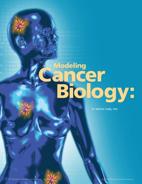
Modeling Cancer Biology - Biomedical Computation Review
Modeling Cancer Biology - Biomedical Computation Review
Modeling Cancer Biology - Biomedical Computation Review

You also want an ePaper? Increase the reach of your titles
YUMPU automatically turns print PDFs into web optimized ePapers that Google loves.
<strong>Modeling</strong><br />
<strong>Cancer</strong><br />
<strong>Biology</strong>:<br />
BY KRISTIN COBB, PhD<br />
16 BIOMEDICAL COMPUTATION REVIEW Spring 2007 www.biomedicalcomputationreview.org
The most common test for prostate cancer (known as PSA<br />
screening) misses aggressively growing prostate tumors—the<br />
type typically seen in young patients. It’s a fact that was accepted<br />
by the medical establishment in 2004 only after a seven-year study<br />
of 9000 men appeared in the New England Journal of Medicine. But<br />
Kristin Swanson, PhD, predicted the test’s inadequacy in 2001<br />
using a single differential equation—a “back of the napkin calculation”<br />
that “a high school student could answer.” This is the type of<br />
powerful insight that mathematics can offer cancer biology, says<br />
Swanson, who is an assistant professor of pathology and applied<br />
mathematics at the University of Washington.<br />
Unfortunately, mathematics has remained largely untapped and<br />
under-appreciated in cancer biology. Though mathematicians have<br />
been deriving formulas about cancer for decades, their work has<br />
been confined to mathematical and theoretical biology journals—a<br />
transforming<br />
the fight against cancer<br />
How mathematical models are transforming<br />
set of dense journals that the average biologist doesn’t read.<br />
Biologists are also skeptical: How can cancer, which is so complex<br />
and unpredictable, be reduced to a set of neat equations?<br />
But cancer biology may be at a turning point. Never before has<br />
there been a greater need for the field to embrace mathematics and<br />
computation. As biological data pile up at an astonishing rate, there<br />
is growing recognition that only quantitative approaches can pull it<br />
all together. As a result, quantitative cancer models are slowly making<br />
their way out of the theoretical and math journals and creeping<br />
into mainstream cancer biology. Leading biology journals like Cell<br />
and <strong>Cancer</strong> Research now contain theory sections. And, in 2003, the<br />
NIH established the Integrative <strong>Cancer</strong> <strong>Biology</strong> Program—which<br />
now funds nine inter-disciplinary centers that are applying quantitative<br />
modeling and systems biology approaches to cancer (the ICBPs).<br />
www.biomedicalcomputationreview.org<br />
Spring 2007 BIOMEDICAL COMPUTATION REVIEW 17
These efforts promise enormous pay<br />
off. <strong>Modeling</strong> can streamline wet-lab<br />
experiments; give scientists deeper insight<br />
into how tumors develop, grow, and<br />
spread; and even predict a patient’s prognosis<br />
and optimal treatment regimen.<br />
“Biologists tend to think of modeling<br />
as some sort of magical thing or<br />
black art,” says professor Philip K.<br />
Maini, PhD, director of the Center for<br />
Mathematical <strong>Biology</strong> at Oxford<br />
University. “But we haven’t done anything<br />
extra; we haven’t done any jiggerypokery<br />
or put any voodoo in there.”<br />
Mathematicians simply translate biologist’s<br />
hypotheses into a formal set of<br />
testable equations, he says.<br />
“Biologists are the first people to tell<br />
us that biology is very complicated; it’s<br />
highly non-linear. Yet biologists use verbal<br />
reasoning, which is linear reasoning,<br />
which is the wrong model,” he says.<br />
Mathematical models are needed to<br />
reach beyond where human intuition<br />
and linear thinking can take us, he says.<br />
A CANCER CELL IS BORN:<br />
THE SUBCELLULAR LEVEL<br />
<strong>Cancer</strong> arises through a series of<br />
genetic changes. Mutations in protooncogenes<br />
allow cells to grow and divide<br />
without the need for normal growth signals,<br />
and mutations in tumor suppressor<br />
genes allow cells to evade normal checks<br />
and balances—such as anti-growth signals<br />
and programmed cell death (apoptosis).<br />
Mutations in genes that detect and<br />
repair DNA damage facilitate the<br />
process by upping a cell’s mutation rate.<br />
Stochastic mathematical models help<br />
investigators test hypotheses about how<br />
cancer mutations accumulate. For example,<br />
Natalia Komarova, PhD, associate<br />
professor of mathematics at the<br />
University of California, Irvine, models<br />
the initiating event in colon cancer—the<br />
inactivation of the APC tumor suppressor<br />
gene. Normally, APC causes cells to<br />
enter apoptosis at the end of their<br />
“term” in the colon tissue, which helps<br />
prevent cancer.<br />
Cells in the colon are constantly<br />
exposed to the elements, and thus have<br />
a high risk of mutation. Thus, it is<br />
imperative that colon cells turn over<br />
quickly. The bottom of each microscopic<br />
pit of colon tissue (called a crypt) contains<br />
adult stem cells whose job is to produce<br />
daughter cells to continually<br />
replenish the colon. These daughter<br />
cells climb up the crypt, differentiate<br />
into colon cells, and die off in about a<br />
week. It is a delicate balance, however:<br />
the quick turnover helps prevent cancer<br />
in the daughter cells, but the frequently<br />
dividing stem cells are vulnerable to<br />
accumulating mutations.<br />
DNA-Damage Control. When cells are<br />
exposed to DNA-damaging radiation, they<br />
produce p53, an anti-cancer protein that<br />
causes damaged cells to undergo apoptosis<br />
(programmed cell death). Here, cells<br />
express fluorescently tagged p53 (green)<br />
and Mdm2 (red) following gamma irradiation.<br />
Time-lapse microscopy shows that, following<br />
DNA damage, p53 and Mdm2 levels<br />
undergo a series of pulses that vary in number<br />
from cell to cell. Courtesy of Galit<br />
Lahav's lab, department of Systems <strong>Biology</strong>,<br />
Harvard Medical School.<br />
“Biologists tend to think of modeling as some sort of magical thing or<br />
black art,” says Philip K. Maini, “But we haven’t done anything extra;<br />
we haven’t done any jiggery-pokery or put any voodoo in there.”<br />
What follows are some examples of how<br />
modeling is adding insight to intuition—<br />
from cancer initiation to metastasis and<br />
from the molecular to the patient level.<br />
One question that cannot be reliably<br />
answered experimentally is how many<br />
stem cells are in each crypt. Komarova<br />
tries to answer this question mathematically—by<br />
calculating the optimal number<br />
to minimize a person’s chance of getting<br />
mutations in the APC gene.<br />
“A situation like this is perfect for<br />
the application of modeling because in<br />
the model we can assume that there is<br />
one stem cell or that 50 percent of<br />
them are stem cells and we can see what<br />
happens,” Komarova says. It turns out<br />
that, for young people, having many<br />
stem cells minimizes the chance of<br />
cancer. But for older individuals,<br />
having a few stem cells is the best<br />
strategy. Likely, evolution favored the<br />
optimal strategy for young people, since<br />
evolution acts on those of reproductive<br />
age, she says.<br />
Besides probabilistic models of mutation<br />
“hits,” other researchers model the<br />
signaling pathways involved in growth,<br />
anti-growth, and cell death. Typically,<br />
these models consist of systems of ordinary<br />
differential equations. Each equation<br />
describes the rate of change in the<br />
concentration of a particular enzyme,<br />
substrate, receptor, or signaling molecule<br />
as a function of its production,<br />
degradation, and reaction with other<br />
network players.<br />
For example, Galit Lahav, PhD,<br />
assistant professor of systems biology at<br />
Harvard Medical School, models the<br />
p53 signaling network. p53 is a tumor<br />
suppressor gene that plays a crucial role<br />
in apoptosis, among other important<br />
anti-cancer functions. If specialized sensors<br />
in the cell detect DNA damage (or<br />
other dangers, such as oncogene over-<br />
18 BIOMEDICAL COMPUTATION REVIEW Spring 2007 www.biomedicalcomputationreview.org
Virtual Angiogenesis. In these snapshots from a computer simulation of tumor growth and angiogenesis, the top panels show the presence<br />
and density of tumor cells at time=5; cells in the core of the tumor become quiescent because oxygen cannot reach them. As the tumor<br />
grows, tumor cells secrete angiogenesis factors that cause new blood vessels to grow and supply extra oxygen and red blood cells to the<br />
tumor (bottom panels). Courtesy of Philip K. Maini.<br />
expression), they trigger p53 to initiate a<br />
cascade of events leading to the cell’s<br />
death. More than half of all human cancers<br />
contain a mutation in p53, making<br />
it the most common cancer mutation.<br />
Lahav studies the p53 network both<br />
experimentally and theoretically. “We go<br />
back and forth from the bench to the<br />
computer,” she says.<br />
In the lab, Lahav uses fluorescence<br />
microscopy to measure the changing levels<br />
of p53 and other proteins of interest<br />
(all tagged with fluorescent markers)<br />
after a cell is exposed to DNA-damaging<br />
gamma radiation. On the theoretical<br />
side, she uses a series of ordinary differ-<br />
ential equations to predict the changing<br />
levels of p53 and related proteins, such<br />
as Mdm2, which is involved in a negative<br />
feedback loop that regulates p53.<br />
“The idea of the models is to help us<br />
predict how the network will behave in<br />
response to different treatments and to<br />
suggest new experiments,” she says.<br />
For example, she discovered that levels<br />
of p53 oscillate following gamma irradiation,<br />
and she is using modeling to<br />
help understand these oscillations. If<br />
they are important for apoptosis, then<br />
some cancer drugs may work better if<br />
delivered in pulses rather than continuously,<br />
she says.<br />
THE GROWING TUMOR:<br />
THE CELLULAR LEVEL<br />
Once tumor cells have acquired the<br />
ability to propagate unchecked, they<br />
grow into a small ball of cells—which<br />
mathematicians model as a growing<br />
spheroid. Initially, the tumor feeds on<br />
oxygen and nutrients that diffuse to its<br />
surface. But these supplies cannot penetrate<br />
deep into the tumor, so cells in<br />
the core become dormant or die of<br />
starvation. The limited nutrient supply<br />
curbs the tumor’s growth to about half<br />
a millimeter in diameter—and if the<br />
story ended here, the tumor would be<br />
harmless.<br />
www.biomedicalcomputationreview.org<br />
Spring 2007 BIOMEDICAL COMPUTATION REVIEW 19
Forecasting Invasion. This graphic depiction of a mathematical model developed by Vito Quaranta and Alexander Anderson predicts<br />
whether a tumor will become invasive. The tumor is represented on a two-dimensional grid. Each virtual cell is accounted for on the grid<br />
and its behavior (e.g., growth, movement, death) is tracked based on mathematical functions and partial differential equations. Solving<br />
these equations in sequential time-steps generates a computer simulation of tumor growth and invasion. This approach has the potential<br />
to predict disease outcome based on precise quantities measured in the tumor of a specific patient. The model was described in:<br />
Anderson et al. Cell. 2006 Dec 1;127(5):905-15. Courtesy of the journal Cell. Graphic by Dominic Doyle.<br />
Unfortunately, as cells in the center<br />
become starved of oxygen (hypoxic), they<br />
release chemicals that stimulate angiogenesis—the<br />
growth of new blood vessels.<br />
These chemicals encourage blood vessel<br />
cells (endothelial cells) to migrate toward<br />
the core of the tumor and supply it with<br />
blood. Now the hungry tumor can feed<br />
unhindered. At the same time, the<br />
tumor gains a connection to vessels<br />
throughout the body, giving it an escape<br />
route for metastasis.<br />
One strategy for modeling angiogenesis<br />
is to set up systems of partial differential<br />
equations that describe how the<br />
tumor and vasculature are changing in<br />
both time and space (how their shapes<br />
are changing). For example, Zvia Agur,<br />
PhD, President of the Institute for<br />
Medical BioMathematics in Israel, has<br />
modeled angiogenesis using three interconnected<br />
modules of partial differential<br />
equations. Her equations describe:<br />
the changing volume of tumor cells<br />
(which depends on factors such as oxygen<br />
concentration); the changing volume<br />
of immature blood vessels (which<br />
depends on how quickly tumor cells<br />
release VEGF, a potent angiogenesis factor);<br />
and the changing volume of mature<br />
blood vessels (which depends on molecular<br />
signals that promote maturation).<br />
“The simplest model we could make was<br />
quite complex,” Agur says.<br />
She also set up an experimental system<br />
to validate her model. Her team<br />
implanted small balls of ovary cancer<br />
cells into mice and measured changes in<br />
the size and shape of the tumors and the<br />
blood vessels using MRI. For each indi-<br />
vidual tumor, Agur simulated its expected<br />
growth in the computer and then<br />
compared the simulation results to the<br />
actual results from the lab—and the prediction<br />
was quite good, she says.<br />
She then simulated what would happen<br />
if tumors were treated with antiangiogenesis<br />
drugs, and got a surprising<br />
result: The model showed that treatment<br />
with a single anti-angiogenesis<br />
drug is not sufficient to eliminate a<br />
tumor; rather, combinations of antiangiogenesis<br />
drugs are needed.<br />
“At the time, the anti-angiogenesis drug<br />
Avastin was very much in the news, and<br />
people thought that it could be used on its<br />
own,” Agur says. “Genentech was doing<br />
extensive clinical trials using Avastin<br />
monotherapy, and it took them another<br />
year or so to realize that we were right.”<br />
20 BIOMEDICAL COMPUTATION REVIEW Spring 2007 www.biomedicalcomputationreview.org
INVASION AND METASTASIS:<br />
THE TISSUE LEVEL<br />
For a while, the tumor continues to<br />
grow as a cohesive ball of cells with<br />
smooth edges. At this point, the tumor<br />
is still often curable, as a surgeon can<br />
just scoop it out, says Vito Quaranta,<br />
MD, professor of cancer biology at<br />
Vanderbilt University and also principal<br />
investigator of the Vanderbilt Integrative<br />
<strong>Cancer</strong> <strong>Biology</strong> Program (one of the<br />
nine ICBPs).<br />
But, eventually, some rogue cells<br />
break away from the growing tumor and<br />
invade the local tissue. To become invasive,<br />
tumor cells have to pick up certain<br />
abilities—they must escape cell-to-cell<br />
adhesion, migrate along the extracellular<br />
matrix (the surrounding connective tissue),<br />
and secrete enzymes that digest the<br />
extracellular matrix.<br />
Eventually, these invading cells burrow<br />
their way into the blood or lymph<br />
systems and spread (metastasize) to distant<br />
sites, where they seed new tumors.<br />
Now it is impossible to just reach in and<br />
scoop out the tumor—and the cancer is<br />
much more deadly.<br />
Quaranta, who is an experimentalist,<br />
collaborates with mathematician<br />
Alexander Anderson, PhD, senior lecturer<br />
of mathematics at the University of<br />
Dundee in Scotland, to model the<br />
process of invasion. They use a “hybrid<br />
discrete-continuum” model, which<br />
means molecules and proteins—such as<br />
oxygen and matrix-degrading enzymes—<br />
are modeled as continuous densities,<br />
but cells are modeled as individual, discrete<br />
entities that make autonomous<br />
decisions. Such agent-based models are<br />
computationally intensive, so simulations<br />
are limited to about five million<br />
cells (in contrast, a tumor may have a few<br />
billion cells).<br />
Cells move on a two-dimensional grid<br />
that represents the changing micro-environment—including<br />
the concentrations<br />
of nutrients, enzymes, and extracellular<br />
matrix proteins. Cells have a certain probability<br />
of moving to each adjacent point<br />
on the grid (called a biased random walk).<br />
For example, cells are more likely to move<br />
to regions where oxygen levels are high.<br />
Cells are also allowed to adhere to each<br />
other, migrate, degrade their surrounding<br />
tissue, divide, even die, according to cer-<br />
www.biomedicalcomputationreview.org<br />
<strong>Cancer</strong> Invasion. Starting with only 50 cancerous cells, this mathematical simulation shows<br />
how a tumor grows first into a smooth ball of non-invasive cells and then—under the right<br />
conditions—into an invasive mass that fingers into the surrounding environment. Blue cells<br />
are highly aggressive; orange cells are less aggressive, and brown cells are dead.<br />
Courtesy of Alexander Anderson<br />
Virtual Tumor. A simulation of one half of the whole living tumor cell population (outer half<br />
sphere) and the complete necrotic (dead) tumor cell population (inner sphere). Coloration<br />
relates to cell-adhesion value—cells on the outer surface of the tumor all have zero cell-tocell<br />
adhesion. Courtesy of Alexander Anderson<br />
Spring 2007 BIOMEDICAL COMPUTATION REVIEW 21
THE INTEGRATIVE CANCER<br />
BIOLOGY PROGRAM<br />
Established by the National <strong>Cancer</strong> Institute in 2003, the Integrative<br />
<strong>Cancer</strong> <strong>Biology</strong> Program (ICBP) funds efforts in computational modeling<br />
and systems biology approaches to cancer. “It’s difficult to do this<br />
type of research because you have to do both experimental biology<br />
and sophisticated computational approaches. Pulling those kinds of<br />
groups together really requires a structure like a center,” says<br />
Jennifer Couch, PhD, IT/<strong>Computation</strong>al <strong>Biology</strong> Coordinator for the<br />
ICBPs. “Our vision is always that these centers will sort of form the<br />
locus for the development of a community focused on integrative<br />
cancer biology.” Currently, the ICBP funds nine centers:<br />
Todd Golub, M.D., Dana-Farber <strong>Cancer</strong> Institute,<br />
Boston, Mass.<br />
Identifying protein kinase signatures in cancer.<br />
Joe W. Gray, Ph.D., Lawrence Berkeley National<br />
Laboratory, Berkeley, Cailf.<br />
<strong>Modeling</strong> signaling networks to identify patients for targeted<br />
therapeutics.<br />
Tim H-M Huang, Ph.D., Ohio State University,<br />
Columbus, Ohio.<br />
Epigenetic changes in cancer genomes.<br />
Timothy Kinsella, M.D., University Hospital of<br />
Cleveland, Cleveland, Ohio.<br />
<strong>Modeling</strong> mismatch repair defective malignancies.<br />
Sylvia Plevritis, Ph.D., Stanford University School of<br />
Medicine, Stanford, Cailf.<br />
Regulatory and signaling pathways in neoplastic transformation.<br />
Joseph Nevins, Ph.D., Duke University,<br />
Durham, N.C.<br />
Cell signaling pathways in cell proliferation and oncogenesis.<br />
Thomas Deisboeck, M.D., Massachusetts General<br />
Hospital, Boston, Mass.<br />
Model and simulation of multicellular patterns in cancer.<br />
Richard Hynes, Ph.D., Massachusetts Institute of<br />
Technology, Boston, Mass.<br />
<strong>Modeling</strong> cancer progression.<br />
Vito Quaranta, M.D., Vanderbilt University Medical<br />
Center, Nashville, Tenn.<br />
Model and simulation of cancer invasion.<br />
tain parameters—which Quaranta measures<br />
experimentally—such as speed<br />
of migration and the rate of cell<br />
division. Moreover, as cells divide, they<br />
acquire mutations that make them more<br />
aggressive and invasive (better able to<br />
proliferate, migrate, and enter the surrounding<br />
tissue).<br />
The resulting computer simulation—<br />
which shows a slice of a growing tumor—<br />
looks a bit like a weather forecasting<br />
map, Quaranta says. Virtual cells divide,<br />
move, and change colors to represent<br />
their changing phenotypes—for example,<br />
blue for highly aggressive, orange for less<br />
aggressive, and brown for dead.<br />
Depending on the conditions, tumors<br />
will either grow with smooth margins<br />
(remain non-invasive) or will finger<br />
out into the surrounding tissue<br />
(become invasive).<br />
When they ran their model, they<br />
got a surprising result: “We found that<br />
if the surrounding environment is a<br />
smooth, easy environment, then the<br />
cells tend to be non-invasive. But if<br />
you put pressure on the cells, say by<br />
reducing oxygen or making the landscape<br />
very hard to deal with, then the<br />
tumors become invasive,” Quaranta<br />
says. In gentle conditions, many different<br />
tumor cell phenotypes co-exist, but<br />
when the conditions become harsh<br />
one or two super-aggressive phenotypes<br />
prevail.<br />
Anti-angiogenisis drugs, inflammation,<br />
even chemotherapy and radiation<br />
therapy might create conditions for<br />
aggressive phenotypes to become dominant,<br />
Quaranta says.<br />
Their findings were published in the<br />
December 1 issue of Cell, a leading biology<br />
journal. Anderson says that before<br />
his collaboration with Quaranta he<br />
would never have dreamed of submitting<br />
a paper to Cell.<br />
“There was a bit of a wrestling match<br />
over the exact wording. But that ultimately<br />
paid off because it produced a<br />
paper that was really aimed at their audience,<br />
and that they could understand,”<br />
Anderson recalls.<br />
“Ultimately I’m hoping this is going<br />
to be good for the math biology community,<br />
because if I can get a paper<br />
published in Cell, then why can’t somebody<br />
else?” he adds.<br />
22 BIOMEDICAL COMPUTATION REVIEW Spring 2007 www.biomedicalcomputationreview.org
Brain Tumor Revealed. Only 10 percent of glioma cells are visible on MRI (the bright white<br />
area above); a computer simulation superimposed over the MRI helps doctors visualize the<br />
rest. The yellow/pink/red areas show that glioma cells may have diffused way beyond the<br />
borders of the mass seen on MRI. Courtesy of Kristin Swanson<br />
“It’s a nice change,” Alex Anderson says.<br />
“To have mathematics driving<br />
experimentation, instead of us just always<br />
playing catch up with the biology.”<br />
Quaranta says the partnership has<br />
changed his biology as well. “Our<br />
experiments now are actually driven<br />
by mathematics. So we’re entering an<br />
era of mathematics-driven experimental<br />
biology that is going to be interesting<br />
to see.”<br />
“It’s a nice change,” Anderson says.<br />
“To have mathematics driving experimentation,<br />
instead of us just always playing<br />
catch up with the biology.”<br />
GLIOMAS:<br />
THE PATIENT LEVEL<br />
Kristin Swanson (of the University of<br />
Washington) also works on modeling<br />
tumor invasion, but in glioma—a specific<br />
type of brain tumor that is particularly<br />
invasive and deadly. By the time a<br />
glioma mass is detectable on MRI, invasive<br />
glioma cells have already wandered<br />
far into the brain. Swanson compares it<br />
to an iceberg: the mass you can see represents<br />
only about 10 percent of the<br />
total tumor cells in the brain; the rest are<br />
undetectable, making it impossible to<br />
remove them.<br />
Swanson’s model consists of a series<br />
of partial differential equations that<br />
describe how the mass of glioma cells<br />
spreads within a virtual brain—a threedimensional<br />
lattice complete with areas<br />
of white and grey matter (glioma cells<br />
migrate at different rates in these different<br />
tissues). Her computer simulations<br />
show the changing density of<br />
glioma cells along sections of the virtual<br />
brain—for example, red where the<br />
tumor density is high and blue where<br />
density is low.<br />
A glioma patient’s MRI reveals only<br />
the detectable part of the tumor, so<br />
Swanson uses her simulations to visualize<br />
the undetectable portion and predict<br />
how the tumor will spread.<br />
“Just using diagnostic MRI and this<br />
mathematical model, you can predict<br />
survival with very reasonable accuracy<br />
for an individual patient,” she says.<br />
Her model can also be used to run in silico<br />
clinical trials. “It’s hard to test therapies<br />
for gliomas because patients don’t live long<br />
and you can’t see what’s happening with<br />
most of the tumor,” Swanson says. “But if<br />
you have a model for the expected behavior<br />
of an individual patient’s tumor, then you<br />
can assess the success of therapy relative to<br />
the expected behavior.”<br />
Another investigator working on<br />
gliomas is Thomas S. Deisboeck, MD,<br />
who is assistant professor of radiology at<br />
Massachusetts General Hospital and<br />
Harvard Medical School, as well as principal<br />
investigator of the Center for the<br />
Development of a Virtual Tumor<br />
(CViT), one of the nine ICBPs.<br />
Deisboeck uses a discrete, cell-based<br />
approach, rather than a continuous<br />
approach, to predict how cells will<br />
spread through a three-dimensional virtual<br />
brain. This allows him to connect<br />
what is happening at the subcellular to<br />
the cellular and tissue levels. “Our main<br />
interest is multi-scale, multi-grid, multiresolution<br />
modeling,” he says.<br />
His virtual cells can proliferate,<br />
migrate, die, and respond to the environment<br />
and each other. They also contain<br />
a nucleus, cytoplasm, membrane<br />
and even working biochemical pathways.<br />
The actions of particular biochemical<br />
pathway components can influence the<br />
behavior of the cells and the spread of<br />
the tumor. For example, Deisboeck is<br />
modeling how the EGFR (epidermal<br />
growth factor receptor) pathway acts as<br />
part of a molecular switch that turns<br />
glioma cells from proliferative (dividing)<br />
to migratory (invading local tissue).<br />
Though he eventually hopes to use<br />
his models to improve patient treat-<br />
www.biomedicalcomputationreview.org<br />
Spring 2007 BIOMEDICAL COMPUTATION REVIEW 23
From Patients to Molecules and Back. MRI images from a brain tumor patient (left) are used to build a 3-D in silico model of the growing<br />
tumor (right). Each cancer cell is represented as an autonomous agent that can move in space and change phenotypes (proliferation = blue;<br />
migration = red; quiescence = green). Each cell’s behavior is determined by equations that represent the cell’s intracellular networks, cell-tocell<br />
interactions, and cell-microenvironment interactions. Images Courtesy of Thomas S. Deisboeck. The underlying multi-scale model was<br />
described in Zhang et al. J. Theor Biol. 244(1): 96-107,2007.<br />
ment, his first goal is more modest—to<br />
improve diagnostics and patients’ quality<br />
of life.<br />
“What would be already a very significant<br />
achievement is if you could argue<br />
that instead of taking three MRI images<br />
say over six months, the combination of<br />
in silico modeling with two images would<br />
be just as informative,” he says.<br />
As principal investigator of CViT,<br />
Deisboeck’s broader vision is to build an<br />
online community of cancer modelers<br />
and a toolkit for multi-scale in silico cancer<br />
research. CViT is creating new infrastructure,<br />
including a digital model<br />
repository that will allow people to share<br />
and combine models (www.cvit.org).<br />
BRIDGING THE DIVIDE<br />
The above examples share a common<br />
theme—a tight link between the<br />
lab or clinic and the computer. But<br />
these examples are still the exception<br />
rather than the rule. The major obstacle<br />
in bringing modeling to cancer<br />
biology remains the lack of communication<br />
between modelers and experimentalists.<br />
On the one hand, biologists and clinicians<br />
tend to be mathematically illiterate<br />
and fearful of mathematics, says<br />
Robert A. Gatenby, MD, professor of<br />
radiology and applied mathematics at<br />
the University of Arizona.<br />
On the other hand, mathematicians<br />
tend to neglect the biology, he<br />
says. “Mathematicians will set up<br />
equations and then they’ll do uniqueness<br />
theorems and things like that,<br />
which are very mathematical<br />
approaches but utterly meaningless<br />
biologically. This just reinforces the<br />
biologists’ opinion that this is meaningless<br />
and can’t be even remotely<br />
helpful to them.”<br />
Getting these two groups to<br />
speak a common language and<br />
embrace a common objective is<br />
a major challenge. But efforts like<br />
the Integrative <strong>Cancer</strong> <strong>Biology</strong><br />
Program are helping to bridge this<br />
divide and to train a new generation<br />
of scientists who are eager to cross<br />
disciplines.<br />
“A lot of the students nowadays<br />
don’t want to get locked into just<br />
one field; they are looking for these<br />
multi-connections between a lot of<br />
disciplines. They may be engineering<br />
majors, but they want to know<br />
something about biology,” says<br />
Daniel Gallahan, PhD, Project<br />
Director of the Integrative <strong>Cancer</strong><br />
<strong>Biology</strong> Program at the NIH.<br />
“That’s been a pleasant surprise to<br />
me and it’s something I see as a<br />
critical component for the future<br />
of this effort.”<br />
“Maybe in one or two generations,<br />
we’ll have experimental biologists who<br />
are fluent in the language of mathematics,”<br />
agrees Vito Quaranta of Vanderbilt<br />
University.<br />
THE CUTTING EDGE<br />
Quaranta believes that a new era of<br />
cancer biology is fast approaching. “The<br />
way we do experimental oncology is<br />
going to change dramatically as these<br />
mathematics-driven simulations become<br />
more and more common place,” he says.<br />
As quantitative modeling moves from<br />
the margins of cancer biology to the<br />
mainstream, it is also presenting cuttingedge<br />
challenges for modelers.<br />
“It’s raising issues that mathematicians<br />
and modelers have never had to<br />
face before,” says Philip Maini of<br />
Oxford University. For example, how<br />
do you model the mechanics of a<br />
growing tissue? How do you build<br />
multi-scale models that are accurate<br />
across different biological and time<br />
scales? How simple or complex is the<br />
optimal model?<br />
“It’s a very interesting time for graduate<br />
students and post-docs to be<br />
involved, because it’s an area that’s<br />
now really beginning to take off,”<br />
Maini says. “Yet it isn’t so far developed<br />
that you can’t immediately start<br />
making inroads.” ■<br />
24 BIOMEDICAL COMPUTATION REVIEW Spring 2007 www.biomedicalcomputationreview.org