Modeling Cancer Biology - Biomedical Computation Review
Modeling Cancer Biology - Biomedical Computation Review
Modeling Cancer Biology - Biomedical Computation Review

Create successful ePaper yourself
Turn your PDF publications into a flip-book with our unique Google optimized e-Paper software.
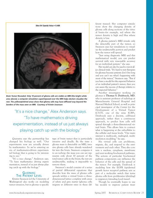
Brain Tumor Revealed. Only 10 percent of glioma cells are visible on MRI (the bright white<br />
area above); a computer simulation superimposed over the MRI helps doctors visualize the<br />
rest. The yellow/pink/red areas show that glioma cells may have diffused way beyond the<br />
borders of the mass seen on MRI. Courtesy of Kristin Swanson<br />
“It’s a nice change,” Alex Anderson says.<br />
“To have mathematics driving<br />
experimentation, instead of us just always<br />
playing catch up with the biology.”<br />
Quaranta says the partnership has<br />
changed his biology as well. “Our<br />
experiments now are actually driven<br />
by mathematics. So we’re entering an<br />
era of mathematics-driven experimental<br />
biology that is going to be interesting<br />
to see.”<br />
“It’s a nice change,” Anderson says.<br />
“To have mathematics driving experimentation,<br />
instead of us just always playing<br />
catch up with the biology.”<br />
GLIOMAS:<br />
THE PATIENT LEVEL<br />
Kristin Swanson (of the University of<br />
Washington) also works on modeling<br />
tumor invasion, but in glioma—a specific<br />
type of brain tumor that is particularly<br />
invasive and deadly. By the time a<br />
glioma mass is detectable on MRI, invasive<br />
glioma cells have already wandered<br />
far into the brain. Swanson compares it<br />
to an iceberg: the mass you can see represents<br />
only about 10 percent of the<br />
total tumor cells in the brain; the rest are<br />
undetectable, making it impossible to<br />
remove them.<br />
Swanson’s model consists of a series<br />
of partial differential equations that<br />
describe how the mass of glioma cells<br />
spreads within a virtual brain—a threedimensional<br />
lattice complete with areas<br />
of white and grey matter (glioma cells<br />
migrate at different rates in these different<br />
tissues). Her computer simulations<br />
show the changing density of<br />
glioma cells along sections of the virtual<br />
brain—for example, red where the<br />
tumor density is high and blue where<br />
density is low.<br />
A glioma patient’s MRI reveals only<br />
the detectable part of the tumor, so<br />
Swanson uses her simulations to visualize<br />
the undetectable portion and predict<br />
how the tumor will spread.<br />
“Just using diagnostic MRI and this<br />
mathematical model, you can predict<br />
survival with very reasonable accuracy<br />
for an individual patient,” she says.<br />
Her model can also be used to run in silico<br />
clinical trials. “It’s hard to test therapies<br />
for gliomas because patients don’t live long<br />
and you can’t see what’s happening with<br />
most of the tumor,” Swanson says. “But if<br />
you have a model for the expected behavior<br />
of an individual patient’s tumor, then you<br />
can assess the success of therapy relative to<br />
the expected behavior.”<br />
Another investigator working on<br />
gliomas is Thomas S. Deisboeck, MD,<br />
who is assistant professor of radiology at<br />
Massachusetts General Hospital and<br />
Harvard Medical School, as well as principal<br />
investigator of the Center for the<br />
Development of a Virtual Tumor<br />
(CViT), one of the nine ICBPs.<br />
Deisboeck uses a discrete, cell-based<br />
approach, rather than a continuous<br />
approach, to predict how cells will<br />
spread through a three-dimensional virtual<br />
brain. This allows him to connect<br />
what is happening at the subcellular to<br />
the cellular and tissue levels. “Our main<br />
interest is multi-scale, multi-grid, multiresolution<br />
modeling,” he says.<br />
His virtual cells can proliferate,<br />
migrate, die, and respond to the environment<br />
and each other. They also contain<br />
a nucleus, cytoplasm, membrane<br />
and even working biochemical pathways.<br />
The actions of particular biochemical<br />
pathway components can influence the<br />
behavior of the cells and the spread of<br />
the tumor. For example, Deisboeck is<br />
modeling how the EGFR (epidermal<br />
growth factor receptor) pathway acts as<br />
part of a molecular switch that turns<br />
glioma cells from proliferative (dividing)<br />
to migratory (invading local tissue).<br />
Though he eventually hopes to use<br />
his models to improve patient treat-<br />
www.biomedicalcomputationreview.org<br />
Spring 2007 BIOMEDICAL COMPUTATION REVIEW 23