Role of Special Histochemical Stains in Staining ... - Dako
Role of Special Histochemical Stains in Staining ... - Dako
Role of Special Histochemical Stains in Staining ... - Dako
You also want an ePaper? Increase the reach of your titles
YUMPU automatically turns print PDFs into web optimized ePapers that Google loves.
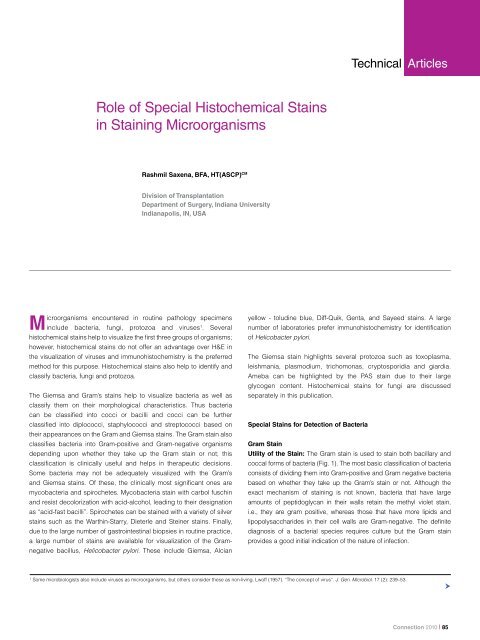
Technical Articles<br />
<strong>Role</strong> <strong>of</strong> <strong>Special</strong> <strong>Histochemical</strong> <strong>Sta<strong>in</strong>s</strong><br />
<strong>in</strong> Sta<strong>in</strong><strong>in</strong>g Microorganisms<br />
Rashmil Saxena, BFA, HT(ASCP) CM<br />
Division <strong>of</strong> Transplantation<br />
Department <strong>of</strong> Surgery, Indiana University<br />
Indianapolis, IN, USA<br />
Microorganisms encountered <strong>in</strong> rout<strong>in</strong>e pathology specimens<br />
<strong>in</strong>clude bacteria, fungi, protozoa and viruses 1 . Several<br />
histochemical sta<strong>in</strong>s help to visualize the first three groups <strong>of</strong> organisms;<br />
however, histochemical sta<strong>in</strong>s do not <strong>of</strong>fer an advantage over H&E <strong>in</strong><br />
the visualization <strong>of</strong> viruses and immunohistochemistry is the preferred<br />
method for this purpose. <strong>Histochemical</strong> sta<strong>in</strong>s also help to identify and<br />
classify bacteria, fungi and protozoa.<br />
The Giemsa and Gram’s sta<strong>in</strong>s help to visualize bacteria as well as<br />
classify them on their morphological characteristics. Thus bacteria<br />
can be classified <strong>in</strong>to cocci or bacilli and cocci can be further<br />
classified <strong>in</strong>to diplococci, staphylococci and streptococci based on<br />
their appearances on the Gram and Giemsa sta<strong>in</strong>s. The Gram sta<strong>in</strong> also<br />
classifies bacteria <strong>in</strong>to Gram-positive and Gram-negative organisms<br />
depend<strong>in</strong>g upon whether they take up the Gram sta<strong>in</strong> or not; this<br />
classification is cl<strong>in</strong>ically useful and helps <strong>in</strong> therapeutic decisions.<br />
Some bacteria may not be adequately visualized with the Gram’s<br />
and Giemsa sta<strong>in</strong>s. Of these, the cl<strong>in</strong>ically most significant ones are<br />
mycobacteria and spirochetes. Mycobacteria sta<strong>in</strong> with carbol fusch<strong>in</strong><br />
and resist decolorization with acid-alcohol, lead<strong>in</strong>g to their designation<br />
as “acid-fast bacilli”. Spirochetes can be sta<strong>in</strong>ed with a variety <strong>of</strong> silver<br />
sta<strong>in</strong>s such as the Warth<strong>in</strong>-Starry, Dieterle and Ste<strong>in</strong>er sta<strong>in</strong>s. F<strong>in</strong>ally,<br />
due to the large number <strong>of</strong> gastro<strong>in</strong>test<strong>in</strong>al biopsies <strong>in</strong> rout<strong>in</strong>e practice,<br />
a large number <strong>of</strong> sta<strong>in</strong>s are available for visualization <strong>of</strong> the Gramnegative<br />
bacillus, Helicobacter pylori. These <strong>in</strong>clude Giemsa, Alcian<br />
yellow - tolud<strong>in</strong>e blue, Diff-Quik, Genta, and Sayeed sta<strong>in</strong>s. A large<br />
number <strong>of</strong> laboratories prefer immunohistochemistry for identification<br />
<strong>of</strong> Helicobacter pylori.<br />
The Giemsa sta<strong>in</strong> highlights several protozoa such as toxoplasma,<br />
leishmania, plasmodium, trichomonas, cryptosporidia and giardia.<br />
Ameba can be highlighted by the PAS sta<strong>in</strong> due to their large<br />
glycogen content. <strong>Histochemical</strong> sta<strong>in</strong>s for fungi are discussed<br />
separately <strong>in</strong> this publication.<br />
<strong>Special</strong> <strong>Sta<strong>in</strong>s</strong> for Detection <strong>of</strong> Bacteria<br />
Gram Sta<strong>in</strong><br />
Utility <strong>of</strong> the Sta<strong>in</strong>: The Gram sta<strong>in</strong> is used to sta<strong>in</strong> both bacillary and<br />
coccal forms <strong>of</strong> bacteria (Fig. 1). The most basic classification <strong>of</strong> bacteria<br />
consists <strong>of</strong> divid<strong>in</strong>g them <strong>in</strong>to Gram-positive and Gram negative bacteria<br />
based on whether they take up the Gram’s sta<strong>in</strong> or not. Although the<br />
exact mechanism <strong>of</strong> sta<strong>in</strong><strong>in</strong>g is not known, bacteria that have large<br />
amounts <strong>of</strong> peptidoglycan <strong>in</strong> their walls reta<strong>in</strong> the methyl violet sta<strong>in</strong>,<br />
i.e., they are gram positive, whereas those that have more lipids and<br />
lipopolysaccharides <strong>in</strong> their cell walls are Gram-negative. The def<strong>in</strong>ite<br />
diagnosis <strong>of</strong> a bacterial species requires culture but the Gram sta<strong>in</strong><br />
provides a good <strong>in</strong>itial <strong>in</strong>dication <strong>of</strong> the nature <strong>of</strong> <strong>in</strong>fection.<br />
1<br />
Some microbiologists also <strong>in</strong>clude viruses as microorganisms, but others consider these as non-liv<strong>in</strong>g. Lw<strong>of</strong>f (1957). “The concept <strong>of</strong> virus”. J. Gen. Microbiol. 17 (2): 239–53.<br />
<br />
Connection 2010 | 85
Figure 1. Photomicrograph <strong>of</strong> ulcerated sk<strong>in</strong><br />
sta<strong>in</strong>ed with Gram’s sta<strong>in</strong>. The purple sta<strong>in</strong><br />
represents gram-positive bacteria which are<br />
seen as clumps (arrowhead) or as separate<br />
clusters <strong>of</strong> cocci (arrows). Everyth<strong>in</strong>g other<br />
than gram-positive bacteria is sta<strong>in</strong>ed p<strong>in</strong>k by<br />
the carbol fusch<strong>in</strong> countersta<strong>in</strong>. The underly<strong>in</strong>g<br />
structure <strong>of</strong> the sk<strong>in</strong> cannot be seen.<br />
Figure 2. Giemsa sta<strong>in</strong>ed section show<strong>in</strong>g<br />
a gastric pit conta<strong>in</strong><strong>in</strong>g Helicobacter pylori<br />
which appear as delicate, slightly curved<br />
rod-shaped purple organisms (arrowheads).<br />
The stomach is <strong>in</strong>flamed and shows many<br />
neutrophils (arrows). The background is<br />
sta<strong>in</strong>ed light p<strong>in</strong>k by the eos<strong>in</strong> countersta<strong>in</strong>.<br />
86 | Connection 2010
“<br />
Silver sta<strong>in</strong>s are very sensitive for the sta<strong>in</strong><strong>in</strong>g<br />
<strong>of</strong> bacteria and therefore most useful for<br />
bacteria which do not sta<strong>in</strong> or sta<strong>in</strong> weakly<br />
with the Grams and Giemsa sta<strong>in</strong>s.<br />
”<br />
components be<strong>in</strong>g azure A and B. Although the polychromatic sta<strong>in</strong> was<br />
first used by Romanowsky to sta<strong>in</strong> malarial parasites, the property <strong>of</strong><br />
polychromasia is most useful <strong>in</strong> sta<strong>in</strong><strong>in</strong>g blood smears and bone marrow<br />
specimens to differentiate between the various hemopoeitic elements.<br />
Nowadays, the Giemsa sta<strong>in</strong> is made up <strong>of</strong> weighted amounts <strong>of</strong> the<br />
azures to ma<strong>in</strong>ta<strong>in</strong> consistency <strong>of</strong> sta<strong>in</strong><strong>in</strong>g which cannot be atta<strong>in</strong>ed if<br />
methylene blue is allowed to “mature” naturally.<br />
Pr<strong>in</strong>ciples <strong>of</strong> Sta<strong>in</strong><strong>in</strong>g: The method consists <strong>of</strong> <strong>in</strong>itial sta<strong>in</strong><strong>in</strong>g <strong>of</strong> the<br />
bacterial slide with crystal violet or methyl violet which sta<strong>in</strong> everyth<strong>in</strong>g<br />
blue. This is followed by Gram’s or Lugol’s iod<strong>in</strong>e made up <strong>of</strong> iod<strong>in</strong>e<br />
and potassium iodide, which act by allow<strong>in</strong>g the crystal violet to adhere<br />
to the walls <strong>of</strong> gram-positive bacteria. Decolorization with an acetonealcohol<br />
mixture washes away the methyl violet which is not adherent to<br />
bacterial cell walls. At this stage, Gram-positive bacteria sta<strong>in</strong> blue while<br />
the Gram-negative bacteria are colorless. A carbol fusch<strong>in</strong> counter-sta<strong>in</strong><br />
is then applied which sta<strong>in</strong>s the Gram-negative bacteria p<strong>in</strong>k.<br />
Modifications: The Brown-Hopps and Brown-Brenn sta<strong>in</strong>s are<br />
modifications <strong>of</strong> the Gram sta<strong>in</strong> and are used for demonstration <strong>of</strong> gram<br />
negative bacteria and rickettsia.<br />
Modifications: The Diff-Quick and Wright’s sta<strong>in</strong>s are modifications <strong>of</strong><br />
the Giemsa sta<strong>in</strong>.<br />
Carbol Fusch<strong>in</strong> Acid-Alcohol Sta<strong>in</strong><br />
Utility <strong>of</strong> the Sta<strong>in</strong>: The carbol fusch<strong>in</strong> sta<strong>in</strong> helps to identify<br />
mycobacteria which are bacilli conta<strong>in</strong><strong>in</strong>g thick waxy cell walls (Lat<strong>in</strong>,<br />
myco=wax). Several mycobacteria can cause human disease; the<br />
two most significant ones are M. tuberculosis and M.leprae caus<strong>in</strong>g<br />
tuberculosis and leprosy respectively. Mycobacteria have large amounts<br />
<strong>of</strong> a lipid called mycolic acid <strong>in</strong> their cell walls which resists both sta<strong>in</strong><strong>in</strong>g<br />
as well as decolorization by acid-alcohol once sta<strong>in</strong><strong>in</strong>g has been<br />
achieved. The latter property is responsible for the commonly used<br />
term “acid-fast bacilli”. Mycobacteria cannot be sta<strong>in</strong>ed by the Gram<br />
sta<strong>in</strong> because it is an aqueous sta<strong>in</strong> that cannot penetrate the lipid-rich<br />
mycobacterial cell walls.<br />
Giemsa Sta<strong>in</strong><br />
Utility <strong>of</strong> the Sta<strong>in</strong>: The Giemsa is used to sta<strong>in</strong> a variety <strong>of</strong><br />
microorganisms <strong>in</strong>clud<strong>in</strong>g bacteria and several protozoans. Like the<br />
Gram sta<strong>in</strong>, the Giemsa sta<strong>in</strong> allows identification <strong>of</strong> the morphological<br />
characteristics <strong>of</strong> bacteria. However, it does not help further classification<br />
<strong>in</strong>to Gram-negative or Gram-positive bacteria. The Giemsa sta<strong>in</strong><br />
is also useful to visualize H. pylori (Fig. 2, 3; See also Fig. 4 for a<br />
high resolution H&E sta<strong>in</strong> show<strong>in</strong>g Giardia). The Giemsa also sta<strong>in</strong>s<br />
atypical bacteria like rickettsia and chlamydiae which do not have the<br />
peptidoglycan walls typical <strong>of</strong> other bacteria and which therefore do not<br />
take up the Gram sta<strong>in</strong>. The Giemsa sta<strong>in</strong> is used to visualize several<br />
protozonas such as toxoplasma, leishmania, plasmodium, trichomonas,<br />
cryptosporidia and giardia.<br />
Pr<strong>in</strong>ciples <strong>of</strong> Sta<strong>in</strong><strong>in</strong>g: The Giemsa sta<strong>in</strong> belongs to the class <strong>of</strong><br />
polychromatic sta<strong>in</strong>s which consist <strong>of</strong> a mixture <strong>of</strong> dyes <strong>of</strong> different hues<br />
which provide subtle differences <strong>in</strong> sta<strong>in</strong><strong>in</strong>g. When methylene blue is<br />
prepared at an alkal<strong>in</strong>e pH, it spontaneously forms other dyes, the major<br />
Pr<strong>in</strong>ciples <strong>of</strong> Sta<strong>in</strong><strong>in</strong>g: The mycobacteral cell walls are sta<strong>in</strong>ed by<br />
carbol fusch<strong>in</strong> which is made up <strong>of</strong> basic fusch<strong>in</strong> dissolved <strong>in</strong> alcohol<br />
and phenol. Sta<strong>in</strong><strong>in</strong>g is aided by the application <strong>of</strong> heat. The organisms<br />
sta<strong>in</strong> p<strong>in</strong>k with the basic fusch<strong>in</strong>. Sta<strong>in</strong><strong>in</strong>g is followed by decolorisation<br />
<strong>in</strong> acid-alcohol; mycobacteria reta<strong>in</strong> the carbol fusch<strong>in</strong> <strong>in</strong> their cell wall<br />
whereas other bacteria do not reta<strong>in</strong> carbol fusch<strong>in</strong>, which is extracted<br />
<strong>in</strong>to the acid-alcohol. Countersta<strong>in</strong><strong>in</strong>g is carried out by methylene blue.<br />
Mycobacteria sta<strong>in</strong> bright p<strong>in</strong>k with basic fusch<strong>in</strong> and the background<br />
sta<strong>in</strong>s a fa<strong>in</strong>t blue. Care has to be taken to not over-counter sta<strong>in</strong> as this<br />
may mask the acid-fast bacilli.<br />
Modifications: The 2 commonly used methods for sta<strong>in</strong><strong>in</strong>g <strong>of</strong> M.<br />
tuberculosis are the Ziehl-Neelsen and K<strong>in</strong>youn’s acid fast sta<strong>in</strong>s. The<br />
Fite sta<strong>in</strong> is used for sta<strong>in</strong><strong>in</strong>g <strong>of</strong> M.leprae which has cell walls that are<br />
more susceptible to damage <strong>in</strong> the deparaff<strong>in</strong>ization process. The Fite<br />
procedure thus <strong>in</strong>cludes peanut oil <strong>in</strong> the deparaff<strong>in</strong>ization solvent to<br />
protect the bacterial cell wall. The acid used for decolorization <strong>in</strong> the Fite<br />
procedure is also weaker (Fig. 5).<br />
<br />
Connection 2010 | 87
Figure 3. Giemsa sta<strong>in</strong>ed section <strong>of</strong> small<br />
<strong>in</strong>test<strong>in</strong>al mucosa show<strong>in</strong>g clusters <strong>of</strong> Giardia<br />
which sta<strong>in</strong> purple (arrows) <strong>in</strong> the crypts. The<br />
background is sta<strong>in</strong>ed fa<strong>in</strong>t p<strong>in</strong>k by the eos<strong>in</strong><br />
countersta<strong>in</strong>.<br />
Figure 4. An H&E section <strong>of</strong> an <strong>in</strong>test<strong>in</strong>al crypt<br />
show<strong>in</strong>g clusters <strong>of</strong> Giardia (arrowheads).<br />
The oval shape and cluster<strong>in</strong>g gives them a<br />
“tumbl<strong>in</strong>g leaves” appearance. Fa<strong>in</strong>t nuclei<br />
can be seen <strong>in</strong> some organisms (arrow).<br />
88 | Connection 2010
Figure 5. Ziehl-Neelsen sta<strong>in</strong>ed section <strong>of</strong><br />
lymph node. The p<strong>in</strong>k color demonstrates<br />
clusters <strong>of</strong> mycobacteria sta<strong>in</strong>ed with carbolfusch<strong>in</strong><br />
(arrows). The sta<strong>in</strong> has resisted<br />
decolorisation by acid-alcohol. Other cells <strong>in</strong><br />
the background are sta<strong>in</strong>ed light blue by the<br />
methylene blue countersta<strong>in</strong>.<br />
Figure 6. Warth<strong>in</strong>-Starry sta<strong>in</strong> <strong>of</strong> stomach<br />
conta<strong>in</strong><strong>in</strong>g Helicobacter pylori which appear<br />
as black and slightly curved, rod-like<br />
bacteria (arrows). The background is sta<strong>in</strong>ed<br />
light yellow.<br />
<br />
Connection 2010 | 89
Microorganism Preferred <strong>Sta<strong>in</strong>s</strong> Disease<br />
Bacteria<br />
Bacteria, usual Gram, Giemsa Wide variety <strong>of</strong> <strong>in</strong>fections<br />
Mycobacteria tuberculosis Ziehl-Neelsen, K<strong>in</strong>youn’s Tuberculosis<br />
Mycobacteria lepra Fite Leprosy<br />
Rickettsia Giemsa Rocky Mounta<strong>in</strong> Spotted fever, typhus fever<br />
Chlamydia Giemsa Sexually transmitted disease, pneumonia<br />
Legionella Silver sta<strong>in</strong>s Pneumonia<br />
Spirochetes Silver sta<strong>in</strong>s Syphilis, leptospirosis, Lyme’s disease<br />
Bartonella Warth<strong>in</strong>-Starry Cat-scratch disease<br />
Helicobacter pylori<br />
Giemsa, Diff-Quik, Alcian-yellow<br />
Tolud<strong>in</strong>e blue, Silver sta<strong>in</strong>s<br />
Inflammation <strong>of</strong> stomach, stomach ulcers<br />
Protozoa<br />
Giardia Giemsa “traveller’s diarrhea”<br />
Toxoplasma Giemsa Toxoplasmosis <strong>in</strong> immunocomprised hosts<br />
Cryptosporidium Giemsa Diarrhea <strong>in</strong> AIDS patients<br />
Leishmania Giemsa Sk<strong>in</strong> <strong>in</strong>fections, severe generalized<br />
<strong>in</strong>fection with anemia and wast<strong>in</strong>g<br />
Plasmodium Giemsa Malaria<br />
Trichomonas Trichomonas Vag<strong>in</strong>al <strong>in</strong>fection<br />
Ameba PAS sta<strong>in</strong> Diarrhea, liver abscess<br />
Table 1. Provides a summary <strong>of</strong> some special sta<strong>in</strong>s used <strong>in</strong> detect<strong>in</strong>g microorganisms.<br />
90 | Connection 2010
Silver <strong>Sta<strong>in</strong>s</strong> (Warth<strong>in</strong> Starry Sta<strong>in</strong>, Dieterle, Ste<strong>in</strong>er <strong>Sta<strong>in</strong>s</strong>)<br />
Utility <strong>of</strong> the <strong>Sta<strong>in</strong>s</strong>: Silver sta<strong>in</strong>s are very sensitive for the sta<strong>in</strong><strong>in</strong>g <strong>of</strong><br />
bacteria and therefore most useful for bacteria which do not sta<strong>in</strong> or<br />
sta<strong>in</strong> weakly with the Grams and Giemsa sta<strong>in</strong>s. Although they can be<br />
used to sta<strong>in</strong> almost any bacteria, they are tricky to perform and are<br />
therefore reserved for visualiz<strong>in</strong>g spirochetes, legionella, bartonella<br />
and H. pylori.<br />
Pr<strong>in</strong>ciples <strong>of</strong> Sta<strong>in</strong><strong>in</strong>g: Spirochetes and other bacteria can b<strong>in</strong>d<br />
silver ions from solution but cannot reduce the bound silver. The slide<br />
is first <strong>in</strong>cubated <strong>in</strong> a silver nitrate solution for half an hour and then<br />
“developed” with hydroqu<strong>in</strong>one which reduces the bound silver to a<br />
visible metallic form. The bacteria sta<strong>in</strong> dark-brown to black while the<br />
background is yellow (Fig. 6).<br />
Auram<strong>in</strong>e O- Rhodam<strong>in</strong>e B Sta<strong>in</strong><br />
The auram<strong>in</strong>e O-rhodam<strong>in</strong>e B sta<strong>in</strong> is highly specific and sensitive for<br />
mycobateria. It also sta<strong>in</strong>s dead and dy<strong>in</strong>g bacteria not sta<strong>in</strong>ed by the<br />
acid-fast sta<strong>in</strong>s. The mycobacteria take up the dye and show a reddishyellow<br />
fluorescence when exam<strong>in</strong>ed under a fluorescence microscope.<br />
Summary<br />
<strong>Histochemical</strong> sta<strong>in</strong>s available for demonstrat<strong>in</strong>g microorganisms<br />
<strong>in</strong>clude Giemsa sta<strong>in</strong>, Grams sta<strong>in</strong>, carbol fusch<strong>in</strong> acid-alcohol sta<strong>in</strong><br />
and a variety <strong>of</strong> silver sta<strong>in</strong>s such as Warth<strong>in</strong>-Starry, Dieterle and Ste<strong>in</strong>er<br />
sta<strong>in</strong>s. The Gram sta<strong>in</strong> allows classification <strong>of</strong> bacteria <strong>in</strong>to Grampositive<br />
and Gram-negative bacteria. The acid-alcohol sta<strong>in</strong> allows<br />
classification <strong>of</strong> bacilli <strong>in</strong>to acid-fast and non-acid-fast bacilli. These are<br />
both cl<strong>in</strong>ically useful classifications. The silver sta<strong>in</strong>s are very sensitive<br />
and help to visualize difficult-to-sta<strong>in</strong> bacteria. Most protozoans are<br />
sta<strong>in</strong>ed by the Giemsa sta<strong>in</strong>.<br />
Glossary<br />
Bacteria are unicellular organisms that do not conta<strong>in</strong> a nucleus or other membrane-bound<br />
organelles. Most bacteria have a rigid cell wall composed <strong>of</strong> peptidoglycan. Although there<br />
are exceptions, bacteria come <strong>in</strong> 3 basic shapes: round (cocci), rod-like (bacilli) and spiral<br />
(spirochetes). Bacteria cause a variety <strong>of</strong> <strong>in</strong>fections <strong>in</strong> various organs.<br />
Bartonella is a Gram-negative bacillus which causes cat-scratch disease. The bacilli are<br />
transmitted to humans by cat-bite or cat-scratch.<br />
Chlamydia are Gram-negative bacteria which are unusual because they do not have typical<br />
bacterial cell walls. They are obligate <strong>in</strong>tracellular parasites which means that they can<br />
only survive with<strong>in</strong> cells. Chlamydia cause sexually transmitted diseases and pneumonia<br />
<strong>in</strong> humans.<br />
Helicobacter pylori is a Gram-negative bacteria which causes <strong>in</strong>flammation (gastritis) and<br />
ulcers <strong>of</strong> the stomach. The name derives from the Greek “helix” for spiral and “pylorus” for<br />
the distal end <strong>of</strong> the stomach.<br />
Legionella is a Gram-negative bacillus so named because it caused an outbreak <strong>of</strong><br />
pneumonia <strong>in</strong> people attend<strong>in</strong>g a 1976 convention <strong>of</strong> the American Legion <strong>in</strong> Philadelphia.<br />
The organism was unknown till then and was subsequently named Legionella. It causes<br />
pneumonia.<br />
Mycobacteria are bacilli that have a thick and waxy (Lat<strong>in</strong>, myco = wax) cell wall composed<br />
<strong>of</strong> a lipid called mycolic acid. This cell wall is responsible for the hard<strong>in</strong>ess <strong>of</strong> this organism<br />
as well as for its sta<strong>in</strong><strong>in</strong>g characteristics. The waxy cell wall is hydrophobic and resists<br />
sta<strong>in</strong><strong>in</strong>g with aqueous sta<strong>in</strong>s like the Gram and Giemsa sta<strong>in</strong>s. It also resists decolorisation<br />
once sta<strong>in</strong>ed. Mycobacteria causes tuberculosis, leprosy and <strong>in</strong>fections <strong>in</strong> patients<br />
with AIDS.<br />
Protozoa (Greek, proton = first; zoa = animal) are unicellular organisms that have a<br />
membrane-bound nucleus and other complex membrane-bound organelles.<br />
Rickettsia are Gram-negative bacteria that like Chlamydia lack typical cell walls and are<br />
obligate <strong>in</strong>tracellular parasites. The rickettsial diseases are primary diseases <strong>of</strong> animals<br />
(zoonosis) such as the deer which are transmitted to humans by bites <strong>of</strong> <strong>in</strong>sects like fleas<br />
and ticks. Rickettsial diseases <strong>in</strong>clude typhus fever and Rocky Mounta<strong>in</strong> Spotty Fever.<br />
Spirochetes are long Gram-negative bacilli with tightly-coiled helical shapes. Spirochetes<br />
cause syphilis, leptospirosis and Lyme’s disease.<br />
Connection 2010 | 91


