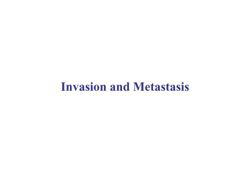
Invasion and metastasis - Experimental Oncology Graduate Study
Invasion and metastasis - Experimental Oncology Graduate Study
Invasion and metastasis - Experimental Oncology Graduate Study

Create successful ePaper yourself
Turn your PDF publications into a flip-book with our unique Google optimized e-Paper software.
<strong>Invasion</strong> <strong>and</strong> Metastasis
What is <strong>metastasis</strong>?<br />
Tumors take many years before detection (>10 9 cells)- particularly if tumor<br />
growing in an extensible space.<br />
Only when tumor begins to compromise function of the organ does it evoke<br />
symptoms. Most primary tumors can be surgically excised <strong>and</strong> account for<br />
less than 10% of cancer deaths.<br />
For 90% of patients - primary successfully excised but patients die as a<br />
result of disease at sites distant to that of primary.<br />
Metastasis is the truly lethal event in cancer <strong>and</strong> the process we know least<br />
about.<br />
Underst<strong>and</strong>ing <strong>metastasis</strong> has the greatest potential to extend patient<br />
survival.
Cell 144, March 4, 2011 p 646-674 2011
Mutations in the cancer cell
Metastatic disease<br />
non Hodgkins<br />
Lymphoma (NHL):<br />
-CT scan (blue)<br />
-PET scan (yellow) – FDG<br />
accumulates in regions of<br />
active metabolism
Tumor cells metastasize through lymph <strong>and</strong><br />
blood vessels<br />
Pancreatic cancer cells within<br />
lymphatic vessel<br />
Breast cancer in<br />
lymphatic drainage
Metastases disrupt organ functions<br />
Breast cancer metastasizes to bone (pain <strong>and</strong> skeletal collapse);<br />
brain, lungs, liver (function).<br />
Other cancers spread to different organs.<br />
Some cancers have very high rates of <strong>metastasis</strong>-while others<br />
hardly metastasize at all.<br />
Underst<strong>and</strong>ing the basis for these differences could lead to<br />
control over tumor spread.
Size of primary correlates with risk of breast<br />
cancer <strong>metastasis</strong><br />
1589 tumors<br />
followed for up to<br />
46 years.
If larger tumors associated with increased risk of<br />
<strong>metastasis</strong> then is <strong>metastasis</strong> a late event (mutations<br />
in <strong>metastasis</strong> genes)?<br />
Simplistic deduction:<br />
Since 4% breast cancers under 1 cm ~ mutant p53 alleles.<br />
And 42% breast cancers over 3 cm ~ mutant p53 cancers acquire further<br />
mutations as they grow.<br />
Mutations that favor <strong>metastasis</strong> may therefore arise as late events.<br />
Just as likely:<br />
a) Cells in large <strong>and</strong> small tumors equally capable of metastasizing but<br />
large tumors seed more cells<br />
b) Large tumors contain more proliferative cells <strong>and</strong> mutations that<br />
influence proliferation influence <strong>metastasis</strong>.<br />
Difficult to predict when <strong>metastasis</strong> first occurs.
If the metastatic phenotype arises early then most<br />
tumors will have already metastasized by the time of<br />
detection<br />
Consensus: most tumors do metastasize early<br />
<strong>and</strong> give rise to micrometastases that are too<br />
small to detect.<br />
Aggressive therapy given soon after detection<br />
of primary may therefore reduce the incidence<br />
of metastatic disease.
How do tumors spread?<br />
The concept of the invasion-<strong>metastasis</strong> cascade: 6<br />
distinct steps<br />
Probability of an individual cancer cell completing cascade is very<br />
small
Invasive<br />
lobular<br />
mammary<br />
carcinoma<br />
progressing in<br />
single file from<br />
left to right<br />
through<br />
channels they<br />
have carved in<br />
stroma<br />
Step 1: <strong>Invasion</strong>
More typically cells invade as a posse<br />
Melanoma cells moving as a cohort<br />
through collagen matrix. Adherens<br />
junctions red (E-cadherin).<br />
β1 integrins<br />
metalloproteinases
Breach of basement membrane<br />
Inflammatory<br />
cells<br />
Squamous<br />
carcinoma<br />
Basement<br />
membrane
Step 2 Intravasation: transport thro<br />
vasculature<br />
Hazards:<br />
Anoikis (homelessness)- loss of substrate<br />
contact-triggers apoptosis.<br />
Shear forces.<br />
Lack of stromal support (survival factors).<br />
Exposure to cells of the immune system.<br />
Exposure to platelets (beneficial).
Cell 147: October 14 2011<br />
p275-292
Routes of<br />
circulation<br />
Tumors usually<br />
lodge in first capillary<br />
bed they encounter<br />
Tumors that attract<br />
platelets ~ may lodge in<br />
wider vessels such as<br />
arteriole
Step 3:<br />
extravasation<br />
tumor clump<br />
vessel
Tumors attach to vessel walls thro<br />
microthrombi<br />
Localized exposure of<br />
basement membrane provides<br />
attachment points for<br />
potential microthrombi<br />
attachment<br />
red blood cells<br />
platelets
Tumors stimulate microthrombi<br />
Within seconds of<br />
entering blood stream<br />
tumors stimulate<br />
thrombus formation<br />
(platelets <strong>and</strong> red blood<br />
cells).<br />
Tissue factor activates<br />
thrombin <strong>and</strong> converts<br />
fibrinogen to fibrin.<br />
Mice lacking platelets<br />
(Nf-E2-) do not support<br />
<strong>metastasis</strong> of malignant<br />
melanoma C <strong>and</strong> D.<br />
Tumor cell<br />
platelet
How efficient is <strong>metastasis</strong>?<br />
Mice carrying primary tumors of 1<br />
gm (10 9 cells)<br />
10 6 cells enter circulation per day.<br />
However less than 5 tumors result.<br />
Metastatic inefficiency.
Step 4: colonization<br />
Colonization - the rate-limiting step.<br />
Unlike the primary tumor tissues lack stroma that<br />
provide support. Consequently most micrometastases<br />
remain dormant for long periods or die before they can<br />
give rise to 2ndary tumors.
Evolution of metastatic ability can occur<br />
outside the primary tumor
The<br />
evolution of<br />
colonizing<br />
ability
Micrometastases (MM) detectable in bone<br />
marrow<br />
A<br />
Micro<strong>metastasis</strong> of:<br />
A. colon cancer<br />
B. breast cancer<br />
in bone-marrow<br />
detected using anti-cytokeratin<br />
antibodies<br />
1% normal have +ve cells cf 36%<br />
breast cancer<br />
(stage I, II, III).<br />
1 colony per 10 5 to 10 6<br />
mesenchymal cells<br />
B
Does marrow micro<strong>metastasis</strong> predict<br />
relapse?<br />
Patients with MM at time of surgery have higher relapse rates.<br />
Breast cancer MM increase risk of developing metastatic disease 4 to 10 fold.<br />
Colon cancer patients 90% patients who lack MM alive at 15 months compared to<br />
30% of those with marrow MM<br />
Cancer esophagus – 79-88% pts have MM in rib marrow at resection
Can MM remain dormant?<br />
Are MM truly dormant?<br />
Fluorescently-labeled cells lose fluorescence<br />
with each division. Breast cancer cells injected<br />
into portal vein lodge in liver were viable <strong>and</strong><br />
had not lost fluorescence 11 weeks later.<br />
Cells could be excised, cultured <strong>and</strong> retained<br />
tumorigenicity when introduced<br />
subcutaneously.<br />
Cells were resistant to chemotherapy that<br />
killed growing cells.
What we have learned so far<br />
<strong>Invasion</strong>-<strong>metastasis</strong> cascade has multiple steps.<br />
As many changes in phenotype involved as precede inititial tumor formation.<br />
Cells readily enter circulation <strong>and</strong> seed distant sites. However, only a small<br />
fraction grow.<br />
Nevertheless vast numbers of cells seeded by growing tumors ensure that some of<br />
these will eventually grow.<br />
Clinically these pose a major threat to survival of patient.<br />
Important question:<br />
To what extent do the steps represent changes in gene expression. Are there<br />
<strong>metastasis</strong> specific genes <strong>and</strong> their suppressors?
Epithelial-mesenchymal transition<br />
(a wolf in sheep’s clothing)<br />
Migration <strong>and</strong> motility require epithelial cells to lose their differentiated adhesive<br />
nature <strong>and</strong> become mesenchyme-like.
EMT<br />
Upper panel: Epithelial cells in monolayer culture exposed to metalloproteinase MMP-3.<br />
Cells lose cytokeratins (red) <strong>and</strong> E-cadherin <strong>and</strong> acquire vimentin (green) fibronectin <strong>and</strong> N-cadherin become motile<br />
<strong>and</strong> invasive.<br />
Become indistinguishable from mesenchyme.<br />
Lower panel: Wounding MCF10A (pre-malignant) monolayer –induction of vimentin (red) at wound site. Vimentin<br />
lost once cells revert to epithelial.
Embryogenesis <strong>and</strong> EMT<br />
Ectodermal-mesodermal transition<br />
at gastrulation<br />
Delamination neuroepithelial cells<br />
from neural tube to form melanocytes,<br />
peripheral NS
Can we see EMT in tumors?<br />
Loss of E-cadherin (brown) from membrane of<br />
cells at invasive edge of colon carcinoma<br />
(arrows)
β-catenin translocates to nuclei
Biochemical changes accompanying EMT<br />
Expressing Twist transcription factor in MDCK cells induces<br />
fibronectin <strong>and</strong> vimentin
Confirmation by immunoblot
E cadherin N-cadherin switch plays a pivotal<br />
role in EMT<br />
E-cadherin to N-cadherin switch permits new associations with mesenchymal cells.<br />
Similarly gastrulation as well as HGF embryonic ectodermal layer—emigration <strong>and</strong><br />
dermal precursors from primitive dermomyotome. Homotypic interactions permit<br />
invasion into <strong>and</strong> establishment of epithelial cells in mesenchyme. N-cadherin bonds<br />
weaker than E-cadherin.
HGF (stromal factor) induces scattering in<br />
MDCK cells
EMT induced by stromal signals <strong>and</strong><br />
therefore reversible
Why do 2ndary tumors resemble primary tumors<br />
<strong>and</strong> not mesenchyme?<br />
Reversibility may be the key<br />
Histopathology of primary <strong>and</strong><br />
secondary tumors often strikingly<br />
similar.<br />
In fact used by pathologists to<br />
a) Identify tumors of unknown<br />
origin<br />
<strong>and</strong><br />
b) Dismiss the concept of EMT!
Exposing the wolf<br />
EMT at invasive edge of human<br />
mammary epithelial cells in mouse<br />
host.<br />
Mouse stroma<br />
Human vimentin
How do stromal cells control EMT?<br />
Role of TGF-β
Macrophages: role in invasion <strong>and</strong> <strong>metastasis</strong><br />
+/op mice make CSF1 <strong>and</strong> recruit macrophages to tumors-mets form in lungs<br />
op/op mice lack CSF1 <strong>and</strong> cannot recruit macrophages- mets do not form in lungs
Signals that trigger EMT
Embryonic transcription factors<br />
programming EMT
Transient expression Slug in wound healing<br />
Monolayer of<br />
keratinocytes<br />
scraped to induce<br />
wound
Expression EMT-inducing transcription factors in<br />
tumors<br />
Slug binds to<br />
E-box sequence<br />
in E-cad promoter
Similarities between EMT embryogenesis <strong>and</strong><br />
tumor progression
Extracellular proteases <strong>and</strong> invasiveness<br />
Mammary carcinoma in MMTV-polyoma middle T mouse<br />
proteolysis<br />
fibroblasts<br />
collagen IV cleavage<br />
(inhibited by MMP<br />
inhibitors)
Podosomes <strong>and</strong> focal degradation of ECM<br />
(MT1-MMP)<br />
Actin (red)<br />
FITC - fibronectin (green)<br />
Degradation fibronectin (black)<br />
Colocalization podosome <strong>and</strong> degradation (arrow)
Protease cascade induced by stroma
Rho-C <strong>and</strong> <strong>metastasis</strong><br />
(epithelial morphology)<br />
(fibroblast morphology)
Metastatic dissemination via lymphatics
Metastatic tropisms-seed <strong>and</strong> soil or<br />
hydrodynamics?
Colorectal cell <strong>metastasis</strong>: a matter of<br />
drainage
Metastasis to bone (osteotropic <strong>metastasis</strong>)<br />
requires the subversion of osteoblasts <strong>and</strong> osteoclasts<br />
Osteoclasts (arrowed) excavating jaw bone
Vicious cycle of osteoclast <strong>metastasis</strong><br />
Parathyroid hormonerelated<br />
peptide
Are there <strong>metastasis</strong> genes?
Cell 147: October 14 2011 p275-292
Cell 147: October 14 2011 p275-292
Science 25 March 2011: Vol. 331 no. 6024 pp. 1559-<br />
1564
Science 25 March 2011: Vol. 331 no. 6024 pp. 1559-<br />
1564
Science 25 March 2011: Vol. 331 no. 6024 pp. 1559-<br />
1564
Cell 144, March 4, 2011 p 646-674 2011
Cell 144, March 4, 2011 p 646-674 2011
Leading Edge Review Tumor Metastasis: Molecular Insights <strong>and</strong> Evolving<br />
Paradigms<br />
Scott Valastyan1,2,4,* <strong>and</strong> Robert A. Weinberg1,2,3,*<br />
Cell 147: October 14 2011 p275-292
Science 25 March 2011: Vol. 331 no. 6024 pp.<br />
1559-1564 DOI: 10.1126/science.1203543<br />
REVIEW:<br />
A Perspective on Cancer Cell Metastasis Christine<br />
L. Chaffer1,3,* Robert A. Weinberg1,2,3,*
Hallmarks of Cancer: The Next Generation Douglas Hanahan1,2,* <strong>and</strong><br />
Robert A. Weinberg3,*<br />
1The Swiss Institute for <strong>Experimental</strong> Cancer Research (ISREC), School of Life Sciences, EPFL, Lausanne CH-<br />
1015, Switzerl<strong>and</strong> 2The Department of Biochemistry & Biophysics, UCSF, San Francisco, CA 94158, USA<br />
3Whitehead Institute for Biomedical Research, Ludwig/MIT Center for Molecular <strong>Oncology</strong>, <strong>and</strong> MIT<br />
Department of Biology, Cambridge, MA 02142, USA<br />
Cell 144, March 4, 2011 p 646-674 2011