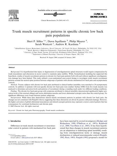
Trunk muscle recruitment patterns in specific chronic low back pain ...
Trunk muscle recruitment patterns in specific chronic low back pain ...
Trunk muscle recruitment patterns in specific chronic low back pain ...

You also want an ePaper? Increase the reach of your titles
YUMPU automatically turns print PDFs into web optimized ePapers that Google loves.
Cl<strong>in</strong>ical Biomechanics 20 (2005) 465–473<br />
www.elsevier.com/locate/cl<strong>in</strong>biomech<br />
<strong>Trunk</strong> <strong>muscle</strong> <strong>recruitment</strong> <strong>patterns</strong> <strong>in</strong> <strong>specific</strong> <strong>chronic</strong> <strong>low</strong> <strong>back</strong><br />
pa<strong>in</strong> populations<br />
Sheri P. Silfies a, *, Dawn Squillante b , Philip Maurer b ,<br />
Sarah Westcott c , Andrew R. Karduna d<br />
a Rehabilitations Sciences Biomechanics Laboratory, Drexel University, 245 North 15th Street, Philadelphia, PA19102-1192, USA<br />
b Booth, Bartolozzi, Balderston Orthopaedics, Pennsylvania Hospital, Philadelphia, PA, USA<br />
c University of Puget Sound, Tacoma, WAand Lake Wash<strong>in</strong>gton School District, Redmond, WA, USA<br />
d Department of Human Physiology, University of Oregon, Eugene, OR, USA<br />
Received 30 August 2004; accepted 26 January 2005<br />
Abstract<br />
Background. It is hypothesized that <strong>in</strong>jury or degeneration of osteoligamentous sp<strong>in</strong>al structures would require compensation by<br />
trunk musculature and alterations <strong>in</strong> motor control to ma<strong>in</strong>ta<strong>in</strong> sp<strong>in</strong>e stability. While, biomechanical model<strong>in</strong>g has supported this<br />
hypothesis, studies of <strong>muscle</strong> <strong>recruitment</strong> <strong>patterns</strong> <strong>in</strong> <strong>chronic</strong> <strong>low</strong> <strong>back</strong> pa<strong>in</strong> patients both with and without significant osteoligamentous<br />
damage have been limited. This study utilized a non-randomized case-control design to <strong>in</strong>vestigate trunk <strong>muscle</strong> <strong>recruitment</strong><br />
<strong>patterns</strong> around the neutral sp<strong>in</strong>e position between subgroups of patients with <strong>chronic</strong> mechanical <strong>low</strong> <strong>back</strong> pa<strong>in</strong> and asymptomatic<br />
controls.<br />
Methods. Twenty subjects with <strong>chronic</strong> <strong>low</strong> <strong>back</strong> pa<strong>in</strong> attributed to cl<strong>in</strong>ical lumbar <strong>in</strong>stability were matched to 20 asymptomatic<br />
controls. In addition 12 patients with non-<strong>specific</strong> <strong>chronic</strong> <strong>low</strong> <strong>back</strong> pa<strong>in</strong> were studied. Surface EMG from five trunk <strong>muscle</strong>s was<br />
analyzed to determ<strong>in</strong>e activation levels and <strong>patterns</strong> of <strong>recruitment</strong> dur<strong>in</strong>g a stand<strong>in</strong>g reach under two different load<strong>in</strong>g conditions.<br />
F<strong>in</strong>d<strong>in</strong>gs. The <strong>chronic</strong> <strong>low</strong> <strong>back</strong> pa<strong>in</strong> group with symptoms attributed to cl<strong>in</strong>ical <strong>in</strong>stability demonstrated significantly higher activation<br />
levels of the external oblique and rectus abdom<strong>in</strong>us <strong>muscle</strong>s and <strong>low</strong>er abdom<strong>in</strong>al synergist ratios than the control group. No<br />
significant differences were found between patient subgroups.<br />
Interpretation. While these data demonstrate altered <strong>muscle</strong> <strong>recruitment</strong> <strong>patterns</strong> <strong>in</strong> patients with <strong>chronic</strong> <strong>low</strong> <strong>back</strong> pa<strong>in</strong>, the<br />
changes are not consistent with PanjabiÕs theory suggest<strong>in</strong>g that these alterations are driven by passive subsystem damage. However,<br />
the higher activation of global abdom<strong>in</strong>al musculature and altered synergist <strong>patterns</strong> may represent a motor control pattern that has<br />
consequences for cont<strong>in</strong>ued dysfunction and <strong>chronic</strong> pa<strong>in</strong>.<br />
Ó 2005 Elsevier Ltd. All rights reserved.<br />
Keywords: Chronic <strong>low</strong> <strong>back</strong> pa<strong>in</strong>; Electromyography; <strong>Trunk</strong> <strong>muscle</strong> coord<strong>in</strong>ation<br />
1. Introduction<br />
Differences <strong>in</strong> trunk <strong>muscle</strong> <strong>recruitment</strong> or neuromuscular<br />
control <strong>in</strong> patients with mechanical <strong>low</strong> <strong>back</strong> pa<strong>in</strong><br />
* Correspond<strong>in</strong>g author.<br />
E-mail address: silfies@drexel.edu (S.P. Silfies).<br />
have been reported by several <strong>in</strong>vestigators (Hodges and<br />
Richardson, 1999; OÕSullivan et al., 1997a; Radebold<br />
et al., 2000; Van Dieen et al., 2003). It has been hypothesized<br />
that these changes <strong>in</strong> <strong>muscle</strong> <strong>recruitment</strong> <strong>patterns</strong><br />
are an adaptation to underly<strong>in</strong>g sp<strong>in</strong>al <strong>in</strong>stability result<strong>in</strong>g<br />
from osteoligamentous laxity or damage, <strong>muscle</strong><br />
dysfunction or reduced neuromuscular control (Panjabi,<br />
1992; Paris, 1985). Panjabi (1992) proposed a model for<br />
0268-0033/$ - see front matter Ó 2005 Elsevier Ltd. All rights reserved.<br />
doi:10.1016/j.cl<strong>in</strong>biomech.2005.01.007
466 S.P. Silfies et al. / Cl<strong>in</strong>ical Biomechanics 20 (2005) 465–473<br />
a sp<strong>in</strong>al stabilization system which partitioned the<br />
responsibility for jo<strong>in</strong>t stability and movement <strong>in</strong>to three<br />
subsystems: a passive subsystem (connective tissue,<br />
bones, and <strong>in</strong>tervertebral discs), an active subsystem of<br />
<strong>muscle</strong>s and tendons, and a neural (motor) control subsystem.<br />
Panjabi further hypothesized that sp<strong>in</strong>al <strong>in</strong>stability<br />
created by dysfunction of the passive support<br />
system, result<strong>in</strong>g <strong>in</strong> loss of control or excessive motion<br />
of a sp<strong>in</strong>al segments neutral zone, would trigger compensation<br />
strategies by trunk musculature under the<br />
guidance of the neural control systems. The objective<br />
of the compensation would be to ma<strong>in</strong>ta<strong>in</strong> sp<strong>in</strong>al stability<br />
(Panjabi, 1992).<br />
Biomechanical model<strong>in</strong>g and experimental studies<br />
have demonstrated that trunk <strong>muscle</strong> co-contraction is<br />
necessary for sp<strong>in</strong>al stability particularly <strong>in</strong> neutral upright<br />
postures even <strong>in</strong> the healthy sp<strong>in</strong>e (Cholewicki<br />
et al., 1997; Granata et al., 2001). Moreover, reduction<br />
of a modelÕs passive stiffness component predicts that<br />
<strong>muscle</strong> activation would <strong>in</strong>crease to ma<strong>in</strong>ta<strong>in</strong> stability<br />
of a sp<strong>in</strong>e (Cholewicki et al., 1997). Gardner-Morse<br />
and Stokes (2001) lend further support to this hypothesis,<br />
by demonstrat<strong>in</strong>g that a 10% reduction <strong>in</strong> segmental<br />
stiffness can compromise sp<strong>in</strong>e stability. They further<br />
suggest that this reduction <strong>in</strong> segmental stiffness, <strong>in</strong> conjunction<br />
with poor neuromuscular control and reduction<br />
<strong>in</strong> <strong>muscle</strong> stiffness could result <strong>in</strong> cl<strong>in</strong>ical<br />
<strong>in</strong>stability. These model<strong>in</strong>g predictions are supported<br />
by data from animal models (Kaigle et al., 1995; Wilke<br />
et al., 1995) and through experiments us<strong>in</strong>g healthy <strong>in</strong>dividuals,<br />
who upon challenges to trunk stability responded<br />
by <strong>in</strong>creas<strong>in</strong>g <strong>muscle</strong> co-contraction (Granata<br />
and Orishimo, 2001). This co-contraction is particularly<br />
necessary around the neutral sp<strong>in</strong>e position and dur<strong>in</strong>g<br />
<strong>low</strong> load conditions (Cholewicki and McGill, 1996).<br />
While numerous <strong>in</strong>vestigators have reported activation<br />
pattern differences <strong>in</strong> patients with non-<strong>specific</strong><br />
mechanical <strong>low</strong> <strong>back</strong> pa<strong>in</strong>, the hypothesis that changes<br />
<strong>in</strong> trunk <strong>muscle</strong> <strong>recruitment</strong> <strong>patterns</strong> are an adaptation<br />
to underly<strong>in</strong>g passive subsystem damage that results <strong>in</strong><br />
an <strong>in</strong>creased neutral zone, segmental hypermobility,<br />
and/or cl<strong>in</strong>ical sp<strong>in</strong>al <strong>in</strong>stability has not been systematically<br />
<strong>in</strong>vestigated (Lariviere et al., 2000; Newcomer<br />
et al., 2002; Van Dieen et al., 2003). The few <strong>in</strong>vestigators<br />
who studied patients with radiographic f<strong>in</strong>d<strong>in</strong>gs<br />
associated with cl<strong>in</strong>ical lumbar <strong>in</strong>stability (i.e., spondylolisthesis)<br />
have found differences <strong>in</strong> <strong>muscle</strong> <strong>recruitment</strong>;<br />
however, these f<strong>in</strong>d<strong>in</strong>gs were demonstrated dur<strong>in</strong>g the<br />
performance of a <strong>specific</strong> therapeutic exercise or nonfunctional<br />
activity (L<strong>in</strong>dgren et al., 1993; OÕSullivan<br />
et al., 1997a; Sihvonen et al., 1997). These <strong>in</strong>vestigators<br />
did not study <strong>muscle</strong> activation <strong>patterns</strong> of the trunk<br />
flexors and extensors simultaneously nor did they consistently<br />
address <strong>muscle</strong> co-contraction or synergist ratios<br />
(i.e., trunk flexors/extensors, <strong>in</strong>ternal oblique/rectus<br />
abdom<strong>in</strong>us).<br />
On the basis of the assumption that <strong>chronic</strong> mechanical<br />
<strong>low</strong> <strong>back</strong> pa<strong>in</strong> (CLBP) patients with significant passive<br />
subsystem damage adapt <strong>muscle</strong> <strong>recruitment</strong> to<br />
compensate for the loss of sp<strong>in</strong>al stability, we have formulated<br />
several hypotheses regard<strong>in</strong>g <strong>muscle</strong> activation<br />
levels and <strong>patterns</strong>. Patients with <strong>chronic</strong> mechanical<br />
<strong>low</strong> <strong>back</strong> pa<strong>in</strong> attributed to cl<strong>in</strong>ical lumbar <strong>in</strong>stability<br />
(CLBP I ) from significant passive subsystem damage<br />
would demonstrate <strong>in</strong>creased <strong>muscle</strong> activation and<br />
greater co-contraction of the trunk muscular than<br />
asymptomatic controls dur<strong>in</strong>g a functional reach<strong>in</strong>g task.<br />
In addition, work by Bergmark (1989) and Panjabi et al.<br />
(1989) suggests that <strong>muscle</strong> architecture plays a role <strong>in</strong><br />
effective sp<strong>in</strong>e stability. They found through biomechanical<br />
model<strong>in</strong>g that activation of segmentally <strong>in</strong>sert<strong>in</strong>g<br />
<strong>muscle</strong>s would be more effective at <strong>in</strong>creas<strong>in</strong>g stability<br />
than multi-segmental <strong>muscle</strong> <strong>in</strong>sert<strong>in</strong>g on the thorax<br />
and pelvis. Based upon this work, we also hypothesized<br />
that synergist <strong>muscle</strong> ratios represented by activation of<br />
segmental relative to multi-segmental <strong>muscle</strong>s when act<strong>in</strong>g<br />
synergistically (i.e., <strong>in</strong>ternal oblique/rectus abdom<strong>in</strong>us)<br />
would be higher <strong>in</strong> the CLBP I group as an attempted to<br />
<strong>in</strong>crease stability. To further establish if passive subsystem<br />
damage associated with f<strong>in</strong>d<strong>in</strong>gs of cl<strong>in</strong>ical lumbar<br />
<strong>in</strong>stability was the determ<strong>in</strong>ate of <strong>muscle</strong> pattern<br />
changes, a separate subset of patients with non-<strong>specific</strong><br />
<strong>chronic</strong> mechanical <strong>low</strong> <strong>back</strong> pa<strong>in</strong> (CLBP N ) was compared<br />
to the CLBP I group. Pattern differences between<br />
CLBP I and CLBP N ; would also lend support to the idea<br />
that unique impairments exist between these subgroups<br />
of the <strong>chronic</strong> <strong>low</strong> <strong>back</strong> pa<strong>in</strong> population. These hypotheses<br />
were tested by record<strong>in</strong>g activity of ten trunk <strong>muscle</strong>s<br />
dur<strong>in</strong>g functional reach under two load<strong>in</strong>g conditions.<br />
2. Methods<br />
2.1. Participants<br />
A total of 39 participants with recurrent or <strong>chronic</strong><br />
<strong>low</strong> <strong>back</strong> pa<strong>in</strong> were recruited from an orthopedic surgery<br />
practice and completed the test<strong>in</strong>g protocol. Inclusion<br />
criteria were current pa<strong>in</strong> episode greater than 3 months,<br />
primary compla<strong>in</strong>t of <strong>back</strong> and not leg pa<strong>in</strong>, and <strong>in</strong>ability<br />
to work or perform essential activities of daily liv<strong>in</strong>g<br />
secondary to pa<strong>in</strong>. All of these <strong>in</strong>dividuals had failed to<br />
resolve their symptoms <strong>in</strong> a course of conservative care,<br />
which <strong>in</strong>cluded medical management, as well as physical<br />
rehabilitation. Potential participants were excluded if<br />
they had prior sp<strong>in</strong>e surgery, structural deformities or<br />
neurological f<strong>in</strong>d<strong>in</strong>gs <strong>in</strong>dicat<strong>in</strong>g radiculopathy. The data<br />
from seven subjects were elim<strong>in</strong>ated from this analysis<br />
secondary to demonstration of a high degree of psychosocial<br />
<strong>in</strong>volvement (three out of five positive f<strong>in</strong>d<strong>in</strong>gs on<br />
WaddellÕs signs (Waddell, 1987) or <strong>in</strong>consistency <strong>in</strong> performance<br />
dur<strong>in</strong>g the cl<strong>in</strong>ical exam<strong>in</strong>ation or test<strong>in</strong>g).
S.P. Silfies et al. / Cl<strong>in</strong>ical Biomechanics 20 (2005) 465–473 467<br />
The data from the rema<strong>in</strong><strong>in</strong>g 32 CLBP participants<br />
were separated <strong>in</strong>to the two groups. Twenty CLBP<br />
patients met the criteria for significant passive subsystem<br />
damage with moderate to severe degenerative disc disease<br />
(DDD) on magnetic resonance imag<strong>in</strong>g (MRI) and positive<br />
<strong>low</strong> pressure discography at one or more correspond<strong>in</strong>g<br />
levels. Although not a criteria for admission to this<br />
group, three of these <strong>in</strong>dividuals had documented spondylolisthesis<br />
at a segmental level correspond<strong>in</strong>g to their<br />
DDD and positive discography. The underly<strong>in</strong>g assumption<br />
was that damage to the major stabiliz<strong>in</strong>g structure of<br />
the sp<strong>in</strong>al segment (disc) resulted <strong>in</strong> an <strong>in</strong>creased segmental<br />
neutral zone as described by Panjabi et al. (1988). Evidence<br />
support<strong>in</strong>g the relationship between DDD,<br />
positive discography and sp<strong>in</strong>al segmental hypermobility<br />
is offered <strong>in</strong> several studies (Eisenste<strong>in</strong> et al., 1999;<br />
Mimura et al., 1994; Tanaka et al., 2001). These medical<br />
f<strong>in</strong>d<strong>in</strong>gs <strong>in</strong> conjunction with cl<strong>in</strong>ical exam<strong>in</strong>ation results<br />
placed these <strong>in</strong>dividuals <strong>in</strong>to the cl<strong>in</strong>ical lumbar <strong>in</strong>stability<br />
(CLBP I ) group. The recommended medical management<br />
of these 20 <strong>in</strong>dividuals was sp<strong>in</strong>al fusion. The<br />
rema<strong>in</strong><strong>in</strong>g 12 patients were diagnosed with non-<strong>specific</strong><br />
mechanical <strong>low</strong> <strong>back</strong> pa<strong>in</strong> (CLBP N ). They demonstrated<br />
DDD consistent with age-related changes and no evidence<br />
of spondylolysis or spondylolisthesis on MRI. In<br />
addition, these subjects demonstrated either negative discography<br />
(10/12) or negative flexion–extension films<br />
(2/12) which decreased the suspicion of an <strong>in</strong>creased neutral<br />
zone or segmental hypermobility.<br />
Asymptomatic controls (n = 20) were matched by<br />
age, sex and body mass <strong>in</strong>dex to the CLBP I group.<br />
The control subjects reported no history of <strong>low</strong> <strong>back</strong><br />
pa<strong>in</strong> that required medical assessment or limited function<br />
for more than 3 days. Standard anterior–posterior<br />
and lateral flexion–extension views were completed on<br />
the control subjects to rule out degenerative changes<br />
deemed abnormal for the subjectÕs age or evidence of<br />
an asymptomatic segmental hypermobility. All participants<br />
were evaluated by the same physician (P.M.), with<br />
that physician read<strong>in</strong>g all imag<strong>in</strong>g studies, perform<strong>in</strong>g<br />
the discography procedures and rul<strong>in</strong>g out other medical<br />
diagnoses. Descriptive <strong>in</strong>formation for the participants<br />
is outl<strong>in</strong>ed <strong>in</strong> Table 1. There were no significant differences<br />
between the three groups based upon age or body<br />
mass <strong>in</strong>dex. Pa<strong>in</strong> (11-po<strong>in</strong>t numeric pa<strong>in</strong> rat<strong>in</strong>g scale),<br />
self-report disability (Roland–Morris disability questionnaire)<br />
and cl<strong>in</strong>ical measures were not significantly<br />
different between the two CLBP subgroups (Table 1).<br />
This study was approved by the Institutional Review<br />
Board of Drexel University and all subjects signed an <strong>in</strong>formed<br />
consent prior to participation.<br />
2.2. Instrumentation<br />
Bipolar, pre-amplified surface electromyography<br />
(sEMG) electrodes (CMMR >100 dB, bandwidth 6–<br />
29 kHz, 300–380 ga<strong>in</strong>, <strong>in</strong>ter-electrode distance 35 mm;<br />
Motion Control, Inc., Salt Lake City, UT, USA) were<br />
applied over five trunk <strong>muscle</strong>s bilaterally: <strong>in</strong>ternal oblique<br />
(IO) (midway between the anterior superior iliac<br />
sp<strong>in</strong>e and pubic tubercle above the <strong>in</strong>gu<strong>in</strong>al ligament),<br />
external oblique (EO) (15 cm lateral to umbilicus), rectus<br />
abdom<strong>in</strong>us (RA) (3 cm lateral to umbilicus), lumbar<br />
erector sp<strong>in</strong>ae (ES) (3 cm lateral to midl<strong>in</strong>e, centered at<br />
the level of the L2 sp<strong>in</strong>ous process), lumbar multifidus<br />
(LM) (2 cm lateral to midl<strong>in</strong>e, centered at the level of<br />
the L5 sp<strong>in</strong>ous process) and ground over right lateral<br />
malleolus. Light sk<strong>in</strong> abrasion and cleans<strong>in</strong>g with alcohol<br />
preceded application of electrodes with conduction<br />
gel and double-sided foam tape. Electrodes placement<br />
was consistent with previous studies (Cholewicki et al.,<br />
1997; Ng and Richardson, 1996). Muscle activity was recorded<br />
at 1248 Hz. Raw sEMG signals were band pass<br />
filtered (Bessel high pass at 10 Hz and a Butterworth<br />
<strong>low</strong> pass at 750 Hz) and differentially amplified with a<br />
ga<strong>in</strong> of 1500–3800 to achieve 3–5 V peak to peak activity<br />
dur<strong>in</strong>g the reference contractions.<br />
K<strong>in</strong>ematic data related to the sp<strong>in</strong>e position were collected<br />
(40 Hz) us<strong>in</strong>g a 3 Space Fastrak (Polhemus Incorporated,<br />
Colchester, VT, USA) with a lightweight<br />
magnetic receiver directly mounted to the sk<strong>in</strong> over<br />
the L1 sp<strong>in</strong>ous process with double-sided adhesive tape.<br />
The Polhemus transmitter def<strong>in</strong>ed the global reference<br />
frame. K<strong>in</strong>ematic data represent<strong>in</strong>g trunk position were<br />
def<strong>in</strong>ed relative to the subjectÕs neutral stand<strong>in</strong>g posture.<br />
Raw sEMG and k<strong>in</strong>ematic data were simultaneously<br />
collected through a custom LabVIEW program<br />
(National Instrument, Aust<strong>in</strong>, TX, USA) and digitally<br />
stored.<br />
2.3. Test<strong>in</strong>g procedures<br />
Normalization of trunk flexor and extensor <strong>muscle</strong><br />
activity was completed us<strong>in</strong>g submaximal isometric contractions.<br />
Each abdom<strong>in</strong>al group was normalized to the<br />
highest activation level produced dur<strong>in</strong>g the isometric<br />
hold (5 s) of either a gravity resisted abdom<strong>in</strong>al crunch<br />
or crunch with rotation. Extensor <strong>muscle</strong>s were normalized<br />
to the highest activation level achieved dur<strong>in</strong>g submaximal<br />
isometric contraction <strong>in</strong> a modified stand<strong>in</strong>g<br />
position (20% of subjectÕs lumbar flexion) with hip and<br />
pelvic motion restra<strong>in</strong>ed. The K<strong>in</strong>-Com (Chattecx<br />
Corp., Chattanooga, TN, USA) <strong>back</strong> test<strong>in</strong>g unit was<br />
modified for this purpose. The target submaximal force<br />
was calculated us<strong>in</strong>g 40% of the subjectÕs body weight.<br />
This calculation was modified from research by Mayer<br />
et al. (1985) and pilot work with similar <strong>chronic</strong> <strong>low</strong><br />
<strong>back</strong> pa<strong>in</strong> subjects.<br />
The functional task, a forward reach<strong>in</strong>g activity, was<br />
performed for three cont<strong>in</strong>uous trials start<strong>in</strong>g <strong>in</strong> a position<br />
of trunk extension (Fig. 1). The reach<strong>in</strong>g task<br />
was completed hold<strong>in</strong>g the upper extremities at 90° of
468 S.P. Silfies et al. / Cl<strong>in</strong>ical Biomechanics 20 (2005) 465–473<br />
Table 1<br />
Descriptive statistics (mean (SD)) and cl<strong>in</strong>ical characteristics of the <strong>chronic</strong> <strong>low</strong> <strong>back</strong> pa<strong>in</strong> and control subjects<br />
Control (n = 20) Cl<strong>in</strong>ical <strong>in</strong>stability (n = 20) Non-<strong>specific</strong> (n = 12) Significance a (P-value)<br />
Sex 4F, 16M 4F, 16M 7F, 5M –<br />
Age (years) 40.6 (8.9) 42.9 (8.7) 44.3 (5.9) NS/NS<br />
Body mass <strong>in</strong>dex (kg/m 2 ) 25.2 (2.8) 26.8 (5.4) 29.5 (7.5) NS/NS<br />
Positive discography (# segments) NA 2.4 (1.0) NA –<br />
Pa<strong>in</strong> location (% <strong>back</strong> pa<strong>in</strong> only) NA 60% 75% NS<br />
Current symptoms onset (years) NA 2.8 (3.7) 3.8 (3.1) NS<br />
NPRS b pre-test (0–10) NA 4.7 (2.3) 4.3 (2.3) NS<br />
NPRS post-test (0–10) NA 5.4 (2.7) 5.1 (2.7) NS<br />
Lumbar flexion (cm) 6.4 (2.6) 6.4 (3.8) 5.9 (2.4) NS<br />
Functional reach (cm) 38.1 (11.7) 32.6 (7.5) 29.7 (9.9) 0.022/NS<br />
<strong>Trunk</strong> extensor strength (N) 342.2 (148.7) 250.8 (112.3) 202.2 (75.8) 0.002/NS<br />
<strong>Trunk</strong> flexion (% pa<strong>in</strong>ful) NA 29% 10% NS<br />
Return to stand<strong>in</strong>g from flexion (% pa<strong>in</strong>ful) NA 48% 40% NS<br />
Return to stand<strong>in</strong>g (% aberrant motion) 9% 48% 30% 0.005/NS<br />
Lumbo-pelvic rhythm (% reversal) 5% 38% 10% 0.013/NS<br />
Extension h<strong>in</strong>ge (%) 23% 57% 70% 0.005/NS<br />
RMQ c (0–24) 0.5 (0.21) 11.1 (4.4) 11.5 (4.6) 0.000/NS<br />
SF-36 d (physical component score) 55.9 (5.9) 34.7 (7.3) 36.5 (10.8) 0.000/NS<br />
SF-36 (mental component score) 51.2 (9.6) 43.6 (14.0) 45.5 (11.7) 0.035/NS<br />
a Comparison of comb<strong>in</strong>ed <strong>chronic</strong> <strong>low</strong> <strong>back</strong> pa<strong>in</strong> to control subjects/comparison of two <strong>chronic</strong> <strong>low</strong> <strong>back</strong> pa<strong>in</strong> groups; NS <strong>in</strong>dicat<strong>in</strong>g not<br />
significant.<br />
b Numeric pa<strong>in</strong> rat<strong>in</strong>g scale (higher score <strong>in</strong>dicates more pa<strong>in</strong>ful condition).<br />
c Roland–Morris disability questionnaire (higher score <strong>in</strong>dicates greater disability).<br />
d Medical Outcomes Study Short Form (SF-36) Health Status Profile (norm based to general US population mean 50, SD 10; <strong>low</strong>er score <strong>in</strong>dicates<br />
reduced health status).<br />
Fig. 1. This series depicts the no load condition of the functional reach<strong>in</strong>g task. Panel A: the start position. Panel B: midrange or neutral position.<br />
Panel C: the maximum forward position at target. Subjects moved through a range of approx. 15° of trunk extension to 15° of flexion with respect to<br />
the global reference frame of the transmitter. Subjects were given a visual target to mark standardized reach<strong>in</strong>g distance (pole, (C)).<br />
shoulder flexion. Subjects selected a comfortable stance<br />
with<strong>in</strong> foot pr<strong>in</strong>ts shoulder width apart. The hips and<br />
pelvis were free to move. The excursion of the reach<br />
was standardized at 50% of the participantÕs forward<br />
reach<strong>in</strong>g distance, determ<strong>in</strong>ed by Functional Reach<br />
(Nakamura et al., 1988). Speed of movement was standardized<br />
(6 s cadence; approximately 10°/s) to control<br />
for its effects on <strong>muscle</strong> activation levels (Luoto et al.,<br />
1996). This movement was relatively s<strong>low</strong> as it<br />
amounted to tak<strong>in</strong>g 2 s to reach forward approximately<br />
6–8 <strong>in</strong>. to a target. Data were collected throughout the<br />
reach<strong>in</strong>g motion.<br />
The task was completed under two conditions with a<br />
m<strong>in</strong>imum of 1 m<strong>in</strong>ute of rest between no load (Fig. 1)<br />
and hold<strong>in</strong>g a 5 lb sandbag with both hands. This protocol<br />
was developed based upon work by Cholewicki and<br />
McGill (1996) <strong>in</strong>dicat<strong>in</strong>g that <strong>in</strong> upright tasks with little<br />
<strong>muscle</strong> demands, such as stand<strong>in</strong>g with no load, the<br />
sp<strong>in</strong>e functions close to the threshold of buckl<strong>in</strong>g. The<br />
no load condition provided a suitable model for test<strong>in</strong>g<br />
the motor control systemÕs ability to provide general<br />
trunk stability, while the additional load <strong>in</strong>creased the<br />
stability challenge.<br />
2.4. Data management and analysis<br />
To determ<strong>in</strong>e sEMG signal amplitude, the raw<br />
sEMG data from a basel<strong>in</strong>e rest<strong>in</strong>g signal, the reference
S.P. Silfies et al. / Cl<strong>in</strong>ical Biomechanics 20 (2005) 465–473 469<br />
contractions and reach<strong>in</strong>g activity was first filtered us<strong>in</strong>g<br />
an algorithm adapted from Am<strong>in</strong>ian et al. (1988) to reduce<br />
heart rate artifact. The signal was further filtered<br />
us<strong>in</strong>g root-mean-square (RMS) process<strong>in</strong>g with a time<br />
constant of 62 ms and then basel<strong>in</strong>e rest<strong>in</strong>g levels were<br />
subtracted. The RMS sEMG signal for each <strong>muscle</strong> correspond<strong>in</strong>g<br />
to the neutral sp<strong>in</strong>e position (0° of trunk<br />
flexion) was extracted from the reach<strong>in</strong>g data and computed<br />
for each trial. The signal was then averaged over<br />
the three trials and between correspond<strong>in</strong>g left and right<br />
trunk <strong>muscle</strong>s. Normalized <strong>muscle</strong> activation was calculated<br />
us<strong>in</strong>g the submaximal isometric contraction for<br />
each <strong>muscle</strong> group creat<strong>in</strong>g a percent activation.<br />
Co-contraction (flexors/extensors) and abdom<strong>in</strong>al (IO/<br />
RA, EO/RA) and extensor (LM/ES) synergist <strong>patterns</strong><br />
were calculated us<strong>in</strong>g the RMS values to create synergist<br />
ratios.<br />
The <strong>muscle</strong> <strong>recruitment</strong> <strong>patterns</strong> described <strong>in</strong> this paper<br />
represent trunk <strong>muscle</strong> activation and <strong>patterns</strong> of<br />
co-contraction at 0° of trunk flexion, dur<strong>in</strong>g the forward<br />
phase of the reach<strong>in</strong>g motion. Comparisons were made<br />
between groups and load<strong>in</strong>g conditions. To address<br />
the hypotheses related to differences <strong>in</strong> <strong>muscle</strong> <strong>recruitment</strong><br />
pattern between asymptomatic control and the<br />
CLBP I group, a repeated measures ANOVA with between-subject<br />
factor of group (asymptomatic and<br />
CLBP I ) and with<strong>in</strong>-subject/repeated factor of load (no<br />
load, 5 lb load) was used. This analysis was completed<br />
on 20 matched pairs of participants. Two planned comparisons<br />
us<strong>in</strong>g orthogonal contrasts were employed to<br />
test the <strong>specific</strong> hypotheses related to group difference.<br />
The first tested the hypothesis regard<strong>in</strong>g differences between<br />
the subgroups of CLBP patients and the second<br />
tested differences between the asymptomatic control<br />
group and the comb<strong>in</strong>ed CLBP groups. The purpose<br />
of the second comparison was to contrast our f<strong>in</strong>d<strong>in</strong>gs<br />
to previously published studies from other laboratories.<br />
Activation parameters for each <strong>muscle</strong> group were evaluated<br />
<strong>in</strong>dependently with significance level set at<br />
P 6 0.05 for each analysis.<br />
3. Results<br />
3.1. Muscle activation levels<br />
Fig. 2 provides group mean <strong>muscle</strong> activations with<br />
standard error for the no load and 5 lb load condition<br />
at 0° of trunk flexion. The rectus abdom<strong>in</strong>us<br />
(F 1,36 = 5.226, P = 0.0001) and external oblique<br />
(F 1,35 = 18.541, P = 0.028) <strong>muscle</strong>s had significantly<br />
higher activation levels <strong>in</strong> the CLBP I group compared<br />
to matched asymptomatic controls. There was a significant<br />
ma<strong>in</strong> effect for load <strong>in</strong> all <strong>muscle</strong> groups except the<br />
external oblique (Table 2). A significant load x group<br />
<strong>in</strong>teraction (F 1,38 = 6.406, P = 0.016) for the lumbar<br />
(A)<br />
14.0<br />
Percent EMG<br />
(B)<br />
12.0<br />
10.0<br />
8.0<br />
6.0<br />
4.0<br />
2.0<br />
0.0<br />
Percent EMG<br />
100.0<br />
90.0<br />
80.0<br />
70.0<br />
60.0<br />
50.0<br />
40.0<br />
30.0<br />
20.0<br />
10.0<br />
0.0<br />
Control<br />
CLBP-Instability<br />
CLBP Non-Specific<br />
IO EO RA IO EO RA<br />
No Load<br />
Load<br />
LM ES LM ES<br />
No Load<br />
Load<br />
Fig. 2. Group mean and standard error of the normalized <strong>muscle</strong><br />
activation levels <strong>in</strong> the no load and 5 lb load conditions. Panel A:<br />
abdom<strong>in</strong>al <strong>muscle</strong> groups: <strong>in</strong>ternal oblique (IO), external oblique (EO)<br />
and rectus abdom<strong>in</strong>us (RA). Panel B: extensor <strong>muscle</strong> groups: lumbar<br />
multifidus (LM) and erector sp<strong>in</strong>ae (ES).<br />
Table 2<br />
Results of the repeated measure ANOVA for <strong>muscle</strong> activation levels<br />
and ratios between the <strong>chronic</strong> mechanical <strong>low</strong> <strong>back</strong> pa<strong>in</strong> <strong>in</strong>stability<br />
subgroup (n = 20) and matched asymptomatic control group (n = 20)<br />
ANOVA<br />
Ma<strong>in</strong> effect<br />
(group)<br />
Ma<strong>in</strong> effect<br />
(load)<br />
Interaction<br />
Muscle groups<br />
Internal oblique (IO) 0.217 0.012 0.633<br />
External oblique (EO) 0.028 0.183 0.522<br />
Rectus abdom<strong>in</strong>us (RA) 0.0001 0.0008 0.172<br />
Lumbar multifidus (LM) 0.962 0.0004 0.016<br />
Erector sp<strong>in</strong>ae (ES) 0.748 0.0004 0.486<br />
Synergist ratios<br />
IO/RA 0.059 0.033 0.609<br />
EO/RA 0.006 0.318 0.964<br />
LM/ES 0.986 0.005 0.782<br />
Flexors/extensors 0.822 0.000 0.181<br />
Data are presented as P-values.<br />
multifidus was found due to a 28% <strong>in</strong>crease <strong>in</strong> activation<br />
<strong>in</strong> the control group, but only a 14% <strong>in</strong>crease <strong>in</strong> the<br />
CLBP I group. Planned comparisons between the CLBP I<br />
and CLBP N groups demonstrated no significant differences<br />
(Table 3). The comb<strong>in</strong>ed CLBP groups demonstrated<br />
significantly higher levels of normalized <strong>muscle</strong><br />
activation than the asymptomatic control group for
470 S.P. Silfies et al. / Cl<strong>in</strong>ical Biomechanics 20 (2005) 465–473<br />
Table 3<br />
Results of planned contrast for <strong>muscle</strong> activation levels or ratios<br />
between the two <strong>chronic</strong> mechanical <strong>low</strong> <strong>back</strong> pa<strong>in</strong> subgroups and<br />
comb<strong>in</strong>ed <strong>chronic</strong> <strong>low</strong> <strong>back</strong> pa<strong>in</strong> (n = 32) and control (n = 20) groups<br />
Planned contrasts condition<br />
CLBP vs.<br />
control<br />
the rectus abdom<strong>in</strong>us and external oblique <strong>in</strong> the both<br />
the no load and 5 lb conditions.<br />
3.2. Muscle co-activation <strong>patterns</strong><br />
CLBP I a vs.<br />
CLBP N<br />
b<br />
No load Load No load Load<br />
Muscle groups<br />
Internal oblique (IO) 0.321 0.721 0.579 0.520<br />
External oblique (EO) 0.036 0.024 0.345 0.842<br />
Rectus abdom<strong>in</strong>us (RA) 0.0002 0.0003 0.357 0.628<br />
Lumbar multifidus (LM) 0.083 0.325 0.976 0.879<br />
Erector sp<strong>in</strong>ae (ES) 0.205 0.979 0.097 0.426<br />
Synergist ratios<br />
IO/RA 0.025 0.139 0.632 0.266<br />
EO/RA 0.028 0.041 0.860 0.406<br />
LM/ES 0.435 0.997 0.425 0.809<br />
Flexors/extensors 0.405 0.399 0.690 0.441<br />
Data are presented as P-values.<br />
a Chronic <strong>low</strong> <strong>back</strong> pa<strong>in</strong> subgroup def<strong>in</strong>ed as cl<strong>in</strong>ical <strong>in</strong>stability.<br />
b Chronic <strong>low</strong> <strong>back</strong> pa<strong>in</strong> subgroup def<strong>in</strong>ed as non-<strong>specific</strong>.<br />
Fig. 3 provides group mean co-activation ratios with<br />
standard error for the no load and 5 lb load condition at<br />
0° of trunk flexion. The EO/RA ratio (F 1,37 = 8.612,<br />
P = 0.006) was significantly <strong>low</strong>er, with a trend toward<br />
a <strong>low</strong>er IO/RA ratio (F 1,34 = 3.813, P = 0.059) for the<br />
CLBP I group compared to the asymptomatic controls.<br />
There was a ma<strong>in</strong> effect for load with decreases <strong>in</strong> all ratios<br />
except the EO/RA. This <strong>in</strong>cludes a significant decrease<br />
<strong>in</strong> the co-contraction ratio (Table 2). Planned<br />
comparisons between the CLBP I and CLBP N groups<br />
demonstrated no significant differences (Table 3). The<br />
comb<strong>in</strong>ed CLBP groups demonstrated a significantly<br />
<strong>low</strong>er IO/RA ratio (no load) and EO/RA ratio (no load,<br />
5 lb) than the asymptomatic control group (Table 3).<br />
4. Discussion<br />
4.1. Muscle activation levels and <strong>patterns</strong><br />
This study compared trunk <strong>muscle</strong> <strong>recruitment</strong> pattern<br />
between two subgroups of CLBP patients and<br />
asymptomatic control us<strong>in</strong>g both normalized <strong>muscle</strong><br />
activation and RMS sEMG <strong>patterns</strong> of co-activation.<br />
The f<strong>in</strong>d<strong>in</strong>gs demonstrate differences <strong>in</strong> activation strategies<br />
of the CLBP I subgroup and matched control subjects,<br />
but not between the CLBP subgroups themselves.<br />
Us<strong>in</strong>g both normalized <strong>muscle</strong> activity and RMS ratios<br />
of activation to describe <strong>muscle</strong> <strong>recruitment</strong> al<strong>low</strong>ed us<br />
to address the limitations associated with EMG normalization<br />
and to also look at <strong>patterns</strong> of synergistic <strong>muscle</strong><br />
activation (Edgerton et al., 1996; Van Dieen et al.,<br />
2003). We choose to normalize our EMG amplitude to<br />
standardized submaximal isometric contraction because<br />
of reported <strong>in</strong>tolerance to maximal resistance and significant<br />
<strong>in</strong>trasubject variability for maximum voluntary<br />
isometric contractions <strong>in</strong> patients with <strong>low</strong> <strong>back</strong> pa<strong>in</strong><br />
(Yang and W<strong>in</strong>ter, 1983). However, these methodological<br />
features limit direct comparison of activity levels<br />
with studies normaliz<strong>in</strong>g to maximal voluntary contractions<br />
or submaximal references us<strong>in</strong>g different activities<br />
or trunk positions. With<strong>in</strong> these limitations, and others,<br />
we believe our data add to the current knowledge of<br />
neuromuscular control <strong>patterns</strong> <strong>in</strong> <strong>specific</strong> subgroups<br />
of patients with CLBP dur<strong>in</strong>g a standardized reach<strong>in</strong>g<br />
task.<br />
The data from this study supported portions of our<br />
hypotheses related to differences between our CLBP I<br />
and matched control group. Muscle activation was generally<br />
higher <strong>in</strong> the CLBP I group with significantly high-<br />
35<br />
30<br />
Control<br />
CLBP- Instability<br />
CLBP- Non Specific<br />
25<br />
EMG Ratio<br />
20<br />
15<br />
10<br />
5<br />
0<br />
IO/RA<br />
EO/RA LM/ES FLEX/EXT IO/RA EO/RA LM/ES FLEX/EXT<br />
No Load<br />
Load<br />
Fig. 3. Group mean and standard error of the synergist ratios <strong>in</strong> the no load and 5 lb load conditions. Synergist ratios: <strong>in</strong>ternal oblique/rectus<br />
abdom<strong>in</strong>us (IO/RA), external oblique/rectus abdom<strong>in</strong>us (EO/RA) and lumbar multifidus/erector sp<strong>in</strong>ae (LM/ES), and co-contraction ratio def<strong>in</strong>ed as<br />
abdom<strong>in</strong>als/extensors (FLEX/EXT).
S.P. Silfies et al. / Cl<strong>in</strong>ical Biomechanics 20 (2005) 465–473 471<br />
er activation of rectus abdom<strong>in</strong>us and external oblique<br />
and a <strong>low</strong>er EO/RA synergist ratio. While, there appears<br />
to be <strong>in</strong>creased <strong>muscle</strong> activity as either a compensation<br />
for or a precipitator of passive subsystem damage, the<br />
co-contraction ratio (flexors/extensors) was not significantly<br />
different between groups. The abdom<strong>in</strong>al synergist<br />
ratios although significantly different, did not<br />
support our hypothesis that coord<strong>in</strong>ated response from<br />
the segmental trunk <strong>muscle</strong>s relative to the multisegmental<br />
<strong>muscle</strong> would raise those synergist ratios.<br />
We found few studies where the <strong>in</strong>vestigators <strong>specific</strong>ally<br />
assessed trunk <strong>muscle</strong> <strong>recruitment</strong> <strong>patterns</strong> <strong>in</strong> patients<br />
with a diagnosis of cl<strong>in</strong>ical lumbar <strong>in</strong>stability. Our f<strong>in</strong>d<strong>in</strong>gs<br />
co<strong>in</strong>cide with data from OÕSullivan et al. (1997a)<br />
who reported a significantly <strong>low</strong>er IO/RA ratio <strong>in</strong> CLBP<br />
patients with spondylolisthesis when perform<strong>in</strong>g <strong>specific</strong><br />
abdom<strong>in</strong>al isometric exercises. In our study, the <strong>low</strong>er<br />
abdom<strong>in</strong>al synergist ratios resulted from greater activation<br />
of the rectus abdom<strong>in</strong>us relative to external or<br />
<strong>in</strong>ternal oblique <strong>muscle</strong> <strong>recruitment</strong>. Although we did<br />
not f<strong>in</strong>d significant differences <strong>in</strong> our subjects extensor<br />
<strong>muscle</strong> <strong>patterns</strong>, L<strong>in</strong>dgren et al. (1993) reported differences<br />
<strong>in</strong> segmental extensor activation <strong>in</strong> stable vs unstable<br />
segments, and Sihvonen et al. (1991) reported a<br />
<strong>low</strong>er ratio of activity for the erector sp<strong>in</strong>ae <strong>muscle</strong>s<br />
(activation averaged over the entire flexion/extension<br />
phase of motion) <strong>in</strong> their subjects with <strong>chronic</strong> <strong>low</strong> <strong>back</strong><br />
pa<strong>in</strong>. The extensor ratio was <strong>low</strong>est <strong>in</strong> their subgroup of<br />
CLBP subjects (25/87) diagnosed with segmental hypermobility<br />
on flexion–extension radiographs.<br />
Us<strong>in</strong>g the results of our planned comparison between<br />
the comb<strong>in</strong>ed CLBP subgroups and asymptomatic control<br />
subjects we are able to further discuss our data relative<br />
to previously published studies. While our f<strong>in</strong>d<strong>in</strong>gs<br />
are generally supported by Chiou et al. (1998), who also<br />
found greater activation around neutral stand<strong>in</strong>g of the<br />
rectus abdom<strong>in</strong>us and external oblique <strong>muscle</strong>s <strong>in</strong> their<br />
<strong>low</strong> <strong>back</strong> pa<strong>in</strong> subjects, additional comparisons are limited<br />
by methodological differences and the lack of<br />
description of their <strong>low</strong> <strong>back</strong> pa<strong>in</strong> subjects. Sihvonen<br />
et al. (1991) also reported a decreased ratio of extensor<br />
activity <strong>in</strong> their non-<strong>specific</strong> CBLP subjects. While,<br />
Van Dieen et al. (2003) did not f<strong>in</strong>d a significant difference<br />
<strong>in</strong> the IO/RA ratio, between their group of non<strong>specific</strong><br />
CLBP subjects and controls; they did report a<br />
significantly higher co-contraction ratio (flexors/extensors)<br />
and ratio of lumbar to thoracic erector sp<strong>in</strong>ae<br />
activity. However, it should be noted that <strong>in</strong> addition<br />
to their block<strong>in</strong>g pelvic/hip motion dur<strong>in</strong>g test<strong>in</strong>g, their<br />
CLBP group was different from ours relative to pa<strong>in</strong><br />
<strong>in</strong>tensity (<strong>low</strong>er), self-reported disability (less) and work<br />
status (work<strong>in</strong>g) at the time of their data collection.<br />
To date, no optimal pattern of activation has been<br />
experimentally determ<strong>in</strong>ed and results of biomechanical<br />
model<strong>in</strong>g studies do not <strong>in</strong>dicate that one particular<br />
<strong>muscle</strong> group is the best stabilizer of the lumbar sp<strong>in</strong>e<br />
(Cholewicki and VanVliet, 2002). Several studies do<br />
demonstrate that <strong>in</strong>creased co-contraction (flexors/<br />
extensors) and higher synergists ratios (IO/RA, lumbar/thoracic<br />
ES) result <strong>in</strong> enhanced sp<strong>in</strong>e stability particularly<br />
<strong>in</strong> upright neutral postures (Cholewicki et al.,<br />
1997; Granata and Orishimo, 2001; Van Dieen et al.,<br />
2003). In addition, Gardner-Morse and Stokes (1998)<br />
demonstrated through model<strong>in</strong>g that the abdom<strong>in</strong>al<br />
<strong>muscle</strong>s may play a more important role than the extensors<br />
<strong>in</strong> provid<strong>in</strong>g trunk stability. Therefore, it is plausible<br />
that an altered abdom<strong>in</strong>al <strong>recruitment</strong> pattern may<br />
lead to deficiencies <strong>in</strong> sp<strong>in</strong>e stability.<br />
Several authors have suggested that lumbar stability<br />
is ma<strong>in</strong>ta<strong>in</strong>ed by segmental <strong>muscle</strong>s and/or a coord<strong>in</strong>ated<br />
response of segmental and multi-segmental synergist<br />
<strong>muscle</strong> groups (Cholewicki and VanVliet, 2002;<br />
Crisco and Panjabi, 1991). In light of this work, we propose<br />
that the trunk <strong>muscle</strong> <strong>recruitment</strong> pattern demonstrated<br />
by our CLBP subjects reflects a muted<br />
response of segmental musculature (<strong>in</strong> our study the<br />
IO and LM) and reliance upon multi-segmental <strong>muscle</strong><br />
activation. This pattern of <strong>recruitment</strong> may suggest<br />
<strong>muscle</strong> or motor control impairment and represent an<br />
<strong>in</strong>ability to successfully meet the demand for ma<strong>in</strong>ta<strong>in</strong><strong>in</strong>g<br />
sp<strong>in</strong>al stability. These f<strong>in</strong>d<strong>in</strong>gs are consistent with<br />
recent research that <strong>in</strong>dicated dysfunction of the segmental<br />
abdom<strong>in</strong>al musculature (<strong>in</strong>ternal oblique and<br />
transverse abdom<strong>in</strong>us) <strong>in</strong> CLBP patients dur<strong>in</strong>g perturbation<br />
(Hodges and Richardson, 1998). Additionally,<br />
atrophy and altered function of the trunk extensors, particularly<br />
the lumbar multifidus, has been reported<br />
(Hides et al., 1996; Hides et al., 1994). We acknowledged<br />
that there is an ongo<strong>in</strong>g debate related to the ability to<br />
accurately determ<strong>in</strong>e lumbar multifidus <strong>muscle</strong> function<br />
us<strong>in</strong>g surface electrodes (Stokes et al., 2003). Thus, our<br />
f<strong>in</strong>d<strong>in</strong>gs, relative to <strong>in</strong>dependent activation of the LM<br />
may be contam<strong>in</strong>ated by crosstalk with the erector<br />
sp<strong>in</strong>ae.<br />
The second hypothesis related to proposed differences<br />
between the two CLBP subgroups was not upheld by the<br />
data. The absence of this difference suggests that cl<strong>in</strong>ical<br />
lumbar <strong>in</strong>stability, as def<strong>in</strong>ed <strong>in</strong> this study (moderate to<br />
severe DDD and positive discography), may not be<br />
the determ<strong>in</strong>ant of the alterations <strong>in</strong> <strong>muscle</strong> activation<br />
<strong>patterns</strong>. These pattern changes could be the result of<br />
other factors (altered mechanoreceptor <strong>in</strong>formation,<br />
<strong>muscle</strong> atrophy, reflex <strong>in</strong>hibition or pa<strong>in</strong>) common to<br />
the two CLBP subgroups (Brumagne et al., 2000; Hides<br />
et al., 1996; Sterl<strong>in</strong>g et al., 2001). This is a reasonable<br />
alternative hypothesis given that our CLBP subgroups<br />
did not demonstrate differences <strong>in</strong> time s<strong>in</strong>ce symptom<br />
onset, pa<strong>in</strong> <strong>in</strong>tensity or location, lumbar flexion ROM,<br />
cl<strong>in</strong>ically observed movement <strong>patterns</strong> or disability level.<br />
In addition, our operational def<strong>in</strong>ition of cl<strong>in</strong>ical lumbar<br />
<strong>in</strong>stability, while suggestive of underly<strong>in</strong>g neutral zone<br />
changes or segmental hypermobility, does not directly
472 S.P. Silfies et al. / Cl<strong>in</strong>ical Biomechanics 20 (2005) 465–473<br />
measure these parameters. Thus it is possible that our<br />
CLBP groups were not significantly different <strong>in</strong> this<br />
attribute. Faced with these f<strong>in</strong>d<strong>in</strong>g we also ran an additional<br />
comparison of CLBP subgroups, this time separat<strong>in</strong>g<br />
them based upon the number of lumbar<br />
segments demonstrat<strong>in</strong>g degenerative change (one level<br />
vs. greater than one level). The hypothesis be<strong>in</strong>g that<br />
those <strong>in</strong>dividuals with a greater amount of passive subsystem<br />
damage would demonstrate a different <strong>recruitment</strong><br />
pattern. Aga<strong>in</strong> we found no differences between<br />
the subgroups.<br />
4.2. Effects of load<br />
The response of the musculature to an <strong>in</strong>creased<br />
external load resulted <strong>in</strong> an expected significant <strong>in</strong>crease<br />
<strong>in</strong> activation level for both the CLBP and control<br />
groups. The abdom<strong>in</strong>al <strong>muscle</strong>s on average <strong>in</strong>creased<br />
activation by 1–3% while the extensors <strong>in</strong>creased by<br />
13–31%. The <strong>in</strong>crease <strong>in</strong> activity due to <strong>in</strong>creased load<br />
is consistent with previously reported f<strong>in</strong>d<strong>in</strong>gs <strong>in</strong> CLBP<br />
subjects and asymptomatic <strong>in</strong>dividuals (Huang et al.,<br />
2001; Ross et al., 1993). The greater <strong>in</strong>crease by extensor<br />
<strong>muscle</strong> activity was expected due to the <strong>in</strong>creased trunk<br />
flexion moment. The reason for the lack of significantly<br />
<strong>in</strong>creased <strong>muscle</strong> activity of the external oblique or the<br />
lumbar multifidus <strong>in</strong>teraction (control groups approximate<br />
twofold <strong>in</strong>crease over CLBP I group) cannot be directly<br />
expla<strong>in</strong>ed and its effect on sp<strong>in</strong>al stability are<br />
unclear. These f<strong>in</strong>d<strong>in</strong>gs may be associated with use of<br />
adaptive movement <strong>patterns</strong> to decrease the external<br />
flexion moment, an <strong>in</strong>ability to further <strong>in</strong>crease activation<br />
secondary to <strong>muscle</strong> <strong>in</strong>hibition, or an altered<br />
<strong>recruitment</strong> pattern <strong>in</strong> response to tissue <strong>in</strong>jury, pa<strong>in</strong><br />
or avoidance behavior. It may be that the general <strong>in</strong>crease<br />
<strong>in</strong> all trunk <strong>muscle</strong> activity satisfied the need for<br />
any additional stability through compression forces<br />
alone (Cholewicki et al., 2000). Future studies would<br />
benefit from the calculation of sp<strong>in</strong>al stability or stiffness<br />
achieved by <strong>specific</strong> <strong>muscle</strong> <strong>recruitment</strong> <strong>patterns</strong>.<br />
The synergist and co-contraction (flexors/extensors)<br />
ratios were affected by the load <strong>in</strong>crease, with the exception<br />
of EO/RA. Thus, the additional 5 lb load <strong>in</strong> the<br />
hands significantly changed the <strong>recruitment</strong>, but did<br />
not assist <strong>in</strong> differentiation of groups with the exception<br />
of the lumbar multifidus response discussed previously.<br />
The decreased synergist and co-contraction ratios were<br />
primarily the result of a greater relative <strong>in</strong>crease <strong>in</strong> the<br />
multi-segmental <strong>muscle</strong> response (RA, ES) and load<br />
shar<strong>in</strong>g.<br />
5. Conclusions<br />
The data from our subjects does not support the theory<br />
that passive subsystem damage drives the <strong>muscle</strong><br />
<strong>recruitment</strong> <strong>patterns</strong> of patients with CLBP. While the<br />
altered abdom<strong>in</strong>al <strong>recruitment</strong> <strong>patterns</strong> demonstrated<br />
by our CLBP patients suggest reliance on multi-segmental<br />
abdom<strong>in</strong>al musculature, the 1–2% mean group difference<br />
<strong>in</strong> <strong>in</strong>dividual <strong>muscle</strong> activation, although statically<br />
significant, may have limited cl<strong>in</strong>ical implications. However,<br />
we believe the synergists ratios are a better and<br />
more mean<strong>in</strong>gful <strong>in</strong>dicator of trunk motor control, particularly<br />
given the issues surround<strong>in</strong>g normalization of<br />
EMG data. The model proposed by Panjabi (1992)<br />
would suggest that these pattern changes were driven<br />
by the need to provide <strong>in</strong>creased sp<strong>in</strong>al stiffness around<br />
the subjectÕs neutral sp<strong>in</strong>e position. The CLBP subjects<br />
<strong>in</strong> our study <strong>in</strong>creased trunk <strong>muscle</strong> activity overall<br />
which would serve to enhance trunk stability, however<br />
no difference <strong>in</strong> the co-contraction ratio and their synergist<br />
pattern of a <strong>low</strong>er IO/RA might suggest they were<br />
not successful at achiev<strong>in</strong>g the goal. Perhaps our CLBP<br />
subjects represent those <strong>in</strong>dividuals who are unable to<br />
adequately compensate for their sp<strong>in</strong>al dysfunction<br />
‘‘non-copers’’ and this has resulted <strong>in</strong> their <strong>chronic</strong> symptoms<br />
and prolonged functional limitations. This is supported<br />
by their history of long stand<strong>in</strong>g <strong>low</strong> <strong>back</strong> pa<strong>in</strong><br />
and moderate to severe functional limitations as per<br />
self-report disability scores. As such, <strong>in</strong>terventions that<br />
address trunk <strong>muscle</strong> <strong>recruitment</strong> strategies, particularly<br />
relative activation levels of the abdom<strong>in</strong>al musculature<br />
may be an important component of a therapeutic exercise<br />
program for these <strong>in</strong>dividuals. At this time cl<strong>in</strong>icians<br />
and researchers are theoriz<strong>in</strong>g that improved activation<br />
of the segmental trunk <strong>muscle</strong>s with a goal of achiev<strong>in</strong>g<br />
higher segmental to multi-segmental synergist ratios of<br />
activation is the most efficient means of atta<strong>in</strong><strong>in</strong>g needed<br />
trunk stability (Van Dieen et al., 2003), reduc<strong>in</strong>g pa<strong>in</strong><br />
and improv<strong>in</strong>g function (Hides et al., 2001; OÕSullivan<br />
et al., 1997b; Rasmussen-Barr et al., 2003).<br />
Acknowledgments<br />
The authors would like to thank Dr. Susan Smith for<br />
her manuscript review and editorial suggestions. This<br />
study was supported <strong>in</strong> part by grants from the Orthopaedic<br />
Section of the American Physical Therapy Association<br />
and the US Department of Education, National<br />
Institute on Disability and Rehabilitation Research.<br />
References<br />
Am<strong>in</strong>ian, K., Ruffieux, C., et al., 1988. Filter<strong>in</strong>g by adaptive sampl<strong>in</strong>g<br />
(FAS). Med. Biol. Eng. Comput. 26, 658–662.<br />
Bergmark, A., 1989. Stability of the lumbar sp<strong>in</strong>e. A study <strong>in</strong><br />
mechanical eng<strong>in</strong>eer<strong>in</strong>g. Acta Orthop. Scand. Suppl. 230, 1–54.<br />
Brumagne, S., Cordo, P., et al., 2000. The role of parasp<strong>in</strong>al <strong>muscle</strong><br />
sp<strong>in</strong>dles <strong>in</strong> lumbosacral position sense <strong>in</strong> <strong>in</strong>dividuals with and<br />
without <strong>low</strong> <strong>back</strong> pa<strong>in</strong>. Sp<strong>in</strong>e 25, 989–994.
S.P. Silfies et al. / Cl<strong>in</strong>ical Biomechanics 20 (2005) 465–473 473<br />
Chiou, W.-K., Lee, Y.-H., et al., 1998. Use of the surface EMG<br />
coactivational pattern for functional evaluation of trunk <strong>muscle</strong>s <strong>in</strong><br />
subjects with and without <strong>low</strong>-<strong>back</strong> pa<strong>in</strong>. Int. J. Ind. Ergonom. 23,<br />
51–60.<br />
Cholewicki, J., McGill, S.M., 1996. Mechanical stability of the <strong>in</strong> vivo<br />
lumbar sp<strong>in</strong>e: implications for <strong>in</strong>jury and <strong>chronic</strong> <strong>low</strong> <strong>back</strong> pa<strong>in</strong>.<br />
Cl<strong>in</strong>. Biomech. (Bristol, Avon) 11, 1–15.<br />
Cholewicki, J., Panjabi, M.M., et al., 1997. Stabiliz<strong>in</strong>g function of<br />
trunk flexor–extensor <strong>muscle</strong>s around a neutral sp<strong>in</strong>e posture.<br />
Sp<strong>in</strong>e 22, 2207–2212.<br />
Cholewicki, J., Simons, A.P., et al., 2000. Effects of external trunk<br />
loads on lumbar sp<strong>in</strong>e stability. J. Biomech. 33, 1377–1385.<br />
Cholewicki, J., VanVliet, J.J.T., 2002. Relative contribution of trunk<br />
<strong>muscle</strong>s to the stability of the lumbar sp<strong>in</strong>e dur<strong>in</strong>g isometric<br />
exertions. Cl<strong>in</strong>. Biomech. (Bristol, Avon) 17, 99–105.<br />
Crisco III, J.J., Panjabi, M.M., 1991. The <strong>in</strong>tersegmental and<br />
multisegmental <strong>muscle</strong>s of the lumbar sp<strong>in</strong>e. A biomechanical<br />
model compar<strong>in</strong>g lateral stabiliz<strong>in</strong>g potential. Sp<strong>in</strong>e 16, 793–799.<br />
Edgerton, V.R., Wolf, S.L., et al., 1996. Theoretical basis for<br />
pattern<strong>in</strong>g EMG amplitudes to assess <strong>muscle</strong> dysfunction. Med.<br />
Sci. Sports Exer. 28, 744–751.<br />
Eisenste<strong>in</strong>, S., Summers, B., et al., 1999. Invasive provocation study.<br />
In: Szpalski, M., Gunzburg, R., Pope, M. (Eds.), Lumbar<br />
Segmental Instability. Lipp<strong>in</strong>cott Williams & Wilk<strong>in</strong>s, Philadelphia,<br />
pp. 45–51.<br />
Gardner-Morse, M.G., Stokes, I.A., 1998. The effects of abdom<strong>in</strong>al<br />
<strong>muscle</strong> coactivation on lumbar sp<strong>in</strong>e stability. Sp<strong>in</strong>e 23, 86–91,<br />
discussion 91-2.<br />
Gardner-Morse, M.G., Stokes, I.A., 2001. <strong>Trunk</strong> stiffness <strong>in</strong>creases<br />
with steady-state effort. J. Biomech. 34, 457–463.<br />
Granata, K.P., Orishimo, K.F., 2001. Response of trunk <strong>muscle</strong><br />
coactivation to changes <strong>in</strong> sp<strong>in</strong>al stability. J. Biomech. 34, 1117–<br />
1123.<br />
Granata, K.P., Orishimo, K.F., et al., 2001. <strong>Trunk</strong> <strong>muscle</strong> coactivation<br />
<strong>in</strong> preparation for sudden load. J. Electromyogr. K<strong>in</strong>esiol. 11,<br />
247–254.<br />
Hides, J.A., Jull, G.A., et al., 2001. Long-term effects of <strong>specific</strong><br />
stabiliz<strong>in</strong>g exercises for first-episode <strong>low</strong> <strong>back</strong> pa<strong>in</strong>. Sp<strong>in</strong>e 26,<br />
E243–E248.<br />
Hides, J.A., Richardson, C.A., et al., 1996. Multifidus <strong>muscle</strong> recovery<br />
is not automatic after resolution of acute, first-episode <strong>low</strong> <strong>back</strong><br />
pa<strong>in</strong>. Sp<strong>in</strong>e 21, 2763–2769.<br />
Hides, J.A., Stokes, M.J., et al., 1994. Evidence of lumbar multifidus<br />
<strong>muscle</strong> wast<strong>in</strong>g ipsilateral to symptoms <strong>in</strong> patients with acute/<br />
subacute <strong>low</strong> <strong>back</strong> pa<strong>in</strong>. Sp<strong>in</strong>e 19, 165–172.<br />
Hodges, P.W., Richardson, C.A., 1998. Delayed postural contraction<br />
of transversus abdom<strong>in</strong>us <strong>in</strong> <strong>low</strong> <strong>back</strong> pa<strong>in</strong> associated with<br />
movement of the <strong>low</strong>er limb. J. Sp<strong>in</strong>al Disord. 11, 46–56.<br />
Hodges, P.W., Richardson, C.A., 1999. Altered trunk <strong>muscle</strong> <strong>recruitment</strong><br />
<strong>in</strong> people with <strong>low</strong> <strong>back</strong> pa<strong>in</strong> with upper limb movement at<br />
different speeds. Arch. Phys. Med. Rehab. 80, 1005–1012.<br />
Huang, Q.M., Andersson, E., et al., 2001. Intramuscular myoelectric<br />
activity and selective coactivation of trunk <strong>muscle</strong>s dur<strong>in</strong>g lateral<br />
flexion with and without load. Sp<strong>in</strong>e 26, 1465–1472.<br />
Kaigle, A.M., Holm, S.H., et al., 1995. Experimental <strong>in</strong>stability <strong>in</strong> the<br />
lumbar sp<strong>in</strong>e. Sp<strong>in</strong>e 20, 421–430.<br />
Lariviere, C., Gagnon, D., et al., 2000. The comparison of trunk<br />
<strong>muscle</strong>s EMG activation between subjects with and without<br />
<strong>chronic</strong> <strong>low</strong> <strong>back</strong> pa<strong>in</strong> dur<strong>in</strong>g flexion–extension and lateral bend<strong>in</strong>g<br />
tasks. J. Electromyogr. K<strong>in</strong>esiol. 10, 79–91.<br />
L<strong>in</strong>dgren, K.A., Sihvonen, T., et al., 1993. Exercise therapy effects on<br />
functional radiographic f<strong>in</strong>d<strong>in</strong>gs and segmental electromyographic<br />
activity <strong>in</strong> lumbar sp<strong>in</strong>e <strong>in</strong>stability. Arch. Phys. Med. Rehab. 74,<br />
933–939.<br />
Luoto, S., Hupli, M., et al., 1996. Isok<strong>in</strong>etic performance capacity of<br />
trunk <strong>muscle</strong>s. Part II. Coefficient of variation <strong>in</strong> isok<strong>in</strong>etic<br />
measurement <strong>in</strong> maximal effort and <strong>in</strong> submaximal effort. Scand.<br />
J. Rehab. Med. 28, 207–210.<br />
Mayer, T.G., Smith, S.S., et al., 1985. Quantification of lumbar<br />
function. Part 2. Sagittal plane trunk strength <strong>in</strong> <strong>chronic</strong> <strong>low</strong>-<strong>back</strong><br />
pa<strong>in</strong> patients. Sp<strong>in</strong>e 10, 765–772.<br />
Mimura, M., Panjabi, M.M., et al., 1994. Disc degeneration affects the<br />
multidirectional flexibility of the lumbar sp<strong>in</strong>e. Sp<strong>in</strong>e 19, 1371–<br />
1380.<br />
Nakamura, D., Holm, M., et al., 1988. Measures of balance and fear<br />
of fall<strong>in</strong>g <strong>in</strong> the elderly: a review. Phys. Occup. Ther. Geriatr. 15,<br />
17–32.<br />
Newcomer, K.L., Jacobson, T.D., et al., 2002. Muscle activation<br />
<strong>patterns</strong> <strong>in</strong> subjects with and without <strong>low</strong> <strong>back</strong> pa<strong>in</strong>. Arch. Phys.<br />
Med. Rehab. 83, 816–821.<br />
Ng, J.K., Richardson, C.A., 1996. Reliability of electromyographic<br />
power spectral analysis of <strong>back</strong> <strong>muscle</strong> endurance <strong>in</strong> healthy<br />
subjects. Arch. Phys. Med. Rehab. 77, 259–264.<br />
OÕSullivan, P., Twomey, L., et al., 1997a. Altered <strong>patterns</strong> of<br />
abdom<strong>in</strong>al <strong>muscle</strong> activation <strong>in</strong> patients with <strong>chronic</strong> <strong>low</strong> <strong>back</strong><br />
pa<strong>in</strong>. Aust. J. Physiother. 43, 91–98.<br />
OÕSullivan, P.B., Phyty, G.D., et al., 1997b. Evaluation of <strong>specific</strong><br />
stabiliz<strong>in</strong>g exercise <strong>in</strong> the treatment of <strong>chronic</strong> <strong>low</strong> <strong>back</strong> pa<strong>in</strong> with<br />
radiologic diagnosis of spondylolysis or spondylolisthesis. Sp<strong>in</strong>e 22,<br />
2959–2967.<br />
Panjabi, M.M., 1992. The stabiliz<strong>in</strong>g system of the sp<strong>in</strong>e. Part I.<br />
Function, dysfunction, adaptation, and enhancement. J. Sp<strong>in</strong>al<br />
Disord. 5, 383–389, discussion 397.<br />
Panjabi, M., Abumi, K., et al., 1989. Sp<strong>in</strong>al stability and <strong>in</strong>tersegmental<br />
<strong>muscle</strong> forces. A biomechanical model. Sp<strong>in</strong>e 14, 194–200.<br />
Panjabi, M., Brown, M., et al., 1988. Intr<strong>in</strong>sic disc pressure as a<br />
measure of <strong>in</strong>tegrity of the lumbar sp<strong>in</strong>e. Sp<strong>in</strong>e 13, 913–917.<br />
Paris, S.V., 1985. Physical signs of <strong>in</strong>stability. Sp<strong>in</strong>e 10, 277–279.<br />
Radebold, A., Cholewicki, J., et al., 2000. Muscle response pattern to<br />
sudden trunk load<strong>in</strong>g <strong>in</strong> healthy <strong>in</strong>dividuals and <strong>in</strong> patients with<br />
<strong>chronic</strong> <strong>low</strong> <strong>back</strong> pa<strong>in</strong>. Sp<strong>in</strong>e 25, 947–954.<br />
Rasmussen-Barr, E., Nilsson-Wikmar, L., et al., 2003. Stabiliz<strong>in</strong>g<br />
tra<strong>in</strong><strong>in</strong>g compared with manual treatment <strong>in</strong> sub-acute and <strong>chronic</strong><br />
<strong>low</strong>-<strong>back</strong> pa<strong>in</strong>. Manual Ther. 8, 233–241.<br />
Ross, E.C., Parnianpour, M., et al., 1993. The effects of resistance<br />
level on <strong>muscle</strong> coord<strong>in</strong>ation <strong>patterns</strong> and movement profile dur<strong>in</strong>g<br />
trunk extension. Sp<strong>in</strong>e 18, 1829–1838.<br />
Sihvonen, T., L<strong>in</strong>dgren, K.A., et al., 1997. Movement disturbances of<br />
the lumbar sp<strong>in</strong>e and abnormal <strong>back</strong> <strong>muscle</strong> electromyographic<br />
f<strong>in</strong>d<strong>in</strong>gs <strong>in</strong> recurrent <strong>low</strong> <strong>back</strong> pa<strong>in</strong>. Sp<strong>in</strong>e 22, 289–295.<br />
Sihvonen, T., Partanen, J., et al., 1991. Electric behavior of <strong>low</strong> <strong>back</strong><br />
<strong>muscle</strong>s dur<strong>in</strong>g lumbar pelvic rhythm <strong>in</strong> <strong>low</strong> <strong>back</strong> pa<strong>in</strong> patients and<br />
healthy controls. Arch. Phys. Med. Rehab. 72, 1080–1087.<br />
Sterl<strong>in</strong>g, M., Jull, G., et al., 2001. The effect of musculoskeletal pa<strong>in</strong> on<br />
motor activity and control. 2, 135–145.<br />
Stokes, I.A., Henry, S.M., et al., 2003. Surface EMG electrodes do not<br />
accurately record from lumbar multifidus <strong>muscle</strong>s. Cl<strong>in</strong>. Biomech.<br />
(Bristol, Avon) 18, 9–13.<br />
Tanaka, N., An, H., et al., 2001. The relationship between disc<br />
degeneration and flexibility of the lumbar sp<strong>in</strong>e. Sp<strong>in</strong>e J. 1, 47–56.<br />
Van Dieen, J.H., Cholewicki, J., et al., 2003. <strong>Trunk</strong> <strong>muscle</strong> <strong>recruitment</strong><br />
<strong>patterns</strong> <strong>in</strong> patients with <strong>low</strong> <strong>back</strong> pa<strong>in</strong> enhance the stability<br />
of the lumbar sp<strong>in</strong>e. Sp<strong>in</strong>e 28, 834–841.<br />
Waddell, G., 1987. 1987 Volvo award <strong>in</strong> cl<strong>in</strong>ical sciences. A new<br />
cl<strong>in</strong>ical model for the treatment of <strong>low</strong>-<strong>back</strong> pa<strong>in</strong>. Sp<strong>in</strong>e 12, 632–<br />
644.<br />
Wilke, H.J., Wolf, S., et al., 1995. Stability <strong>in</strong>crease of the lumbar<br />
sp<strong>in</strong>e with different <strong>muscle</strong> groups. A biomechanical <strong>in</strong> vitro study.<br />
Sp<strong>in</strong>e 20, 192–198.<br />
Yang, J.F., W<strong>in</strong>ter, D.A., 1983. Electromyography reliability <strong>in</strong><br />
maximal and submaximal isometric contractions. Arch. Phys.<br />
Med. Rehab. 64, 417–420.