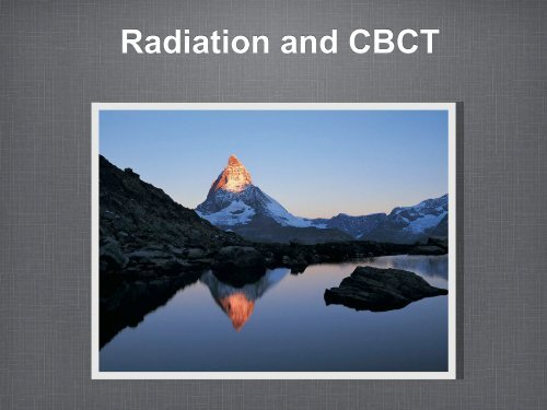
Radiation and CBCT
Radiation and CBCT
Radiation and CBCT

Create successful ePaper yourself
Turn your PDF publications into a flip-book with our unique Google optimized e-Paper software.
<strong>Radiation</strong> <strong>and</strong> <strong>CBCT</strong>
what is a normal radioactive<br />
dose??<br />
We live in a radioactive world!<br />
We can not see or feel it but it is all around us.<br />
The st<strong>and</strong>ard measurement is the mrem.<br />
An average annual dose of 620 mrem is common.<br />
International st<strong>and</strong>ards allow up to 5000 mrems.<br />
Medical <strong>and</strong> Dental xrays increase the normal dose.
environmental radiation<br />
american Nuclear society July 2010<br />
Cosmic radation at sea level 62 (mrems).<br />
Elevation above sea level add 2, 5, 9, 15, 21, 29,<br />
40, 53, 70 (mrems) for each 1000 additional feet.<br />
Atlantic Coasts add 16 (mrems).<br />
Colorado Plateau Area (Denver) 63 (mrems).<br />
Stone, Brick or Concrete Building 7 (mrems).<br />
From food , water <strong>and</strong> air 268 (mrem).
Entry Dose<br />
• Entry Dose is a measured amount of radiation at the<br />
tissue surface as the radiation enters the body. It is<br />
easily quantifiable using thermoradiolucent sensors<br />
on the surface of the skin.
medical/dental radiation<br />
Absorbed dose: fundamental quantity for describing<br />
effects of radiation in a tissue. It is the energy<br />
deposited in the volume of tissue the beam passes<br />
through divided by the mass of the tissue.<br />
(joules/kilogram) joules = xray energy, kilogram =<br />
mass or weight of tissue. 1 joule/kilogram = 1 gray<br />
(Gy)<br />
In older terms 1 Gy = 100 rad..<br />
1 Gy = 1,000,000 microsieverts
Medical/dental<br />
radiation<br />
Equivalent dose: Biological effects in tissue vary by<br />
the type of radiation given whether it is xrays, gamma<br />
rays, beta rays, neutrons of other particles. Each type<br />
of radiation interacts with tissues differently. This<br />
difference in reaction with tissues is described by a<br />
“radiation weighting factor” for each type of<br />
radiation. The “absorbed dose” (in Gy) averaged over<br />
the entire tissue is multiplied by the “radiation<br />
weighting factor” = the “equivalent dose. The<br />
equivalent dose is measured by the sievert (Sv).
Sv Gy <strong>and</strong> <strong>Radiation</strong><br />
weighting factors<br />
For x ray energy encountered in CT the radiation<br />
weighting factor is 1.0.<br />
equivalent dose (Sv) = absorbed dose (Gy) x radation<br />
weighting factor 1 (for CT)<br />
so for CT absorbed dose in a tissue in Gy is equal to<br />
equivalent dose in Sv.
effective dose<br />
or risk of cancer<br />
The risk of cancer from an equivalent dose depends<br />
on the organ receiving the dose. Each tissue has a<br />
specific tissue weighting factor based on its weight,<br />
density <strong>and</strong> other characteristics that account for the<br />
variations in cancer risk.<br />
The product of equivalent dose x tissue weighting<br />
factor = effective dose. It is a calculated<br />
measurement not a measured quantity.<br />
Effective dose is also measured by the sievert (Sv).
eview<br />
Absorbed dose: 1 joule/kilogram = 1Gy<br />
Equivalent dose: radiation weighting factor x<br />
absorbed dose (Gy) = equivalent dose (Sv).<br />
Effective dose: tissue weighting factor x equivalent<br />
dose. (Sv).
Conversions<br />
<strong>Radiation</strong> Units<br />
The rem (rem) is replaced by the sievert (Sv)<br />
1 kilorem (krem) = 10 sievert (Sv)<br />
1 rem (rem) = 10 millisievert (mSv)<br />
1 millirem (mrem) = 10 microsievert (µSv)<br />
1 microrem (µrem) = 10 nanosievert (nSv)<br />
The sievert (Sv) replaces the rem (rem)<br />
1 sievert (Sv) = 100 rem (rem)<br />
1 millisievert (mSv) = 100 millirem (mrem)<br />
1 microsievert (µSv) = 100 microrem (µrem)<br />
1 nanosievert (nSv) = 100 nanorem (nrem<br />
Multiples<br />
kilo (k) 1,000 = 10^3 = 1 thous<strong>and</strong><br />
hecto (h) 100 = 10^2 = 1 hundred<br />
deka (da)10 = 10 = ten<br />
1<br />
deci (d) 0.1 =10^-1 = 1 tenth<br />
centi (c) 0.01 = 10^-2 = 1 hundredth<br />
milli (m) 0.001 = 10^-3 = 1 thous<strong>and</strong>th<br />
micro (µ) 0.000 001 = 10^-6 = 1 millionth
Online converter<br />
online unit converter pro @<br />
http://online.unitconverterpro.com
New vs old numbers<br />
A somewhat new article (2008) by John Ludwig <strong>and</strong><br />
others in Oral Surg Oral Med Pathol Radiol Endod<br />
2008 has brought to light new radiation dose<br />
numbers that differ from current numbers noted by<br />
some <strong>CBCT</strong> manufacturers.<br />
What do they mean to the doctor <strong>and</strong> patient?
Things to know<br />
The ICRP - International Commission of Radiological<br />
Protection.<br />
ICRP has recently revised the tissue weighting<br />
factors <strong>and</strong> have included the salivary gl<strong>and</strong>s <strong>and</strong> the<br />
brain as a weighted tissue (remember: tissue<br />
weighting x equivalent dose = effective dose.<br />
Old effective doses were from 1990 new doses are<br />
from the 2007 recommendations.
x ray is cumulative<br />
Because x-ray risks are cumulative doctors need<br />
strategies to reduce dose.<br />
2007 recommendations have not been adequately<br />
reported.<br />
Tissue weighted factors: 2 new independent tissues,<br />
brain <strong>and</strong> salivary gl<strong>and</strong>s <strong>and</strong> remainder tissues from<br />
11 total to 14 total.
Organs included in the study<br />
Individual organs used for calculating E or effective dose<br />
(tissue weighting factor x equivalent dose).<br />
Bone Marrow, esophagus, thyroid, bone surface, skin<br />
dose, brain, salivary gl<strong>and</strong>s, oral mucosa, lymphatic<br />
nodes, muscle, <strong>and</strong> extrathoracic regions.<br />
Remember effective dose is a calculated amount not an<br />
actual measurement. If you examine the charts in the<br />
article you will see in Table V. very high numbers in the<br />
columns. Using the equation above gives us the<br />
effective doses we see in Table VI.
comparing the 1990 <strong>and</strong> the<br />
2007<br />
FOV<br />
Increase in effective<br />
dose<br />
Large NewTom 3G 42 68 62%<br />
Medium<br />
Manufacturer<br />
Effective dose<br />
1990 micro sv<br />
Effective dose<br />
2007 micr sv<br />
Classic i-CAT<br />
extended scan 125 235 87%<br />
Next Generation<br />
Portrait mode 37 74 100%<br />
Classic i-CAT<br />
st<strong>and</strong>ard scan 47 102 112%<br />
Next Generation<br />
L<strong>and</strong>scape mode 36 87 139%<br />
Galileos default<br />
exposure 28 70 148%<br />
Galileos maximum<br />
exposure 52 128 148%<br />
Somatron 64<br />
MDCT 453 860 90%<br />
Somatron 64<br />
MDCT care dose 285 534 87%
FOV Manufacturer 1990 effective dose 2007 effective dose % change<br />
Small<br />
Promax 3D<br />
small adult 151 488 224%<br />
Promax 3D<br />
large adult 203 488 222%<br />
PreXion 3D<br />
st<strong>and</strong>ard 512 66 189 187%<br />
PreXion 3D<br />
high res 1024 154 388 151%<br />
PreXion 3D<br />
V-11 38 (85) New Mode<br />
Large FOV had an 83% increase<br />
Medium FOV had a 115% increase<br />
Small FOV had a 196% increase
Effective doses of the i-CAT<br />
using several modes<br />
British J of radiology 2009 Jan:82(973):35-40<br />
This study was done in the UK <strong>and</strong> shows similar results as<br />
the study we just reviewed. It is more complete as it gives<br />
values for many of the different scans that can be done on<br />
the i-CAT.<br />
1990 ICRP 2007 ICRP<br />
Full Head 92.8 206.2<br />
13 cm jaws 39.5 133.9<br />
6cm high res m<strong>and</strong> 47.2 188.5<br />
6cm high res max 18.5 93.3<br />
6cm st<strong>and</strong>ard m<strong>and</strong> 23.9 96.2<br />
6cm st<strong>and</strong>ard max<br />
9.7 58.9
Evaluating the new data<br />
Greater increases in effective doses are seen in the smaller FOV This is due to the<br />
concentration of the beam or focus in areas where weighted tissues are found. These effects<br />
are diluted in the larger FOV scans.<br />
The comparison in terms of multiples of panoramics has surprising results. Even with the<br />
increase in effective doses of <strong>CBCT</strong> the multiples of panorexes decreases. It can be<br />
explained similarly to small FOV <strong>CBCT</strong> scans. Panoramic scans have rotational centers in<br />
the center of the floor of the mouth for scanning the anterior jaws. This rotation center<br />
coincides with the location of the parotid, subm<strong>and</strong>ibular <strong>and</strong> sublingual gl<strong>and</strong>s continuously<br />
exposing the same areas to radiation. The effective dose from this panoramic imaging will<br />
be higher than the more even distribution of the <strong>CBCT</strong>.<br />
Average Small FOV Scanner<br />
Scan to Panorex 1990 10<br />
Scan to Panorex 2007 5
Risks of <strong>CBCT</strong><br />
Still 1.5 to 12X less than MDCT.<br />
Considered a dose sparing technique.<br />
ALARA as-low-as-reasonably-achievable should<br />
always be considered.<br />
Dose vs Diagnostic task should be evaluated.<br />
1990 vs 2007 where do we go?????
Has <strong>Radiation</strong> Really<br />
Increased??<br />
• No. All machines still produce the same amount of<br />
radiation. We are using new parameters to measure<br />
effective dose. All numbers listed should be<br />
considered <strong>and</strong> averaged. Effective dose will vary<br />
based on the area of the scan <strong>and</strong> the mode<br />
incorporated in the scan acquisition.<br />
• Remember that resolution <strong>and</strong> radiation go h<strong>and</strong> in<br />
h<strong>and</strong>. To achieve the best image quality of any<br />
machine the higher dose of radiation is required.
Other Factors in Clear Images<br />
• <strong>Radiation</strong> in not monochromatic. Some photons are weaker <strong>and</strong><br />
do not reach the sensor.<br />
• Absorption <strong>and</strong> reflection of photons in the target tissues.<br />
• Noise to Signal ratio: the actual energy reaching the sensor is<br />
the signal. With low energies used in conebeam the noise ratio<br />
can increase rendering a “fuzzy” image.<br />
• Focal Point improves image quality: smaller = better.<br />
• Pixel size of the sensor.<br />
• Software processing.
<strong>Radiation</strong> in Perspective<br />
• As we see from the tests, radiation can vary from 60-<br />
500 msev with the typical scan.<br />
• <strong>Radiation</strong> treatment for a head <strong>and</strong> neck tumor<br />
approachs 250,000 msev.<br />
• We owe our patients the lowest possible dose with<br />
the corresponding acceptable diagnostic image.
Conclusion<br />
• This is not meant to be an all inclusive report on<br />
radiation. It should stimulate you the doctor to learn<br />
more about the radiation <strong>and</strong> the diagnostic<br />
information you getting from your current radiology<br />
practices. It is our obligation to use not the lowest<br />
radiation, but the best radiology mode to acquire the<br />
most complete <strong>and</strong> diagnostic information for our<br />
patients. Learn more <strong>and</strong> prosper.<br />
• Dr Dan McEowen<br />
e-mail: drdan1310@gmail.com