Respiratory Distress of the Newborn: Causes and ... - Trinity Health
Respiratory Distress of the Newborn: Causes and ... - Trinity Health
Respiratory Distress of the Newborn: Causes and ... - Trinity Health

You also want an ePaper? Increase the reach of your titles
YUMPU automatically turns print PDFs into web optimized ePapers that Google loves.
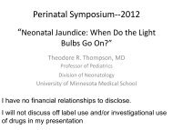
<strong>Respiratory</strong> <strong>Distress</strong> <strong>of</strong> <strong>the</strong> <strong>Newborn</strong>:<br />
<strong>Causes</strong> <strong>and</strong> Treatment<br />
<strong>Respiratory</strong> <strong>Distress</strong> <strong>of</strong> <strong>the</strong><br />
<strong>Newborn</strong>: <strong>Causes</strong> <strong>and</strong> Treatment<br />
<strong>Trinity</strong> <strong>Health</strong><br />
Perinatal Medicine <strong>and</strong> Women’s <strong>Health</strong>care<br />
33 rd Annual Symposium<br />
Minot, ND<br />
September 21, 2012<br />
Tom George, MD<br />
Pr<strong>of</strong>essor <strong>of</strong> Pediatrics<br />
Medical Director, NICU, University <strong>of</strong> Minnesota Amplatz Children’s Hospital<br />
Associate Director, Pediatric Residency Program<br />
Objectives<br />
At <strong>the</strong> end <strong>of</strong> this presentation, <strong>the</strong> attendee<br />
will be able to<br />
• Identify common causes <strong>of</strong> neonatal respiratory<br />
distress<br />
• Underst<strong>and</strong> how to evaluate <strong>and</strong> methods to treat<br />
neonatal respiratory distress<br />
• Underst<strong>and</strong> new issues in <strong>the</strong> treatment <strong>of</strong> neonatal<br />
respiratory distress<br />
Acute neonatal deterioration<br />
<strong>Respiratory</strong> causes<br />
• Upper airway<br />
• Pulmonary disease<br />
• Lower airway disease<br />
• Pulmonary compression<br />
1
<strong>Respiratory</strong> <strong>Distress</strong> <strong>of</strong> <strong>the</strong> <strong>Newborn</strong>:<br />
<strong>Causes</strong> <strong>and</strong> Treatment<br />
Acute neonatal deterioration<br />
<strong>Respiratory</strong> causes<br />
• Upper airway obstruction<br />
– nose: choanal stenosis<br />
– mouth/jaw: large tongue<br />
– larynx: cord paralysis<br />
– trachea: tracheomalacia, tracheal stenosis<br />
– extrinsic: cystic hygroma, vascular ring<br />
Interpreting neonatal x-rays<br />
• Also going to use this opportunity to<br />
review neonatal radiographic pathology<br />
Approach to reading x-rays<br />
• Make sure <strong>the</strong> x-ray belongs to <strong>the</strong> patient<br />
• Evaluate technique<br />
– What is <strong>the</strong> quality <strong>of</strong> <strong>the</strong> x-ray?<br />
– Technique- importance <strong>of</strong> positioning<br />
Rotation<br />
Lordosis<br />
2
<strong>Respiratory</strong> <strong>Distress</strong> <strong>of</strong> <strong>the</strong> <strong>Newborn</strong>:<br />
<strong>Causes</strong> <strong>and</strong> Treatment<br />
What is <strong>the</strong> quality <strong>of</strong> <strong>the</strong> x-ray?<br />
Extrinsic<br />
Clutter<br />
Normal chest x-ray<br />
(Before we look at pulmonary pathology)<br />
Also can <strong>of</strong>ten see thymic shadow in newborns<br />
Acute neonatal deterioration<br />
<strong>Respiratory</strong> causes - alveolar disease<br />
• Transient tachypnea <strong>of</strong> <strong>the</strong> newborn<br />
• Bacterial pneumonia<br />
• Surfactant deficiency - RDS<br />
3
<strong>Respiratory</strong> <strong>Distress</strong> <strong>of</strong> <strong>the</strong> <strong>Newborn</strong>:<br />
<strong>Causes</strong> <strong>and</strong> Treatment<br />
Transient tachypnea <strong>of</strong> <strong>the</strong> newborn<br />
Early<br />
• Retained Alveolar Fluid<br />
• Often a retrospective diagnosis<br />
12 h later<br />
Group B Strep Pneumonia<br />
early<br />
12 hrs later<br />
Group B Strep Pneumonia<br />
early<br />
12 hrs later<br />
4
<strong>Respiratory</strong> <strong>Distress</strong> <strong>of</strong> <strong>the</strong> <strong>Newborn</strong>:<br />
<strong>Causes</strong> <strong>and</strong> Treatment<br />
Surfactant Deficiency (RDS)<br />
• ‘Ground glass’ appearance<br />
• Air bronchograms<br />
• Symmetric disease<br />
• Predominantly in preterm<br />
infants, though it can occur<br />
in term infants, especially<br />
those delivered by C-<br />
section without labor, or<br />
elective sections before 39<br />
weeks<br />
Acute neonatal deterioration<br />
<strong>Respiratory</strong> causes - lower airway<br />
• Meconium Aspiration Syndrome<br />
– Secondary Surfactant Inactivation<br />
Early Meconium Aspiration<br />
• Diffuse patchy<br />
infiltrates<br />
• Hyperinflation that<br />
places <strong>the</strong>m at risk<br />
for airleaks<br />
• At risk for PPHN<br />
5
<strong>Respiratory</strong> <strong>Distress</strong> <strong>of</strong> <strong>the</strong> <strong>Newborn</strong>:<br />
<strong>Causes</strong> <strong>and</strong> Treatment<br />
Late Meconium Aspiration<br />
Phrenic Nerve Injury<br />
• Brachial plexus injury<br />
• Can have associated phrenic nerve injury<br />
leading to respiratory distress<br />
Right Diaphragm Paralysis<br />
6
<strong>Respiratory</strong> <strong>Distress</strong> <strong>of</strong> <strong>the</strong> <strong>Newborn</strong>:<br />
<strong>Causes</strong> <strong>and</strong> Treatment<br />
Air Leak Syndromes/<br />
Primary Lung Compression<br />
• Pneumothorax<br />
• Congenital Diaphragmatic Hernia<br />
• Cystic Adenomatoid Malformation<br />
• Pleural Effusion<br />
• Thoracic Compression<br />
Right tension pneumothorax<br />
• Presents with acute<br />
hypoxemia, also possible<br />
bradycardia, hypotension<br />
• Decreased BS on right<br />
• Needle thoracentesis<br />
Right Pneumothorax<br />
after needle thoracentesis<br />
7
<strong>Respiratory</strong> <strong>Distress</strong> <strong>of</strong> <strong>the</strong> <strong>Newborn</strong>:<br />
<strong>Causes</strong> <strong>and</strong> Treatment<br />
Congenital Diaphragmatic Hernia<br />
• Acute respiratory distress at birth<br />
• Intubation <strong>and</strong> NG to suction<br />
Congenital Adenomatoid Malformation<br />
Pleural Effusion<br />
8
<strong>Respiratory</strong> <strong>Distress</strong> <strong>of</strong> <strong>the</strong> <strong>Newborn</strong>:<br />
<strong>Causes</strong> <strong>and</strong> Treatment<br />
Thoracic Compression<br />
Objectives<br />
At <strong>the</strong> end <strong>of</strong> this presentation, <strong>the</strong> attendee<br />
will be able to<br />
• Identify common causes <strong>of</strong> neonatal respiratory<br />
distress<br />
• Underst<strong>and</strong> how to evaluate <strong>and</strong> methods to treat<br />
neonatal respiratory distress<br />
• Underst<strong>and</strong> new issues in <strong>the</strong> treatment <strong>of</strong> neonatal<br />
respiratory distress<br />
Evaluation <strong>and</strong> Stabilization <strong>of</strong> <strong>the</strong><br />
<strong>Newborn</strong> Infant in <strong>Respiratory</strong> <strong>Distress</strong><br />
• Establish Airway (suctioning, mask,<br />
endotracheal tube)<br />
• Evaluate Breathing (bag if needed)<br />
• Maintain Circulation<br />
• Administer Drugs<br />
– Antibiotics - ampicillin <strong>and</strong> gentamicin<br />
(minimize cefotax use due to resistance)<br />
– IV flluids with dextrose<br />
• Maintain Environment (<strong>the</strong>rmal regulation)<br />
9
<strong>Respiratory</strong> <strong>Distress</strong> <strong>of</strong> <strong>the</strong> <strong>Newborn</strong>:<br />
<strong>Causes</strong> <strong>and</strong> Treatment<br />
Evaluation <strong>and</strong> Stabilization <strong>of</strong> <strong>the</strong><br />
Neonate with <strong>Respiratory</strong> <strong>Distress</strong><br />
• History, physical examination<br />
• Vital signs, with blood pressure<br />
• Pulse oximetry—SaO 2 / Arterial blood gases<br />
• Chest x-ray<br />
• CBC with differential, platelet count<br />
• Blood culture, o<strong>the</strong>r cultures as indicated<br />
Assessment <strong>of</strong> <strong>the</strong> acutely ill newborn<br />
• <strong>Respiratory</strong>:<br />
– Tachypnea – rate > 70-80 breaths/minute<br />
– Grunting, retractions, flaring <strong>of</strong> alae nasi<br />
– Reduced air entry<br />
– Apnea<br />
– Cyanosis<br />
–Stridor<br />
Acute neonatal deterioration<br />
Assessment <strong>of</strong> <strong>the</strong> acutely ill newborn<br />
• Objective assessments<br />
– Vitals signs<br />
• If worried about a newborn, should be<br />
continuously monitored with cardiac/apnea<br />
monitors<br />
– Frequent reassessment<br />
10
<strong>Respiratory</strong> <strong>Distress</strong> <strong>of</strong> <strong>the</strong> <strong>Newborn</strong>:<br />
<strong>Causes</strong> <strong>and</strong> Treatment<br />
Evaluation <strong>and</strong> Stabilization<br />
<strong>Respiratory</strong> Support for <strong>the</strong> Neonate<br />
<strong>Respiratory</strong> support should be provided for <strong>the</strong> infant<br />
with respiratory distress, especially if hypoxemia is<br />
present<br />
• Blow-by oxygen<br />
• Nasal cannula<br />
• Oxyhood<br />
• Mechanical ventilation<br />
Evaluation <strong>and</strong> Stabilization<br />
<strong>Respiratory</strong> Support for <strong>the</strong> Neonate<br />
• Blow-by oxygen:<br />
Acutely used for a non-intubated infant with<br />
hypoxemia<br />
Evaluation <strong>and</strong> Stabilization<br />
<strong>Respiratory</strong> Support for <strong>the</strong> Neonate<br />
• Nasal cannula - limited FiO 2 that can be delivered<br />
– STOP ROP effective FiO 2 conversion tables<br />
– OxygenbyNC.pdf<br />
• High Flow Nasal Cannula (HFNC)<br />
– To deliver CPAP, need higher flow <strong>of</strong> 3-4 lpm<br />
– Effective in some situations, particularly for transitioning<br />
infants <strong>of</strong>f PPV with resolving RDS<br />
– Should be cautious about use in babies for initial<br />
treatment <strong>of</strong> RDS<br />
11
<strong>Respiratory</strong> <strong>Distress</strong> <strong>of</strong> <strong>the</strong> <strong>Newborn</strong>:<br />
<strong>Causes</strong> <strong>and</strong> Treatment<br />
Evaluation <strong>and</strong> Stabilization<br />
<strong>Respiratory</strong> Support for <strong>the</strong> Neonate<br />
• Oxyhood - delivers FiO 2 <strong>of</strong> up to 90 - 100%<br />
Evaluation <strong>and</strong> Stabilization<br />
<strong>Respiratory</strong> Support for <strong>the</strong> Neonate<br />
• Mechanical ventilation<br />
– For acute deterioration, or for worsening clinical<br />
condition<br />
Evaluation <strong>and</strong> Stabilization<br />
<strong>Respiratory</strong> Support for <strong>the</strong> Neonate<br />
Indications for Mechanical Ventilation<br />
• <strong>Respiratory</strong> acidosis<br />
(pH < 7.25 with increasing PaCO 2 )<br />
• Hypoxemia –<br />
saturations < ~ 85%, PaO 2 < 50 mmHg in FiO 2 > 60%<br />
• Apnea <strong>of</strong> prematurity<br />
12
<strong>Respiratory</strong> <strong>Distress</strong> <strong>of</strong> <strong>the</strong> <strong>Newborn</strong>:<br />
<strong>Causes</strong> <strong>and</strong> Treatment<br />
Endotracheal Tube:<br />
Size, Length by Weight, Gestational age<br />
Weight (g)<br />
Below 1000 1<br />
Gestational<br />
Age (wks)<br />
Below 28<br />
Tube Size<br />
(mm)<br />
(Gestational Age ÷<br />
2.5 10)<br />
Number at<br />
Lip to Tip<br />
6 + wt (cm)<br />
6.5<br />
1000-2000<br />
28-34<br />
3.0<br />
7-8<br />
2000-3000<br />
34-38<br />
3.0-3.5<br />
8-9<br />
Above 3000<br />
Above 38<br />
3.5-4.0<br />
>9<br />
Intubation: Atropine (0.02 mg/kg IV) → Morphine (0.1 mg/kg IV)<br />
→ Rocuronium(0.6 mg/kg IV)<br />
Endotracheal Tube in <strong>the</strong> Esophagus<br />
• Poor chest movement<br />
• Poor response to intubation - no<br />
improvement in color, heart rate, tone,<br />
respiratory effort<br />
• No mist in tube<br />
• Crying sounds around tube<br />
Endotracheal Tube in <strong>the</strong> Esophagus<br />
• Air heard entering stomach/gastric<br />
distension<br />
• No breath sounds<br />
•CO 2 detector - no expired CO 2<br />
– (Remains purple)<br />
13
<strong>Respiratory</strong> <strong>Distress</strong> <strong>of</strong> <strong>the</strong> <strong>Newborn</strong>:<br />
<strong>Causes</strong> <strong>and</strong> Treatment<br />
Goals <strong>of</strong> respiratory support<br />
• Maintain adequate tissue oxygenation<br />
• Allow recovery without causing additional<br />
lung injury<br />
Evaluation <strong>and</strong> Stabilization<br />
Mechanical Ventilation<br />
• Continuous positive airway pressure (CPAP)<br />
• Synchronized CPAP (SiPAP/rated CPAP)<br />
• Conventional mechanical ventilation (SIMV)<br />
• High frequency ventilation<br />
Evaluation <strong>and</strong> Stabilization<br />
Continuous Positive Airway Pressure<br />
• CPAP maintains end-expiratory airway pressure,<br />
usually at 5 - 8 cm H 2 O:<br />
– increases resting lung volume thus improving<br />
oxygenation <strong>and</strong> ventilation<br />
– maintains upper airway patency<br />
– improves distribution <strong>of</strong> ventilation<br />
14
<strong>Respiratory</strong> <strong>Distress</strong> <strong>of</strong> <strong>the</strong> <strong>Newborn</strong>:<br />
<strong>Causes</strong> <strong>and</strong> Treatment<br />
Evaluation <strong>and</strong> Stabilization<br />
Continuous Positive Airway Pressure<br />
• Several methods to deliver CPAP:<br />
Nasal prongs<br />
Nasopharyngeal<br />
HFNC to deliver CPAP<br />
NIPPV<br />
• Effective in decreasing apneic events in infants<br />
on CPAP in premature infants (Lin 1998)<br />
• Most delivery systems are asynchronous <strong>and</strong> so<br />
use <strong>of</strong> higher rates should be considered<br />
carefully<br />
• Might prevent need for reintubation<br />
• Lower rate <strong>of</strong> BPD (Ramanathan 2012)<br />
• Not indicated for treatment <strong>of</strong> acute RDS<br />
Evaluation <strong>and</strong> Stabilization<br />
Conventional Mechanical Ventilation<br />
Initial settings:<br />
• PIP (Peak Inspiratory Pressure) or TV (tidal volume):<br />
– 16 - 30 cm H 2 O pressure, adjust based on infant size,chest wall movement<br />
– Or TV instead <strong>of</strong> PIP for volume control ( ~ 5 cc/kg)<br />
• PEEP (Positive end-expiratory pressure):<br />
– 4 - 7 cm H 2 O pressure<br />
• IT (Inspiratory time): 0.35 - 0.5 seconds<br />
• <strong>Respiratory</strong> rate: 20 - 40 breaths per minute<br />
• Monitor response<br />
– oxygenation (saturations <strong>and</strong> amount <strong>of</strong> FiO 2 required)<br />
– ventilation (blood gas)<br />
15
<strong>Respiratory</strong> <strong>Distress</strong> <strong>of</strong> <strong>the</strong> <strong>Newborn</strong>:<br />
<strong>Causes</strong> <strong>and</strong> Treatment<br />
Oxygenation<br />
• Oxygenation (saturations, PaO 2 ):<br />
- primarily determined by <strong>the</strong><br />
Mean Airway Pressure (MAP)<br />
• MAP can be increased by:<br />
- increasing PIP, PEEP <strong>and</strong>/or IT<br />
Ventilation<br />
• Ventilation (PaCO 2 ):<br />
- primarily determined by <strong>the</strong> total<br />
volume <strong>of</strong> air that passes in <strong>and</strong> out <strong>of</strong> <strong>the</strong><br />
alveoli, - <strong>the</strong> tidal volume (TV) delivered by<br />
<strong>the</strong> ventilator<br />
• TV typically increased by:<br />
- increasing <strong>the</strong> RR<br />
- can also PIP or PEEP<br />
Goals <strong>of</strong> Mechanical Ventilation<br />
Depends on <strong>the</strong> disease process:<br />
•In RDS:<br />
Oxygenation: saturation ~ 85 - 95%<br />
(PaO 2 50 - 80 mmHg)<br />
Ventilation: PaCO 2 45 - 60 mmHg<br />
in RDS as long as pH > 7.25<br />
• In PPHN/MAS:<br />
Sats > 90%; PaCO 2 more normal - 40-50 mmHg<br />
16
<strong>Respiratory</strong> <strong>Distress</strong> <strong>of</strong> <strong>the</strong> <strong>Newborn</strong>:<br />
<strong>Causes</strong> <strong>and</strong> Treatment<br />
<strong>Respiratory</strong> - Customized Goals<br />
• Preterm infant<br />
– Hypocarbia associated with PVL<br />
– Hyperoxia associated with ROP<br />
– Volutrauma associated with pneumothorax, IVH <strong>and</strong> BPD<br />
• Term infant with pulmonary hypertension<br />
– Hypercarbia <strong>and</strong> hypoxemia increase PVR <strong>and</strong> shunting<br />
• Term infant with congenital heart disease<br />
– Vary parameters to optimize tissue perfusion <strong>and</strong><br />
oxygenation, generally normal pCO 2<br />
Synchronized Intermittent<br />
Mechanical Ventilation<br />
• Synchronizes IMV breaths by sensing a<br />
spontaneous breath <strong>and</strong> delivers a mechanical<br />
breath synchronized with <strong>the</strong> infants inspiratory<br />
phase<br />
• Sensing is typically via flow sensing systems<br />
• Still set TV or PIP, PEEP, IT <strong>and</strong> RR<br />
SIMV- proven <strong>and</strong> potential benefits<br />
• Studies have found:<br />
– improved oxygenation <strong>and</strong> ventilation in infants<br />
crossed-over between SIMV <strong>and</strong> IMV, with<br />
higher PaO 2 <strong>and</strong> lower PaCO 2<br />
– less cerebral blood flow velocity variability,<br />
particularly <strong>of</strong> concern in premature infants<br />
• Decreased agitation with “fighting <strong>the</strong> vent”<br />
• Decreased need for sedation/paralysis<br />
17
<strong>Respiratory</strong> <strong>Distress</strong> <strong>of</strong> <strong>the</strong> <strong>Newborn</strong>:<br />
<strong>Causes</strong> <strong>and</strong> Treatment<br />
It’s not just <strong>the</strong> <strong>the</strong> respiratory support<br />
• Nutrition<br />
• Optimizing PEEP/MAP<br />
• Fluid management<br />
• Addressing infectious disease issues<br />
• Surfactant for post-surfactant slump<br />
Katz & Klein, J Perinat 2006;26:414<br />
Objectives<br />
At <strong>the</strong> end <strong>of</strong> this presentation, <strong>the</strong> attendee<br />
will be able to<br />
• Identify common causes <strong>of</strong> neonatal respiratory<br />
distress<br />
• Underst<strong>and</strong> how to evaluate <strong>and</strong> methods to treat<br />
neonatal respiratory distress<br />
• Underst<strong>and</strong> new issues in <strong>the</strong> treatment <strong>of</strong> neonatal<br />
respiratory distress<br />
Changing trends<br />
• Use <strong>of</strong> early CPAP to avoid intubation<br />
• iNO in preterm infants<br />
• Oxygen use in <strong>the</strong> DR<br />
18
<strong>Respiratory</strong> <strong>Distress</strong> <strong>of</strong> <strong>the</strong> <strong>Newborn</strong>:<br />
<strong>Causes</strong> <strong>and</strong> Treatment<br />
CPAP<br />
• SUPPORT trial<br />
– Significant percentage <strong>of</strong> infants who don’t need<br />
intubation with early, optimal CPAP to prevent collapse<br />
<strong>of</strong> alveoli<br />
– A trial <strong>of</strong> nasal CPAP should be considered as an<br />
alternative to routine DR intubation<br />
• If intubation is needed:<br />
– consider early extubation after surfactant to optimal<br />
CPAP in preterm infants<br />
Inhaled Nitric Oxide<br />
• Valuable tool in <strong>the</strong> treatment <strong>of</strong> Persistent Pulmonary<br />
Hypertension <strong>of</strong> <strong>the</strong> <strong>Newborn</strong><br />
• PPHN occurs in ~1/1400 live births causing severe<br />
<strong>and</strong> labile hypoxemia<br />
• FDA approved for hypoxemic respiratory failure in<br />
term <strong>and</strong> near-term infants, decreases need for ECMO<br />
• iNO diffuses from alveolus to pulmonary vascular<br />
smooth muscle<br />
• increases cGMP levels<br />
• leads to muscle relaxation <strong>and</strong> vasodilation<br />
Inhaled Nitric Oxide in Preterm Infants<br />
• 3 large trials<br />
– One study shows benefit in larger babies, one study shows<br />
decreased CLD (iNO started later), one shows no benefit<br />
– Questions remain about when to start, how long to treat, dose<br />
– NICH consensus 2010: routine use not recommended<br />
– Select use for premature infants with pulmonary<br />
hyptertension/pulmonary hypoplasia with pulmonary<br />
hypertension<br />
• TOLSURF trial now ongoing<br />
– iNO combined with 5 doses <strong>of</strong> late surfactant starting<br />
DOL 7-14 for intubated patients < 28 weeks<br />
19
<strong>Respiratory</strong> <strong>Distress</strong> <strong>of</strong> <strong>the</strong> <strong>Newborn</strong>:<br />
<strong>Causes</strong> <strong>and</strong> Treatment<br />
Oxygen use in <strong>the</strong> DR<br />
• NRP recommendations:<br />
Current evidence is insufficient to resolve<br />
all questions regarding supplemental<br />
oxygen use during neonatal resuscitation<br />
Pre-ductal <strong>and</strong> Post-ductal O2 Saturation in<br />
<strong>Health</strong>y Term Neonates after Birth<br />
Mariani et al J Peds 2007<br />
Oxygen use in <strong>the</strong> DR – term infants<br />
• Research suggests that 21% oxygen might be<br />
as effective as 100% oxygen<br />
• Recommendations:<br />
- start resuscitation with room air<br />
- <strong>the</strong>n oximetry could guide <strong>the</strong><br />
concentration <strong>of</strong> oxygen needed<br />
20
<strong>Respiratory</strong> <strong>Distress</strong> <strong>of</strong> <strong>the</strong> <strong>Newborn</strong>:<br />
<strong>Causes</strong> <strong>and</strong> Treatment<br />
SpO2 values in <strong>the</strong> first 20 minutes after birth in extremely low gestational age<br />
neonates r<strong>and</strong>omly assigned to <strong>the</strong> low-oxygen (Lox) group (initial FIO2 after<br />
birth: 30%) or <strong>the</strong> high-oxygen (Hox) group (initial FIO2 after birth: 90%)<br />
Escrig, R. et al.<br />
Pediatrics<br />
Copyright ©2008 American Academy <strong>of</strong> Pediatrics<br />
Oxygen use in <strong>the</strong> DR – preterm infants<br />
• Optimum saturation in preterm infants in <strong>the</strong><br />
first minutes <strong>of</strong> life is not known<br />
• A high oxygen saturation in preterm infants<br />
might be damaging<br />
• Titrate oxygen to achieve balance <strong>of</strong> need for<br />
sufficient oxygen to correct hypoxemia<br />
against excessive oxygen levels<br />
Oxygen use in <strong>the</strong> DR – preterm infants<br />
• Delivery centers have placed blenders in <strong>the</strong> DR<br />
• Some start at 21% <strong>and</strong> increase<br />
• Some start at < 100% <strong>and</strong> as needed<br />
• Awareness <strong>of</strong> saturations is important<br />
• Even a few breaths <strong>of</strong> high oxygen results in<br />
inflammatory cytokines<br />
• Effect on retinal vessels<br />
21
<strong>Respiratory</strong> <strong>Distress</strong> <strong>of</strong> <strong>the</strong> <strong>Newborn</strong>:<br />
<strong>Causes</strong> <strong>and</strong> Treatment<br />
AAP recommendations<br />
• Oxygen blenders in <strong>the</strong> DR<br />
• One team member attaches oximeter probe<br />
to right wrist<br />
• Start oxygen at < 100%; we start at 30%.<br />
Summary<br />
• Reviewed common causes <strong>of</strong> neonatal<br />
respiratory distress<br />
• Reviewed how to evaluate <strong>and</strong> treat neonatal<br />
respiratory distress<br />
• Reviewed newer issues in <strong>the</strong> treatment <strong>of</strong><br />
neonatal respiratory distress<br />
Thank you<br />
Questions/comments welcome!<br />
tgeorge@umn.edu<br />
22