
You also want an ePaper? Increase the reach of your titles
YUMPU automatically turns print PDFs into web optimized ePapers that Google loves.
Volume 13<br />
Number 1<br />
April <strong>2013</strong><br />
Published by<br />
European<br />
Wound Management<br />
Association<br />
SUPPLEMENT<br />
23 rd Conference of the<br />
European Wound Management Association<br />
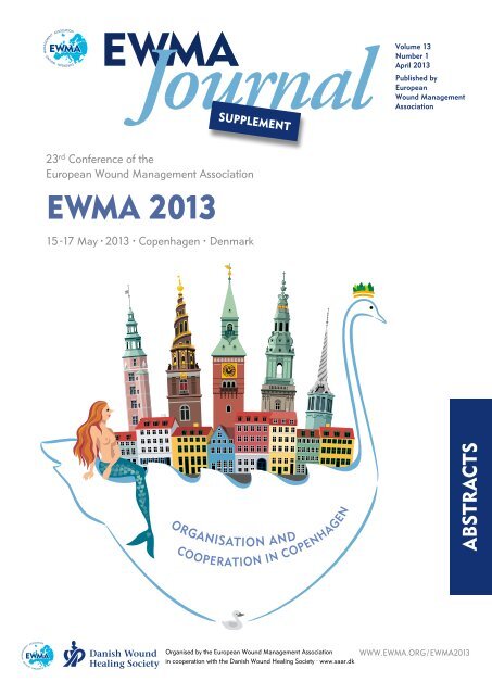
<strong>EWMA</strong> <strong>2013</strong><br />
15 -17 May · <strong>2013</strong> · Copenhagen · Denmark<br />
ABSTRACTS<br />
Organised by the European Wound Management Association<br />
in cooperation with the Danish Wound Healing Society · www.saar.dk<br />
WWW.<strong>EWMA</strong>.ORG / <strong>EWMA</strong><strong>2013</strong>
Dechronifying with PolyHeal<br />
An innovative technology<br />
for treating hard to heal wounds<br />
NCM<br />
TECHNOLOGY<br />
NCM*<br />
TECHNOLOGY<br />
NCM<br />
TECHNOLOGY<br />
Proven efficacy in 4 clinical studies<br />
• 64.5% of wounds reached > 75% granulation within 4 weeks of treatment<br />
• Up to 40% wound area reduction within 4 weeks<br />
• 3 out of 10 wounds closed<br />
Safe and well tolerated<br />
Easy to use<br />
Indicated for the treatment of wounds of various etiologies<br />
<br />
www.polyheal.com
European Wound<br />
Management Association<br />
Executive Committee<br />
Jan Apelqvist, President<br />
Zena Moore, Immediate Past President<br />
Salla Seppanen, President Elect<br />
Corrado M. Durante, Treasurer<br />
Gerrolt Jukema, Recorder<br />
Dubravko Huljev, Honorary Secretary<br />
Council Members<br />
Paulo Alves<br />
Magdalena Annersten Gershater<br />
Sue Bale, <strong>EWMA</strong> Journal Editor<br />
Barbara E. den Boogert-Ruimschotel<br />
Mark Collier<br />
Javorka Delic<br />
Ann-Mari Fagerdahl<br />
Georgina Gethin<br />
Luc Gryson<br />
Nada Kecelj-Leskovec<br />
Martin Koschnick<br />
Sebastian Probst<br />
Elia Ricci<br />
Rytis Rimdeika<br />
Robert Strohal<br />
Jose Verdu Soriano<br />
Address<br />
<strong>EWMA</strong> Secretariat<br />
Nordre Fasanvej 113, 2<br />
DK-2000 Frederiksberg<br />
Denmark<br />
Tel.: +45 7020 0305<br />
ewma@ewma.org<br />
www.ewma.org<br />
Danish Wound Healing Society<br />
Council<br />
Eskild W. Henneberg, President<br />
Susan Bermark, Vice President<br />
Maria Plaschke, Secretary<br />
Jens Lykke Sørensen, Treasurer<br />
Rolf Jelnes<br />
Bo Jørgensen<br />
Annette V. Norden<br />
Ann Brockdorff<br />
Anne Marie Rasmussen<br />
Aksel Jes Bomberg<br />
Address<br />
Danish Wound Healing Society<br />
Cypresvej 18<br />
DK-3450 Alleroed<br />
Denmark<br />
dsfs@mail.tele.dk<br />
www.saar.dk<br />
CONFERENCE ORGANISATION<br />
Scientific Committee<br />
Gerrolt Jukema, Recorder<br />
Paulo Alves, <strong>EWMA</strong><br />
Jan Apelqvist, <strong>EWMA</strong><br />
Corrado Durante, <strong>EWMA</strong><br />
Francisco P. G. Fernandez, GNEAUPP<br />
Finn Gottrup, DSFS<br />
Eskild W. Henneberg, DSFS<br />
Pedro L. Pancorbo Hidalgo, GNEAUPP<br />
Pablo Lopez Casanova, GNEAUPP<br />
Rolf Jelnes, DSFS<br />
Zena Moore, <strong>EWMA</strong><br />
Sebastian Probst, <strong>EWMA</strong><br />
Bente Ramskover, DSFS<br />
Rytis Rimdeika, <strong>EWMA</strong><br />
José Verdú Soriano, GNEAUPP<br />
Index<br />
SUPPLEMENT<br />
Conference Organisations 1<br />
<strong>EWMA</strong> Council 2<br />
Welcome to <strong>EWMA</strong> <strong>2013</strong> 3<br />
About the conference logo 4<br />
<strong>EWMA</strong> Documents 5<br />
The <strong>EWMA</strong> University<br />
Conference Model (UCM) 6<br />
<strong>EWMA</strong> Corporate Sponsor<br />
Contact Data 8<br />
About <strong>EWMA</strong> 9<br />
Submit your paper to<br />
<strong>EWMA</strong> Journal 10<br />
Become a member of <strong>EWMA</strong> 10<br />
Oral Presentations Overview 13<br />
Oral Presentations 19<br />
Poster Presentations Overview 118<br />
Poster Presentations 125<br />
E-Poster Presentations Overview 244<br />
Find <strong>EWMA</strong> on<br />
Local Organising Committee<br />
Susanne Aagaard<br />
Susan Bermark<br />
Finn Gottrup<br />
Else Godsk Vestergaard<br />
Eskild W. Henneberg<br />
Jens Lykke Sørensen<br />
E-Poster Presentations 249<br />
Author Index 332<br />
<strong>EWMA</strong> <strong>2013</strong><br />
COPENHAGEN<br />
15-17 May · <strong>2013</strong><br />
CONFERENCE SECRETARIAT<br />
<strong>EWMA</strong> Secretariat<br />
Nordre Fasanvej 113, 2<br />
DK-2000 Frederiksberg<br />
Denmark<br />
Tel.: +45 7020 0305<br />
ewma@ewma.org<br />
www.ewma.org/ewma<strong>2013</strong><br />
Danish Wound<br />
Healing Society<br />
1
<strong>EWMA</strong><br />
Council<br />
Jan Apelqvist<br />
President<br />
Salla Seppänen<br />
President Elect<br />
Zena Moore<br />
Immediate Past President<br />
Corrado M. Durante<br />
Treasurer<br />
Gerrolt Jukema<br />
Scientific Recorder<br />
Dubravko Huljev<br />
Secretary<br />
Paulo Alves<br />
Sue Bale<br />
<strong>EWMA</strong> Journal Editor<br />
Barbara E.<br />
den Boogert-Ruimschotel<br />
Mark Collier<br />
Javorka Delic<br />
Ann-Mari Fagerdahl<br />
Georgina Gethin<br />
Magdalena Annersten<br />
Gershater<br />
Nada Kecelj-Leskovec<br />
Martin Koschnick<br />
Sebastian Probst<br />
Elia Ricci<br />
Rytis Rimdeika<br />
José Verdú Soriano<br />
Robert Strohal<br />
CO-OPERATING ORGANISATIONS’ BOARD<br />
Esther Armans Moreno, AEEVH<br />
Christian Thyse, AFISCeP.be<br />
Tommaso Bianchi, AISLeC<br />
Roberto Cassino, AIUC<br />
Aníbal Justiniano, APTFeridas<br />
Gilbert Hämmerle, AWA<br />
Jan Vandeputte, BEFEWO<br />
Vladislav Hristov, BWA<br />
Els Jonckheere, CNC<br />
Lenka Veverková, CSLR<br />
Ivana Vranjkovic, CWA<br />
Arne Buss, DGfW<br />
Bo Jørgensen, DSFS<br />
Heidi Castrén, FWCS<br />
Pedro Pacheco, GAIF<br />
J. Javier Soldevilla, GNEAUPP<br />
Christian Münter, ICW<br />
Aleksandra Kuspelo, LBAA<br />
Susan Knight, LUF<br />
Loreta Pilipaityte, LWMA<br />
Corinne Ward, MASC<br />
Hunyadi János, MSKT<br />
Suzana Nikolovska, MWMA<br />
Anne Wilson, NATVNS<br />
Kristin Bergersen, NIFS<br />
Louk van Doorn, NOVW<br />
Arkadiusz Jawień, PWMA<br />
Severin Läuchli, SAfW (DE)<br />
Hubert Vuagnat, SAfW (FR)<br />
Goran D. Lazovic, SAWMA<br />
Mária Hok, SEBINKO<br />
F. Xavier Santos <strong>Here</strong>dero, SEHER<br />
Sylvie Meaume, SFFPC<br />
Susanne Dufva, SSIS<br />
Jozefa Košková, SSOOR<br />
Leonid Rubanov, STW (Belarus)<br />
Guðbjörg Pálsdóttir, SUMS<br />
Cedomir Vucetic, SWHS Serbia<br />
Magnus Löndahl, SWHS Sweden<br />
Alison Hopkins, TVS<br />
Jasmina Begić-Rahić, URuBiH<br />
Zoya Ishkova, UWTO<br />
Barbara E. den Boogert-Ruimschotel, V&VN<br />
Julie Jordan O’Brien, WMAI<br />
Skender Zatriqi, WMAK<br />
Nada Kecelj Leskovec, WMAS<br />
Mustafa Deveci, WMAT<br />
<strong>EWMA</strong> JOURNAL SCIENTIFIC REVIEW PANEL<br />
Paulo Jorge Pereira Alves, Portugal<br />
Caroline Amery, UK<br />
Jan Apelqvist, Sweden<br />
Sue Bale, UK<br />
Michelle Briggs, UK<br />
Stephen Britland, UK<br />
Mark Collier, UK<br />
Rose Cooper, UK<br />
Javorka Delic, Serbia<br />
Corrado Maria Durante, Italy<br />
Bulent Erdogan, Turkey<br />
Ann-Mari Fagerdahl, Sweden<br />
Madeleine Flanagan, UK<br />
Milada Franců, Czech Republic<br />
Peter Franks, UK<br />
Francisco P. García-Fernández, Spain<br />
Magdalena Annersten Gershater, Sweden<br />
Georgina Gethin, Ireland<br />
Luc Gryson, Belgium<br />
Eskild W. Henneberg, Denmark<br />
Alison Hopkins, UK<br />
Gabriela Hösl, Austria<br />
Dubravko Huljev, Croatia<br />
Gerrolt Jukema, Netherlands<br />
Nada Kecelj, Slovenia<br />
Klaus Kirketerp-Møller, Denmark<br />
Zoltán Kökény, Hungary<br />
Martin Koschnick, Germany<br />
Severin Läuchli, Schwitzerland<br />
Maarten J. Lubbers, Netherlands<br />
Sylvie Meaume, France<br />
Zena Moore, UK<br />
Christian Münter, Germany<br />
Andrea Nelson, UK<br />
Pedro L. Pancorbo-Hidalgo, Spain<br />
Hugo Partsch, Austria<br />
Patricia Price, UK<br />
Sebastian Probst, Schwitzerland<br />
Elia Ricci, Italy<br />
Rytis Rimdeika, Lithuania<br />
Zbigniew Rybak, Poland<br />
Salla Seppänen, Finland<br />
José Verdú Soriano, Spain<br />
Robert Strohal, Austria<br />
Richard White, UK<br />
Carolyn Wyndham-White, Switzerland<br />
Gerald Zöch, Austria<br />
2
Dear Participant<br />
We are pleased to welcome you to the 23rd Conference of the European Wound<br />
Management Association in Copenhagen: <strong>EWMA</strong> <strong>2013</strong>. This conference is being held<br />
in cooperation with the Danish Wound Healing Society (DSFS).<br />
<strong>EWMA</strong> <strong>2013</strong> is dedicated to sharing and debating the latest knowledge and developments<br />
in wound management. During the 3 exciting conference days, participants will<br />
experience a diverse programme that includes keynote sessions, free paper sessions,<br />
workshops, full-day streams, guest sessions, and sponsored satellite symposia.<br />
The conference theme, Organisation and Cooperation in Copenhagen, reflects the fact<br />
that the quintessence of successful wound management requires cooperation across<br />
several different caregiver professions as well as cooperation between caregivers and<br />
patients. This theme will be emphasised at the opening plenary session, which focuses<br />
on the importance of multi disciplinary approaches to wound care.<br />
<strong>EWMA</strong> <strong>2013</strong> will include new topics of importance to the European wound community<br />
as well as topics that have had huge appeal during previous <strong>EWMA</strong> conferences.<br />
The key sessions in <strong>2013</strong> will address several <strong>EWMA</strong> focus areas, including antimicrobials,<br />
wound care in home care settings, and patient safety aspects of wound care.<br />
Other exciting key sessions will offer presentations on regenerative medicine,<br />
nutrition, leg ulcers, diabetic foot, and evidence in wound care.<br />
Following the full-day streams will offer participants the chance to experience more<br />
in-depth presentations and discussions within a particular field. The full-day symposia<br />
topics include the Nordic Diabetic Foot Symposium (Thursday, 16 May),<br />
E-Health Day (Friday, 17 May), and the Russian Speaking Symposium (Wednesday<br />
and Thursday, 15 and 16 May). Furthermore, a symposium on wound care in<br />
resource-poor settings, which will take place on Thursday, 16 May,<br />
is one of the many highlights of this year’s conference.<br />
<strong>EWMA</strong> workshops are often interactive and provide participants with an opportunity<br />
to address and elaborate on particular aspects of the themes of the individual sessions.<br />
This year’s workshops will cover topics on debridement, dressings and topical agents,<br />
palliative care, cancer wounds, compression bandaging, maggot therapy,<br />
and pressure ulcer prevention.<br />
Thanks to abstract submissions from around the world, we assembled an extensive<br />
free paper and poster programme that offers more than 120 free paper presentations<br />
and more than 400 poster presentations.<br />
Because the <strong>EWMA</strong> is a multidisciplinary European association,<br />
the <strong>EWMA</strong> conference attracts participants from many different countries and areas<br />
of clinical expertise. Consequently, many diverse points of view on the organisation of<br />
wound management are shared every year at this conference. We will do our best to<br />
create an inspiring scientific environment in Copenhagen, as well as networking<br />
activities and opportunities to exchange data and experiences.<br />
In addition to the scientific aspects of the conference, don’t forget to experience the<br />
capital of Denmark, with its refreshing, unpretentious, and dynamic environment.<br />
A warm welcome to Copenhagen!<br />
Gerrolt Jukema Jan Apelqvist Eskild W. Henneberg<br />
<strong>EWMA</strong> Scientific Recorder <strong>EWMA</strong> President DSFS President<br />
3
ABOUT THE CONFERENCE LOGO<br />
The conference logo is a composition of significant buildings<br />
in Copenhagen and the two fairy tales The Little Mermaid<br />
and The Ugly Duckling written by the world-renowned<br />
Danish author, fairy tale writer, and poet H. C. Andersen.<br />
The Little Mermaid<br />
“Far out in the ocean the water is as blue as the petals of<br />
the loveliest cornflower, and as clear as the purest glass.<br />
But it is very deep too. It goes down deeper than any<br />
anchor rope will go, and many, many steeples would<br />
have to be stacked one on top of another to reach from<br />
the bottom to the surface of the sea. It is down there that<br />
the sea folk live …”<br />
Throughout his entire life H. C. Andersen was fascinated<br />
by imaginative creatures and beings, which he wrote about<br />
in many of his fairytales. Fairytales like The Little Mermaid<br />
from 1836 have entertained both children and adults with its<br />
fairytale form and its eternal themes like love, life and death.<br />
Just as they have inspired musicals, cartoons and sculptures<br />
– one of the most famous examples include Disney’s cartoon<br />
about the little mermaid and Edvard Eriksen’s world-famous<br />
bronze sculpture from 1913, situated at Langeline in<br />
Copenhagen.<br />
The Ugly Duckling<br />
“Do you think this is the whole world?” their mother asked.<br />
“Why it extends on and on, clear across to the other side of<br />
the garden and right on into the parson’s field, though that is<br />
further than I have ever been. I do hope you are all hatched,”<br />
she said as she got up. “No, not quite all. The biggest egg<br />
still lies here. How much longer is this going to take? I am<br />
really rather tired of it all,” she said, but she settled back on<br />
her nest.”<br />
Visit <strong>EWMA</strong><br />
on our<br />
Social Media<br />
platforms<br />
Follow us and get the latest<br />
updates about<br />
the <strong>EWMA</strong> <strong>2013</strong> Conference<br />
as well as other <strong>EWMA</strong> activities:<br />
www.facebook.com/<br />
<strong>EWMA</strong>.Wound<br />
www.linkedin.com/company/<br />
european-wound-managementassociation<br />
Twitter: @ewmatweet<br />
In the fairytale from 1843 a little bird is born among ducks in<br />
a duck-yard. The bird is different from the others and gets<br />
picked on, but in the end of the fairytale it sees its reflection<br />
and discovers that it has become a beautiful swan. “The Ugly<br />
Duckling” became one of Andersen’s most beloved fairytales<br />
and has been published all over the world.<br />
The entire stories can be read at www.andersen.sdu.dk<br />
4
<strong>EWMA</strong> DOCUMENTS<br />
<strong>EWMA</strong> publications in <strong>2013</strong>:<br />
<strong>EWMA</strong> document on Debridement<br />
The document was published in January <strong>2013</strong><br />
and is currently being translated into five languages.<br />
It offers a clarification of the principal<br />
role of debridement and defines the possibilities<br />
and limitations for standard and new debridement<br />
options.<br />
<strong>EWMA</strong> Document:<br />
Debridement<br />
An updated overview and clarification<br />
of the principle role of debridement<br />
A <strong>EWMA</strong> Document<br />
JWC <strong>EWMA</strong>_final.indd 1 07/02/<strong>2013</strong> 11:02<br />
<strong>EWMA</strong> Document on Antimicrobials<br />
This document will meet the on-going<br />
discussion across Europe concerning<br />
the issues and controversies of the use<br />
of antimicrobials in wound treatment.<br />
The document will be launched at<br />
the <strong>EWMA</strong> <strong>2013</strong> Conference.<br />
Clinical study guidelines<br />
on non healing wounds<br />
The guidelines will include a checklist with relevant<br />
research questions, frequent mistakes and links<br />
to other relevant sources of information. With these<br />
guidelines the POG group aims to support the<br />
recommendations included in the 2010 POG<br />
document on evidence and outcomes:<br />
Outcomes in controlled and comparative<br />
studies on non healing wounds<br />
– Recommendations to improve quality of<br />
evidence in wound management<br />
Upcoming projects in 2014:<br />
Home Care – Wound Care and Multidisciplinary Treatment<br />
Home Care – Wound Care will outline a list of recommendations<br />
for the treatment of patients with wounds in their<br />
own homes. The project is an attempt to anticipate the<br />
future challenges of different European health care systems<br />
due to demographic and public health developments.<br />
The Multidisciplinary Treatment project will<br />
promote the use of the multidisciplinary<br />
approach to wound care as well as identified<br />
any current challenges and barriers in<br />
the use of multidisciplinary teams.<br />
Both projects are expected to be launched at the <strong>EWMA</strong> 2014 Conference in Madrid.<br />
All <strong>EWMA</strong> Documents can be downloaded from www.ewma.org<br />
For further details contact:<br />
<strong>EWMA</strong> Secretariat, Nordre Fasanvej 113, 2000 Frederiksberg, Denmark · Tel: +45 7020 0305 · Fax: +45 7020 0315 · ewma@ewma.org<br />
5
THE <strong>EWMA</strong> UNIVERSITY<br />
CONFERENCE MODEL (UCM)<br />
<strong>EWMA</strong> <strong>2013</strong><br />
COPENHAGEN<br />
15-17 May · <strong>2013</strong><br />
The <strong>EWMA</strong> UCM programme offers students of wound management from institutes<br />
of higher education across Europe the opportunity to take part of their academic<br />
studies whilst participating in the <strong>EWMA</strong> Conference.<br />
The opportunity of participating in the <strong>EWMA</strong> UCM is available to all teaching institutions<br />
with wound management courses for health professionals.<br />
The UCM programme at the <strong>EWMA</strong> <strong>2013</strong> Conference in Copenhagen will offer networking<br />
opportunities between the students from various UCM groups, UCM Lectures<br />
as well as assignments and workshops arranged specifically for the UCM students.<br />
<strong>EWMA</strong> strongly encourages teaching institutions and students from all countries to<br />
benefit from the possibilities of international networking and access to lectures by many<br />
of the most experienced wound management experts in the world.<br />
Yours sincerely<br />
Zena Moore,<br />
Chair of the Education Committee, Immediate Past President<br />
Participating institutions:<br />
Donau Universität Krems<br />
Austria<br />
HUB Brussels<br />
Belgium<br />
Universidade Católica Portuguesa<br />
Porto, Portugal<br />
Haute École de Santé<br />
Geneva, Switzerland<br />
Lithuanian University of Health Sciences<br />
Lithuania<br />
University of Hertfordshire<br />
United Kingdom<br />
Metropolitan University College<br />
Denmark<br />
6<br />
For further information about the <strong>EWMA</strong> UCM, please visit the Education section at www.ewma.org<br />
or contact the <strong>EWMA</strong> Secretariat at ewma@ewma.org.
24 th <strong>EWMA</strong>·GNEAUPP<br />
Conference of the<br />
European Wound Management Association<br />
X<br />
Simposio Nacional sobre<br />
Úlceras por Presión y Heridas Crónicas<br />
Bilingual<br />
English & Spanish<br />
14-16 May 2014 2014<br />
adrid · Spain · España<br />
Organizers<br />
European Wound Management<br />
Association<br />
Asociación Europea<br />
para el manejo de las heridas<br />
Spanish Group for the study and<br />
advice on pressure ulcers and<br />
chronic wounds<br />
Grupo Nacional para el Estudio y<br />
Asesoramiento en Úlceras<br />
por Presión y Heridas Crónicas<br />
Sergio Juán Jordán Foundation<br />
for investigation and<br />
study on chronic wounds<br />
Fundación Sergio Juán Jordán<br />
para la Investigación y el Estudio<br />
de las Heridas Crónicas<br />
Nordre Fasanvej 113, 2 nd floor<br />
2000 Frederiksberg C, Denmark<br />
T. +45 70 20 03 05 · F. +45 70 20 03 15<br />
ewma@ewma.org<br />
plaça gal·la placídia 1, 9º 2ª esc. A · 08006 barcelona, spain<br />
tel. +34 934 161 220 · fax +34 934 158 466<br />
gneaupp2014ewma@bocemtium.com
Corporate A<br />
Corporate Sponsor Contact Data<br />
BSN medical GmbH<br />
www.bsnmedical.com<br />
www.cutimed.com<br />
Paul Hartmann AG<br />
www.hartmann.info<br />
Ferris Mfg. Corp.<br />
www.PolyMem.eu<br />
Coloplast<br />
www.coloplast.com<br />
KCI Europe Holding B.V.<br />
www.kci-medical.com<br />
Wound Management<br />
Smith & Nephew Medical Ltd<br />
www.smith-nephew.com/wound<br />
ConvaTec Europe<br />
www.convatec.com<br />
Lohmann & Rauscher<br />
www.lohmann-rauscher.com<br />
Sorbion AG<br />
www.sorbion.com<br />
Flen Pharma NV<br />
www.flenpharma.com<br />
Mölnlycke Health Care Ab<br />
www.molnlycke.com<br />
Systagenix Wound Management<br />
www.systagenix.com<br />
Corporate B<br />
ArjoHuntleigh<br />
www.ArjoHuntleigh.com<br />
DryMax<br />
www.absorbest.se/<br />
drymax-woundcare<br />
SastoMed<br />
www.sastomed.com<br />
3M Health Care<br />
www.mmm.com<br />
Abbott Nutrition<br />
www.abbottnutrition.com<br />
B. Braun Medical<br />
www.bbraun.com<br />
MediWound Ltd.<br />
www.mediwound.com<br />
Söring Gmb<br />
www.soering.com<br />
Advanced BioHealing, Inc.<br />
www.AdvancedBioHealing.com<br />
Chemviron<br />
www.chemvironcarbon.com<br />
Nutricia Advanced<br />
Medical Nutrition<br />
www.nutricia.com<br />
Stryker<br />
www.stryker.com<br />
ABIGO Medical AB<br />
www.abigo.se<br />
Curea Medical GmbH<br />
www.curea-medical.de<br />
Organogenesis<br />
Switzerland GmbH<br />
www.organogenesis.com<br />
Laboratoires Urgo<br />
www.urgo.com<br />
AOTI Ltd.<br />
www.aotinc.net<br />
Drawtex<br />
www.drawtex.com<br />
Phytoceuticals<br />
www.1wound.info<br />
Welcare Industries SPA<br />
www.welcaremedical.com<br />
8
About <strong>EWMA</strong><br />
The European Wound Management<br />
Association (<strong>EWMA</strong>) was founded in<br />
1991, and the association works to<br />
promote the advancement of education<br />
and research into native epidemiology,<br />
pathology, diagnosis, prevention<br />
and management of wounds of all<br />
aetiologies.<br />
<strong>EWMA</strong> is an umbrella organisation<br />
linking wound management associations<br />
across Europe and a multidisciplinary<br />
group bringing together<br />
individuals and organisations interested<br />
in wound management.<br />
<strong>EWMA</strong> works to reach its objectives by<br />
being an educational resource, holding<br />
conferences, supporting/carrying out<br />
international projects related to wound<br />
management, actively supporting the<br />
implementation of existing knowledge<br />
within wound management, providing<br />
information and publications on all<br />
aspects of wound management.<br />
<strong>EWMA</strong> Secretariat,<br />
Nordre Fasanvej 113, 2.<br />
DK-2000 Frederiksberg<br />
Denmark<br />
Tel: +45 7020 0305<br />
Fax: +45 7020 0315<br />
ewma@ewma.org<br />
Management of<br />
the Diabetic Foot<br />
5th Pisa International Diabetic<br />
Foot Course, 2 - 5 October <strong>2013</strong><br />
Pisa, Italy<br />
This 4 day theoretical course & practical<br />
training gives participants a thorough introduction<br />
to all aspects of diagnosis, management<br />
and treatment of the diabetic foot.<br />
Lectures will be combined with practical<br />
sessions held in the afternoon at the diabetic<br />
foot clinic at the Pisa University Hospital.<br />
Lectures will be in agreement with the<br />
International Consensus on the Diabetic Foot<br />
& Practical Guideline on the Management<br />
and Prevention on the Diabetic Foot.<br />
EUROPEAN<br />
· COURSE<br />
·MANAGEMENT· ASSOCIATION<br />
· WOUND<br />
ENDORSED BY<br />
·<br />
This course is endorsed by <strong>EWMA</strong>.<br />
www.ewma.org<br />
www.diabeticfootcourses.org<br />
9
Submit your paper to <strong>EWMA</strong> Journal<br />
Published by<br />
EUROPEAN<br />
WOUND MANAGEMENT<br />
ASSOCIATION<br />
www.ewma.org<br />
Editorial Board<br />
Sue Bale, Editor<br />
Jan Apelqvist<br />
Georgina Gethin<br />
Martin Koschnick<br />
Marco Romanelli<br />
Rytis Rimdeika<br />
José Verdú Soriano<br />
Rita Gaspar Videira<br />
Salla Seppänen<br />
Make a difference in clinical practice<br />
Become a Member of <strong>EWMA</strong><br />
Benefits of your <strong>EWMA</strong> Membership:<br />
n You make a difference in clinical practice within wound management in Europe<br />
n Right to vote and stand for <strong>EWMA</strong> Council<br />
n <strong>EWMA</strong> Journal send directly to you two times a year<br />
n <strong>EWMA</strong> news and statements send directly to you<br />
n A discount on your registration fee for <strong>EWMA</strong> Conferences<br />
n Right to apply for <strong>EWMA</strong> travel grants<br />
n Yearly membership fee € 25<br />
n Yearly membership fee for members of cooperating organisations € 10<br />
Please register as a <strong>EWMA</strong> member at WWW.<strong>EWMA</strong>.ORG<br />
<strong>EWMA</strong> Secretariat<br />
Nordre Fasanvej 113,<br />
2000 Frederiksberg,<br />
Denmark<br />
Tel: +45 7020 0305<br />
Fax: +45 7020 0315<br />
ewma@ewma.org<br />
www.ewma.org<br />
10
11th Scientific Meeting of the<br />
2nd International Course on<br />
The Neuropathic<br />
Osteoarthropathic Foot<br />
(Charcot Foot Course)<br />
Advanced Postgraduate Course,<br />
Rheine, Germany<br />
10 -12 April, 2014<br />
Diabetic Foot<br />
Study Group<br />
of the EASD<br />
20-22 September <strong>2013</strong><br />
Sitges, Spain<br />
Conference theme<br />
Advancement<br />
of knowledge<br />
on all aspects of<br />
diabetic foot care<br />
The international course will be based on the<br />
expertise gathered from 12 consecutive years of<br />
providing national courses on the Diabetic Foot.<br />
The main focus are practical sessions in small<br />
groups to train the diagnostic and treatment skills<br />
necessary for the interdisciplinary treatment of<br />
Charcot patients.<br />
The course will be held at the Mathias-Spital in<br />
Rheine.<br />
The courses are open to anyone involved in<br />
the treatment or management of Neuropathic<br />
Osteoarthropathic Foot patients.<br />
Main subjects during conference:<br />
Epidemiology<br />
Basic and clinical science<br />
Diagnostics<br />
Classification<br />
Foot clinics<br />
Biomechanics, Osteoarthropathy<br />
Orthopaedic surgery<br />
Infection<br />
Revascularisation<br />
Uraemia<br />
Wound healing/outcome<br />
Number of participants: 25-50<br />
Language: English<br />
www.charcotfootcourses.org<br />
www.dfsg.org<br />
11
COOPERATING ORGANISATIONS<br />
AEEVH<br />
Spanish Association of Vascular Nursing<br />
and Wounds, www.aeevh.es<br />
AFIScep.be<br />
Francophone Nurses’ Association in Stoma<br />
Therapy, Wound Healing and Wounds<br />
www.afiscep.be<br />
AISLeC<br />
Italian Nurses’ Cutaneous Wounds<br />
Association<br />
www.aislec.it<br />
AIUC<br />
Italian Association for the study<br />
of Cutaneous Ulcers<br />
www.aiuc.it<br />
APTFeridas<br />
Portuguese Association<br />
for the Treatment of Wounds<br />
www.aptferidas.com<br />
AWA<br />
Austrian Wound Association<br />
www.a-w-a.at<br />
BEFEWO<br />
Belgian Federation of Woundcare<br />
www.befewo.org<br />
BWA<br />
Bulgarian Wound Association<br />
www.woundbulgaria.org<br />
CNC<br />
Clinical Nursing Consulting – Wondzorg<br />
www.wondzorg.be<br />
CSLR<br />
Czech Wound Management Society<br />
www.cslr.cz<br />
CWA<br />
Croatian Wound Association<br />
www.huzr.hr<br />
DGfW<br />
German Wound Healing Society<br />
www.dgfw.de<br />
DSFS<br />
Danish Wound<br />
Healing<br />
www.saar.dkSociety<br />
Danish Wound Healing Society<br />
FWCS<br />
Finnish Wound Care Society<br />
www.suomenhaavanhoitoyhdistys.fi<br />
GAIF<br />
Associated Group of Research in Wounds<br />
www.gaif.net<br />
GNEAUPP<br />
National Advisory Group for the Study of<br />
Pressure Ulcers and Chronic Wounds<br />
www.gneaupp.org<br />
International Partner Organisations<br />
ICW<br />
Chronic Wounds Initiative<br />
www.ic-wunden.de<br />
LBAA<br />
Latvian Wound Treating Organisation<br />
LUF<br />
The Leg Ulcer Forum<br />
www.legulcerforum.org<br />
LWMA<br />
Lithuanian Wound<br />
Management Association<br />
www.lzga.lt<br />
MASC<br />
Maltese Association of Skin and<br />
Wound Care<br />
www.mwcf.madv.org.mt/<br />
MSKT<br />
Hungarian Wound Care Society<br />
www.euuzlet.hu/mskt/<br />
MWMA<br />
Macedonian Wound<br />
Management Association<br />
NATVNS<br />
National Association of Tissue Viability<br />
Nurses, Scotland<br />
NIFS<br />
Norwegian Wound Healing<br />
Association<br />
www.nifs-saar.no<br />
NOVW<br />
Dutch Organisation of<br />
Wound Care Nurses<br />
www.novw.org<br />
PWMA<br />
Polish Wound Management Association<br />
www.ptlr.pl<br />
SAfW<br />
Swiss Association for Wound Care<br />
www.safw.ch<br />
SAfW<br />
Swiss Association for Wound Care<br />
www.safw-romande.ch<br />
SAWMA<br />
Serbian Advanced Wound Management<br />
Association<br />
www.lecenjerana.com<br />
SEBINKO<br />
Hungarian Association for the<br />
Improvement in Care of Chronic Wounds<br />
and Incontinentia<br />
www.sebinko.hu<br />
SEHER<br />
The Spanish Society of Wounds<br />
www.sociedadespanolaheridas.es<br />
SFFPC<br />
The French and Francophone<br />
Society of Wounds and Wound Healing<br />
www.sffpc.org<br />
SSiS<br />
Swedish Wound Care Nurses Association<br />
www.sarsjukskoterskor.se<br />
SSOOR<br />
Slovak Association for Wound Care<br />
www.ssoor.sk<br />
STW Belarus<br />
Society for the Treatment of Wounds<br />
(Gomel, Belarus)<br />
www.burnplast.gomel.byety<br />
SUMS<br />
Iceland Wound Healing Society<br />
www.sums-is.org<br />
SWHS<br />
Serbian Wound Healing Society<br />
www.lecenjerana.com<br />
SWHS<br />
Swedish Wound Healing Society<br />
www.sarlakning.se<br />
TVS<br />
Tissue Viability Society<br />
www.tvs.org.uk<br />
URuBiH<br />
Association for Wound Management of<br />
Bosnia and Herzegovina<br />
www.urubih.ba<br />
UWTO<br />
Ukrainian Wound Treatment<br />
Organisation<br />
www.uwto.org.ua<br />
V&VN<br />
Decubitus and Wound Consultants,<br />
Netherlands<br />
www.venvn.nl<br />
WMAI<br />
Wound Management Association of<br />
Ireland<br />
www.wmai.ie<br />
WMAK<br />
Wound Management Association of<br />
Kosova<br />
WMAS<br />
Wound Management Association<br />
Slovenia<br />
www.dors.si<br />
WMAT<br />
Wound Management Association Turkey<br />
www.yaradernegi.net<br />
For more information about <strong>EWMA</strong>’s Cooperating Organisations please visit www.ewma.org<br />
Associated Organisations<br />
12<br />
AWMA<br />
Australian Wound<br />
Management Association<br />
www.awma.com.au<br />
AAWC<br />
Association for the<br />
Advancement of Wound Care<br />
www.aawconline.org<br />
Debra International<br />
Dystrophic Epidermolysis<br />
Bullosa Research Association<br />
www.debra-international.org<br />
EFORT<br />
European Federation of<br />
National Associations of Orthopaedics<br />
and Traumatology<br />
www.efort.org<br />
ILF<br />
International Lymphoedema<br />
Framework<br />
www.lympho.org<br />
NZWCS<br />
New Zealand Wound Care<br />
Society<br />
www.nzwcs.org.nz<br />
SILAUHE<br />
Iberolatinoamerican Society of<br />
Ulcers and Wounds<br />
www.silauhe.org<br />
SOBENFeE<br />
Brazilian Wound Management<br />
Association<br />
www.sobenfee.org.br<br />
Leg Club<br />
Lindsay Leg Club Foundation<br />
www.legclub.org<br />
LSN<br />
The Lymphoedema<br />
Support Network<br />
www.lymphoedema.org/lsn
Oral Presentations Overview<br />
Bold = presenting author<br />
1 A MULTIDISCIPLINARY APPROACH – THE LIGHT AT THE<br />
END OF THE TUNNEL<br />
Zena Moore<br />
2 HOW WE WORK MULTIDISCIPLINARY IN AUSTRALIA<br />
William McGuiness<br />
3 WHAT ARE THE MAJOR CONTROVERSIES IN WORKING<br />
MULTIDISCIPLINARY IN USA?<br />
Robert Snyder<br />
4 THE PATIENT PERSPECTIVE OF THE<br />
MULTIDISCIPLINARY APPROACH AND HOW TO SECURE<br />
PATIENT SAFETY<br />
Beth Lilja<br />
5 Examples of multidisciplinary collaboration<br />
from cancer and palliative care<br />
Mogens Grønvold<br />
6 MECHANISM OF INHIBITION OF WOUND HEALING<br />
CHALLENGING PATIENTS OUTCOME<br />
Marjana Tomic-Canic<br />
7 REGENERATIVE MEDICINE IN BURN WOUND HEALING:<br />
AIMING FOR THE PERFECT SKIN<br />
Magda Ulrich<br />
8 DISTINCT CONTRIBUTION OF STEM AND PROGENITOR<br />
CELLS TO EPIDERMAL MAINTENANCE<br />
Guilhem Mascré, Sophie Dekoninck, Benjamin Drogat,<br />
Khalil Kass Youssef, Sylvain Brohée,<br />
Panagiota A. Sotiropoulou, Benjamin D. Simons,<br />
Cédric Blanpain<br />
9 TREATMENT OF PATIENTS WITH PYODERMIA<br />
GANGRENOSUM (PG): 48 CASES<br />
Sergey Goryunov, Sergey Zhidkikh<br />
10 FIRST RESULTS FROM A MULTICENTRIC EVALUATION<br />
OF THE W.A.R. (Wounds-At-Risk)-SCORE OF 970<br />
PATIENTS WITH CHRONIC LEG ULCERS<br />
Finja Jockenhöfer, Maren Stoffels-Weindorf, Joachim<br />
Dissemond<br />
11 ANALYSIS OF THE RECURRENCE OF VENOUS<br />
ULCERATION DURING 5-YEAR FOLLOW-UP<br />
Arkadiusz Jawien, Maria Szewczyk, Paulina Moscicka,<br />
Justyna Cwajda-Bialasik, Elzbieta Hancke<br />
12 VENUS IV (VENOUS LEG ULCER STUDY IV): A<br />
RANDOMISED CONTROLLED TRIAL OF COMPRESSION<br />
HOSIERY VERSUS COMPRESSION BANDAGING IN THE<br />
TREATMENT OF VENOUS LEG ULCERS<br />
Jo Dumville, Rebecca Ashby, Rhian Gabe, Shehzad Ali,<br />
Pedro Saramago, Una Adderley, J Martin Bland, Nicky Cullum,<br />
Cynthia Iglesias, Marta Soares, Nikki Stubbs, David Torgerson<br />
13 SINGLE USE NEGATIVE PRESSURE WOUND THERAPY<br />
(SU-NPWT) FOR THE TREATMENT OF CHRONIC LOWER<br />
LEG WOUNDS<br />
John Lantis, Jamie Schwartz, Ema Avdagic, Cynthia Gendics<br />
14 TOPICAL APPLICATION OF HAEMOGLOBIN TO<br />
PROMOTE THE WOUND HEALING OF PATIENTS WITH<br />
ULCUS CRURIS VENOSUM IN A PROSPECTIVE, SINGLE<br />
BLINDED RANDOMIZED CLINICAL STUDY<br />
Peter Engels<br />
15 IS IT TIME TO RE-APPRAISE THE ROLE OF<br />
COMPRESSION IN NON-HEALING VENOUS LEG<br />
ULCERS?<br />
Julian Guest, Charles Hildegard, Keith Cutting<br />
16 REDUCING SURGICAL SITE INJECTIONS. COMPARATIVE<br />
ECONOMIC EVALUATION OF THE USE OF A SURGICAL<br />
FILM DRESSING IN THE MANAGEMENT OF POST-<br />
OPERATIVE SURGICAL WOUNDS. AN INEXEPENSIVE<br />
AND SIMPLE SOLUTION TO A COSTLY PROBLEM<br />
Joan-Enric Torra i Bou, Ana Abejón Arroyo,<br />
Pablo López Casanova, José Verdú Soriano<br />
17 THE IRRESISTIBLE FORCE OF LONG FIBRE ACTIVATED<br />
CARBON CLOTH ON COLONISED WOUND OUTCOMES<br />
Martin Tadej, Cathie Bree-Aslan, Sylvie Hampton,<br />
Aaron Knowles<br />
18 EFFICACY OF VARIOUS TOPICAL ANTIMICROBIAL<br />
AGENTS IN DIFFERENT TIME PERIODS AFTER<br />
BACTERIAL CONTAMINATION OF BURN WOUND<br />
Marianna Hajska, Livia Slobodnikova, Helena Hupkova,<br />
Jan Koller<br />
19 ASSESSMENT OF PERSPECTIVES AND PRACTICES OF<br />
US WOUND CARE SPECIALISTS WITH REGARD TO<br />
INFECTION ASSESSMENT AND TREATMENT<br />
Robert Snyder, Lorna McInroy, David Leaper, Rachel Benson,<br />
Breda Cullen<br />
20 BIOFILM PHENOTYPES ASSOCIATED WITH INFECTION-<br />
RELATED WOUND CONDITIONS IN RAT MODELS<br />
Mayumi Asada, Gojiro Nakagami, Hiroshi Sagara,<br />
Takeo Minematsu, Hiromi Sanada<br />
21 ASSESSING THE BIOFILM PREVENTION AND<br />
ERADICATION ABILITY OF FOUR ANTIMICROBIAL<br />
AGENTS USING SINGLE AND MULTI SPECIES ASSAYS<br />
Keith Cutting, Ojan Assadian<br />
22 W.A.R. AND W.I.R.E. – NEW PREDICTION SCORES FOR<br />
EARLY IDENTIFICATION OF INFECTION AND<br />
CHRONIFICATION<br />
Thomas Wild, Paul Jhass, Matthias Augustin,<br />
Thomas Eberlein<br />
23 THE ROLE OF TOPICAL NEGATIVE PRESSURE FOR THE<br />
TREATMENT OF DEEP STERNAL WOUND INFECTION:<br />
SINGLE CENTER EXPERIENCE FROM THE NEONATAL<br />
AGE TO THE OCTOGENARIAN<br />
Marisa De Feo, Veronica D’oria, Ester Della Ratta,<br />
Giuseppe Petrone, Andrea Petraio, Fabio Ursomando,<br />
Giuseppe Caianiello, Alessandro Della Corte, Pasquale Santè,<br />
Gianantonio Nappi<br />
24 BIOMECHANICAL MODELING OF MICROCLIMATE<br />
FACTORS AND THEIR EFFECT ON SKIN INTEGRITY<br />
Amit Gefen<br />
25 MICROCLIMATE AND PRESSURE ULCERS:<br />
FACT, FICTION OR UNSURE?<br />
Michael Clark<br />
26 HOW DOES IMMOBILITY AFFECT THE SKIN BARRIER?<br />
Jan Kottner, Gabor Dobo, Ulrike Blume-Peytavi<br />
27 THE USE OF 3D PHOTOGRAPHY IN THE ASSESSMENT<br />
OF MILITARY WOUNDS<br />
Steven Jeffery<br />
28 SOFT SILICONE DRESSINGS* DECREASE THE SEVERITY<br />
OF ACUTE RADIATION-INDUCED SKIN REACTIONS<br />
POST-MASTECTOMY<br />
Dean Paterson, Prashika Poonman, Noelle Bennett,<br />
Ruth Peszynski, Meredith van Beekhuizen, Marieke Jasperse,<br />
Patries Herst<br />
29 PERISTOMAL COMPLICATIONS IN OLD AGE –<br />
RETROSPECTIVE ANALYSIS<br />
Andrea Pokorná, Monika Antonová<br />
30 THE USE OF HUMAN AMNIOTIC MEMBRANE AS A<br />
PRIMARY DRESSING MATERIAL IN ACUTE AND CHRONIC<br />
WOUNDS<br />
Mohammad Khaleel Baghdadi, Tauqeer Ahmed Malik, Ahmed<br />
Afandi, Nashat Ghandoura,<br />
Kareemuddin Mohammad Majid, Samia Faraj Mushara<br />
31 EVALUATION OF 1% HYDROGEN PEROXIDE CREAM (HP)<br />
VERSUS PETROLATUM AND UNTREATED CONTROLS IN<br />
OPEN WOUNDS IN HEALTHY HORSES: RANDOMIZED,<br />
BLINDED CONTROL STUDY<br />
Tamás Tóth, Hans Broström, Viveca Båverud,<br />
Ulf Emanuelson, Elisabeth Bagge, Tommy Karlsson,<br />
Kerstin Bergvall<br />
<strong>EWMA</strong> <strong>2013</strong><br />
COPENHAGEN<br />
15-17 May · <strong>2013</strong><br />
Danish Wound<br />
Healing Society<br />
13
ORAL PRESENTATIONS OVERVIEW<br />
32 REDESIGNING WOUND ASSESSMENT AND<br />
MANAGEMENT DOCUMENTATION IN AN ACUTE CARE<br />
FACILITY<br />
Bernadette McNally, Anne Gardner<br />
33 WOUND DEBRIDEMENT IN CHILDREN’S PRACTICE<br />
Ruben Nalbandyan, Valery Mitish,<br />
Pavel Medinskiy Andrey Nikonov<br />
34 NURSE-PATIENT CONSULTATIONS IN PRIMARY CARE –<br />
DO PATIENTS DISCLOSE THEIR CONCERNS?<br />
Julie Green<br />
35 EFFECTS OF A SPECIFIC ARGININE-ENRICHED ORAL<br />
NUTRITIONAL SUPPLEMENT ON THE HEALING<br />
PROCESS OF CHRONIC WOUNDS IN NON-<br />
MALNOURISHED PATIENTS: A MULTICENTER CASE<br />
STUDY IN THE NETHERLANDS<br />
Jacques Neyens, Armand Rondas, Martin van Leen,<br />
Jos Schols<br />
36 HYPERBARIC OXYGENATION IN SURGICAL TREATMENT<br />
OF PATIENTS WITH DIABETIC FOOT<br />
Andrey Anikin, Goryunov Sergei<br />
37 OPINION AND ATTITUDES ABOUT CHRONIC WOUNDS<br />
AND COMPRESSION DEVICES<br />
Tamara Sinozic, Jadranka Kovacevic<br />
38 ELDERLY RESIDENTS’ NUTRITIONAL CARE FROM<br />
MANAGEMENT POINT OF VIEW<br />
Kirsi Kiviniemi<br />
39 RELATING SF-12 SURVEY RESULTS TO A VALUE OF LIFE<br />
IN PATIENTS WITH WOUNDS<br />
Theresa Hurd<br />
40 DEVELOPMENT AND VALIDATION OF THE “WOUND-<br />
QOL”, A SHORT QUESTIONNAIRE FOR THE<br />
ASSESSMENT OF HEALTH-RELATED QUALITY OF LIFE<br />
IN CHRONIC WOUNDS<br />
Matthias Augustin, Christine Blome, Katrin Baade,<br />
Kristina Heyer, Patricia Price, Katharina Herberger,<br />
Michael Engelhardt, Sebastian Debus<br />
41 HOW MUCH EXPERIENCE AND EDUCATION IS NEEDED<br />
TO EFFECTIVELY APPLY COMPRESSION THERAPY<br />
Stella Amesz, Annelies van Zandbergen, Peter Schlejen<br />
42 IMPACT OF STRUCTURED EDUCATIONAL INTERVENTION<br />
ON PREVENTION OF PRESSURE ULCERS IN BEDRIDDEN<br />
ORTHOPEDIC PATIENTS-A RANDOMIZED CONTROLLED<br />
TRIAL<br />
Soundappan Kathirvel, Amarjeet Singh,<br />
Mandeep Singh Dhillon, Sukhpal Kaur, Sonu Goel<br />
43 WOUND MANAGEMENT – THE EDUCATIONAL<br />
PREPARATION OF UNDERGRADUATE NURSING<br />
STUDENTS<br />
Mariama Seray-Wurie, Beverley Brathwaite<br />
44 AN EVALUATION OF THE IMPLEMENTATION OF A NEW<br />
SKIN BARRIER REGIME ACROSS A UK PRIMARY CARE<br />
ORGANISATION<br />
Jackie Stephen-Haynes<br />
45 THE ROLE OF A STRUCTURED EDUCATIONAL<br />
PROGRAMME IN ENHANCING THE KNOWLEDGE OF<br />
NURSES IN WOUND ASSESSMENT AND<br />
DOCUMENTATION<br />
Mounia Sabasse, Shyarlin Ruba<br />
46 NURSING CARE TO THE FRONT REACTIONS CAUSED BY<br />
RADIOTHERAPY<br />
Roselie Corcini Pinto, Fernanda Silva de Souza Rodrigues,<br />
Nanci Felix Mesquita, Leila Maria de Abreu Jaggi, Neiro<br />
Waechter da Motta<br />
47 PATIENTS SAFETY IN GENERAL<br />
Beth Lilja<br />
48 ZERO TOLERANCE – UK EXPERIENCES<br />
Hamish Laing<br />
49 HOME CARE – WHAT ARE THE ISSUES AND<br />
CHALLENGES OF WOUND-TREATMENT<br />
Sebastian Probst<br />
50 USING A HEALTH TECHNOLOGY ASSESSMENT TOOL<br />
FOR PATIENTS WITH PRESSURE ULCERS IN THE HOME<br />
CARE SETTING<br />
Inger Futtrup, Anne Lee, Iben Fasterholdt, Jørgen Lauridsen,<br />
Jens Lykke Sørensen<br />
51 ECONOMIC ASPECTS OF HOME CARE – EXPERIENCES<br />
FROM A DANISH UNIVERSITY HOSPITAL<br />
Iben Fasterholdt<br />
52 EVALUATING USABILITY AND CLEANING EFFECT OF<br />
HYDROACTIVE COMBINED SAP & PHMB WOUND<br />
DRESSING IN HOME CARE SETTINGS<br />
Stefan Krasnik, Peter Kurz<br />
53 TELEMEDICINE FOR WOUND MANAGEMENT IN HOME<br />
CARE SETTINGS<br />
Kian Zarchi, Gregor B.E. Jemec<br />
54 THE BURDEN OF WOUND CARE ON HOME CARE<br />
NURSES<br />
Kian Zarchi, Maja F. Hansen, Hanne Hansen,<br />
Gregor B.E. Jemec<br />
55 COLLABORATION VIA TELEMEDICINE:<br />
FOLLOW UP THE PATIENT AT HOME<br />
Ingebjørg Irgens, Hanne Haugland, Sørli Hilde<br />
56 PRESSURE ULCER WOUND MANAGEMENT BASED<br />
ON SMART PHONE APPLICATION<br />
Chanyeong Heo, Boyeoun Yu<br />
57 IMPROVING WOUND MANAGEMENT OUTCOMES<br />
IN RESIDENTIAL AGED CARE<br />
William McGuiness, Carol Baines<br />
58 WOUND MANAGEMENT E-LEARNING COURSES IN<br />
HOMECARE, IMPLEMENTATION CHALLENGES &<br />
OPPORTUNITIES<br />
Helle Simonsen<br />
59 PRESSURE ULCERS AS A RISK FACTOR OF DISCHARGE<br />
TO ACUTE CARE UNIT IN OLDER HOSPITAL-AT-HOME<br />
PATIENTS IN NEED OF GERIATRIC MANAGEMENT AND<br />
REHABILITATION AFTER ACUTE ILLNESS<br />
Miquel Àngel Mas Bergas, Sebastià J Santaeugènia<br />
Gonzàlez, Sara Gamez Vera, Veronica Delgado Castel<br />
60 COMMUNITY NURSING CARE – CHALLENEGES AND<br />
POTENTIALS IN MULTIDISCIPLINARY APPROACH IN<br />
WOUND CARE<br />
Mirna Žulec<br />
61 DISTANCE LEARNING PROGRAMMES OF STUDY:<br />
WHAT ARE THE INGREDIENTS FOR SUCCESS?<br />
Samantha Holloway<br />
62 BLENDED LEARNING IN A NORWEGIAN POSTGRADUATE<br />
WOUND MANAGEMENT COURSE<br />
Edda Johansen<br />
63 PICTURE DRIVEN EDUCATION<br />
Barbara den Boogert<br />
64 E-LEARNING FOR MEDICAL STUDENTS<br />
Severin Läuchli<br />
65 PATIENTS’ EXPERIENCES OF NEGATIVE PRESSURE<br />
WOUND THERAPY: A SYSTEMATIC REVIEW<br />
Dominic Upton, Abbye Andrews<br />
66 COLD PLASMA WELDING SYSTEM FOR SURGICAL SKIN<br />
CLOSURE – IN VIVO PORCINE FEASIBILITY<br />
ASSESSMENT<br />
Josef Haik, Oren Weissman, Amnon Lam, Michael Maller,<br />
Moti Harats.<br />
67 ARTERIOGENESIS IN ISCHEMIC WOUNDS USING<br />
ARTERIAL ASSIST COMPRESSION PUMPS<br />
Edward Arkans, Andrew Nicolaides, Paul van Bemmelen,<br />
Christoffer Lattimer<br />
68 WOUND DECHRONIFICATION WITH NEGATIVELY<br />
CHARGED MICROSPHERES – FINAL RESULTS OF A<br />
RANDOMIZED, PROSPECTIVE, DOUBLE BLIND,<br />
MULTICENTERED STUDY<br />
Yaron Shoham, Leonid Kogan, Jerry Weiss, Eran Tamir, Yuval<br />
Krieger, Yoav Barnea, Eli Regev, Natalia Haikin,<br />
Amir Inbal, Alexander Bogdanov-Berezovsky,<br />
Eldad Silberstein, Gabriel Zeilig<br />
69 BURN AND POST-TRAUMATIC SCAR TREATMENT<br />
Agostino Bruno, Marco Palombo, Lucio Fasciani,<br />
Giancarlo delli Santi, Tiziana Pagliarini, Marco Schirosi,<br />
Simone Moroni, Paolo Palombo<br />
70 MONOCHROMATIC PHOTOTHERAPY ENHANCES<br />
HEALING RATE IN DIABETIC FOOT ULCERS<br />
Magnus Löndahl, Stefan Sjöberg, Jan Apelqvist<br />
14
71 PRESSURE TIME INTEGRAL OF COMPRESSION DEVICES<br />
TO EVALUATE OEDEMA REDUCTION<br />
Hugo Partsch, Mosti Giovanni<br />
72 EFFECTIVENESS OF AN ACELLULAR SYNTHETIC<br />
MATRIX IN THE TREATMENT OF HARD-TO-HEAL LEG<br />
ULCERS<br />
Keith Harding, Pat Aldons, Helen Edwards, Micheal Stacey,<br />
Kathleen Finlayson, Michelle Gibb, Liz Jenkins, Gary Shooter,<br />
Derek Van Lonkhuyzen, Emily Lynam, Zee Upton,<br />
Eva-Lisa Heinrichs<br />
73 RESULTS FROM A MULTICENTER EUROEAN<br />
EXPERIENCE FOLLOW-UP PROGRAM OF CHRONIC<br />
WOUNDS TREATED WITH NEGATIVELY CHARGED<br />
MICROSPHRES (NCM*) TECHNOLOGY<br />
Joachim Dissemond, Wolfgang Vanscheidt, Ralf Peter<br />
74 WOUND COVERAGE USING AUTOGRAFT OF ADIPOSE-<br />
DERIVED STROMAL VASCULAR FRACTION CELLS<br />
Seung-Kyu Han, Ye-Na Lee, Seong-Ho Jeong,<br />
Woo-Kyung Kim<br />
75 FIRST DUTCH EVIDENCE-BASED GUIDELINE ON ACUTE<br />
WOUND CARE<br />
Dirk Ubbink, Fleur Brömann, Hester Vermeulen<br />
76 WHICH FACTORS PREDICT ACUTE WOUND HEALING IN<br />
A WOUND EXPERTISE CENTRE?<br />
Dirk Ubbink, Anne Eskes, Huub Brull, Hester Vermeulen<br />
77 WOUND HEALING IN PREMATURE AND FULL TERM<br />
NEONATES<br />
Angela Meszes, Gyula Tálosi, Krisztina Máder, Judit Kiss,<br />
Csilla Sánta, Judit Vasas, Hajnalka Orvos, Sándor Túri,<br />
Lajos Kemény, Zsanett Csoma<br />
78 EXPERIENCE IN THE TREATMENT OF THE<br />
POSTOPERATIVE WOUNDS OF PATIENS WITH<br />
ANAEROBIC ABSCESS (AA)<br />
Michail Egorkin<br />
79 THE DEVELOPMENT AND IMPLEMENTATION OF A<br />
HOSPITAL WIDE SKIN TEAR MANAGEMENT PLAN<br />
Melissa Ward<br />
80 TREATMENT OF ABDOMINAL WALL DEFECTS:<br />
A CHALLENGE FOR SURGEON<br />
Lenka Veverkova, Jan Žák, Petr Vlček, Katerina Krejsova<br />
81 TREATMENT OF ACUTE WOUNDS AT PARTIAL<br />
DEFFECTS ON EXTREMITIES<br />
Cedomir Vucetic, Javorka Delic, Sasa Borojevic,<br />
Jelena Jeremic, Goran Tulic, Radovan Manojlovic,<br />
Boris Ukropina, Bojan Karovic, Zvonko Carevic<br />
82 RESULTS OF A NATIONAL MULTICENTER TRIAL WITH<br />
A FOAM DRESSING IMPREGNATED WITH A MATRIX-<br />
METALLOPROTEINASES-INHIBITOR IN OUTPATIENTS<br />
WITH CHRONIC WOUNDS<br />
Karl-Christian Muenter, Steffen Luetzkendorf, Udo Moeller<br />
83 PREDICTING THE LIKELIHOOD OF DELAYED HEALING:<br />
A VENOUS LEG ULCER RISK ASSESSMENT TOOL<br />
Christina Parker, Helen Edwards, Kathleen Finlayson<br />
84 THE INFLUENCE OF THE MEASURING SYSTEMS FOR<br />
SELECTION OF READY MADE COMPRESSION STOCKING<br />
BELOW KNEE<br />
Susan Nørregaard, Susan Bermark, Finn Gottrup<br />
85 QUALITY OF LIFE IN PATIENTS WITH LOWER LIMB<br />
ULCERATION – SKINDEX-29 QUESTIONNAIRE STUDY<br />
Arkadiusz Jawien, Justyna cwajda-Bialasik,<br />
Maria T.Szewczyk, Paulina Moscicka<br />
86 SUPPORTIVE BIO-OCCLUSIVE ALGINATE DRESSING<br />
WITH MEDICAL CHESTNUT HONEY IN TREATMENT OF<br />
INFECTED VENOUS ULCERS<br />
Nada Kecelj Leskovec, Sandra Marinović Kulišić,<br />
Tanja Planinšek Ručigaj<br />
87 ENABLING SELF-MANAGEMENT TO PREVENT VENOUS<br />
LEG ULCER RECURRENCE<br />
Suzanne Kapp, Charne Miller<br />
88 USE OF A SILICONE BORDER SACRUM DRESSING TO<br />
REDUCE PRESSURE ULCER FORMATION IN CRITICALLY<br />
ILL PATIENTS: A RANDOMIZED CLINICAL TRIAL<br />
Peggy Kalowes, Melanie Li, Carole Carlson, Leslie Carr,<br />
Leonora Llantero, Diana Lukaszka, Kelly Martinez,<br />
Rowena Tan-Manrique, Lety Sia-McGee, Valerie Messina,<br />
Adele Sanddusky<br />
89 RISK INDICATORS FOR PRESSURE ULCER<br />
DEVELOMNENT IN ACUTE AND LONG TERM CARE<br />
Esa Soppi, Ansa Iivanainen, Pasi Korhonen<br />
90 100 DAYS FREE – ELIMINATING AVOIDABLE PRESSURE<br />
ULCERS<br />
Vanessa Mcdonagh<br />
91 THE IMPLEMENTATION OF THE STRATEGIC HEALTH<br />
AMBITION 1: THE ELIMINATION OF AVOIDABLE<br />
PRESSURE ULCERS ACROSS A UK PRIMARY CARE<br />
ORGANISATION<br />
Jackie Stephen-Haynes<br />
92 CLINICAL IMPACT OF PRESSURE ULCERS IN PATIENTS<br />
ADMITTED IN A REHABILITATION UNIT OF AN<br />
INTERMEDIATE CARE HOSPITAL<br />
Miguel Angel Mas, Manoli García Lázaro,<br />
Anna Maria Alventosa Cortés, Albert Monterde Martínez,<br />
Alícia Gutiérrez Benito, Margarita Álvaro Pardo<br />
93 EXPOSURES TO PRESSURE INJURIES, A PROSPECTIVE<br />
COHORTE STUDY<br />
Anne-Birgitte Vogelsang<br />
94 CELL THERAPY AND TISSUE REMODELING:<br />
FIBROBLAST OR FIBROBLAST?<br />
Bernard Coulomb<br />
95 THE ROLE OF CELLULAR SENESCENCE IN TISSUE<br />
HOMEOSTASIS AND IN CELL REPLACEMENT THERAPIES<br />
Dimitris Kletas<br />
96 INFLUENCE OF TOPICAL NEGATIVE PRESSURE<br />
THERAPY ON FORMATION OF NEW GRANULATION<br />
TISSUE<br />
Gerrolt Jukema, Michael S.Timmers<br />
97 MAGGOT THERAPY IN A WOUND HEALING CENTRE<br />
Finn Gottrup<br />
98 MAGGOTS: THE (RE)SEARCH FOR EVIDENCE<br />
Gerrolt Jukema<br />
99 MAGGOTS FOR TREATMENT OF TRAUMA INJURIES<br />
Wim Fleischmann<br />
100<br />
101<br />
102<br />
103<br />
104<br />
105<br />
106<br />
107<br />
108<br />
109<br />
110<br />
111<br />
CHANGES IN THE SURROUNDING SKIN WHEN TREATING<br />
WITH MAGGOTS<br />
Tonny Karlsmark<br />
HONORARY LECTURER OF THE <strong>EWMA</strong> CONFERENCE<br />
<strong>2013</strong> COPENHAGEN: FINN GOTTRUP<br />
NUTRITIONAL STATUS:<br />
ASSESSMENT AND RISK STRATIFICATION<br />
Alessandro Laviano<br />
NUTRITION TREATMENT AND WOUND HEALING<br />
Lubos Sobotka<br />
SPECIFIC NUTRITIONAL SUPPORT: NUTRACEUTICS<br />
AND MOLECULAR MECHANISMS<br />
Miriam Theila<br />
REVASCULARISATION IN THE DIABETIC FOOT:<br />
WHY IS A MULTIDISCIPLINARY APPROACH ESSENTIAL?<br />
Gerd Rümenapf<br />
REVASCULARISATION OF THE ISCHEMIC DIABETIC<br />
FOOT ULCER – WHERE IS THE EVIDENCE<br />
Robert Hinchliffe<br />
WHEN TO REVASCULARIZE?<br />
Mauri Lepäntalo<br />
FACTORS RELATED TO OUTCOME OF NEUROISCHEMIC/<br />
ISCHEMIC FOOT ULCER IN DIABETIC PATIENTS<br />
Targ Elgzyri<br />
<strong>EWMA</strong> ANTIMICROBIAL DOCUMENT<br />
Finn Gottrup, Jan Apelqvist, Zena Moore, Sebastian Probst,<br />
Rose Cooper, Thomas Bjansholt, Edgar Peters<br />
WHERE ARE WE NOW AND WHERE ARE WE GOING –<br />
PRESENT REALITY AND FUTURE POTENTIAL<br />
Kevin Dean<br />
PEOPLE, PROCESS, AND TECHNOLOGY: INTEGRATING<br />
IT INTO CARE DELIVERY<br />
Hal Wolf<br />
<strong>EWMA</strong> <strong>2013</strong><br />
COPENHAGEN<br />
15-17 May · <strong>2013</strong><br />
Danish Wound<br />
Healing Society<br />
15
112<br />
113<br />
114<br />
115<br />
116<br />
117<br />
118<br />
119<br />
AN EVIDENCE GENERATING IMPLEMENTATION<br />
PROCESS – THE TELEMEDICINE SOLUTION FOR<br />
DIABETIC FOOT ULCERS IN THE REGION OF SOUTHERN<br />
DENMARK<br />
Knud Yderstræde<br />
CASE FOR SCOTLAND – TECHNOLOGY PART OF<br />
NORMAL SERVICE<br />
Anne Reoch<br />
MOBILE WOUND HEALING CENTER USING<br />
TELEMEDECINE: ANALYSIS OF A DATABASE INCLUDING<br />
5795 PATIENTS AND PERSPECTIVES<br />
Luc Teot, C. Trial, J. Lan, E. Riba, S. Palmier<br />
THE INFLAMMATORY RESPONSE IS REGARDED AS THE<br />
FIRST OF A NUMBER OF OVERLAPPING PROCESSES<br />
THAT CONSTITUTE WOUND HEALING<br />
Judit Daróczy<br />
THE PATHOPHYSIOLOGICAL IMPACT OF SMOKING,<br />
SMOKING CESSATION AND NICOTINE REPLACEMENT<br />
THERAPY ON WOUND HEALING<br />
Lars Tue Sørensen<br />
STERILE AND BACTERIAL BURDENED ACUTE WOUND:<br />
PARAMETERS OF LUMINOL-DEPENDENT<br />
CHEMILUMINESCENCE OF WOUND FLUID<br />
Yuliya Yarets, Tatjana Petrenko<br />
A RANDOMISED CONTROLLED TRIAL OF LARVAL<br />
THERAPY FOR THE DEBRIDEMENT OF LEG ULCERS<br />
Elizabeth Mudge, Patricia Price, Keith Harding<br />
EFFECTIVENESS OF OCTENIDINE-BASED DRESSING IN<br />
ERADICATION OF PERSISTENT BACTERIA COLONIZING<br />
VENOUS ULCERS<br />
Marzenna Bartoszewicz, Danuta Smutnicka, Anna Secewicz,<br />
Adam Feliks Junka, Beata Maczynska, Grzegorz Krasowski,<br />
Patrycja Szymczyk, Kamila Ligas<br />
131<br />
132<br />
133<br />
134<br />
135<br />
136<br />
137<br />
138<br />
ASSESSMENT OF COMORBIDITY OF PERIPHERAL<br />
ARTERIAL DISEASE WITH VENOUS INSUFFICIENCY IN<br />
DIABETIC PATIENTS<br />
Zohreh Annabestani, Shahrzad Mohseni,<br />
Mohammad Reza Mohajeri Tehrani, Zahra ShayGanmehr,<br />
Maryam Aboee Rad, Majid Moini<br />
TREATMENT OF HARD-TO-HEAL DIABETIC FOOT<br />
ULCERS WITH A LEUCOCYTE AND PLATELET-RICH<br />
FIBRIN PATCH – A PROSPECTIVE SCANDINAVIAN<br />
MULTICENTER STUDY<br />
Bo Jørgensen, Magnus Löndahl, Lise Tarnow,<br />
Anna Marie Nielsen, Morten Michelsen, Anders Nilsson,<br />
Mariusz Zakrzewski, Tonny Karlsmark<br />
RECONSTRUCTIVE SURGERY IN DIABETIC FOOT<br />
PATIENTS<br />
Michael Schintler, Anna Vasilyeva, Darious Parvizi,<br />
Stephan Spendel, Lars Kamolz<br />
ATYPICAL WOUNDS AND ATYPICAL CAUSES<br />
Karsten Fogh, Jes Velling<br />
PAIN MANAGEMENT REGARDING NON-HEALING<br />
WOUNDS FROM NURSES VIEWPOINT<br />
Andrea Pokorná, Markéta Koutná<br />
CANCER AS A COMPLICATION OF EPIDERMOLYSIS<br />
BILLOSA IN BRAZIL<br />
Vania Declair Cohen, Luiz Gustavo Balaguer Cruz<br />
PAIN CONTROL AT DRESSING CHANGE IN RECESSIVE<br />
DYSTROPHYC EPIDERMOLYSIS BULLOSA CHILDREN<br />
Vania Declair Cohen, Luiz Gustavo Balaguer Cruz<br />
ADJUVANT CHEMOTHERAPY REDUCES THE INCIDENCE<br />
OF ABDOMINAL HYPERTROPHIC SCARRING<br />
FOLLOWING IMMEDIATE TRAM BREAST<br />
RECONSTRUCTION<br />
Eun Key Kim, Woo Shik Jeong, Jin Sup Eom, Taik Jong Lee<br />
ORAL PRESENTATIONS OVERVIEW<br />
120<br />
121<br />
122<br />
123<br />
124<br />
125<br />
126<br />
127<br />
128<br />
129<br />
130<br />
CYTOLOGICAL SIGNS OF THE PATIENTS’ WOUNDS<br />
BIOPTATES, PREPARED FOR SKIN GRAFTING<br />
Yuliya Yarets, Ivan Stepanenko, Leonid Rubanov<br />
TISSUE-ENGINEERED DERMIS GRAFT AFTER REMOVAL<br />
OF BASAL CELL CARCINOMA ON FACE<br />
Seung-Kyu Han<br />
MICROBIOLOGICAL EVALUATION OF ANTIMICROBIAL<br />
DRUGS ACTIVITY FOR LOCAL TREATMENT OF BURN<br />
WOUNDS<br />
Andrey Alekseev, Michael Krutikov, Alexandr Bobrovnikov,<br />
Raisa Terekhova<br />
THE ANTISEPTIC HYDROGEL WOUND GEL* AND THE<br />
PROCESS OF WOUND HEALING: INTERIM ANALYSIS OF<br />
A PROSPECTIVE CASE CONTROLLED CLINICAL STUDY<br />
Dr. Braun, Gilbert Hämmerle<br />
PROFILING OF MOISTURE STATUS IN VENOUS LEG<br />
ULCERS<br />
Joshua Burke, Mustafa Khanbhai, Charles McCollum, Patricia<br />
Connolly<br />
TELEMEDICIN CAN BE USED AS A TOOL FOR<br />
COMMUNICATION BETWEEN THE PRIMARY HEALTH<br />
CARE SECTOR AND HOSPITAL SECTOR – QUALITATIVE<br />
DATA<br />
Camilla Bak Nielsen, Wilja Dam, Karsten Fogh<br />
A CLUSTER RANDOMISED TRIAL OF THE LEG ULCER<br />
PREVENTION PROGRAMME (LUPP) IN VENOUS LEG<br />
ULCER PATIENTS WITHIN AN IRISH COMMUNITY CARE<br />
SETTING<br />
Emer Shanley, Zena Moore<br />
STEM CELL THERAPY (CT) IN COMPLEX TREATMENT OF<br />
CHRONIC WOUNDS (CW)<br />
Sergey Zhidkikh, Sergey Goryunov, Alexandr Prividencev,<br />
Yuriy Shestakov<br />
PRESENTATION OF LUP PROJECT (WMAS)<br />
Nada Kecelj Leskovec<br />
AN ANALYSIS OF DIABETES RELATED LOWER LIMB<br />
AMPUTATIONS IN A LARGE URBAN TEACHING<br />
HOSPITAL IN IRELAND<br />
Pauline Wilson, Corey Gillan<br />
EFFECT OF NORMOBARIC OXYGEN THERAPY ON<br />
TISSUE OXYGENATION IN DIABETIC FOOT ULCER<br />
Ye-Na Lee, Seoung-Kyu Han<br />
139<br />
140<br />
141<br />
142<br />
143<br />
144<br />
145<br />
146<br />
147<br />
QUALITY OF CARE OF PATIENTS WITH CHRONIC<br />
LYMPHOEDEMA BASED ON GUIDELINES AND PATIENT-<br />
REPORTED OUTCOMES<br />
Matthias Augustin, Christine Blome, Katharina Herberger,<br />
Kristina Heyer, Angelika Sandner, Friederich Altheide,<br />
Karl Christian Münter, Wolf Rüdiger Gottlieb, Sebastian Debus<br />
THE ACCURANCY OF SUBJECTIVE ESTIMATES TYPES<br />
OF TISSUE IN THE CHRONIC WOUNDS<br />
Ivana Vranjkovic, Dubravko Huljev<br />
STANDARD AND APPARATUS METHODS OF PRE-<br />
PROCEDURAL CHRONIC WOUND TREATMENT: DYNAMIC<br />
OF NEUTROPHILS FUNCTION<br />
Yuliya Yarets, Natallia Gusakova, Janna Zubkova<br />
A NOVEL RAPID ENZYMATIC DEBRIDEMENT BASED<br />
MINIMALLY INVASIVE MODALITY FOR BURN WOUND<br />
MANAGEMENT: A MULTI-CENTER RCT<br />
Yaron Shoham, Yuval Krieger, Alexander Bogdanov-<br />
Berezovsky, Eldad Silberstein, Adam Singer<br />
EFFICACY OF PLATELET-RICH PLASMA FOR THE<br />
TREATMENT OF CHRONIC WOUNDS<br />
Vladimir Obolenskiy, Darya Ermolova, Leonid Laberko<br />
MODERN TECHNOLOGIES FOR LOCAL CONSERVATIVE<br />
TREATMENT IN BURNED PATIENTS<br />
Andrey Alekseev, Alexandr Bobrovnikov, Michael Krutikov,<br />
Murman Lagvilava, Vitaliy Bogdanov<br />
A NEW APPROACH FOR CHILDREN BURN SCARS<br />
Agostino Bruno, Marco Palombo, Giancarlo delli Santi,<br />
Lucio Fasciani, Tiziana Pagliarini, Paolo Palombo<br />
THE CORRELATION BETWEEN ULTRASOUND FINDINGS<br />
AND CLINICAL ASSESSMENT OF PRESSURE RELATED<br />
ULCERS: IS THE EXTENT OF INJUY GREATER THAN<br />
WHAT IS PREDICTED?<br />
Kristen Aliano, Christopher Low, Steve Stavrides,<br />
Johnathan Luchs, Thomas Davenport<br />
THE BORDER TRIAL: A PROSPECTIVE RANDOMISED<br />
CONTROLLED TRIAL OF THE EFFECTIVENESS OF<br />
MULTI-LAYER SILICONE DRESSINGS IN PREVENTING<br />
INTENSIVE CARE UNIT PRESSURE ULCERS<br />
Nick Santamaria, Marie Gerdtz, Theresa Vassiliou,<br />
Jonathan Knott, Stepanie DeVincentis, Sarah Sage,<br />
Ai Wei Ng, Jane McCann, Amy Freeman, Wei Liu<br />
16
148<br />
149<br />
DOES THE TREATMENT OF LEG ULCERS NEED TO BE<br />
FINANCIAL FAILURE?<br />
Grzegorz Krasowski, Arkadiusz Jawień, Zbigniew Rybak,<br />
Artur Kurzeja, Sławomir Rowiński, Wajda Robert,<br />
Marek Glinka, Jarosław Kalemba, Maciej Miodoński,<br />
Małgorzata Olejniczak-Nowakowska,<br />
Katarzyna Seweryn-Serkis<br />
OCCURRENCE AND SPECIFIC RISK FACTORS OF<br />
PRESSURE ULCERS IN ADULT ICU – A COHORT STUDY<br />
Maarit Ahtiala<br />
166<br />
167<br />
IN VITRO EVALUATION OF THE FLUID DISTRIBUTION IN<br />
DIFFERENT WOUND DRESSINGS DURING NEGATIVE<br />
PRESSURE WOUND THERAPY (NPWT)<br />
Cornelia Wiegand, Steffen Springer, Martin Abel, Peter Ruth,<br />
Uta-Christina Hipler<br />
CLINICAL AND ECONOMIC EFFECTIVENESS OF THE<br />
NEGATIVE PRESSURE WOUND THERAPY IN ACUTE AND<br />
CHRONIC WOUNDS TREATMENT<br />
Vladimir Obolenskiy, Alexander Ermolov, Dmitriy Sychev,<br />
Grigoriy Rodoman<br />
150<br />
151<br />
152<br />
153<br />
154<br />
SERVICE EVALUATION OF A RAPID RISK<br />
IDENTIFICATION TOOL FOR PRESSURE ULCER<br />
PREVENTION – A PILOT STUDY<br />
Mike Ellis<br />
LOBBYING GOVERNMENT TO SUPPORT CLIENTS<br />
SUFFERING FROM VENOUS LEG ULCERATION:<br />
STRATEGIES AND OUTCOMES ACHIEVED BY THE<br />
AUSTRALIAN WOUND MANAGEMENT ASSOCIATION<br />
(AWMA) CAMPAIGN<br />
William McGuiness<br />
CHALLENGING SITUATIONS – EPIDERMOLYSIS<br />
BULLOSA (EB) AND PRESENTATION OF THE WOUND<br />
CARE GUIDELINES<br />
Jackie Denyer, Liz Pillay<br />
TIME TO HEALING FOOT ULCERS AMONG PATIENTS<br />
WITH TYPE 1 AND TYPE 2 DIABETES HAVE DECREASED<br />
IN THE PERIOD 2002-2010<br />
Anne Rasmussen, Annemette Nielsen, Thomas Almdal,<br />
Kirsten Engelhard Nielsen, Ulla Bjerre-Christensen,<br />
Per Holstein<br />
DO PEOPLE WITH DIABETES HAVE A GREATER RISK OF<br />
DEVELOPING ACTIVE DIABETIC FOOT DISEASE WHEN<br />
LIVING WITHIN AN URBAN POPULATION?<br />
Pauline Wilson, Meave Corcoran, Marie Louise Healy<br />
168<br />
169<br />
170<br />
171<br />
172<br />
NPWT AND ANTIBIOTIC TREATMENT: A COMPLEX<br />
THERAPEUTIC STRATEGY TO TREAT DIFFICULT TO<br />
HEAL WOUNDS IN SEVERE PROSTHETIC JOINT<br />
INFECTIONS<br />
Ciro Pempinello, Aldo Bova, Fiorella Martucci,<br />
Raffaele Pempinello<br />
THE ROLE OF VACUUM IN THE TREATMENT OF<br />
METHICILLIN-RESISTANT DEEP STERNAL WOUND<br />
INFECTION<br />
Marisa De Feo, Veronica D›Oria, Ester Della Ratta,<br />
Marco Montibello, Alessandro Della Corte, Pasquale Santè,<br />
Gianantonio Nappi<br />
NPWT IN THE TREATMENT OF ACUTE AND CHRONIC<br />
WOUNDS<br />
Boris Chaparian, Sergey Gorunov, Sergey Zhidkikh<br />
NEGATIVE PRESSURE THERAPY ASSOCIATED WITH<br />
ARTIFICIAL DERMIS FOR NECROTIZING FASCIITIS<br />
SURGICAL TREATMENT<br />
Eric Dantzer, Safia Abed<br />
NEGATIVE PRESSURE WOUND THERAPY WITH<br />
INSTILLATION (NPWTI) BETTER REDUCES POST<br />
DEBRIDEMENT BIOBURDEN IN CHRONICALLY INFECTED<br />
LOWER EXTREMITY WOUNDS THAN NPWT ALONE<br />
John Lantis, Cynthia Gendics, Jamie Schwartz, Ema Avdagic<br />
155<br />
156<br />
READMISSIONS OF PATIENTS WITH DIABETES AND<br />
FOOT ULCERS AFTER INFRA-POPLITEAL BYPASS<br />
SURGERY: ATTACKING THE PROBLEM BY AN<br />
INTEGRATED CASE MANAGEMENT MODEL<br />
Gerhard Rümenapf, Stephan Morbach, Klaus Amendt,<br />
Norbert Nagel<br />
SETTING THE STANDARDS FOR DIABETIC FOOT CARE-<br />
DEVELOPMENT OF A DIABETIC FOOT COMPETENCY<br />
FRAMEWORK<br />
Joanne Mccardle, Matthew Young<br />
173<br />
174<br />
AN OPEN, PROSPECTIVE, CLINICAL EVALUATION TO<br />
DETERMINE THE CLINICAL EFFICACY OF A NEW<br />
NEGATIVE PRESSURE WOUND THERAPY (NPWT)<br />
SYSTEM WITH SOFT PORT TECHNOLOGY AFTER<br />
PARTIAL DAIBETIC FOOT AMPUTATION<br />
Cynthia Gendics, John Lantis, Jamie Schwartz, Ema Advagic,<br />
Amy Fuller<br />
A RETROSPECTIVE COMPARISON OF TWO<br />
NPWT SYSTEMS<br />
Theresa Hurd, Kim Deroo, Sarah Maloney<br />
157<br />
158<br />
TRANSCUTANEOUS OXYGEN TENSION (TCPO2)<br />
MEASUREMENTS – REPRODUCIBILITY PLUS<br />
PREDICTABILITY OF AMPUTATION ON DIABETIC<br />
PATIENTS WITH FOOT ULCERS<br />
Anna Marie Nielsen<br />
TREATMENT RESULTS OF SEPTIC COMPLICATIONS AND<br />
MIXED FORMS OF NEUROPATHIC DIABETIC FOOT<br />
Tamara Tamm, M.S. Popov, O.V. Danilova, A.V. Pasechnik<br />
175<br />
176<br />
VACUUM THERAPY OF POSTSURGICAL WOUND<br />
COMPLICATION IN PATIENTS WITH TUMORS OF THE<br />
SKIN AND SOFT TISSUES<br />
A.V. Khazov, M.D. Khanevich<br />
APPROPRIATE DIFFERENT WBP METHODS AT CHRONIC<br />
WOUNDS DISCREPANT ON ETHYOLOGY AND<br />
EXPRESSIVENESS OF INFLAMMATION<br />
Leonid Rubanov, Yulia Yarets<br />
159<br />
160<br />
161<br />
162<br />
163<br />
164<br />
165<br />
HELPING TO RAISE THE QUALITY OF RESEARCH<br />
EVIDENCE IN WOUND MANAGEMENT: LESSONS WE<br />
HAVE LEARNT<br />
Patricia Price<br />
UPDATE ON EVIDENCE BASED PRACTISE – WHERE ARE<br />
WE NOW<br />
Andrea Nelson<br />
WHAT KIND OF EVIDENCE DO WE NEED FOR<br />
INVESTMENT IN E-HEALTH?<br />
Kristian Kidholm<br />
HOW TO ASSESS THE TRANSFERABILITY OF RESULTS<br />
FROM STUDIES OF E-HEALTH?<br />
Anne-Kristine Dyrvig<br />
THE AWARENESS OF EFFECTIVE MULTIDISCIPLINARY<br />
TEAM WORK IN PREVENTION AND SUCCESSFULNESS<br />
OF CARE OF LEG ULCER PATIENTS<br />
Olle Nelzen<br />
The differential diagnosis in chronic<br />
leg ulcers<br />
INELASATIC OR ELASTIC COMPRESSION BANDAGES,<br />
WHICH TO PREFER<br />
Giovanni Mosti<br />
177<br />
180<br />
181<br />
182<br />
183<br />
184<br />
CHRONIC WOUNDS AND WOUNDS BED PREPARATION<br />
METHODS – TESTS OF LABORATORY SUPPORT<br />
Yuliya Yarets, Leonid Rubanov, Natallia Shauchenka<br />
ЭФФЕКТИВНОСТЬ НОВОЙ ХИДРОКОЛОИДНОЙ<br />
ПОВЯЗКИ C ПЕНОЙ* ПРИ ЛЕЧЕНИИ ТРОФИЧЕСКИХ<br />
ЯЗВ<br />
Ingrida Asakiene, Ugne Yarilinayte<br />
ПРИЧИНЫ НЕУДОВЛЕТВОРИТЕЛЬНЫХ РЕЗУЛЬТАТОВ<br />
ЛЕЧЕНИЯ РАН: ПУТИ ПРОФИЛАКТИКИ<br />
РЕГЕНЕРАТОРНЫХ НАРУШЕНИЙ<br />
AG Baindurashvili, O.V. Philippova, I.V. Krasnogorskiy,<br />
K.A. Afonichev<br />
Х ИРУРГИЧЕСКОЕ ЛЕЧЕНИЕ ДЕТЕЙ С<br />
ПОСЛЕОЖОГОВЫМИ РУБЦОВЫМИ ДЕФОРМАЦИЯМИ<br />
K. A. Afonichev, O.V. Filippova, A.G. Baindurashvili<br />
LOOSE DERMAL-FAT AUTOPLASTIC AS AN EFFECTIVE<br />
WAY OF TREATMENT OF THE EXTENSIVE<br />
POSTOPERATIVE WOUNDS DEFECTS OF THE FOOT OF<br />
THE PATIENS WITH DIABETES MELITUS<br />
Svyrydov Mykola<br />
ПОВРЕЖДЕНИЯ МЯГКИХ ТКАНЕЙ У ДЕТЕЙ С<br />
ПОСЛЕДСТВИЯМИ СПИННОМОЗГОВЫХ ГРЫЖ<br />
AG Baindurashvili, S.V. Ivanov, V.M. Kenis<br />
<strong>EWMA</strong> <strong>2013</strong><br />
COPENHAGEN<br />
15-17 May · <strong>2013</strong><br />
Danish Wound<br />
Healing Society<br />
17
185<br />
186<br />
187<br />
188<br />
189<br />
190<br />
191<br />
192<br />
193<br />
TАКТИКА АНТИБАКТЕРИАЛЬНОЙ ТЕРАПИИ И<br />
ПРОФИЛАКТИКИ ИНФЕКЦИИ В КОМПЛЕКСНОМ<br />
ХИРУРГИЧЕСКОМ ЛЕЧЕНИИ ОЖОГОВЫХ РАН<br />
L. Shlyk, K.M. Krylov<br />
ЗНАЧЕНИЕ ПОДГОТОВКИ ПАЦИЕНТОВ В<br />
ПРОФИЛАКТИКЕ ПОСЛЕОПЕРАЦИОННЫХ<br />
ОСЛОЖНЕНИЙ В УСЛОВИЯХ ДЕТСКОГО<br />
ХИРУРГИЧЕСКОГО СТАЦИОНАРА<br />
AG Baindurashvili, T Caleb<br />
ХИРУРГИЯ ОЖОГОВ И РАН В РЕСПУБЛИКЕ БЕЛАРУСЬ<br />
Leonid Rubanov, B.T. Leshchenko<br />
УСПЕШНОЕ ЛЕЧЕНИЕ РАН: ХИРУРГИЧЕСКАЯ ТАКТИКА<br />
И ЛАБОРАТОРНЫЙ МЕНЕДЖМЕНТ<br />
Yuliya Yarets, L.N. Rubanov, N.I. Shevchenko<br />
РЕВАСКУЛЯРИЗИРУЮЩИЕ ОПЕРАЦИИ ПРИ ГНОЙНЫХ<br />
ДЕФЕКТАХ ВЕРХНИХ КОНЕЧНОСТЕЙ<br />
G.P. Kozinets, A.A. Millstone<br />
ИСПОЛЬЗОВАНИЕ АУТОЛОГИЧНЫХ<br />
МЕЗЕНХИМАЛЬНЫХ КЛЕТОК КОСТНОГО МОЗГА В<br />
ЛЕЧЕНИИ РАН<br />
Tamara Grigorieva, Elena Schegelskaya, Elena Markelov,<br />
Helen Savva Leonardovna<br />
ХИРУРГИЧЕСКОЕ ЛЕЧЕНИЕ ДЕФЕКТОВ КОЖИ И<br />
МЯГКИХ ТКАНЕЙ ПРИ ПРОЛЕЖНЯХ<br />
TG Grigorieva, Gregory Anatoliyvych Oleinik,<br />
Aslan A Tsogoev, Yuri Pavlovich Kolesnik<br />
ХИРУРГИЧЕСКОЕ ЛЕЧЕНИЕ РАСПРОСТРАНЕННЫХ<br />
ОЖОГОВЫХ РАН<br />
G.P. Kozinets, O.N. Kovalenko<br />
ТАКТИКА ЛЕЧЕНИЯ ГЛУБОКИХ ОЖОГОВ У ДЕТЕЙ<br />
G.P. Kozinets, O.N. Kovalenko<br />
ORAL PRESENTATIONS OVERVIEW<br />
18
ORAL PRESENTATIONS<br />
Where ever the Conference Organisation has encountered<br />
a brand name in an abstract text, the brand name<br />
has been changed to its generic name.<br />
The Conference Organisation takes no responsibility<br />
for any possible misunderstandings occurred.<br />
19
ORAL PRESENTATIONS<br />
1<br />
Opening Session: Multidisciplinarity and Organisation of Care<br />
A multidisciplinary approach – the light at the end of the tunnel<br />
Zena Moore 1<br />
1 Royal College of Surgeons in Ireland (Dublin, Ireland)<br />
The World Health Organisation (2004) stresses the importance of multidisciplinary<br />
approaches to patient care delivery in order to maximise health and social gain. In<br />
wound care this is fundamentally important as no one profession has all the required<br />
skills required to address the complex needs of patients with wounds (Gottrup et al.<br />
2001). Indeed, lack of integrated care systems and functioning multidisciplinary teams<br />
compounds the suffering of patients and increases demands on already overstretched<br />
health budgets (Moore & Cowman 2005). Conversely, structured multidisciplinary<br />
interventions, such as interdisciplinary collaboration and education, improve patient<br />
outcomes and overall health service delivery (Apelqvist & Larsson 2000). <strong>EWMA</strong><br />
considers active collaboration and integrated working of all members of the<br />
multidisciplinary team as central to success in wound management. As such, a main<br />
objective of <strong>EWMA</strong> is to develop strategies for the development of multidisciplinary<br />
treatment guidelines, with a specific focus on the dissemination, implementation and<br />
evaluation of such guidelines. This presentation will provide an overview of the <strong>EWMA</strong><br />
strategy for a multidisciplinary approach and its associated project activities.<br />
References: Apelqvist J & Larsson J (2000): What is the most effective way to reduce incidence of amputation in<br />
the diabetic foot? Diabetes/Metabolism Research and Reviews 16, S75-S83.<br />
Gottrup F, Holstein P, Jorgensen B, Lohman M & Karlsmark T (2001): A new concept of a multidisciplinary wound<br />
healing centre and national expert function of wound healing. Archives of Surgery 136, 765-772.<br />
Moore Z & Cowman S (2005): The need for EU standards in wound care: an Irish survey. Wounds UK 1, 20-28.<br />
World Health Organisation (2004) Patient safety. World Health Organisation, Geneva. Available at: http://www.<br />
who.int/patientsafety/about/en/index.html (accessed 25/03/<strong>2013</strong>).<br />
OPENING SESSION: MULTIDISCIPLINARITY AND ORGANISATION OF CARE<br />
2<br />
How we work multidisciplinary in Australia<br />
Opening Session: Multidisciplinarity and Organisation of Care<br />
William McGuiness 1<br />
1 La Trobe University (Melbourne, Australia)<br />
The need for multidisciplinary wound care is well documented within Australian literature<br />
(Vu, Harris et al. 2007; Bergin, Gurr et al. 2012). Whilst the benefits to the patient are<br />
evident the pragmatics of achieving multidisciplinary wound management within<br />
Australian is challenging. Australian health care is funded by both State (local) and<br />
Federal (national) governments. State governments predominately fund acute care<br />
agencies; the Federal government fund the community sector. As a result wound<br />
management services in Australia have developed two distinct methods of<br />
multidisciplinary care.<br />
Within the acute care setting some agencies provide wound management clinics. Each<br />
clinic is staffed with varying forms of multidisciplinary teams often linked to the services<br />
provided by the hospital. Access to multidisciplinary wound care teams for the patient is<br />
therefore driven by ‘luck’ related to their geographic location than by specific need.<br />
In the community sector patents access multidisciplinary care via a referral mechanism.<br />
The ‘gate keeper’ for referrals is the local general medical practitioner (GP). For<br />
example, a patient being managed by a community nurse requires a review by a<br />
vascular surgeon. The nurse would first refer the patient to the local GP who would then<br />
decide if a referral to the vascular surgeon was warranted and if so refer on. At best the<br />
community referral system results in lengthy time delays for patients and at worst can<br />
mean that patients are not given access to specialised services required. The<br />
presentation provides further details of each.<br />
Bergin, S. M., J. M. Gurr, et al. (2012). «Australian Diabetes Foot Network: management of diabetes-related foot<br />
ulceration – a clinical update.» Medical Journal of Australia 197(4): 226-229.<br />
Vu, T., A. Harris, et al. (2007). «Cost-effectiveness of multidisciplinary wound care in nursing homes: a pseudorandomized<br />
pragmatic cluster trial.» Family Practice 24(4): 372-379.<br />
20
OPENING SESSION: MULTIDISCIPLINARITY AND ORGANISATION OF CARE<br />
Opening Session: Multidisciplinarity and Organisation of Care<br />
3<br />
WHAT ARE THE MAJOR CONTROVERSIES IN WORKING MULTIDISCIPLINARY<br />
IN USA?<br />
Robert Snyder 1<br />
1 Wound Healing and Hyperbaric Center at University Hospital (Tamarac, Florida,<br />
United States)<br />
Research supports the premise that a multidisciplinary approach to wound management<br />
is both effective and cost efficient. However challenges exist in formulating these groups<br />
including ensuring continuum of care, prevention of departmentalizing physician<br />
services, and lack of certain essential skill sets in a particular community, among others.<br />
This presentation will discuss an overview of the multidisciplinary concept and the<br />
unique challenges and controversies clinicians and hospitals may face in the United<br />
States.<br />
4<br />
Opening Session: Multidisciplinarity and Organisation of Care<br />
The patient perspective of the multidisciplinary approach<br />
and how to secure patient safety<br />
Beth Lilja 1<br />
1<br />
Danish Society for Patient Safety (Copenhagen, Denmark)<br />
Patient safety is a relatively new health care discipline that emphasises reporting,<br />
analysis, and prevention of medical errors, which often leads to adverse health care<br />
events.<br />
The frequency and magnitude of avoidable adverse patient events was not well known<br />
until the 1990s, where multiple countries started reporting staggering numbers of<br />
patients harmed and killed by medical errors. Today, the World Health Organization<br />
refers to patient safety as an endemic concern as health care errors impact 1 in every 10<br />
patients around the world.<br />
Although the quality and safety movement were born in the more financial robust times<br />
the work has never been more important than it is now. Many organisations actually see<br />
the quality and safety work as a solution to a lot of the challenges, which we face in our<br />
health care system today. In order to develop a sustainable and safe health care system,<br />
several initiatives needs to be undertaken:<br />
1. Empowerment of patients. The more informed and empowered patients are, the more<br />
likely it is that they choose a less invasive treatment.<br />
2. The is an urgent need to develop capability and capacity to improve work processes,<br />
in order to deliver the right treatment to all patients 24/7. The elimination of pressure<br />
ulcers is a good example of how the quality and safety work has developed.<br />
3. Reduce waste and inefficiencies.<br />
4. Eliminate unintended harm to the patients. In order to do so it is important to know<br />
what goes wrong. An important tool for this is reporting systems. The Danish Act on<br />
Patient Safety passed Parliament in June 2003, and on January 1, 2004, Denmark<br />
became the first country to introduce nation-wide mandatory reporting. The reporting<br />
system is intended purely for learning and frontline personnel cannot experience<br />
sanctions for reporting.<br />
The talk will focus on these above-mentioned issues and examples of how these have<br />
been implemented will be demonstrated.<br />
<strong>EWMA</strong> <strong>2013</strong><br />
COPENHAGEN<br />
15-17 May · <strong>2013</strong><br />
Danish Wound<br />
Healing Society<br />
21
ORAL PRESENTATIONS<br />
5<br />
Opening Session: Multidisciplinarity and Organisation of Care<br />
Examples of multidisciplinary collaboration from cancer and<br />
palliative care<br />
Mogens Grønvold 1<br />
1 Department of Public Health, Faculty of Health and Medical Sciences, Copenhagen<br />
University (Copenhagen, Denmark)<br />
Multidisciplinary collaboration plays an important role in cancer and palliative care.<br />
This presentation reports on the experiences in Denmark at three levels:<br />
1. Based on succesful experiences in breast and lung cancer, diagnosis specific<br />
multidiciplinary groups were established covering the whole cancer area, and this<br />
facilitated national implementation of 'packages' leading to faster diagnosis and start<br />
of treatment.<br />
2. Using the same model for palliative care, a truly multiprofessional organisation was<br />
established leading to development of multidisciplinary clinical guidelines, education<br />
programs, and research collaboration.<br />
3. Within the same organisation, a national database was established measuring<br />
several quality indicators including the proportion of patients discussed at<br />
multidisciplinary conferences. Thus, within the cancer field multidisciplinary<br />
organisations and collaboration have been succesful elements in quality<br />
improvement.<br />
KEY SESSION: REGENERATIVE MEDICINE<br />
6<br />
Key Session: Regenerative Medicine<br />
Mechanism of inhibition of wound healing challenging patients<br />
outcome<br />
Marjana Tomic-Canic 1<br />
1 University of Miami, Miller School of Medicine (Coral Gables, Miami, United States)<br />
Chronic wounds, diabetic foot ulcers, venous ulcers and pressure ulcers, represent a<br />
clinical challenge with high morbidity, mortality, and cost. The development of new<br />
treatment approaches is essential and is limited by the current understanding of<br />
molecular mechanisms that inhibit wound healing. Furthermore, a better understanding<br />
is needed to foresee which wounds will respond to standard care and which require<br />
more aggressive interventions. A summary of new knowledge regarding inhibition of<br />
wound healing in patients will be presented as well as utilization of this knowledge to<br />
develop feasible, quantifiable, tissue specific biomarkers that can predict healing<br />
outcomes in patients. Furthermore, new treatment approaches including allogeneic and<br />
autologous progenitor cells will be discussed.<br />
22
Key Session: Regenerative Medicine<br />
7<br />
Regenerative medicine in burn wound healing: aiming for the<br />
perfect skin<br />
Magda Ulrich 1<br />
1 Association of Dutch Burn Centres (Beverwijk, Netherlands).<br />
Healing of full thickness wounds, such as burn wounds, is still complicated by<br />
(hypertrophic) scar formation and contraction. Standard treatment is transplantation with<br />
autologous split thickness skin graft. In extended burns the grafts have to be widely<br />
meshed because of limited donor sites. This often results in a poor functional and<br />
cosmetic outcome. Application of cultured autologous keratinocytes (CK) may enhance<br />
wound closure and improve scars. In 1979 the first epidermal substitute, a confluent<br />
epithelial sheet, was developed. These CEA (cultured Epidermal Autografts) have been<br />
used in burn patients with variable success.<br />
Due to the variation in efficacy new strategies have been employed and currently the<br />
application of preconfluent proliferating keratinocytes is considered a better strategy.<br />
In addition to the epidermal grafts, the outcome of healing may improve with the<br />
application of dermal substitutes. Over the past decades several scaffolds to mimic the<br />
dermis have been developed. These substitutes can be supplemented with growth<br />
factors and cells.<br />
8<br />
Distinct contribution of stem and progenitor cells<br />
to epidermal maintenance<br />
Key Session: Regenerative Medicine<br />
Guilhem Mascré 1 , Sophie Dekoninck 1 , Benjamin Drogat 1 , Khalil Kass Youssef 1 ,<br />
Sylvain Brohée 1,2 , Panagiota A. Sotiropoulou 1 , Benjamin D. Simons 3,4 , Cédric Blanpain 4,5<br />
1 Université Libre de Bruxelles, IRIBHM, (Bruxelles, Belgium);<br />
2 Université Libre de Bruxelles, Machine Learning Group (Bruxelles, Belgium);<br />
3 Cavendish Laboratory, Department of Physics, University of Cambridge (Cambridge,<br />
United Kingdom);<br />
4 The Wellcome Trust/Cancer Research UK Gurdon Institute, University of Cambridge<br />
(Cambridge, United Kingdom);<br />
5 WELBIO, Université Libre de Bruxelles (Bruxelles, Belgium).<br />
The skin interfollicular epidermis (IFE) is the first barrier against the external environment<br />
and its maintenance is critical for survival. Two seemingly opposite theories have been<br />
proposed to explain IFE homeostasis. One posits that IFE is maintained by long-lived<br />
slow-cycling stem cells (SCs) that give rise to transit-amplifying (TA) cell progeny, while<br />
the other suggests that homeostasis is achieved by a single committed progenitor (CP)<br />
population that balances stochastic fate. <strong>Here</strong>, we probed the cellular heterogeneity<br />
within the IFE using two different inducible CREER targeting IFE progenitors.<br />
Quantitative analysis of clonal fate data and proliferation dynamics demonstrate the<br />
existence of two distinct proliferative cell compartments arranged in a hierarchy involving<br />
slow-cycling SCs and CP cells. Following wounding, only SCs contribute substantially to<br />
the repair and long-term regeneration of the tissue, while CP cells make a minimal and<br />
transient contribution.<br />
The discovery of mesenchymal stem cells (MSC), especially the presence of these stem<br />
cells in subcutaneous fat has opened new opportunities for cell based tissue<br />
engineering.<br />
Several papers have shown that MSC reduce fibrosis. Initially the general idea was that<br />
MSC were incorporated into the damaged tissues and differentiated into the tissue<br />
specific cells. However it is now becoming clear that these cells exert their main<br />
therapeutic effect through paracrine actions and their immune regulatory features, and to<br />
a lesser extent through the incorporation into the damaged tissue.<br />
KEY SESSION: REGENERATIVE MEDICINE<br />
<strong>EWMA</strong> <strong>2013</strong><br />
COPENHAGEN<br />
15-17 May · <strong>2013</strong><br />
Danish Wound<br />
Healing Society<br />
23
ORAL PRESENTATIONS<br />
9<br />
Free Paper Session: Leg Ulcers I<br />
Treatment of patients with Pyodermia Gangrenosum (PG): 48 cases.<br />
Sergey Goryunov 1 , Sergey Zhidkikh 1<br />
1 Municipal Clinical Hospital # 15 n.a. O.M. Filatov, Department of Contaminated Surgery<br />
#17 (Moscow, Russia).<br />
Aim: To assess the results of treatment of 48 patients with PG<br />
Methods: 48 cases of PG were analysed, in 14,6 % PG was associated with Crohn’s<br />
disease, 20,8% – with tumors, 14,6% – with Collagenoses, 22,9% were due to other<br />
diseases and 27,1% of patients had an idiopathic form. 75% of patients were women,<br />
25% were men. Average age was 53,2±4,8 yr. Ulcerative lesions localized on lower<br />
extremities in 71%, on a trunk in 13,3% and in 15,7% in other areas. In 93% cases<br />
patients had multiple ulcers. The square of lesions varied from 0,5 to 350 cm 2 . Every<br />
patient underwent biopsy and bacteriologic examination. All patients were treated with<br />
glucocorticoides (Prednisolone 1 mg/kg), 10,4% of them required pulse therapy, in<br />
14,6% of cases the combined therapy with methotrexate was used. During the acute<br />
phase 45,8% of patients went through a radical surgical treatment. Autodermoplasty was<br />
made in 37,5% of cases after the remission had been reached; in 88,9% a complete<br />
engraftment of skin rags was observed.<br />
Results: Stable remission was reached in 85,4% of cases. In 45,8% ulcerative defects<br />
revitalized completely, in 39,6% – decreased considerably. 22 patients had been<br />
followed for 1-13years, a stable remission was observed in 81,8%, 18,2% of patients<br />
had a recurrence of PG.<br />
Conclusion: Active surgical treatment followed by an autodermoplasty is beneficial in<br />
85,4% cases of PG.<br />
FREE PAPER SESSION: LEG ULCERS I<br />
10<br />
Free Paper Session: Leg Ulcers I<br />
FIRST Results FROM A MULTICENTRIC EVALUATION OF THE W.A.R.<br />
(WOUNDS-AT-RISK)-SCORE OF 970 PATIENTS WITH CHRONIC LEG<br />
ULCERS<br />
Finja Jockenhöfer 1 , Maren Stoffels-Weindorf 1 , Joachim Dissemond 1<br />
1 Universitätsklinikum Essen (Essen, Germany).<br />
Aim: Wound infections are one of the most feared complications in patients<br />
complicated or chronic wounds. For the evaluation of the individual risk for wound<br />
infections currently the W.A.R.-score was established as an interdisciplinary expert<br />
consensus.<br />
Methods: The W.A.R.-score is a clinical questioner in which points are given for<br />
individual criteria. A total score ≥3 points indicate a recommendation for an antiseptic<br />
treatment.<br />
Results: Overall the data of 970 patients (553 male; 417 female) with chronic leg<br />
ulcers from 10 wound centres in different regions of Germany were evaluated. The<br />
age was 10-100 years (mean 69.8 years), duration of the wounds was up to 68 years<br />
(mean 41.1 month), wound size was 1-736 cm 2 (mean 42.8 cm 2 ). In total a W.A.R.-<br />
score ≥3 could be determined in 26.9% of the patients. Moreover we were able to<br />
demonstrate significant differences of the W.A.R.-scores with regard to different<br />
regions, to bacterial colonisation and entities of the chronic wounds.<br />
Conclusion: Our multicentre analysis is the first clinical evaluation of the new<br />
developed W.A.R.-score. We could demonstrate that the W.A.R.-score is an easy to<br />
perform instrument to identify a subpopulation from patients with chronic leg ulcers<br />
who have – according to an expert’s estimation – a higher risk for wound infections.<br />
As a clinical consequence all patients with a W.A.R.-score of ≥3 points should receive<br />
an antiseptic therapy.<br />
24
FREE PAPER SESSION: LEG ULCERS I<br />
Free Paper Session: Leg Ulcers I<br />
11<br />
Analysis of the recurrence of venous ulceration during 5-year<br />
follow-up<br />
Arkadiusz Jawien 1 , Maria Szewczyk 2 , Paulina Moscicka 2 , Justyna Cwajda-Bialasik 2 ,<br />
Elzbieta Hancke 2<br />
1 Collegium Medicum, University of Nicolai Copernicus, Department of Vascular Surgery<br />
and Angiology (Bydgoszcz, Poland);<br />
2 Collegium Medicum, University of Nicolai Copernicus, Department of Surgical Nursing<br />
(Bydgoszcz, Poland).<br />
Aim: Recurrence of successfully healed venous leg ulceration is a major disappointing<br />
factor not only for the patient but also for the treating team. The aim of this study was to<br />
identify sociodemographic, and clinical predictors of recurrent venous ulceration during<br />
5-year follow-up in patients with primary healed venous leg ulcer.<br />
Methods: Our study included 346 patients with venous leg ulceration. Among them were<br />
55 individuals (15.9%) with recurrence. The role of analyzed variables to determine the<br />
risk factors for recurrence of venous leg ulceration was verified using univariate analysis<br />
of logistic regression to assess the odds ratios (OR) of recurrent ulceration.<br />
Results: The following were identified as the risk factors of recurrent ulceration (ranked<br />
in descending order): the history of previously occurred recurrent ulceration (OR=9.15),<br />
presence of inflammation in the ulceration (OR=3.54), occurrence of erysipelas<br />
(OR=3.44), poor emotional status of patient (OR=3.30), low level of knowledge regarding<br />
one’s health (OR=2.68), low level of therapeutic (OR=2.41) and physical independence<br />
(OR=2.30), inadequate level of hygiene (OR=2.12), tendency to recurrence score based<br />
on CEAP scale (OR=2.12), low level of knowledge regarding the desirable healthcare<br />
(OR=2.00), and wrong pattern of family life (OR=1.81)<br />
Conclusion: Sociodemographic factors seem to play an important role in causing<br />
recurrence of venous leg ulceration but can be easily improved by better patient’s<br />
education and psychological status.<br />
12<br />
Free Paper Session: Leg Ulcers I<br />
VENUS IV (VENOUS LEG ULCER STUDY IV): A RANDOMISED CONTROLLED<br />
TRIAL OF COMPRESSION HOSIERY VERSUS COMPRESSION BANDAGING<br />
IN THE TREATMENT OF VENOUS LEG ULCERS<br />
Jo Dumville 1 , Rebecca Ashby 1 , Rhian Gabe 1 , Shehzad Ali 1 , Pedro Saramago 1 ,<br />
Una Adderley 2 , J Martin Bland 1 , Nicky Cullum 3 , Cynthia Iglesias 1 , Marta Soares 1 ,<br />
Nikki Stubbs 4 , David Torgerson 1<br />
1 University of York (York, United Kingdom);<br />
2 University of Leeds (Leeds, United Kingdom);<br />
3 University of Manchester (Manchester, United Kingdom);<br />
4 St Mary’s Hospital (Leeds, United Kingdom).<br />
Aim: To compare the clinical effectiveness of two layer hosiery with the four layer<br />
bandage in the treatment of venous leg ulcers.<br />
Methods: A multi-centred two arm randomised controlled trial (randomisation via remote<br />
telephone system) recruiting UK adult patients with at least one venous leg ulcer and an<br />
ankle brachial pressure index ≥ 0.8. The intervention group received two layer hosiery<br />
and the control group the four layer bandage (both treatments designed to deliver 40<br />
mmHg of compression at the ankle). The primary outcome measure was time to healing<br />
of the ‘reference’ ulcer (blinded assessment). A number of secondary outcome measures<br />
including ulcer recurrence were also assessed.<br />
Results: In total, 457 participants were randomised. There was no evidence of a<br />
difference between trial treatments in terms of time to ulcer healing (hazard ratio 0.99:<br />
95% CI 0.79 to 1.25: p=0.96). Participants in the two layer hosiery group demonstrated<br />
fewer ulcer recurrences compared with the four layer bandage group (14.4% vs. 23.3%)<br />
and time ulcer recurrence was significantly shorter in the four layer bandage group<br />
(hazard ratio =0.56: 95% CI: 0.33 to 0.94: p=0.026).<br />
Conclusions/Discussion: Trial data from VenUS IV found no evidence of a difference<br />
in ulcer healing for two layer hosiery and four layer bandage treated venous leg ulcers.<br />
However, there was some evidence that two layer hosiery may reduce ulcer recurrence<br />
rates when compared with the four layer bandage.<br />
<strong>EWMA</strong> <strong>2013</strong><br />
COPENHAGEN<br />
15-17 May · <strong>2013</strong><br />
Danish Wound<br />
Healing Society<br />
25
ORAL PRESENTATIONS<br />
13<br />
SINGLE USE NEGATIVE PRESSURE WOUND THERAPY (SU-NPWT)<br />
FOR THE TREATMENT OF CHRONIC LOWER LEG WOUNDS<br />
Free Paper Session: Leg Ulcers I<br />
John Lantis 1 , Jamie Schwartz 1 , Ema Avdagic 1 , Cynthia Gendics 1<br />
1 St Luke’s-Roosevelt Hospital; Columbia University (New York, United States).<br />
Aim: The management of the chronic lower leg ulcer revolves around the TIME<br />
principals. NPWT has been used extensively in this field. Therefore, we undertook a<br />
prospective trial of an affordable low profile SU-NPWT on chronic lower extremity<br />
wounds that would usually be deemed too small or superficial for traditional NPWT.<br />
Methods: A prospective trial of 20 patients was undertaken in which chronic wound of<br />
the lower extremity were treated with SU-NPWT, with the pad being changed twice a<br />
week and the power unit weekly. Wound depth, appearance of granulation tissue,<br />
periwound skin integrity, patient comfort as well as digital photography and planimetry<br />
were carried out at each visit. Patients with VLU were treated under 4 layer wrap, while<br />
DFUs were treated with offloading shoes.<br />
Results: 80% were VLU, while 20% were DFU, with 10% being HIV+. The average<br />
wound duration was 14.2 months, the average size was 23.87 cm 2 (±17.67 cm 2 ). Over<br />
the course of the study the amount of beefy red tissue increased from 42.5% to 58.5%<br />
(p=0.47), while the average size decreased to 17.3 cm 2 (±15.54 cm 2 ) (p=0.069). 10% of<br />
wound closed entirely. Overall the wound became much more superficial, all patients<br />
tolerated the dressing well.<br />
Conclusion: The SU-NPWT appeared to make the wounds much more superficial while<br />
allowing for improved granulation beds, meeting the TIME principals of moisture balance<br />
and epithelial edge migration.<br />
FREE PAPER SESSION: LEG ULCERS I<br />
14<br />
Free Paper Session: Leg Ulcers I<br />
Topical application of haemoglobin to promote the wound healing<br />
of patients with Ulcus cruris venosum in a prospective, single<br />
blinded randomized clinical study<br />
Peter Engels 1<br />
1 EngelsConsult (Bergisch Gladbach, Germany).<br />
Aim: Hypoxia is one of the most critical factors limiting chronic wound healing. Therefore<br />
the improvement of wound oxygenation is getting increasing attention in modern wound<br />
care. Aim of a complementary application of haemoglobin to wounds is to improve and<br />
accelerate wound healing by providing additional oxygen through facilitated diffu-sion to<br />
hypoxic tissue.<br />
Methods: A solution comprising purified haemoglobin was added to the standard wound<br />
care procedure of patients with Ulcus cruris venosum and compared to a second group<br />
without addition of the haemoglobin. Patients were hospitalized two weeks prior to study<br />
initiation. Only pa-tients, who were refractory to the initial treatment in the hospital, were<br />
selected. In each group, 36 patients were included. Duration of treatment was 13 weeks.<br />
Primary end point was reduction of wound size or wound closing during treatment<br />
period.<br />
Results: In the group treated with the additional haemoglobin solution, an average of<br />
53% of wound size reduction was obtained (p
FREE PAPER SESSION: INFECTION<br />
Free Paper Session: Leg Ulcers I<br />
15<br />
IS IT TIME TO RE-APPRAISE THE ROLE OF COMPRESSION IN NON-HEALING<br />
VENOUS LEG ULCERS ?<br />
Julian Guest 1 , Charles Hildegard 2 , Keith Cutting 3<br />
1 Catalyst Health Economics Consultants (Northwood, Middlesex, United Kingdom);<br />
2 Tissue Viability Consultant, CLCH London, Buckinghamshire New University (Uxbridge,<br />
United Kingdom);<br />
3 Buckinghamshire New University (Uxbridge, United Kingdom).<br />
Aim: To evaluate the role of compression in non-healing venous leg ulcers (VLUs) of<br />
>3 months duration.<br />
Methods: Patients’ records from three independent data sets of non-healing VLUs of<br />
>3 months duration were re-analysed. Two data sets were separate audits of clinical<br />
practice and the third comprised patients’ records from a randomised controlled trial.<br />
Some patients in each data set were never treated with compression. The effect of<br />
compression on healing at 6 months was tested with logistic regression.<br />
Results: Patients’ baseline characteristics are summarised in Table 1*. In each data set<br />
patients in the compression and no-compression groups were matched according to size<br />
and age of ulcer, and there were no differences in comorbidities. (see figure).<br />
Comparing the no-compression with the compression groups, the healing rate at 6<br />
months was 68% versus 48% in study 1, 12% versus 6% in study 2 and 26% versus<br />
11% in study 3. Use of compression was found to be an independent predictor of not<br />
healing with an Odds ratio of 0.422, 0.456 and 0.408 in study 1, 2 and 3 respectively.<br />
Conclusions: The healing rate of non-healing VLUs of >3 months duration in the nocompression<br />
groups was double that of VLUs in the compression groups. These findings<br />
need to be evaluated in a prospective study.<br />
*Table not available in abstract book<br />
16<br />
Free Paper Session: Infection<br />
REDUCING SURGICAL SITE INJECTIONS. COMPARATIVE ECONOMIC<br />
EVALUATION OF THE USE OF A SURGICAL FILM DRESSING IN THE<br />
MANAGEMENT OF POST-OPERATIVE SURGICAL WOUNDS. AN INEXEPENSIVE<br />
AND SIMPLE SOLUTION TO A COSTLY PROBLEM<br />
Joan-Enric Torra i Bou 1 , Ana Abejón Arroyo 2 , Pablo López Casanova 3 ,<br />
José Verdú Soriano 4<br />
1 Smith&Nephew Iberia (Sant Joan Despí, Spain);<br />
2 Hospital Clínico de Valladolid (Valladolid, Spain);<br />
3 Hospital de Elche (Elche, Spain);<br />
4 Universidad de Alicante (Alicante, Spain).<br />
Introduction: Surgical site infections (SSI) have been shown to extend hospital stay,<br />
leading to avoidable extended hospital stays and a detrimental impact on patient<br />
quality of life. The dressing* is a surgical film dressing that provides a waterproof,<br />
breathable, bacteria barrier for surgical wounds whilst managing exudate.<br />
Methods: An economic evaluation was conducted alongside an open-label,<br />
controlled trial conducted in 14 centres throughout Spain which compared a surgical<br />
film dressing to the standard treatment with gauze dressings in the management of<br />
surgical wounds.<br />
Results: A total of 411 patients were recruited, 196 treated with gauze and 215 with<br />
OPOV. Patients were well-matched in terms of their demographics and wound<br />
characteristics at baseline.<br />
SSI rates were significantly lower in patients treated with surgical film dressing<br />
compared to gauze dressings (6,6% vs. 1.4%, p=0,006). Patients treated with the<br />
surgical film dressing experienced fewer blisters compared to traditional dressings<br />
(2.3% vs 8.7% p=0,004), erythemas (2.8% vs 12.2% p
ORAL PRESENTATIONS<br />
17<br />
Free Paper Session: Infection<br />
THE IRRESISTIBLE FORCE OF LONG FIBRE ACTIVATED CARBON CLOTH ON<br />
COLONISED WOUND OUTCOMES<br />
Martin Tadej 1 , Cathie Bree-Aslan 1 , Sylvie Hampton 1 , Aaron Knowles 1<br />
1 Wound Healing Centres CIC (Eastbourne, United Kingdom).<br />
Aim: To ascertain the antimicrobial and overall performance of Long Fibre Carbon dressing on<br />
2 cohorts of patients with non-healing colonised wounds.<br />
Methods:<br />
• Subjects were treated for 4 weeks using Long Fibre Carbon dressing<br />
• Swabs were taken from all wounds on days 1, 3 and 7<br />
• pH was tested on each visit<br />
• Photographs were obtained on each visit.<br />
• Subjects seen twice a week initially until 7th day; visits were then weekly<br />
• Lined buckets of tap water were used for leg cleansing<br />
• Qualitative and quantitative data and wound progress was recorded in CRFs<br />
• Wound measurements were obtained with Digital Planimetry.<br />
Results:<br />
• Exudate level decreased at each visit for all subjects and the number of those subjects with<br />
serous exudate rose from 60% to 85%, demonstrating a reduction in bacterial loading.<br />
• 100% of cohort 2 experienced pain reduction during the first week of using the dressing. One<br />
subject expressed a mild increase of pain on day 1 of the evaluation but this returned to base<br />
level of 1 by the second visit.<br />
• Only 20% of subject yielded swabs negative for any organisms on day 7 though visually all<br />
100% of wounds showed reduced slough presence and greater formation of granulating tissue<br />
over the first week and this continued to improve. The bacteria type in the wound changed rather<br />
than reduced but this colonisation did not affect wound healing.<br />
• Clinicians found it easy to apply and remove and rating of the product was 100% that they would<br />
wish to use it again.<br />
• Malodour was significantly reduced as would be expected when bacterial load is reduced.<br />
• Pseudomonas incidence dropped from 2 subjects to 1 for days 3 and 7<br />
• Two subjects were clear of bacterial cultures on swab results by day 7<br />
• Group C Beta-haemolytic streptococcus bacteria incidences were low, one initially then 2 on day<br />
3 before no trace was found on any subjects on day 7. However, both subjects who had group C<br />
on day 3 developed group G on day 7.<br />
• Purulence was only seen in 2 subjects at the start and was eliminated by day 3 though<br />
reoccurred in one subject by the end of the week. Progression from purulent to haemoserous to<br />
more commonly encountered and healthy serous exudates indicates an improving wound bed<br />
moving towards healing.<br />
• Exudate level decreased significantly at successive visit.<br />
• Significant malodour was only encountered on the first visit before use of the long fibre carbon<br />
began with all reports falling to mild or none by day 3.<br />
Conclusions: This evaluation demonstrated some the effectiveness of Long Fibre Carbon in<br />
chronic wounds and supported the idea that colonisation can be reduced and has the possibility of<br />
changing views of how Long Fibre Carbon should be positioned in wound care.<br />
FREE PAPER SESSION: INFECTION<br />
18<br />
Free Paper Session: Infection<br />
EFFICACY OF VARIOUS TOPICAL ANTIMICROBIAL AGENTS IN DIFFERENT TIME<br />
PERIODS AFTER BACTERIAL CONTAMINATION OF BURN WOUND<br />
Marianna Hajska 1 , Livia Slobodnikova 2 , Helena Hupkova 2 , Jan Koller 1<br />
1 Teaching Department of Burns and Reconstructive Surgery, Medical Faculty of<br />
Comenius University (Bratislava, Slovakia);<br />
2 Institute of Microbiology, Medical Faculty of Comenius University (Bratislava, Slovakia).<br />
Aim: In vitro efficacy evaluation of eleven topical antimicrobial agents frequently used in<br />
our burn centre against six multidrug-resistant bacterial strains isolated from patients´<br />
burn wounds.<br />
Material and Methods: A simple and reproducible in vitro model was used to evaluate<br />
the effects of following topical antimicrobials: 1% silver sulfadiazine, 1% acetic acid,<br />
0,2% nitrofurazone, povidone iodine solution, octenidindihydrochloride solution, chloride<br />
solution, 1% acetic acid+1% silver sulfadiazine, manuka honey dressing, 2 silver<br />
impregnated dressings and 1 dressing impregnated with silver and hyaluronic acid.<br />
Different wound models were created (freshly contaminated, 4hours, 6hours and<br />
24hours from contamination). Survival of 6 bacteria – Pseudomonas aeruginosa<br />
(2strains), Staphylococcus aureus, Staphylococcus haemolyticus, Enterococcus faecalis<br />
and Escherichia coli – was evaluated 24-hours after application of the agents.<br />
Results: The efficacy against all strains in planktonic form (freshly contaminated wound<br />
model) was excellent in the majority of the tested agents. The longer was the period<br />
between contamination and application of topical antimicrobial, the higher infectivity of<br />
the agents was observed. The highest efficacy from all the antimicrobials had povidone<br />
iodine and octenidindihydrochloride.<br />
Conclusions: Topical antimicrobial agents play an important role in treatment of burns,<br />
but they should be used according to their efficacy against bacterial strains in patients’<br />
wounds. According to the results obtained 24 hours after bacterial contamination of the<br />
burn wounds, it was not possible to kill the bacteria using topical antimicrobial therapy<br />
only.<br />
28
Free Paper Session: Infection<br />
19<br />
Assessment of perspectives and practices of US wound care<br />
specialists with regard to infection assessment and treatment<br />
Robert Snyder 1 , Lorna McInroy 1 , David Leaper 2 , Rachel Benson 1 , Breda Cullen 1<br />
1 Systagenix (Gatwick, United Kingdom);<br />
2 Cardiff University (Cardiff, United Kingdom).<br />
Aim: To assess the perceptions and methodologies of US wound care specialists in<br />
diagnosing infections in chronic wounds of patients with diabetes.<br />
Methods: The questionnaire was distributed to members of the American Academy of<br />
Wound Management (n=570 response rate 20%). Registered nurses, physical<br />
therapists, physicians, podiatrists, and advances nurse practitioners comprised 85% of<br />
the respondents<br />
Results: The inconsistencies in assessment tools used to diagnose wound infections<br />
were highlighted. When clinical signs of infection are absent, 15% of respondents culture<br />
on first visit, howeverthe majority (74%) would procure cultures when secondary signs<br />
and symptoms were present; indicating culture is primary used to confirm clinical<br />
assessment. Of the perceived gold standard, 32% performed quantitative biopsies and<br />
67.5% stated they did not. Current practices were shown to confirm clinical diagnosis,<br />
but rarely were used to influence treatment decisions initially. As many as 54% of<br />
respondents confirmed they would prescribe a systemic antibiotic without the benefit of<br />
culture or biopsy, and only 43% culture wounds when sensitivities for systemic<br />
antibiotics are required.<br />
20<br />
Free Paper Session: Infection<br />
BIOFILM PHENOTYPES ASSOCIATED WITH INFECTION-RELATED WOUND<br />
CONDITIONS IN RAT MODELS<br />
Mayumi Asada 1 , Gojiro Nakagami 1 , Hiroshi Sagara 2 , Takeo Minematsu 1 ,<br />
Hiromi Sanada 1<br />
1 Department of Gerontological Nursing/Wound Care Management, The University of<br />
Tokyo (Tokyo, Japan);<br />
2<br />
Fine Morphological Analysis Group, Institute of Medical Science, The University of<br />
Tokyo (Tokyo, JA (Tokyo, Japan).<br />
Aim: Bacterial cells have several phenotypic characteristics such as planktonic, sessile<br />
and biofilm. Especially, there is no study that showed the relationship between infectionrelated<br />
wound conditions and biofilm phenotypes. We investigated ultrastructural<br />
morphology of biofilms in wounds with different infection-related conditions using animal<br />
models.<br />
Methods: We have previously developed reproducible rat models of four different<br />
infectious statuses including No inoculation (Control), Colonisation, Delayed healing and<br />
Infection (Asada et al, 2012). We created full thickness wounds and inoculated different<br />
concentrations of Pseudomonas aeruginosa dispersion to produce the four model<br />
groups: Control (O.D.600 = 0.0), Colonisation (O.D.600 = 0.5), Delayed healing<br />
(O.D.600 = 0.75) and Infection (O.D.600 = 1.0). Ultrastructure of biofilm in the wound<br />
bed tissues were observed by scanning electron microscopy (SEM).<br />
Results: The Delayed healing group showed delayed wound area reduction compared<br />
to the Control group and the Infection group showed severe and spreading infection with<br />
the obvious signs of infection. The SEM analyses showed sessile communities of rodshaped<br />
bacteria that reside in a complex matrix in the Infection group. Surprisingly, in the<br />
Delayed healing group, we observed that the wound surfaces were covered by the<br />
matrix but could not identify the bacterial cells. We could not find either planktonic nor<br />
biofilm in the tissues from the Control and Colonisation groups.<br />
Conclusions: Each wound that represented different infection-related wound conditions<br />
had distinctive biofilm phenotypes. These data suggest that status of bacterial biofilms<br />
might contribute to wound chronicity or severity of infection.<br />
Conclusions: Secondary signs and symptoms were important; quantitative biopsies<br />
were not typically used in clinical practice and antibiotics were generally over prescribed.<br />
Deterioration of the wound was ranked the highest clinical signs and symptoms of<br />
infections, Deterioration of the wound was ranked the highest clinical sign and symptom<br />
of wound infection. However, in conclusion this survey has highlighted many<br />
inconsistencies in clinical practice which may be eliminated with a ‘litmus test’, as<br />
advocated by the majority of respondents (82%).<br />
FREE PAPER SESSION: INFECTION<br />
<strong>EWMA</strong> <strong>2013</strong><br />
COPENHAGEN<br />
15-17 May · <strong>2013</strong><br />
Danish Wound<br />
Healing Society<br />
29
ORAL PRESENTATIONS<br />
21<br />
Free Paper Session: Infection<br />
ASSESSING THE BIOFILM PREVENTION AND ERADICATION ABILITY OF FOUR<br />
ANTIMICROBIAL AGENTS USING SINGLE AND MULTI SPECIES ASSAYS<br />
Keith Cutting 1 , Ojan Assadian 2<br />
1 Buckinghamshire New University (Uxbridge, United Kingdom);<br />
2 Medical University of Vienna (Vienna, Austria).<br />
Aim: To investigate the efficacy of wound antiseptics to eradicate biofilm-embedded<br />
bacteria and to prevent biofilm formation.<br />
Methods: Biofilm eradication – Staphylococcus aureus and Pseudomonas aeruginosa<br />
were used to grow biofilms on the pin lid of a 96-well microtitre plate. The 24 hour<br />
biofilms were challenged using serial dilutions of four topical antiseptics for 5 minutes.<br />
Using an appropriate neutralising agent test organisms were removed and survivors<br />
recovered.<br />
Biofilm prevention – The ability of 4 antiseptics to prevent single and multi-species S.<br />
aureus and P. aeruginosa biofilms was assessed using a 96-well microtitre plate. The<br />
antiseptics were tested at 30 minutes, 3, 24, 48 and 72 hours.<br />
Results: Biofilm eradication: All antiseptics were active against planktonic S. aureus.<br />
Antiseptics 3 and 4 completely inhibited bacterial growth in higher dilutions than<br />
antiseptics 1 and 2. Antiseptics 2, 3 and 4 eradicated biofilm-embedded S. aureus after 5<br />
minutes application time. All antiseptics were active against planktonic P. aeruginosa but<br />
only antiseptics 1 and 4 demonstrated ability to eradicate P. aeruginosa in biofilm.<br />
Biofilm prevention: All antiseptics prevented biofilm formation at 30 minute and 3 hour<br />
application time. The antibacterial activity of the agents decreased with time. From 24<br />
hours onwards only antiseptic 4 effectively prevented single specie biofilm formation of<br />
S. aureus or P. aeruginosa. Only antiseptic 4 effectively prevented mixed species<br />
biofilms.<br />
Conclusion: The tested antiseptics performed differently in their activity against biofilms.<br />
Knowledge on the antimicrobial ability of antiseptics is important when used for<br />
treatment or prevention of wound infection.<br />
FREE PAPER SESSION: INFECTION<br />
22<br />
Free Paper Session: Infection<br />
W.A.R. and W.I.R.E. – NEW PREDICTION SCORES FOR EARLY IDENTIFICATION<br />
OF INFECTION AND CHRONIFICATION<br />
Thomas Wild 1 , Paul Jhass 2 , Matthias Augustin 3 , Thomas Eberlein 1<br />
1 German Wound Academy (Hamburg/Dessau, Germany);<br />
2 NHS Kent (Ashford, United Kingdom);<br />
3 Institute for Health Services Research in Dermatology and Nursing University Medical<br />
Center Hambur (Hamburg, Germany).<br />
Aim: A common issue in the routine clinical management of wounds is to correctly<br />
identify level of, and then put in place correct strategies to manage wound infections as<br />
early as possible. Infection leads to complication, including systemic infection,<br />
amputation and sepsis. No objective tools exist to solve this problem. The aim of the<br />
investigation was to develop such a tool with a visual analogue scale.<br />
Methods: In the assessment phase two main groups were identified. Group one<br />
included unselected patients managed by community nurses and group two escalated<br />
patients for management by specialist tissue viability nurses. After stratification of typical<br />
income and outcome parameters a validation process of the WAR (wounds at risk) and<br />
WIRE (wound infection risk evaluation) score document undertaken by Kent Community<br />
Health NHS Trust UK and in Germany in certified wound healing centres with 100<br />
patients in each country<br />
Results: W.A.R. Score was evaluated and validated for early identification and<br />
prevention of wound infection in the community managed patients. If the W.A.R. Score<br />
was positive and then the W.I.R.E. Score was used to indicate the stage of infection<br />
based of the increased level of scrutiny. This enabled early escalation to wound<br />
managed within agreed care pathway by suitably competent clinician.<br />
Conclusion: The use of these tools – visual scores have lead to better indication and<br />
case management for all wound care patients both at community health care level and<br />
then escalation for higher risk patients to sophisticated wound care centres.<br />
30
EPUAP Guest Session: Support Surfaces, Microclimate and Skin Conditions<br />
24<br />
Free Paper Session: Infection<br />
23<br />
BIOMECHANICAL MODELING OF MICROCLIMATE FACTORS AND<br />
THEIR EFFECT ON SKIN INTEGRITY<br />
Amit Gefen 1<br />
THE ROLE OF TOPICAL NEGATIVE PRESSURE FOR THE TREATMENT OF DEEP<br />
STERNAL WOUND INFECTION: SINGLE CENTER EXPERIENCE FROM THE<br />
NEONATAL AGE TO THE OCTOGENARIAN<br />
1 Department of Biomedical Engineering, Faculty of Engineering, Tel Aviv University<br />
(Tel Aviv, Israel).<br />
Marisa De Feo 1 , Veronica D’oria 1 , Ester Della Ratta 1 , Giuseppe Petrone 1 ,<br />
Andrea Petraio 2 , Fabio Ursomando 2 , Giuseppe Caianiello 2 , Alessandro Della Corte 1 ,<br />
Pasquale Santè 1 , Gianantonio Nappi 1<br />
Aim: Biomechanical models of skin in interaction with support surfaces were developed<br />
to determine effects of wetness, ambient temperature and humidity, age, and wrinkles<br />
on mechanical loads in skin in order to understand the effects of microclimate factors on<br />
skin tolerance to superficial pressure ulcers (SPUs)<br />
1 Dpt Cardiothoracic Science Second University of Naples (Naples, Italy);<br />
2 Dpt Cardiovascular Surgery and Transplant, Monaldi Hospital Naples, Italy.<br />
Aim: To review our experience in the treatment of deep sternal wound infections (DSWI)<br />
with topical negative pressure.<br />
Methods: We used mathematical and computational (finite element, FE) modeling. The<br />
mathematical modeling included effects of wetness-related friction on skin shear<br />
stresses and shear strength. Using FE, we further modeled young versus aged skin,<br />
which differ in mechanical properties as well as in depth of wrinkles.<br />
Results: The modeling identified the following factors as such that decrease the<br />
tolerance of skin to SPUs: (i) increase in skin temperature, (ii) increase in ambient<br />
temperature, (iii) increase in relative humidity, (iv) increase in skin/support contact<br />
pressures, and (v) decrease in permeabilities of the materials contacting the skin or<br />
being close to it, e.g. covering sheets of the support and clothing. Compression and<br />
shear with wetness produced the highest skin surface loads. Volumetric exposure of<br />
aged skin to potentially injurious shear stresses was 6-times greater than in the young<br />
skin. Deeper wrinkles caused elevated loads in the stratum corneum (SC).<br />
Methods: Between October 2004 and December 2012, 9903 patients underwent cardiac<br />
surgery at our institution (7488 cases in the adults, 2415 cases in pediatric or neonatal<br />
age). DSWI occurred in 84 patients (0.84%; 58 adults, 52 male, 32 female). Mean age<br />
was 63±25 years (range 6 days-79 years). In adults, cardiac diseases leading to<br />
operation included: 38 coronary artery disease, 9 aortic valve disease, 5 mitral valve<br />
disease, 6 cardiomyopathies; in newborn/children: 8 transposition of the great arteries, 1<br />
hypoplastic/single ventricle, 4 tetralogy of Fallot, 5 interventricular septum defect, 2 aortic<br />
coartation. Diabetes and obesity coexisted in 34 adults. The mean interval between<br />
operation and infection was 27±14 days. Most frequent pathogens were: Staphylococci<br />
Aurei in 33%, Epidermidis 26%.<br />
Conclusions: The modeling is consistent with relevant empirical findings and clinical<br />
observations in the literature, explains them from a basic science aspect, and can be<br />
further developed for designing interventions, safer patient clothing and supports that<br />
consider optimization of microclimate factors. Our findings indicate that wetness, skin<br />
aging, and/or skin wrinkling are all risk factors for SPUs.<br />
Results: One adult and one pediatric patients died during DSWI treatment (2.4%<br />
mortality). Causes of death were multi-organ failure and low output syndrome<br />
respectively. The median healing time was 14±5.5 days in adults (range 7-30 days), 14±<br />
2.3 in pediatric patients (range 11-18 days). DSWI did not recur following topical<br />
negative pressure* treatment in any case. Mean hospital-stay was 29±12 days in the<br />
adult, 29±11 in the pediatric age.<br />
Conclusions: Negative-pressure wound therapy improves the prognosis of poststernotomy<br />
DSWI, reduces the risk of reinfection and the time needed for wound healing.<br />
It can be considered the first choise approach for poststernotomy DSWI.<br />
* VAC<br />
EPUAP GUEST SESSION: SUPPORT SURFACES, MICROCLIMATE AND SKIN CONDITIONS<br />
<strong>EWMA</strong> <strong>2013</strong><br />
COPENHAGEN<br />
15-17 May · <strong>2013</strong><br />
Danish Wound<br />
Healing Society<br />
31
ORAL PRESENTATIONS<br />
EPUAP GUEST SESSION: SUPPORT SURFACES, MICROCLIMATE AND SKIN CONDITIONS<br />
25<br />
EPUAP Guest Session: Support Surfaces, Microclimate and Skin Conditions<br />
Microclimate and Pressure Ulcers: Fact, Fiction or Unsure?<br />
How does immobility affect the skin barrier?<br />
26<br />
EPUAP Guest Session: Support Surfaces, Microclimate and Skin Conditions<br />
Michael Clark 1<br />
Jan Kottner 1 , Gabor Dobos 1 , Ulrike Blume-Peytavi 1<br />
1 Birmingham City University (Birmingham, United Kingdom).<br />
The first conference on pressure ulcers held in Europe was clear upon the steps<br />
required to achieve successful pressure ulcer prevention – ‘We know how to avoid bed<br />
sores and tissue necrosis – maintain the circulation, avoid long continued pressure,<br />
abrasions, extremes of heat and cold, maintain a favourable micro-climate, avoid<br />
irritating fluids and infection’ (Roaf 1976, reprinted 2006). The term ‘micro-climate’ was<br />
coined to reflect local heat and moisture at the skin-support surface interface. However<br />
as time passed the support-surface debate moved away from considerations of microclimate<br />
to discussion of the mechanical loads imposed by the mattress or cushion. In the<br />
last years attention has returned to micro-climate perhaps initially prompted by the work<br />
of the US led Support Surface Standards Initiative (www.npuap.org/s3i.htm). This<br />
presentation will seek to review the avialbale evidence that links microclimate to<br />
pressure ulcer development to highlight several significant gaps in our understanding of<br />
the role of micro-climate in pressure ulcer prevention and management.<br />
References<br />
[1] Roaf R. The causation and prevention of bed sores. J Tissue Viability 2006; 16(2): 6-8. Reprinted from<br />
Bedsore Biomechanics, McMillan Press, 1976.<br />
1 Clinical Research Center for Hair and Skin Science, Department of Dermatology and<br />
Allergy, Charité-U (Berlin, Germany).<br />
Aim: The skin barrier resides mostly in the outermost layer of the skin – the stratum<br />
corneum. Among others the stratum corneum integrity is influenced by intrinsic (e.g.,<br />
advanced age) and by extrinsic factors like mechanical loads and microclimate. The aim<br />
of this explorative pilot study was to quantify possible changes of skin barrier function in<br />
predilection areas for skin damage while lying in bed.<br />
Methods: Skin function characteristics like transepidermal water loss, stratum corneum<br />
hydration, and erythema were measured before and after lying in controlled supine<br />
positions on a standard hospital mattress. All measurements were performed in<br />
temperature and humidity controlled conditions.<br />
Results: Six healthy females between 60 and 70 years participated. During lying skin<br />
surface temperature, transepidermal water loss and stratum corneum hydration<br />
increased. Values nearly returned to baseline after 15 minutes unloading but markedly<br />
differences between skin areas and a relation between loading time and skin barrier<br />
function deterioration was observed.<br />
Conclusions: Enhancement and protection of skin integrity is an important goal when<br />
caring for immobile patients. Results of this study suggest that the skin barrier function<br />
changes while lying in bed. Small scale deformations through tension and shear and<br />
occlusive effects might be responsible for these observations. Functional changes<br />
usually occur before clinical signs are visible. Therefore results suggest that the skin<br />
barrier especially in vulnerable skin conditions (e.g. diabetes, incontinence, obesity,<br />
cachexia) might be directly clinically compromised even in short periods of lying or<br />
sitting.<br />
32
FREE PAPER SESSION: DRESSINGS AND WOUND ASSESSMENT<br />
Free Paper Session: Dressings and Wound Assessment<br />
27<br />
THE USE OF 3D PHOTOGRAPHY IN THE ASSESSMENT OF MILITARY WOUNDS<br />
Steven Jeffery 1<br />
1 The Royal Centre for Defence Medicine (Birmingham, United Kingdom).<br />
Aim: To assess the usefulness of 3D photography in the management of complex<br />
miltary wounds.<br />
Methods: The camera was assessed both at a hospital in the UK which receives military<br />
injuries from around the world, as well as in a British Filed Hospital in Afghanistan.<br />
Results: Wounds of 40 patients were assessed using the 3D camera, 20 in Afghanistan<br />
and a further 20 in the UK. Wound photography was found to be a useful way of<br />
communicating to all members of the team details of the size, position and nature of the<br />
wound without everyone being present at the dressing change. It was also useful for<br />
certain patients to see the images in order to understand the treatment being proposed.<br />
Discussion: The 3D camera has proved to be particularly useful in recording wounds<br />
which do not lie flat in a 2D plane. The 3D images can be manipulated by the viewer,<br />
allowing for a more detailed understanding of the wound topography. The software<br />
easily allows for mapping of both the wound area and volume, giving real numbers to<br />
these assessments. If taken serially, this data can be plotted graphically. This has been<br />
particularly helpful in reassuring some patients that their wounds really are getting<br />
smaller. On occasion it has been helpful in demonstrating to some patients that their<br />
wounds really are not progressing and that a different treatment is necessary.<br />
28<br />
Free Paper Session: Dressings and Wound Assessment<br />
Soft silicone dressings* decrease the severity of acute radiationinduced<br />
skin reactions post-mastectomy<br />
Dean Paterson 1 , Prashika Poonman 2 , Noelle Bennett 3 , Ruth Peszynski 3 ,<br />
Meredith van Beekhuizen 4 , Marieke Jasperse 1 , Patries Herst 1<br />
1 University of Otago (Wellington, New Zealand);<br />
2 Regional Cancer Treatment (Palmerston North, New Zealand);<br />
3 Southern Blood and Cancer Centre (Dunedin, New Zealand);<br />
4 Auckland Radiation Oncology (Auckland, New Zealand).<br />
Aim: This randomized controlled clinical trial set out to investigate whether thin selfadherent<br />
soft silicone dressings were superior to aqueous cream in reducing the severity<br />
of acute radiation-induced skin reactions in patients receiving treatment for breast cancer<br />
post-mastectomy.<br />
Methods: A total of 80 women were recruited from four hospitals in New Zealand with<br />
74 women contributing a full data set for analysis. The first skin area on the chest wall to<br />
develop erythema was randomly divided into two similar halves; one half was treated<br />
with a soft silicone dressing, the other half with aqueous cream. Skin reactions were<br />
assessed using the Radiation-Induced Skin Reaction Assessment Scale (RISRAS).<br />
Results: Compared with aqueous cream, the dressings did not significantly reduce the<br />
incidence of moist desquamation but did reduce the overall severity of skin reactions by<br />
41% (p
ORAL PRESENTATIONS<br />
29<br />
Free Paper Session: Dressings and Wound Assessment<br />
PERISTOMAL COMPLICATIONS IN OLD AGE – RETROSPECTIVE ANALYSIS<br />
Andrea Pokorná 1 , Monika Antonová 2<br />
1 Masary university, Faculty of Medicine, department of Nursing (Brno, Czech Republic);<br />
2 Teaching Hospital Brno (Brno, Czech Republic).<br />
Aim: The contribution is focused on caring for the elderly patients with stoma in relation<br />
to the occurrence of parastomal complications.<br />
The main goal was to evaluate the incidence of peristomal complications in a sample of<br />
the senior population with a stoma and to asses the influence of factors which influence<br />
the observed incidence of these complications (age, level of the self care, cognitive<br />
functions, nutritional status – etc.).<br />
Methods: The retrospective analysis of documentation was used (2006 till 2011). For<br />
the objectification of some measured data standardized rating scales were used (ADL,<br />
BMI, for cognitive function evaluation MMSE and DET Score). For statistical evaluation<br />
Pearson’s chi-square test and Mann Whitney U test were used. The investigation<br />
examined 192 elderly patients over the age of 65 years. Age variance ranged from 65 to<br />
97 years. Average age was 75.18 years (SD 7.42).<br />
Results: Most of the patients had a colostomy (115) rest had ileostomy (77). 138<br />
patients (71.9 %) had peristomal complications. 90% of the complications occurred<br />
within 36 weeks after surgery, leading to the establishment of the stoma, more<br />
complications occurred in ileostomy, which also had an earlier and faster increase in the<br />
incidence of complications. The most common skin complication was maceration (29.7<br />
%).<br />
Conclusions: Patients in the home care services had higher percentage of peristomal<br />
complications (p 0,005). The time period from surgery is statistically significantly<br />
associated with the occurrence of peristomal complications (p 0,005) as well as age<br />
(p 0,000).<br />
FREE PAPER SESSION: DRESSINGS AND WOUND ASSESSMENT<br />
30<br />
Free Paper Session: Dressings and Wound Assessment<br />
THE USE OF HUMAN AMNIOTIC MEMBRANE AS A PRIMARY DRESSING<br />
MATERIAL IN ACUTE AND CHRONIC WOUNDS<br />
Mohammad Khaleel Baghdadi 1 , Tauqeer Ahmed Malik 1 , Ahmed Afandi 1 ,<br />
Nashat Ghandoura 2 , Kareemuddin Mohammad Majid 1 , Samia Faraj Mushara 2<br />
1 King Fahad Armed Forces Hospital (Jeddah, Saudi Arabia);<br />
2 King Fahad General Hospital (Jeddah, Saudi Arabia).<br />
Aim: The use of human amniotic membrane as a primary dressing material in acute and<br />
chronic wounds.<br />
Methods: Human Amniotic Membrane (HAM) is extracted under strict sterile conditions<br />
during an elective C section in an operating room. Written consent is obtained from<br />
HIV1, HIV2, HTLV, HBV, HCV and Syphilis negative mothers. HAM is washed with<br />
normal saline to remove blood clots and then preserved in normal saline in a sterile<br />
container with 160 mg of Gentamicin. Patients are screened for seronegativity. Patients<br />
with Diabetic foot ulcers, pressure ulcers and acute wounds are selected. Swabs for<br />
culture and sensitivity are taken from wounds and from the HAM before application.<br />
Written consent is obtained from recipients. HAM is applied as a primary dressing with<br />
non adherent silicon dressing to protect the membrane. Dressing is done as an OPD<br />
under sterile protocols. Dressing is changed one to two times a week. Photographs are<br />
taken at all stages of wound treatment.<br />
Results: 68 patients are treated with HAM: Diabetic Foot: 35, Crush Injuries:7, Burn:14,<br />
Pressure ulcers 8, Post operative 4. Complete healing was achieved in 95 percent of<br />
patients.<br />
Conclusions: With HAM healing was faster with no adverse affects. HAM is HLA<br />
negative, donates epithelium, has many growth factors, antibacterial properties and has<br />
minimal stroma. It is readily available, easy to harvest, preserve and easy to apply. It is<br />
cost effective as compared to advanced dressings. It reduces patient’s daily visits to<br />
hospital. We are including more patients from other hospitals in this study.<br />
34
Free Paper Session: Dressings and Wound Assessment<br />
31<br />
EVALUATION OF 1% HYDROGEN PEROXIDE CREAM (HP) VERSUS PETROLATUM<br />
AND UNTREATED CONTROLS IN OPEN WOUNDS IN HEALTHY HORSES:<br />
RANDOMIZED, BLINDED CONTROL STUDY<br />
Tamás Tóth 1 , Hans Broström 2 , Viveca Båverud 3 , Ulf Emanuelson 2 , Elisabeth Bagge 3 ,<br />
Tommy Karlsson 2 , Kerstin Bergvall 2<br />
1 University Animal Hospital Equine Clinic, Swedish University of Agricultural Sciences<br />
(Uppsala, Sweden);<br />
2 Dept of Clinical Sciences, Swedish University of Agricultural Sciences (Uppsala,<br />
Sweden);<br />
3 Dept of Bacteriology, National Veterinary Institute, SVA (Uppsala, Sweden).<br />
Aim: To investigate antibacterial effect and impact on wound healing of 1% hydrogen<br />
peroxide (HP) cream.<br />
Methods: Standardized wounds were created in 10 Standardbred mares. Three wounds<br />
were made in each horse. Two wounds were randomly treated with HP or petrolatum<br />
and the third wound served as untreated control. All wounds were assessed daily until<br />
complete epithelization. Protocol data were recorded on day 2, 6, 11, 16, 21 and 28<br />
including clinical scores for inflammation and healing, photoplanimetry for calculating<br />
wound areas and swab cytology to assess bacterial colonization and inflammation.<br />
Bacterial cultures were obtained on day 2, 6 and 16.<br />
Results: Mean time to complete healing for HP treated wounds was 32 days<br />
(95%CI=26.9-37.7), whereas for petrolatum and untreated control wounds 41.6 days<br />
(95%CI=36.2-47.0) and 44.0 days (95%CI=38.6-49.4) respectively. Wound healing<br />
occurred significantly faster in HP wounds compared to both petrolatum (p=0.0004) and<br />
untreated controls (p
ORAL PRESENTATIONS<br />
33<br />
WOUND DEBRIDEMENT IN CHILDREN’S PRACTICE<br />
Free Paper Session: Dressings and Wound Assessment<br />
Ruben Nalbandyan 1 , Valery Mitish 1 Pavel Medinskiy 1 Andrey Nikonov 1<br />
1 Clinical and Research Institute of Urgent Pediatric Surgery and Trauma<br />
(Moscow, Russia).<br />
Aim: To improve the results of treatment, to decrease the terms of hospitalization and<br />
increase considerably the quality of administered aid to children.<br />
Materials and Methods: The present study is based on experience in treatment of 128<br />
children with wounds (26,6%), open fractures of long bones (12,5%), traumatic<br />
amputations of extremities (6,3%), infected wounds (46,9%), pressure ulcers (7,7%),<br />
who underwent treatment in the Children’s clinical and research institute of emergency<br />
surgery and trauma from 2005 to 2011 years. The age of patients ranged from one<br />
month to eighteen years old. The area of wound surface varied between 5 and 30 cm 2 .<br />
The patient treatment was provided according to the principles of active surgical wound<br />
healing method, included:<br />
1) radical debridement;<br />
2) additional treatment of wound with different physical methods;<br />
3) local wound treatment;<br />
4) primary or early immobilization;<br />
5) early plastic and reconstructive surgery.<br />
Results: Debridement was carried out with the anatomical features of affected segment.<br />
Hydrosurgical system (54 patients), ultrasound cavitation of wound (64 patients), wound<br />
treatment with defocused ray of plasmic scalpel (30 patients), VAC- therapy (58 patients)<br />
were used during the surgery in order to facilitate the surgical treatment and improve<br />
results. Hydrosurgical wound treatment was carried out simultaneously with plasty in<br />
24,2 %.<br />
Conclusion: Combined use of traditional and up-to-date methods of wound treatment<br />
has allowed to bring the course of complicated wound process to not complicated one,<br />
reduced the number of repeated debridements and allowed to prepare wound surface<br />
for further plastic closure.<br />
FREE PAPER SESSION: QUALITY OF LIFE AND NUTRITION<br />
34<br />
Nurse-patient consultations in primary care<br />
– do patients disclose their concerns?<br />
Free Paper Session: Quality of Life and Nutrition<br />
Julie Green 1<br />
1 Keele University (Staffordshire, United Kingdom).<br />
Background: Person centred care (PCC) and shared decision making (SDM) are<br />
important for the management of long term conditions. Interventions to improve them<br />
have been inconclusive which may reflect variable disclosure by patients of their<br />
concerns.<br />
Aim: To explore the extent to which patients disclose concerns to their community nurse<br />
during wound care consultations.<br />
Methods: Using unstructured interviews, the issues which impacted on the quality of life<br />
(QoL) of 9 patient participants with chronic venous leg ulceration (CVLU) were elicited.<br />
The interviews were audiotaped, transcribed and, using thematic analysis, the themes<br />
and subthemes were identified and constructed into an ‘observation checklist’. This was<br />
completed during 4 wound care consultations with their District Nurse for 5 of the<br />
interview participants. Each item on the checklist was scored 0 (not raised by the patient<br />
or the nurse) to 5 (raised by the patient and fully addressed by the nurse).<br />
Results: 4 themes and 28 subthemes were identified. During the 20 observed<br />
consultations, the patient participants had 160 opportunities to raise previously identified<br />
pain, exudate and odour symptoms yet did not on 64 (40%) of occasions. They had 28,<br />
32 and 84 opportunities to raise emotional, wound care and daily living issues but did not<br />
on 16 (57%), 3 (9%) and 32 (38%) of occasions.<br />
Conclusion: Overall patients did not raise 38% of their concerns. If these data are<br />
representative, this has profound implications for PCC and SDM models of care, which<br />
are predicated on patients articulating their needs and the training of health carers.<br />
36
FREE PAPER SESSION: QUALITY OF LIFE AND NUTRITION<br />
Free Paper Session: Quality of Life and Nutrition<br />
35<br />
Effects of a specific arginine-enriched oral nutritional<br />
supplement on the healing process of chronic wounds in<br />
non-malnourished patients: a multicenter case study in the<br />
Netherlands<br />
Jacques Neyens 1 , Armand Rondas 1 , Martin van Leen 2 , Jos Schols 1<br />
1 Maastricht University (Maastricht, Netherlands);<br />
2 Avoord (Etten-Leur, Netherlands).<br />
Aim: A series of cases has been conducted to record the effect of a specific arginineenriched<br />
oral nutritional supplement (ONS) in patients with pressure ulcers (PUs), leg<br />
ulcers or diabetic foot ulcers. Primary outcome parameters: 1) wound size 2) patients’<br />
compliance and appreciation of ONS.<br />
Methods: Design: case report study, approved by ethic committee, conducted in two<br />
Dutch nursing homes and one wound centre. Twenty-two non-malnourished patients<br />
with a PU, leg ulcer or diabetic foot ulcer, existing > 3 weeks were included. All<br />
participants were offered 1-3 servings per day of a specific arginine-enriched ONS in<br />
addition to their regular diet and standard wound care, for 12 weeks max. Besides<br />
patient characteristics, information on wound size (cm 2 ), product intake plus appreciation<br />
and photographs were collected every month.<br />
Results: Fourteen females and eight males (mean age: 80) were included. Main<br />
diagnosis: arterial leg ulcer (n=5), venous leg ulcer (n=6), diabetic foot ulcer (n=2) and<br />
PU (n=9). Within 7-12 weeks, complete healing occurred in 11 ulcers, 10 showed a<br />
partial wound size reduction (35% to 75%) and one kept unchanged.<br />
Overall, the daily ONS servings, on average 400 ml, were fully consumed and the<br />
appreciation was good (n=22).<br />
Conclusion: Nutritional support with a specific arginine-enriched ONS seems to be<br />
beneficial for the healing of different types of chronic ulcers in non-malnourished<br />
patients. More research into this field should be performed.<br />
36<br />
Free Paper Session: Quality of Life and Nutrition<br />
Hyperbaric oxygenation in surgical treatment of patients<br />
with diabetic foot<br />
Andrey Anikin 1 , Goryunov Sergei 2<br />
1 City hospital No 56 (Moscow, Russia);<br />
2 City hospital No15 (Moscow, Russia).<br />
Aim: To improve results of treatment of patients from diabetic foot.<br />
Methods: The results of treatment were analyzed in 67 patients with syndromeof<br />
diabetic foot, including 35 patients whose treatment was performed in accordance with<br />
the generally accepted standards (basic group) and 32 patients whose combined<br />
therapy involved hyperbaric oxygenation sessions (control group).<br />
Doppler ultrasound of lower limbs artery was used to study macrohemodynamics.<br />
Microcirculation was examined by transcutaneous oximetry. Microcirculation on lower<br />
limbs we estimated as: compensated (TcPO2>30 mm.hg.), subcompensated (TcPO2<br />
20-30 mm.hg.) and decompensated (TcPO2
ORAL PRESENTATIONS<br />
37<br />
OPINION AND ATTITUDES ABOUT CHRONIC WOUNDS AND<br />
COMPRESSION DEVICES<br />
Free Paper Session: Quality of Life and Nutrition<br />
Tamara Sinozic 1 , Jadranka Kovacevic 1<br />
1 Family practice Tamara Sinožić,dr.med. (Mošćenička Draga, Croatia).<br />
Aim: To examine opinion and attitudes of examinees about chronic wounds and<br />
compression devices<br />
Methods: The research was conducted among patients in waiting areas of 10 family<br />
medicine offices, between 3rd and 14th of December 2012. The research was based on<br />
anonymous questionnaire consisting of 25 questions and three pictures.<br />
Results: 560 patients (RR 57,7%) filled out the questionnaire. Most of the examinees<br />
were 60-year-olds living in a city with families, employed, with a high school degree, and<br />
of average financial situation. The largest number of examines expressed concern when<br />
shown a picture of chronic wound, whereas the smallest number of examinees remained<br />
indifferent. The impact of chronic wound on the quality of life of patients is deemed<br />
considerable and that of compression devices is deemed moderate; whereas treatment<br />
is believed to be long-term and expensive. They would unwillingly share a hospital room<br />
or a working space with such a patient, and half of the examinees think that such<br />
patients should be treated in specialized centers.<br />
Gender, age, place of residence, and level of education are not in positive correlation<br />
with the answers given.<br />
Conclusion: The largest number of examinees have never before come across chronic<br />
wound patients, but nevertheless just looking at the pictures has caused concern with<br />
most of them, as well as the impression of decreased mobility and quality of life of such<br />
patients. Further activities are essential for raising awareness of general population for<br />
problems of these patients.<br />
FREE PAPER SESSION: QUALITY OF LIFE AND NUTRITION<br />
38<br />
Free Paper Session: Quality of Life and Nutrition<br />
ELDERLY RESIDENTS’ NUTRITIONAL CARE FROM MANAGEMENT POINT OF<br />
VIEW<br />
Kirsi Kiviniemi 1<br />
1 Turku City, Municipal Health Care and Social Services Department (Turku, Finland).<br />
Aim: Nutrition is an important issue for elderly residents of long-term care facilities<br />
(LTCFs). This study was conducted by management point of view. The aim was to<br />
describe nutritional care of elderly residents of LTCFs and to provide information to<br />
develop further nutritional care in LTCFs.<br />
Methods: The screening of the elderly residents’ nutritional status was provided by<br />
using the Mini Nutritional Assessment test and Braden scale, and by monitoring<br />
residents’ weight. Additionally the residents’ electronic patient records were analyzed<br />
based on Finnish Care Classification system (FinCC). FinCC is a national classification<br />
of nursing consisting of three systems: the Finnish classification of nursing diagnoses, of<br />
nursing interventions and of nursing outcomes. A total of 1 100 residents’ records were<br />
analyzed using descriptive statistics.<br />
Results: About 20 % of the residents were living in long-term care nursing homes, 40%<br />
were living in long-term care institutions and 20 % were in long-term care hospitals. The<br />
proportion of the nutritional related diagnosis varied 1 to 8 % of all the documented<br />
nursing diagnosis. The most usual diagnosis related to diet, to changes in nutritional<br />
status and to difficulties in swallowing. About 1 to 4 % of the nursing interventions related<br />
to nutritional care. Comparing the results of the MNA test and residents’ weight to the<br />
analysis of the diagnosis and interventions there were inconsistency.<br />
Conclusions: The study discovered important elements to the education program of the<br />
long-term care staff concerning nutritional care and residents quality of life.<br />
38
FREE PAPER SESSION: QUALITY OF LIFE AND NUTRITION<br />
Free Paper Session: Quality of Life and Nutrition<br />
39<br />
RELATING SF-12 SURVEY Results TO A VALUE OF LIFE IN PATIENTS<br />
WITH WOUNDS<br />
Theresa Hurd 1<br />
1 Nursing Practice Solutions Inc. (Stevensville, Canada).<br />
Aim: Policy decisions are influenced by cost-effectiveness analysis and studies to<br />
determine healthcare fiscal responsibility. Patient well-being is an important factor when<br />
considering costs, but no standardized functioning score exists to measure and<br />
deliberate the human and financial costs attributed to managing patients with wounds.<br />
Health policy makers must capture both quality and quantity of life when comparing and<br />
prioritizing health interventions for the prevention and treatment of wounds.<br />
Methods: The Short Form-12 Health Survey (SF-12) is utilized by clinicians to gather<br />
and measure patient perception of their health. The SF-12 score is used as an indicator<br />
for quantifying the compensation value for a patient’s illness and is utilized in<br />
concurrence with the maximum value of life. Using a subjective score, the author has<br />
converted the human and financial cost of a patient’s years of life living with a wound<br />
compared to a year of life lived in perfect health.<br />
Results: Describing the impact of wounds on a large wound population in Canada has<br />
demonstrated costs in the billions of dollars. Extrapolating both the human and financial<br />
cost demonstrates the true impact and burden of managing patients with wounds.<br />
Conclusions: Relating the SF-12 survey results to a value of life allows for insight into<br />
the extensiveness of the exponential cost in terms of years loss of healthy living. The<br />
ability to collect quality data could prove to be a remarkable addition to investigating<br />
patient wellbeing and managing patients with wounds.<br />
40<br />
Free Paper Session: Quality of Life and Nutrition<br />
DEVELOPMENT AND VALIDATION OF THE “WOUND-QOL”, A SHORT<br />
QUESTIONNAIRE FOR THE ASSESSMENT OF HEALTH-RELATED QUALITY OF<br />
LIFE IN CHRONIC WOUNDS<br />
Matthias Augustin 1 , Christine Blome 1 , Katrin Baade 1 , Kristina Heyer 1 , Patricia Price 2 ,<br />
Katharina Herberger 1 , Michael Engelhardt 3 , Sebastian Debus 4<br />
1 Institute for Health Services Research in Dermatology and Nursing, University Clinics<br />
of Hamburg (Hamburg, Germany);<br />
2 School of Healthcare Studies, University of Cardiff (Wales, United Kingdom);<br />
3 Bundeswehrkrankenhaus (Ulm, Germany);<br />
4 Clinic for Vascular Surgery, University Clinics of Hamburg (Hamburg, Germany).<br />
Aim: Development and validation of the “Wound-QoL”, a short questionnaire on healthrelated<br />
quality of life (HRQoL) in chronic wounds.<br />
Methods: The Wound-QoL combines features of three validated instruments, the<br />
Freiburg life quality assessment for wounds (FLQA-w), the Cardiff wound impact scale<br />
(CWIS) and the Würzburg wound score (WWS). The questionnaires were tested for their<br />
psychometric properties and feasibility under routine care (results presented at <strong>EWMA</strong><br />
2012). Deriving from these tests, best-performing items in factor analyses and optimum<br />
formal features of the questionnaires (e.g. scaling) were identified and included in the<br />
new instrument. In particular, patient acceptance was a major criterion for adaptation.<br />
For this, patients were included in the item selection and wording process. Data of the<br />
new questionnaire were derived from a non-interventional routine study on chronic<br />
wounds in routine care on record. In part, data were documented in an online database.<br />
Psychometric assessments included distribution, internal consistency, responsiveness,<br />
and construct validity. Feasibility and patient acceptance were specifically evaluated.<br />
Results: The item selection process resulted in a 17-items solution loading on three<br />
scales. Data analysis on n=165 patients revealed good internal consistency with<br />
Cronbach’s alpha = 0.91. Construct validity and responsiveness were acceptable. Paperbased<br />
as well as electronic recording showed good feasibility.<br />
Conclusion: Wound-QoL is a reliable, sensitive and valid instrument for the assessment<br />
of HRQoL in leg ulcers. It is feasible for use in clinical routine, including online<br />
databases, and in clinical trials.<br />
<strong>EWMA</strong> <strong>2013</strong><br />
COPENHAGEN<br />
15-17 May · <strong>2013</strong><br />
Danish Wound<br />
Healing Society<br />
39
ORAL PRESENTATIONS<br />
41<br />
Free Paper Session: Education<br />
HOW MUCH EXPERIENCE AND EDUCATION IS NEEDED TO EFFECTIVELY<br />
APPLY COMPRESSION THERAPY<br />
Stella Amesz 1 , Annelies van Zandbergen 1 , Peter Schlejen 1,2<br />
1 Wond Expertise Centrum en Zorg Brug (Gouda, Netherlands);<br />
2 Department of surgery, Groene Hart Ziekenhuis (Gouda, Netherlands).<br />
Aim: In the Netherlands, compression therapy is often applied by different types of<br />
professionals, having differing levels of education and experience. Unfortunately,<br />
compression therapy isn’t always executed correctly, which can – in extreme cases –<br />
lead to amputation of a lower limb. The question is: can education level, special<br />
courses, or the amount of experience guarantee the correct application of compression<br />
therapy? Or do we need a different technique to administer this therapy? These are<br />
questions we’d like to answer.<br />
Methods: We asked fifty people of varying levels of education and experience to<br />
administer compression therapy on the same pair of legs. The right leg was treated<br />
with the traditional method, and on the other leg a visual indication was used to gauge<br />
the stress level of the bandage. We judged the bandaging by measuring the pressure<br />
on the leg.<br />
Results: All of the participants had some experience with the traditional method, but<br />
only a few ever used bandages with stress indicators. Neither education level nor<br />
experience proved to be a guarantee for adequately applying compression therapy.<br />
However, using the bandages with the stress indicators led to much better pressure<br />
readings.<br />
Conclusions: The visual indicator on the bandage proved to more effective than either<br />
experience or education.<br />
FREE PAPER SESSION: EDUCATION<br />
42<br />
Free Paper Session: Education<br />
IMPACT OF STRUCTURED EDUCATIONAL INTERVENTION ON PREVENTION OF<br />
PRESSURE ULCERS IN BEDRIDDEN ORTHOPEDIC PATIENTS-A RANDOMIZED<br />
CONTROLLED TRIAL<br />
Soundappan Kathirvel 1 , Amarjeet Singh 1 , Mandeep Singh Dhillon 1 , Sukhpal Kaur 1 ,<br />
Sonu Goel 1<br />
1 PostGraduate Institute of Medical Education and Research (Chandigarh, India).<br />
Aim: Pressure ulcer (PU) is a common, neglected and preventable complication of<br />
bedridden patients at hospital and at home. This randomized controlled trial<br />
(CTRI/2011/07/001862) compares the impact of two structured educational intervention<br />
packages on prevention of PU in bedridden orthopedic patients.<br />
Methods: Ninety two orthopedically bedridden patients with Braden score ≤12 & stage I<br />
PU were block randomized into two groups. One group was offered Prevention Package<br />
1 (PP1), i.e Self Instruction Manual (SIM), one to one training and counseling on PU care<br />
practices to patients and their care givers. Second group was given SIM only (PP2).<br />
Patients were followed daily in hospital till discharge and fortnightly in home for 3 months<br />
for development of PU.<br />
Results: The cumulative incidence of PU was 8.7% in PP1 and 21.7% in PP2 for the<br />
entire study period. Hospital acquired pressure ulcer (HAPU) incidence rate was<br />
6.1/1000 person days in PP1 and 21.8 in PP2. Risk ratio was 2.5 (95% CI 0.89, 7.1,<br />
p-0.07) and rate ratio was 2.77 (95% CI 0.91, 8.4, p-0.04). Kaplan Meier survival curves<br />
for HAPU in PP1 and PP2 were significantly different (p-0.045). PP1 group also showed<br />
significant improvement in knowledge and skills.<br />
Conclusion: Provision of information combined with training of patients and care givers<br />
on preventive practices reduces PU incidence both at hospital and at home. A<br />
comprehensive approach involving administrators, health care professional, patients and<br />
care givers warrants further research.<br />
40
FREE PAPER SESSION: EDUCATION<br />
Free Paper Session: Education<br />
43<br />
Wound Management – The Educational preparation of<br />
undergraduate nursing students<br />
Mariama Seray-Wurie 1 , Beverley Brathwaite 1<br />
1 Middlesex University (London, United Kingdom).<br />
Aim: This is a study of pre- registration third year student nurses who have had 2 years<br />
of both theory and practice. The research was conducted to determine if the amount of<br />
focus on wound management within the curriculum prepared them for clinical practice.<br />
Method: A cross- sectional study of 152 adult, child, mental health finalist nursing<br />
students on the BSc/Diploma Advanced programme were given a short answer<br />
questionnaire,111 students responded.<br />
Results: 95% agreed that knowledge and understanding of wound management is<br />
important in pre-registration educational preparation of which 66% agreed that the<br />
curriculum as a whole prepared them for wound management in clinical practice whilst<br />
44% disagreed. 55% identified clinical practice as where the learning had taken place to<br />
a large extent; classroom teaching was identified to some extent by 37% followed by the<br />
skills lab at 29%.10.8% responded to other and identified self-directed/own study as<br />
where they had learned about wound management.<br />
Conclusion: Results confirmed the curriculum does prepare students for wound<br />
management. Further analysis of the data showed that classroom based learning had<br />
the least impact on their learning as it is seen as a practical skill. Students<br />
acknowledged highly the significance of the role of clinical practice, specialist nurses<br />
such as the Tissue Viability Nurse (TVN), the need for input from TVN’s in the curriculum<br />
and also the need for continuous assessment of knowledge and understanding through<br />
years 1-3 in various settings.<br />
44<br />
Free Paper Session: Education<br />
AN EVALUATION OF THE IMPLEMENTATION OF A NEW SKIN BARRIER REGIME<br />
ACROSS A UK PRIMARY CARE ORGANISATION.<br />
Jackie Stephen-Haynes 1<br />
1 Worcester Health Care NHS Trust and Birmingham City University (Worcester,<br />
United Kingdom).<br />
Aim: To monitor the implementation of a strategic plan to introduce a new skin barrier<br />
range across a primary care organisation with a population of 620,000 and 2000 clinical<br />
staff.<br />
Methods: Education has been offered across the Trust to all staff including risk<br />
assessment, skin assessment, categorization of pressure ulcers and management of<br />
pressure ulcers and the use of skin barrier protection. This included bespoke tools to<br />
support the use of the barrier range and patient information. A post implementation has<br />
been undertaken across the organization using a pre prepared audit tool.<br />
Results: Of 100 randomly selected staff 97% of staff viewed the implementation of the<br />
barrier protection as effective. 96% felt better prepared to deliver effective skin and<br />
barrier protection. 87% were completely satisfied with the education and training.89%<br />
were using the bespoke patient information. The NHS Trust has reported the lowest<br />
number of patients with pressure ulcers in England and has saved over £15000 pa<br />
following the implementation of the regime.<br />
Discussion: Staff have undertaken specific educational support and supported the<br />
strategy across the NHS Trust and demonstrate a high level of knowledge regarding the<br />
use of barrier protection. This is important as the protection of the skin can help prevent<br />
the development of pressure ulcers. Skin management prevention and treatment should<br />
include the use of barrier film protection against corrosive irritants, adhesive devices and<br />
offer skin protection<br />
<strong>EWMA</strong> <strong>2013</strong><br />
COPENHAGEN<br />
15-17 May · <strong>2013</strong><br />
Danish Wound<br />
Healing Society<br />
41
ORAL PRESENTATIONS<br />
45<br />
Free Paper Session: Education<br />
THE ROLE OF A STRUCTURED EDUCATIONAL PROGRAMME IN ENHANCING THE<br />
KNOWLEDGE OF NURSES IN WOUND ASSESSMENT AND DOCUMENTATION<br />
Mounia Sabasse 1 , Shyarlin Ruba 1<br />
1 Dha.Rh (Dubai, Arab Emirates).<br />
Background: Randomized surveys done in an acute tertiary care hospital identified lack<br />
of nurses’ knowledge on wound assessment and documentation. This resulted in wrong<br />
dressing practices like unpacked cavity and tunneled wounds which lead to poor wound<br />
healing rates and prolonged hospital stay.<br />
Aim: To identify nurses’ knowledge in wound assessment and documentation in the<br />
acute hospital setting and to plan strategies to improve the knowledge level.<br />
Method: A survey questionnaire was designed by the wound care nurses and data was<br />
collected from 150 nurses using convenience sampling method. The strategic planning<br />
of interventions was done by balanced score card method using the analyzed data. A<br />
wound assessment competency checklist was created and all the link nurses were<br />
educated and completed competency on wound assessment and documentation. An<br />
electronic data containing all teaching materials regarding wound assessment and<br />
documentation was distributed to the link nurses. They in turn become trainers in their<br />
units. A post project survey was done and the data were analyzed to measure the impact<br />
of the project.<br />
Results: The results indicated that, nurses’ knowledge regarding wound assessment<br />
improved by 30% within 4 months post implementation of the project. Findings also<br />
suggest that the more wounds treated per week significantly impacted on the wound<br />
assessment competence of the nurses.<br />
Conclusions: structured educational progrmme with a competency frame work is<br />
greatly beneficial in improving the knowledge and competence of nurses in wound<br />
assessment and documentation.<br />
FREE PAPER SESSION: EDUCATION<br />
46<br />
Free Paper Session: Education<br />
NURSING CARE TO THE FRONT REACTIONS CAUSED BY RADIOTHERAPY<br />
Roselie Corcini Pinto 1 , Fernanda Silva de Souza Rodrigues 1 , Nanci Felix Mesquita 1 ,<br />
Leila Maria de Abreu Jaggi 1 , Neiro Waechter da Motta 1<br />
1 Serviço de Radioterapia/Hospital Santa Rita da Irmandade Santa Casa de Misericórdia<br />
de Porto Alegre (Porto Alegre, Brazil).<br />
Ionizing radiation preventing and / or destroys tumor cells, from damage caused by<br />
cellular DNA. But even targeting cancer cells, healthy tissues such the lining epithelial<br />
cells are affected, explaining the radiodermatitis. Nurses play a key role in guiding<br />
patient care as necessary during treatment and assistance to those who showed<br />
reaction to treatment.<br />
Objective: To determine the incidence of patients with reactions to radiotherapy service<br />
attended by nurses Radiotherapy Brazilian and describe what kind of reaction was more<br />
frequent.<br />
Method: Study of incidence were analyzed where tables created by nurses to record the<br />
reactions to treatment attended the service. The analysis was performed from<br />
September 2011 to February 2012.<br />
Results: 1294 patients were treated in the period, and 314 of them were attended by<br />
nurses, because they showed some response to treatment. It was observed that 31.2%<br />
(98) of these patients treated breast; 25.5% (80) treated head and neck, 11.5% (36)<br />
treated cervix; 8.3% (26) treated with prostate, 3.8% (12) treated rectum and 19.7%<br />
treated other tumors. Of the patients treated with reaction, 32.4% (102) sought care for<br />
presenting radiodermatitis.<br />
Conclusion: The study shows that 24% of patients who underwent radiotherapy in the<br />
period were seen by nurses because they are showing response to treatment. It was<br />
observed that the greatest number of responses were seen for patients with the<br />
radiodermatitis. This fact demonstrates the importance of nursing consultation and<br />
assistance to those patients with response to radiotherapy, as evidence of the need for<br />
special care and journals.<br />
42
Key Session: Improving Patient Safety – Zero Tolerance to Pressure Ulcers<br />
48<br />
Key Session: Improving Patient Safety – Zero Tolerance to Pressure Ulcers<br />
47<br />
Patients safety in general<br />
Zero Tolerance – UK experiences<br />
Hamish Laing 1<br />
Beth Lilja 1<br />
1 Welsh Centre for Burns and Plastic Surgery (Swansea, United Kingdom)<br />
1 Danish Society for Patient Safety (Copenhagen, Denmark)<br />
Patient safety is a relatively new health care discipline that emphasises reporting,<br />
analysis, and prevention of medical errors, which often leads to adverse health care<br />
events.<br />
NOT AVAILABLE AT TIME OF PRINT<br />
The frequency and magnitude of avoidable adverse patient events was not well known<br />
until the 1990s, where multiple countries started reporting staggering numbers of<br />
patients harmed and killed by medical errors. Today, the World Health Organization<br />
refers to patient safety as an endemic concern as health care errors impact 1 in every 10<br />
patients around the world.<br />
Although the quality and safety movement were born in the more financial robust times<br />
the work has never been more important than it is now. Many organisations actually see<br />
the quality and safety work as a solution to a lot of the challenges, which we face in our<br />
health care system today. In order to develop a sustainable and safe health care system,<br />
several initiatives needs to be undertaken:<br />
1. Empowerment of patients. The more informed and empowered patients are, the more<br />
likely it is that they choose a less invasive treatment.<br />
2. The is an urgent need to develop capability and capacity to improve work processes,<br />
in order to deliver the right treatment to all patients 24/7. The elimination of pressure<br />
ulcers is a good example of how the quality and safety work has developed.<br />
3. Reduce waste and inefficiencies.<br />
4. Eliminate unintended harm to the patients. In order to do so it is important to know<br />
what goes wrong. An important tool for this is reporting systems. The Danish Act on<br />
Patient Safety passed Parliament in June 2003, and on January 1, 2004, Denmark<br />
became the first country to introduce nation-wide mandatory reporting. The reporting<br />
system is intended purely for learning and frontline personnel cannot experience<br />
sanctions for reporting.<br />
The talk will focus on these above-mentioned issues and examples of how these have<br />
been implemented will be demonstrated.<br />
KEY SESSION: IMPROVING PATIENT SAFETY – ZERO TOLERANCE TO PRESSURE ULCERS<br />
<strong>EWMA</strong> <strong>2013</strong><br />
COPENHAGEN<br />
15-17 May · <strong>2013</strong><br />
Danish Wound<br />
Healing Society<br />
43
ORAL PRESENTATIONS<br />
49<br />
Key Session: Home Care -Wound Care<br />
Home Care – What are the issues and challenges of woundtreatment<br />
Sebastian Probst 1<br />
1 Zurich University of Applied Sciences, Department of Health, Institute of Nursing (Zurich,<br />
Switzerland)<br />
The management of chronic wounds in Europe has, during the past decade, gone<br />
through a dramatic shift in the location of service delivery from hospital towards home<br />
care settings. The changes are primarily due to health economic aspects which cause a<br />
drive towards an earlier discharge of hospitalised patients. As a consequence, more<br />
wound patients are treated at home and more patients with a chronic wound develop a<br />
complex pathological picture due to untreated co-morbidities. Today, it is estimated that<br />
around 70-90 % of wound care is conducted in the community, the majority by nurses.<br />
Challenges of managing wounds in the home care setting are that there is a need of<br />
specific knowledge and skills as well as there are no guidelines and recommendations of<br />
minimal requirement of providing best care to patients and their families with non-healing<br />
wounds in the home care setting.<br />
KEY SESSION: HOME CARE -WOUND CARE<br />
50<br />
Key Session: Home Care -Wound Care<br />
Using a Health Technology Assessment tool for patients with<br />
pressure ulcers in the home care setting<br />
Inger Futtrup 1 , Anne Lee 2 , Iben Fasterholdt 2 , Jørgen Lauridsen 2 , Jens Lykke Sørensen 3<br />
1 Odense University Hospital (Odense, Denmark);<br />
2 University of Southern Denmark (Odense, Denmark);<br />
3 Roskilde Hospital (Roskilde, Denmark)<br />
Patients with chronic pressure ulcers are usually treated by nurses in the primary health<br />
care, guided by hospital wound care department specialists. Quality of treatment<br />
depends on compliance to guidelines from the hospital.<br />
Methods: In a randomized setup, the consequences of outgoing wound specialist nurse,<br />
guiding the assessment and treatment in the home (I grp.), were compared to treatment<br />
at the hospital outpatient woundclinic (C grp.)<br />
Results: 85 patients with pressure ulcers Clinical consequences: More ulcers healed in<br />
the (I grp.) 40% versus C grp.: 31% Pain experience was reduced 2 / 3 of the level at<br />
baseline (I grp.) vs reduction of 1 / 2 of the level at baseline (C grp.) Economic<br />
consequences were evaluated in relation to timeconsumption (outpatient clinic, outgoing<br />
specialist and primary nurses), transport, materials and other healthcare services. The<br />
cost pr. patient treated* were 15.250 DKK (I grp.) and 16.000 DKK (C grp.) In order to<br />
avoid hospital visits, patients expressed satisfaction with home visits. They experienced<br />
good cooperation between specialist and primary nurses, a higher degree of<br />
involvement, better quality of wound care. 94% recommended the outgoing specialist<br />
nurse. Primary nurses acknowledged the bed-side supervision. Home visits attributed to<br />
understanding of ulcer etiologi and treatment. Guidelines were adjusted to home<br />
environments and needs of the patients, enhancing prevention and increasing treatment<br />
compliance.<br />
Conclusions: The intervention is viewed as an attractive alternative when outpatient<br />
treatment is a challenge or there is a need for improving treatment quality.<br />
Supported by the National Board of Health.<br />
44
FREE PAPER SESSION: E-HEALTH AND HOME CARE<br />
Key Session: Home Care -Wound Care<br />
51<br />
Economic aspects of home care – experiences from a Danish<br />
University Hospital<br />
Iben Fasterholdt 1<br />
1 Odense University Hospital (Odense, Denmark).<br />
Background: Odense University Hospital (OUH) in Denmark has organised a wound<br />
centre for outpatient treatment of wounds. However, for fragile patients long<br />
transportation to the centre is not optimal.<br />
Objective: To assess the clinical and economical consequences of pressure wound<br />
treatment by an outgoing specialized hospital nurse in the patients’ home – in<br />
comparison with traditional outpatient visits at the hospital.<br />
Methods: A randomised controlled trial was conducted and clinical and economic data<br />
were collected at baseline and each of the following month until wound healing for each<br />
patient. Clinical parameters (e.g. wound size, pain) were analysed by multiple regression<br />
analysis. Cost comparisons were performed including costs related to staff, wound<br />
materials and transportation costs.<br />
Findings: A total of 76 patients were included. The primary clinical outcomes were<br />
wound healing, wound development and pain. The study found no statistically significant<br />
differences. The mean costs per patient were € 100 lower in the intervention group but<br />
the difference was not statistically significant. Sensitivity analysis showed that the cost<br />
reduction could be € 266 lower when treatment is implemented in daily practice.<br />
Implications for the health system/professionals/patients/society: The research group<br />
recommends establishing an option of outgoing treatment of problematic wounds at<br />
larger hospitals. The target group should be patients unable or unwilling to transport<br />
themselves to the outpatient clinic, or where treatment and prevention do not function in<br />
everyday life and where an improved relationship between hospital and municipality is<br />
needed.<br />
52<br />
Free Paper Session: E-health and Home Care<br />
Evaluating usability and cleaning effect of hydroactive combined<br />
SAP & PHMB wound dressing in home care settings<br />
Stefan Krasnik 1 , Peter Kurz 1<br />
1 WPM Wund Pflege Management GmbH (Bad Pirawarth, Austria).<br />
Aim: This study focuses on the effect and usability of hydroactive wound dressings<br />
combined with SAP (superabsorbent polymers) & PHMB (polyhexamethylen-biguanid)<br />
due moist therapy of chronicle wounds in the home care setting. Positive aspects have<br />
been already shown in other studies. These are:<br />
• High cleaning performance<br />
• High tolerance<br />
• Infection protection<br />
• Reduction of microbial loads<br />
• Simple and time-saving application<br />
Methods: A quantitative structural Case Study was established in home care. The data<br />
has been collected in Austria during 3 wound dressing changes with additional<br />
questionnaires and photos. An additional wound swab was done at the beginning and<br />
the end of the study. The wound-dressing changers, specialized on wound treatment<br />
have been chosen by non-profit organizations focused on woundmanagment. The study<br />
was defined as open prospective and non comparative.<br />
Results: The data analysis confirms the results on usability and cleaning effect in the<br />
first step. Photo documentation and questionnaires show<br />
• High cleaning performance<br />
• High tolerance<br />
• Simple application<br />
Photos and statements of wound-dressing changers show a continuous improvement of<br />
the wound situation. Unfortunately the actual sample is too small to make significant<br />
statements concerning reduction of germs. Therefor the sample was already increased<br />
to get a statistical view.<br />
Conclusion: The results approve the positive effect of hydroactive combined SAP &<br />
PHMB wound dressing in home care settings. The sample was already increased to<br />
achieve better evidence and the possibility of significant statements.<br />
<strong>EWMA</strong> <strong>2013</strong><br />
COPENHAGEN<br />
15-17 May · <strong>2013</strong><br />
Danish Wound<br />
Healing Society<br />
45
ORAL PRESENTATIONS<br />
53<br />
Free Paper Session: E-health and Home Care<br />
TELEMEDICINE FOR WOUND MANAGEMENT IN HOME CARE SETTINGS<br />
Kian Zarchi 1 , Gregor B.E. Jemec 1<br />
1 Department of Dermatology, Roskilde Hospital (Roskilde, Denmark).<br />
Aim: To evaluate the feasibility and effectiveness of secondary telemedicine for home<br />
care wound management.<br />
Method: All home care nurses in the municipality Vordingborg, Denmark (pop. 46000),<br />
were trained to use an integrated web-based software, to create, store and transfer<br />
medical records, including clinical photographs. The software allows bilateral web-based<br />
communication between the home care nurse and the Department of Dermatology. All<br />
patients with chronic non-healing wounds (>6 weeks), who were being treated by home<br />
care nurses and who gave consent to participate, were included. The home care nurses<br />
obtained and stored relevant medical data. Data were communicated to and evaluated<br />
at the Department of Dermatology, leading to further diagnostic investigations and<br />
treatment plans executed by the responsible home care nurse.<br />
Results: Between January 1, 2011 and May 1, 2012, 84 patients with 139 non-healing<br />
chronic ulcers were included. Two thirds of the patients (67%) achieved wound healing<br />
within the end of the study (November 2012). In 20%, complete wound healing could not<br />
be achieved due to various reasons, such as the presence of very severe<br />
atherosclerosis and multiple comorbidities, and 13% of patients died during the trial due<br />
to non wound-related reasons. On average, it took 178 days to achieve wound healing.<br />
Significant improvement was achieved in diagnosis of wound-type and treatment plans<br />
in the course of the study.<br />
Conclusion: Secondary telemedicine provides a feasible and effective option in wound<br />
management through collaboration between a specialist centre and empowerment of<br />
home care nurses.<br />
FREE PAPER SESSION: E-HEALTH AND HOME CARE<br />
54<br />
THE BURDEN OF WOUND CARE ON HOME CARE NURSES<br />
Free Paper Session: E-health and Home Care<br />
Kian Zarchi 1 , Maja F Hansen 2 , Hanne Hansen 2 , Gregor B.E. Jemec 1<br />
1 Department of Dermatology, Roskilde Hospital (Roskilde, Denmark);<br />
2 Vordingborg Home Care Nursing (Vordingborg, Denmark).<br />
Aim: To determine the prevalence of wounds in home care patients as an indicator of<br />
the burden of care.<br />
Method: An exploratory retrospective review of patient records was conducted in the<br />
municipality of Vordingborg, Denmark (pop. 46000). Data of all patients who received<br />
wound care from January 1, to December 31, 2011 were analyzed.<br />
Results: A total of 631 patients (1.4% of inhabitants) received wound therapy by home<br />
care nurses. Lower extremity wounds were the most common reason as 32% suffered<br />
from foot ulcer, and 17% from leg ulcer (above the foot). In 7% of cases, pressure ulcer<br />
was registered as the diagnosis. In 91% of cases, wound care was no longer being<br />
provided at the time of the review (summer 2012), of which 69% was due to wound<br />
healing, 11% due to death and in 20%, the reason was not specified. However, half of<br />
the patients (53%) received wound care for more than 6 weeks. Furthermore, one fifth of<br />
patients (20%) received wound care for more than 6 months, and 14% for more than 9<br />
months.<br />
Conclusion: A considerable number of patients receive wound care by home care<br />
nurses, suggesting that wound care constitutes an important function. One fifth of the<br />
patients treated receive therapy for more than 6 months, indicating that a targeted effort<br />
at improving care in a subgroup of patients may alleviate the work-load considerably.<br />
46
FREE PAPER SESSION: E-HEALTH AND HOME CARE<br />
Free Paper Session: E-health and Home Care<br />
55<br />
Collaboration via telemedicine: Follow up the patient at home<br />
Hanne Haugland 1 , Ingebjørg Irgens 1 , Sørli Hilde 1<br />
1 Sunnaas Rehabilitation Hospital (Nesoddtangen, Norway).<br />
Aim: We have arranged for a new service for patients with spinal cord injury and<br />
pressure ulcers. Previously, some of these patients had prolonged hospitalizations.From<br />
March 2012 to January <strong>2013</strong>, 7 patients were offered a pilot outpatient service at home,<br />
in close collaboration with staff in community care. The interaction takes place via<br />
computer-based videoconferencing and webcam.<br />
Methods: Advice and guidance is done «on the fly».<br />
Patient and municipal employees discuss the pressure ulcer with personnel at the<br />
hospital, using secure software and webcam.<br />
If necessary, a plastic surgeon is consulted<br />
A web-based skills package has been developed, containing general education about<br />
spinal cord injury and pressure ulcers, using e-learning course, brochure material and<br />
web lectures.<br />
Group based guidance/supervision has been offered via videoconferencing<br />
New patients and their local support services are offered pressure ulcer preventionconsultations.<br />
Results: Feedback from patients and municipality is very positive. Close dialogue and<br />
interaction creates confidence among patients and healthcare professionals. Preliminary<br />
results show 3 healed and 3 almost healed pressure ulcers among the 7 pilot patients.<br />
Conclusion: Outpatient collaboration between personnel at hospital and staff in<br />
community care via computer-based videoconferencing and webcam is a possible way<br />
of following up spinal cord patients with pressure ulcer. Such follow-up improve<br />
continuity of care and increases the patient’s wellbeing and quality of life.<br />
56<br />
Free Paper Session: E-health and Home Care<br />
PRESSURE ULCER WOUND MANAGEMENT BASED ON SMART PHONE<br />
APPLICATION<br />
Chanyeong Heo 1 , Boyeoun Yu 1<br />
1 Seoul National university bundang hospital (Gyeonggi Province, Korea).<br />
Aim: The aim of this study lies in developing technology of the Store-and-forward<br />
Teleconsultation based on smartphone application “Wound Manager” and evaluate the<br />
usefulness for pressure ulcer. We assessed the effectiveness and analyzed the<br />
treatment effects to confirmed whether the Wound Manager works as the remote<br />
consultation solution for pressure ulcer management.<br />
Methods: For this study, we assessed the 60 patients with pressure ulcer. Compared<br />
Wound Manager with face-to-face counseling to prove the consistent rates of medical<br />
doctor’s assessment by PSST(Pressure sore status tool) standards and dressing<br />
recommendations. We analyzed the patient’s pressure ulcer healing state by PSST total<br />
score after using smartphone based remote consultation solution Wound Manager. And<br />
we assessed the user satisfaction grade by structured questionnaire.<br />
Results: We found out that more than 85 percent of the assessment’s consistent rates<br />
in the total 13 PSST examinations except for the size and discordance rates of the<br />
dressing recommendations were 0 percent. The treatment through telecommunicable<br />
Wound Manager proved positive wound healing effect except for the 6 case of patients<br />
with septic condition. In user satisfaction survey, we found that more than 4 points on<br />
5-point scale in the total 7 factors of satisfaction except for the Ease of Use.<br />
Conclusions: Wound Manager suggests the new effective teleconsultation solution<br />
based on smart phone iTunes for the remote patients and medical doctors. Furthermore,<br />
it provides rapid healing effect to pressure ulcer patients and high satisfaction, by<br />
complement of existing ineffective, unprofessional wound management.<br />
<strong>EWMA</strong> <strong>2013</strong><br />
COPENHAGEN<br />
15-17 May · <strong>2013</strong><br />
Danish Wound<br />
Healing Society<br />
47
ORAL PRESENTATIONS<br />
57<br />
Free Paper Session: E-health and Home Care<br />
Improving wound management outcomes in residential aged care<br />
William McGuiness 1 , Carol Baines 2<br />
1 La Trobe University (Melbourne, Australia);<br />
2 Royal Hobart Hospital (Hobart, Australia).<br />
Alternative care models that achieve required outcomes with less qualified staff are<br />
needed for residential aged care.<br />
Aim: To explore if wound management outcomes could be improved in residential aged<br />
care by providing a program of education and a standardised product. The project<br />
sought to test the outcomes using a sample of qualified nurses to provide base line data<br />
for studies using unqualified personnel.<br />
Method: Wound management outcomes were examined using a review of medical<br />
records. An audit of knowledge was conducted via a questionnaire using case studies<br />
and photo flash cards.<br />
Results: Changes to wound healing rates demonstrated a decrease of skin tears and<br />
pressure ulcers following the intervention. Although product utilisation was varied there<br />
was some indication that the dressings selected were appropriate for skin tears. The<br />
survey of nursing knowledge demonstrated a high level of confidence and knowledge.<br />
Conclusion: An inconsistent approach to wound management was demonstrated in the<br />
medical records reviewed, suggesting an inadequate clinician knowledge. This<br />
assumption was not supported by the knowledge assessment with respondents<br />
demonstrating an awareness of best practice. The former could be associated with a<br />
reluctance to complete the resident’s medical record. Equally, the decrease in skin tears<br />
and pressure ulcers may be attributed to an absent documentation rather than the<br />
intervention. If residential aged care is to move to a less qualified workforce using<br />
protocol driven practice qualified staff will have to ensure documentation is appropriate.<br />
It would seem that further work in this area is needed prior to any further projects being<br />
undertaken.<br />
FREE PAPER SESSION: E-HEALTH AND HOME CARE<br />
58<br />
Free Paper Session: E-health and Home Care<br />
WOUND MANAGEMENT E-LEARNING COURSES IN HOMECARE,<br />
IMPLEMENTATION CHALLENGES & OPPORTUNITIES<br />
Helle Simonsen 1<br />
1 Homecare Nursing (Frederiksberg, Denmark).<br />
Aim: Evaluate the challenges & opportunities of implementing wound management<br />
e-learning courses to a broad specter of health care workers in the homecare area.<br />
Method: Since 2009 e-learning courses in wound care management has been offered to<br />
the about 930 healthcare workers in both homecare and nursing homes in the<br />
community of Frederiksberg. In the homecare area 267 caregivers with none or 1 year of<br />
education, and 100 members of the nursing staff were offered courses adjusted to their<br />
level of previous education. The first year we started with a course about preventing and<br />
managing pressure ulcers, subsequently more courses were made available.<br />
Results: The first year the e-learning courses were read by 70-80% of the caregivers<br />
and by about 66% of the nursing staff. The following year’s attendance decreased and<br />
courses were only attended by about 50% the caregivers and 30% of the nursing staff.<br />
The implementation challenges have been many; for example lack of organizing study<br />
time, over limited access to computers and appropriate study facilities to language<br />
barriers, and more. Despite these challenges we have seen a positive impact on our<br />
wound management, especially less pressure ulcers, and a reduction in frequent<br />
changes of bandages.<br />
Conclusions: Based on the positive impact seen by the education of so few, we should<br />
clearly expect further improvement with better implementation. Right level of resourcing,<br />
appropriate study time and facilities could substantially strengthen all the opportunities<br />
made possible by e-learning.<br />
48
FREE PAPER SESSION: E-HEALTH AND HOME CARE<br />
Free Paper Session: E-health and Home Care<br />
59<br />
Pressure ulcers as a risk factor of discharge to acute care unit<br />
in older hospital-at-home patients in need of geriatric management<br />
and rehabilitation after acute illness.<br />
Miquel Àngel Mas Bergas 1 , Sebastià J Santaeugènia Gonzàlez 1 , Sara Gamez Vera 2 ,<br />
Veronica Delgado Castel 2<br />
1 Department of Geriatric Medicine and Palliative Care. Badalona Serveis Assistencials<br />
(Badalona.Catalonia, Spain);<br />
2 SAID. Badalona Serveis Assistencials (Badalona. Catalonia, Spain).<br />
Aim: There is evidence of the usefulness of multidisciplinary hospital-at-home (HaH)<br />
interventions for older patients with acute geriatric conditions. The aim of this study was<br />
to assess the effect of pressure ulcers on clinical outcomes of these patients.<br />
Methods: A longitudinal pilot study analyzed outcomes of older patients with medical/<br />
surgical conditions consecutively admitted to a HaH unit for Comprehensive Geriatric<br />
Assessment and rehabilitation, based on the presence and evolution of pressure ulcers.<br />
Results: Of 121 patients assessed, 47% had pressure ulcers at admission and 19% at<br />
the end of intervention. Baseline characteristics of the group were: age 82; 81% female;<br />
Charlson Comorbidity Index 2; number of geriatric syndromes 5; length of stay 51 days.<br />
The subgroup of patients with pressure ulcers at admission had higher prevalence of<br />
malnutrition (37 vs 20%), more number of geriatric syndromes (4.2 vs 3.7) and higher<br />
rate of discharge to acute care unit (14 vs 9%). Moreover, we found a higher rate of<br />
discharge to acute care unit (33 vs 8%) in those patients with pressure ulcers at<br />
admission without improvement of this condition at the end of intervention.<br />
Conclusions: Pressure ulcers had negative impact on destination of patients at the end<br />
of intervention in our HaH unit. The presence of pressure ulcers at admission or a bad<br />
evolution of this condition was found associated with an increased rate of admissions to<br />
acute care unit<br />
60<br />
Free Paper Session: E-health and Home Care<br />
COMMUNITY NURSING CARE – CHALLENEGES AND POTENTIALS IN<br />
MULTIDISCIPLINARY APPROACH IN WOUND CARE<br />
Mirna Žulec 1<br />
1 Technical College Bjelovar (Bjelovar, Croatia)<br />
Aim: Community nursing care is main key for wound care management and patient<br />
education. Main goal was to examine needs in community care from nursing point of<br />
view.<br />
Method: Community and home care nurses (totally 63) were asked to fulfill<br />
questionnaire with 10 questions about their impression of wound care knowledge,<br />
cooperation with doctors (general practice doctors and specialist) and their needs in<br />
organizing better wound care management.<br />
Results: 70% claimed that they are up to date with wound care knowledge, more than<br />
80% suggest wound care treatment to general practitioner, 35% patients are referred to<br />
specialist. From that number 40% gets treatment different than suggested.<br />
More than 90% claims that on line suggestions and specialized center for wound care<br />
will be helpful ine their work.<br />
Conclusions: It is obvious and expected that community and home care nurses have<br />
the main role in wound care management in patient home. Also, it is evident that medical<br />
professional are not informed about accurate guidelines in wound care. On line advisory<br />
center or specialized outpatient clinic would be helpful for better wound management<br />
outcomes.<br />
<strong>EWMA</strong> <strong>2013</strong><br />
COPENHAGEN<br />
15-17 May · <strong>2013</strong><br />
Danish Wound<br />
Healing Society<br />
49
ORAL PRESENTATIONS<br />
61<br />
<strong>EWMA</strong> Education Session<br />
Distance Learning Programmes of Study: What are the ingredients<br />
for success?<br />
Samantha Holloway 1<br />
1 Cardiff University (Cardiff, United Kingdom)<br />
The Open University have championed the provision of distance- learning (DL) in the UK<br />
and many higher education institutions (HEI) now offer DL programmes of study.<br />
Definitions of DL vary but perhaps the most useful is, ‘a system which the courses and<br />
support are supplied by various distance media, although there may be face-to-face<br />
elements’ (Simpson 2002). DL programmes can be cost effective for both the HEI and<br />
the student as the requirement to be on-campus is reduced significantly. However the<br />
design of curricula for DL courses requires careful consideration of learning theory with<br />
an emphasis on encouraging the student to concentrate on what they want to learn as<br />
well as meeting the necessary learning outcomes for the programme of study. An<br />
advantage of DL is that the student can study at their own pace which can help to<br />
provide a work-life balance, however the challenges for the individual are finding the<br />
time to study and also avoiding feeling isolated. This presentation will outline the<br />
concept of DL and the curriculum design issues, to include consideration of learning<br />
theory. The Masters in Wound Healing and Tissue Repair programme at Cardiff<br />
University will then be used as an example of how DL theory can be put into practice.<br />
The discussion will include a review of the developments since the programme’s<br />
inception in 1996 as well as a discussion of the strengths and challenges this form of<br />
teaching and learning presents.<br />
<strong>EWMA</strong> EDUCATION SESSION<br />
62<br />
Blended learning in a Norwegian postgraduate wound<br />
management course<br />
<strong>EWMA</strong> Education Session<br />
Edda Johansen 1<br />
1 Buskerud University College (Kongsberg, Norway)<br />
In 2007 clinicians in Norway had identified that wound patients were not receiving “best<br />
practice” care. This resulted in the Norwegian Wound Association requesting Buskerud<br />
University College (HiBu) to develop a formal qualification in wound management in<br />
order to improve patient care.<br />
In September 2008, a 15 ECTS <strong>EWMA</strong>-endorsed postgraduate course was offered at<br />
HiBu. It has since been re-designed and is now a 30 ECTS postgraduate course at<br />
master’s level.<br />
Blended learning, sometimes referred to as the combination of online and face-to-face<br />
learning, can be effective in giving adult students access to lifelong learning. In its aim to<br />
achieve best educational practice this wound course has been continously revised<br />
through an action research project. Intrinsically, online activities are combined with<br />
4 face-to-face meetings, three formative and one summative assignment. The<br />
assignments encourage students to reflect on existing practice, whilst linking scientific<br />
knowledge and patient preferences to strive for evidence based practice and improved<br />
patient care.<br />
Online communication is partly made through peer assessments based on the<br />
assignments and partly through discussions established by students or teachers online.<br />
Experience gained so far has demonstrated that the blended learning approach has had<br />
a clear benefit in facilitating adult students’ access to continuous education. In order to<br />
ensure best practice in wound care education, didactical, personal and workplace related<br />
factors need to be taken into account in planning, carrying out, evaluating and changing<br />
educational practice.<br />
50
64<br />
E-learning for medical students<br />
<strong>EWMA</strong> Education Session<br />
Severin Läuchli 1<br />
1 Department of Dermatology Zürich (Zürich, Switzerland).<br />
Many aspects of today’s world are largely based on new information technologies. It is<br />
therefore a logical consequence that online resources and e-learning programs are<br />
becoming an important part of education. This is also true for the education of medical<br />
students.<br />
One option available are online handbooks with internet based CME such as the<br />
dermatology handbook “Dermokrates” which also contains a detailed chapter on wound<br />
healing. More didactic e-learning programs are ideally based on the elements orientation<br />
– learning – training – feedback – repeating. One example of a very successful<br />
e-learning initiative for medical students in Dermatology is the program “DOIT”<br />
(Dermatology Online with Interactive Technology), founded by Prof. Günter Burg in<br />
collaboration with faculty from several different Universities. It is accessible free of<br />
charge in English, German, French, Italian, Spanish and Portuguese. The core of the<br />
program is a cyber-lecture where all basic information about a subject is given. The new<br />
knowledge can be trained with interactive cases and tested with learning games.<br />
Detailed feedback is given with comments and test scores. The new knowledge can be<br />
repeated with podcasts. The program is very successful with medical students as it<br />
combines didactic qualities with a fun-based motivation.<br />
<strong>EWMA</strong> Education Session<br />
63<br />
Picture driven education<br />
Barbara den Boogert 1<br />
1 Reinier de Graaf Hospital (Delft, Netherlands)<br />
The presentation will focus on the decrease of a patient history according to ALTIS.<br />
What we call the ALTIS questionnaire, I will explain this method later in the presentation.<br />
Treating a patient with a complex wound starts with good diagnosis. In addition, the<br />
waning of a wound assessment conform to the TIME model a useful instrument for<br />
prosecuting and evaluating these wounds. To optimize wound care in this group of<br />
patients it is important to have a holistic view. One of the tasks of a Tissue viability nurse<br />
takes is.The case manager to arrange for the wound care in the hospital and in the<br />
home care or nursing homes.<br />
<strong>EWMA</strong> EDUCATION SESSION<br />
<strong>EWMA</strong> <strong>2013</strong><br />
COPENHAGEN<br />
15-17 May · <strong>2013</strong><br />
Danish Wound<br />
Healing Society<br />
51
ORAL PRESENTATIONS<br />
65<br />
Free Paper Session: Devices and Intervention<br />
PATIENTS’ EXPERIENCES OF NEGATIVE PRESSURE WOUND THERAPY:<br />
A SYSTEMATIC REVIEW<br />
Dominic Upton 1 , Abbye Andrews 1<br />
1 Institute of Health & Society, University of Worcester (Worcester, United Kingdom).<br />
Aim: Research has shown that Negative Pressure Wound Therapy (NPWT) is effective<br />
in healing wounds. However, less is known about patients’ experiences of this treatment.<br />
This study aimed to review the literature regarding patients’ experiences.<br />
Methods: A systematic literature search was carried out using the databases*. The<br />
search covered the period from 2001 to 2012. Twenty-four relevant articles were<br />
reviewed.<br />
Results: From the small number of studies that have explored patients’ experiences, it<br />
is clear that there are a number of challenges with NPWT. Several effects on<br />
psychological wellbeing have been reported, including restriction of patients’ social lives<br />
and a negative impact upon self-esteem and self-image. However, few studies have<br />
explored the impact on patients during the procedure. A significant issue for patients<br />
during NPWT is the pain experienced, which can also lead to psychological stress.<br />
Since research has linked pain and stress with delayed wound healing, this is an<br />
important area for improvement. Current studies and anecdotal reports have indicated<br />
specific factors which may increase pain, stress, and skin trauma during NPWT although<br />
these need to be further investigated.<br />
Conclusions: Despite being an effective treatment, patients’ experiences of NPWT<br />
need to be explored further so that any negative effects of NPWT can be minimised and<br />
wound care improved.<br />
* Academic Search Complete, CINAHL, PsychINFO, PubMed, MEDLINE, and PsychARTICLES<br />
FREE PAPER SESSION: DEVICES AND INTERVENTION<br />
66<br />
Free Paper Session: Devices and Intervention<br />
Cold Plasma Welding System for Surgical Skin Closure<br />
– In Vivo Porcine Feasibility Assessment<br />
Josef Haik 1 , Oren Weissman 2 , Amnon Lam 3 , Michael Maller 3 , Moti Harats 2.<br />
1 Sheba Medical Center & Nara Medical Center Institute (Ramat Gan, Israel);<br />
2 Sheba Medical Institute (Ramat Gan, Israel);<br />
3 IonMed (Yokneam, Israel).<br />
Background: Cold plasma skin welding is a novel technology that bonds skin edges<br />
through welding without the use of synthetic materials or conventional wound<br />
approximation methods. The cold plasma welding system uses a biological solder that is<br />
applied to the edges of a skin incision followed by application of cold plasma energy.<br />
The objectives of this study were to assess the feasibility of cold plasma based welding<br />
system in approximating and fixating skin incisions in comparison to conventional suture<br />
enabled incision closure and evaluate and define optimal plasma welding parameters in<br />
a porcine model.<br />
Methods: Cold plasma welding system* was used on twenty full thickness 20-80<br />
millimeters porcine skin incisions using variable energy parameters. On the 7th<br />
postoperative day the pig was sacrificed and wound healing was compared to incisions<br />
that were approximated with sutures macroscopically and histologically.<br />
Results: In comparison to suture skin closure, the cold plasma welding system in<br />
specific system parameters demonstrated comparable and sometimes favorable wound<br />
healing results histopathologically as well as macroscopically. Plasma welded incisions<br />
exhibited equal appearance in terms of skin alignment and approximation, wound<br />
redness or crust. On macroscopic appearance, the epidermal closure was satisfactory.<br />
Histological assessment revealed no evidence of epidermal integrity damage, thermal<br />
damage or necrosis in all skin welded incisions. No wound healing complications were<br />
detected at all incision sites, incisions that were welded at extreme energy parameters<br />
presented 2nd degree burns.<br />
Conclusions: Cold plasma welding has been shown to be feasible and initial in-vivo<br />
results suggest that it might provide equal if not better healing results than traditional<br />
skin incision closure methods. Using the correct parameters, thermal damage, a major<br />
side effect of previous skin soldering methods, can be avoided. Longer follow up periods<br />
are required for further evaluation of long term results as well as longer incisions and<br />
incision tensile strength.<br />
* IonMed<br />
52
Free Paper Session: Devices and Intervention<br />
67<br />
ARTERIOGENESIS IN ISCHEMIC WOUNDS USING ARTERIAL ASSIST<br />
COMPRESSION PUMPS<br />
Edward Arkans 1 , Andrew Nicolaides 3 , Paul van Bemmelen 2 , Christoffer Lattimer<br />
1 ACI Medical (San Marcos, United States);<br />
2 Vascular Screening Diagnostic Center (Nicosia, Cyprus);<br />
3 Temple University (Philadelphia, United States).<br />
Aim: To review the technology and clinical research supporting formation of collateral<br />
arteries in critically ischemic limbs using a form of intermittent pneumatic compression.<br />
Methods: Randomized controlled trials and physiological studies are reviewed and<br />
discussed.<br />
68<br />
Free Paper Session: Devices and Intervention<br />
WOUND DECHRONIFICATION WITH NEGATIVELY CHARGED MICROSPHERES<br />
– FINAL Results OF A RANDOMIZED, PROSPECTIVE, DOUBLE BLIND,<br />
MULTICENTERED STUDY<br />
Yaron Shoham 1 , Leonid Kogan 2 , Jerry Weiss 3 , Eran Tamir 4 , Yuval Krieger 1 ,<br />
Yoav Barnea 5 , Eli Regev 6 , Natalia Haikin 2 , Amir Inbal 7 ,<br />
Alexander Bogdanov-Berezovsky 1 , Eldad Silberstein 1 , Gabriel Zeilig 8<br />
1 Department of Plastic and Reconstructive Surgery and Burn Unit, Soroka University<br />
Medical Center (Be’er Sheva, Israel);<br />
2 Plastic Surgery Unit, Western Galilee Hospital (Naharia, Israel);<br />
3 Division of Plastic and Reconstructive Surgery, Tel Aviv Sourasky Medical Center<br />
(Tel Aviv, Israel);<br />
4 Maccabi Health Services (Tel Aviv, Israel);<br />
5 Breast Reconstruction Unit, Tel Aviv Sourasky Medical Center (Tel Aviv, Israel);<br />
6 Hard to Heal Wounds Clinic, Sheba Medical Center (Tel Hashomer, Israel);<br />
7 Plastic and Reconstructive Surgery Department, Tel Aviv Sourasky Medical Center (Tel<br />
Aviv, Israel);<br />
8 Neurological Rehabilitation Department, Sheba Medical Center (Tel Hashomer, Israel).<br />
Aim: We report the final analysis of a multicenter, prospective, double-blind, randomized<br />
controlled study comparing the efficacy and safety of Negatively Charged Microspheres*<br />
(NCM) to control (saline soaks) in the treatment of hard-to-heal wounds of different<br />
etiologies.<br />
Methods: Patients with at least one hard-to-heal wound, defined as refractory to healing<br />
for at least four weeks, or wounds with exposed bones, tendons or ligaments, were<br />
eligible for the study and randomized to NCM or control, both applied twice daily for four<br />
weeks. Patients were monitored bi-weekly for eight additional weeks while treated by<br />
standard wound care at the investigators’ discretion. The primary endpoint was defined<br />
as coverage of >75% of the wound area by light-red granulation tissue after four weeks<br />
of treatment.<br />
Results: Fifty-eight patients completed the study, 32 in the NCM group and 26 in the<br />
control group. In the NCM group 46.9% of patients achieved >75% light red granulation<br />
tissue after 4 weeks compared with 15.4% of patients in the control group (p=0.01). The<br />
mean wound surface area in the NCM group was reduced by 39% after 4 weeks<br />
compared with 14.9% in the control group (p=0.02).<br />
Conclusion: This study demonstrates that compared to control treatment, NCM<br />
treatment of hard-to-heal and chronic wounds improves formation of healthy granulation<br />
tissue and reduces wound size by ”dechronifying” the chronic wounds and “kick-starting”<br />
the healing process.<br />
* NCM is distributed as PolyHeal by MediWound LTD<br />
Results: Permanent improvements are demonstrated by healed wounds (97%),<br />
resolved rest pain (100%), limb salvage (86–94% at 1.5 to 3.5 years), increased toe<br />
pressure (15.5mmHg), increased popliteal artery flow (20.5 cm/sec) and formation of<br />
collateral arteries with angiographic evidence.<br />
Discussion: There are few medical treatments available to patients with critical limb<br />
ischemia unable to undergo surgical intervention. A body of evidence has emerged that<br />
shows how collateral artery growth (arteriogenesis) is created. Arterial assist pumps<br />
rapidly apply high pressures to the foot, ankle and calf.<br />
There are several mechanisms of action identified by which these pumps support<br />
arteriogenesis for chronic improvement and acutely increase tissue blood flow for more<br />
immediate ischemic pain relief. 1. The arterial-venous pressure gradient is increased. 2.<br />
The induced endothelial shear stress releases nitric oxide and causes arteriolar<br />
vasodilatation and reduced peripheral resistance. 3. Longer term usage leads to<br />
expression of growth factors responsible for collateral formation and permanent<br />
reduction of ischemia.<br />
Conclusions: Limb salvage studies on non-reconstructable patients show significantly<br />
reduced major amputation rates with wound healing and elimination of rest pain. Earlier<br />
randomized controlled trials showed permanent improvements in pain free walking<br />
distance for claudicants. Results were as good as or better than those obtained in<br />
supervised exercise programs.<br />
FREE PAPER SESSION: DEVICES AND INTERVENTION<br />
<strong>EWMA</strong> <strong>2013</strong><br />
COPENHAGEN<br />
15-17 May · <strong>2013</strong><br />
Danish Wound<br />
Healing Society<br />
53
ORAL PRESENTATIONS<br />
69<br />
Burn and post-traumatic scar treatment<br />
Free Paper Session: Devices and Intervention<br />
Agostino Bruno 1 , Marco Palombo 1 , Lucio Fasciani 1 , Giancarlo delli Santi 1 ,<br />
Tiziana Pagliarini 1 , Marco Schirosi 1 , Simone Moroni 1 , Paolo Palombo 1<br />
1 S Eugenio Hospital (Rome, Italy).<br />
Aim: Burn scars can frequently result in extensive scarring, which may have profound<br />
psychologic impact. Standard scar treatments include:surgery, pressure therapy, silicone<br />
devices, intralesional steroid injections, radiotherapy and pulsed-dye laser treatments.<br />
This study aimed to verify whether lipofilling alone or in combination with ablative and<br />
nonablative fractionated lasers could improve scar remodeling.<br />
Methods: The study was divided into three time phases. First phase/pre-treatment<br />
phase: after careful vision and documentation with photographic record, we made<br />
aesthetic evaluation of the scar (using both the Vancouver scale and the subjective<br />
scale). In the pre-treatment phase punch biopsies were taken. Second phase/treatment<br />
phase: each scar was divided into three zones. Lipofilling alone was performed in the<br />
first area; in the second and third zone non-ablative fractional Erbium:glass 1540nm and<br />
fractional ablative CO2 laser treatments were combined with lipofilling. First laser<br />
session is performed on the same day of lipofilling. Other laser sessions are performed<br />
at a distance of one and two months post lipofilling respectively.Third phase/follow up:<br />
after 7 days in order to observe the tissue repair process; evaluation of scar changing<br />
after one and two months. At the end of this cycle of treatments punch biopsies were<br />
taken from area 1, 2 and 3.<br />
Results: No adverse reactions to surgery and laser treatment were found;a significant<br />
aesthetic and functional improvement were observed.<br />
Conclusions: Lipofilling alone allows a significant aesthetic and functional scar<br />
improvement. The combined use of lasers and lipofilling allows further improvement of<br />
the final result.<br />
FREE PAPER SESSION: DEVICES AND INTERVENTION<br />
70<br />
Free Paper Session: Devices and Intervention<br />
Monochromatic phototherapy enhances healing rate in diabetic<br />
foot ulcers<br />
Magnus Löndahl 1 , Stefan Sjöberg 2 , Jan Apelqvist 3<br />
1 Skane University Hospital (Lund, Sweden);<br />
2 Halmstad Hospital (Halmstad, Sweden);<br />
3 Skane University Hospital (Malmö, Sweden).<br />
Aim: This non-invasive, non-thermal CE-marked medical device*, applying a specific<br />
combination of pulsating monochromatic light, has been shown to enhance healing of<br />
pressure ulcers. The aim of this pilot study was to evaluate the safety and feasibility of<br />
this device on diabetic foot ulcer healing.<br />
Method: Diabetic patients with non-ischemic Wagner grade 1 or 2 ulcers at or below the<br />
ankle were included in this double-blind, placebo-controlled randomised multicentre trial.<br />
Important exclusion criteria were use of immunosuppressive treatment including<br />
steroids(>7.5 mg Prednisone/day), creatine level > 250 μg/L, use of antibiotic treatment<br />
two weeks before inclusion and >40% ulcer area reduction during the four week long<br />
run-in-period. Treatment was given three times a week for the two first weeks, and<br />
thereafter twice a week for up to 20 weeks.<br />
Results: Of the 33 included patients 19 were excluded after the run-in-period. The<br />
median ulcer duration of the remaining 14 ulcers (Wagner grade 1 n=6, grade 2 n=8)<br />
was 46 weeks. 9 patients were randomised to active and 5 to placebotreatment.<br />
Baseline characteristics were similar between groups. Time to 50% ulcer area reduction<br />
was significant lower in the device group (figure, p
72<br />
EFFECTIVENESS OF AN ACELLULAR SYNTHETIC MATRIX IN THE TREATMENT<br />
OF HARD-TO-HEAL LEG ULCERS<br />
Free Paper Session: Devices and Intervention<br />
71<br />
Pressure time integral of compression devices to evaluate<br />
oedema reduction<br />
Free Paper Session: Devices and Intervention<br />
Hugo Partsch 1 , Mosti Giovanni 2<br />
Keith Harding 1 , Pat Aldons 2 , Helen Edwards 3 , Micheal Stacey 4 , Kathleen Finlayson 3 ,<br />
Michelle Gibb 3 , Liz Jenkins 2 , Gary Shooter 3 , Derek Van Lonkhuyzen 3 , Emily Lynam 3 ,<br />
1<br />
Medical University of Vienna (Vienna, Austria);<br />
Zee Upton 3 , Eva-Lisa Heinrichs 5<br />
2<br />
Barbantini Hospital (Lucca, Italy).<br />
1 Department of Wound Healing, Cardiff University (Cardiff, Wales, United Kingdom);<br />
Background: After application of a compression device to a swollen extremity the<br />
pressure drops in a variable fashion, depending on underlying conditions of the treated<br />
area (configuration and consistency of the tissue) and on the properties of the material<br />
2 The Prince Charles Hospital (Chermside, Queensland, Australia);<br />
3 Queensland University of Technology (Brisbane, Queensland, Australia);<br />
4 Department of Surgery, Fremantle Hospital, The University of Western Australia<br />
used (pressure, elasticity).The amount of pressure drop determines the time when the<br />
(Fremantle, Australia);<br />
device needs to be renewed.<br />
5 Tissue Therapies Europe Ltd, Daresbury Innovation Centre (Cheshire, United<br />
Aim: To evaluate the dose-response relationship between the pressure of a<br />
Kingdom).<br />
compression product and its oedema reducing effect not only by measuring the initial<br />
Aim: Hard-to-heal leg ulcers are a major cause of morbidity in the elderly population.<br />
pressure but by calculating a pressure time integral.<br />
Despite improvements in wound care, some wounds will not heal and they present a<br />
Material and Methods: In order to compare the pressure time integral of different<br />
compression bandages and stockings the interface pressures of four compression<br />
devices were measured every afternoon up to one week in 40 patients with leg-oedema (CEAP C3-C6), 10 for each material: a short stretch multilayer compression system*, a significant challenge for patients and healthcare providers. Therefore a multi-centre<br />
cohort study was conducted to evaluate the effectiveness and safety of a synthetic,<br />
acellular matrix protein as an adjunct to standard care in the treatment of hard-to-heal<br />
venous or mixed leg ulcers.<br />
long stretch multilayer compression system**, a double compression stocking (“ulcer<br />
Methods: Primary effectiveness criteria were (1) reduction in wound size evaluated by<br />
kit”) and a modified inelastic bandage***. The area under the pressure curves was<br />
percentage change in wound area and (2) healing as assessed by the number of<br />
calculated by summing up the daily trapezoid areas. Pressure time integrals were also<br />
patients healed by end of the 12 week study. Pain reduction was assessed as a<br />
calculated from several published studies in which volume changes of legs had been<br />
secondary effectiveness criteria using VAS.<br />
measured before and after varying time intervals between 2 days and one week.<br />
Results: A total of 45 patients completed the Study and no difference was observed<br />
Results: The results for the four compression products are summarized in the figure<br />
between cohorts for treatment frequency. Healing was achieved in 35.6% and wound<br />
below. To obtain pressure time integrals with inelastic bandages comparable to less stiff<br />
size decreased in 93.3% of patients. Median wound area percentage reduction was<br />
material the initial pressure after application needs to be much higher. The dose<br />
70.8%. Over 50% of patients reported pain on the first visit and 82.6% of these reported<br />
response relationship between the pressure time integrals and percent volume reduction<br />
no pain at the end of the Study. The median time to first reporting no pain was 14 days<br />
of the oedematous extremity showed that higher values did not correlate with more<br />
after treatment initiation<br />
volume reduction, while lower integrals did.<br />
Conclusion: Time pressure integral is a more meaningful parameter for characterizing<br />
the compression dose than just the initial pressure value after application.<br />
Conclusions: It was concluded that the acellular synthetic matrix protein offers an<br />
effective and safe adjunct to standard care in the treatment of hard- to-heal leg ulcers.<br />
*Rosidal sys ®<br />
**Profore ®<br />
FREE PAPER SESSION: DEVICES AND INTERVENTION<br />
<strong>EWMA</strong> <strong>2013</strong><br />
COPENHAGEN<br />
15-17 May · <strong>2013</strong><br />
Danish Wound<br />
Healing Society<br />
55
ORAL PRESENTATIONS<br />
73<br />
Free Paper Session: Devices and Intervention<br />
Results FROM A MULTICENTER EUROEAN EXPERIENCE FOLLOW-UP<br />
PROGRAM OF CHRONIC WOUNDS TREATED WITH NEGATIVELY CHARGED<br />
MICROSPHRES (NCM*) TECHNOLOGY<br />
Joachim Dissemond 1 , Wolfgang Vanscheidt 2 , Ralf Peter 3<br />
1 University Hospital Essen (Essen, Germany);<br />
2 Clinic of Dermatology, Venerology and Allergology (Freiburg, Germany);<br />
3 Clinic of Dermatology (Freiburg, Germany).<br />
Background: Hard to heal and chronic wounds are an epidemic in the modern era.<br />
Negatively Charged Microspheres (NCM*)-Technology is a simple, topical treatment that<br />
promotes granulation tissue development, reduction of wound’s size and its closure. We<br />
report the outcomes of a large multicenter European wound specialists experience<br />
follow-up using NCM in treating hard to heal chronic wounds.<br />
Aim: To summarize physicians’ experience of treating chronic wounds with NCM*-<br />
technology.<br />
Methods: NCM* treatment was developed by PolyHeal Ltd., and has been used to treat<br />
hard to heal, chronic wounds in wound care centers in Europe. NCM* was topically<br />
(drops) applied twice daily in an outpatient settings.<br />
The monitoring included detailed patient background, wound etiology, duration and<br />
condition, % of wound granulation coverage and healing progress (wound size<br />
reduction).<br />
Results: 197 patients in 70 European centers (9 countries) have completed, on<br />
average, 7 weeks of NCM treatment. The wound size at baseline was 18 cm 2 .<br />
Following NCM* application, 46 % of the patients had >75% of their wound surface<br />
covered with red, healthy granulation tissue (p< 0.01 compared to pre treatment<br />
baseline). In addition, there was an average 50 % reduction in wound surface area after<br />
7 weeks of NCM treatment (p= 0.01). Finally, compared with prior conditions (baseline)<br />
39% of the patients achieved either spontaneous closure or their wounds were ready for<br />
grafting (p< 0.0001).<br />
Conclusions: In real life situation, it appears that NCM* topical application may be an<br />
effective technology to facilitate granulation tissue formation and reduction in wound<br />
size. These results corroborate results of a previous randomized controlled study.<br />
* NCM is manufactured and distributed as PolyHeal by MediWound LTD<br />
FREE PAPER SESSION: ACUTE WOUNDS<br />
74<br />
Free Paper Session: Acute Wounds<br />
WOUND COVERAGE USING AUTOGRAFT OF ADIPOSE-DERIVED STROMAL<br />
VASCULAR FRACTION CELLS<br />
Seung-Kyu Han 1 , Ye-Na Lee 2 , Seong-Ho Jeong 1 , Woo-Kyung Kim 1<br />
1 Korea University College of Medicine (Seoul, Korea);<br />
2 Korea University Guro Hospital (Seoul, Korea).<br />
Aim: In order to utilize cultured cells for clinical purposes, Food and Drug Administrationapproved<br />
facilities and techniques are required and a lengthy culture period is needed.<br />
Fortunately, stromal vascular fraction (SVF) cells obtained from adipose tissue are<br />
relatively easy to harvest in large quantities without cell cultures. The purpose of this<br />
study was to report reliability of SVF cell autografts in wound coverage by presenting our<br />
clinical experience with them.<br />
Methods: Autologous SVF cells were obtained by liposuction from a patient’s abdominal<br />
adipose tissue and incubation with type I collagenase. The SVF cells were dispersed<br />
onto the wound bed and dressing was kept moist until complete wound closure was<br />
achieved. Our study included 33 patients who were treated for the full thickness skin and<br />
soft tissue defects created by trauma or resection of skin cancer. The location of the<br />
defect was as follows: 15 cases on the hand, 10 on the lower extremity, and 8 on the<br />
face. The defect size ranged from 2.6 to 9.7 cm 2 . The patients were followed up for 6 to<br />
23 months.<br />
Results: The SVF cell graft was well taken by all patients. The wounds reepithelialized<br />
after grafting within 23 to 42 days. All patients had satisfactory results in both functional<br />
and cosmetic matters with high quality skin characteristics. No significant scar<br />
contracture was observed. The safety, tolerability, and patient satisfaction was also<br />
excellent.<br />
Conclusion: The autologous SVF cell graft can be used reliably for coverage of wounds<br />
with entire skin and soft tissue defects.<br />
56
FREE PAPER SESSION: ACUTE WOUNDS<br />
Free Paper Session: Acute Wounds<br />
75<br />
FIRST DUTCH EVIDENCE-BASED GUIDELINE ON ACUTE WOUND CARE<br />
Dirk Ubbink 1 , Fleur Brömann 1 , Hester Vermeulen 1<br />
1 Academic Medical Center at the University of Amsterdam (Amsterdam, Netherlands).<br />
Aim: Internationally, many evidence-based guidelines on chronic wound care exist. For<br />
acute wounds only one guideline is available on the prevention of surgical site infection.<br />
We therefore developed a national evidence-based guideline regarding the care for<br />
wounds with an acute aetiology (i.e., trauma or surgery) by primary and secondary care<br />
institutions.<br />
Methods: During 2012, a team of 17 Dutch experts, representing the relevant societies<br />
of medical specialties and nursing organisations involved in wound care, developed the<br />
guideline according to the AGREE-II instrument.<br />
First, the current bottlenecks as perceived by experts and patients in acute wound care<br />
were listed and prioritised.<br />
Second, one member systematically reviewed relevant literature and produced summary<br />
of findings tables, which were discussed among the experts.<br />
Third, based on this discussion, which incorporated the experts’ clinical knowledge and<br />
experience, specific recommendations were formulated. This preliminary guideline was<br />
dispersed among the professional specialties for comments and final approval.<br />
Results: The experts answered the following prioritized issues:<br />
1) cleansing or disinfecting of acute wounds;<br />
2) managing wound pain;<br />
3) instructing patients about the care for their wounds;<br />
4) covering with suitable dressings (see attached flowchart);<br />
5) organising wound care nationally.<br />
A total of 38 specific evidence-based recommendations were formulated.<br />
Conclusions/Discussion: A national evidence-based guideline was developed for<br />
acute wound care, based on prioritised clinical bottlenecks. Broad guideline<br />
implementation is likely to improve quality of care and reduce practice variation. Future<br />
updates of this guideline will widen its scope.<br />
76<br />
WHICH FACTORS PREDICT ACUTE WOUND HEALING IN<br />
A WOUND EXPERTISE CENTRE?<br />
Free Paper Session: Acute Wounds<br />
Dirk Ubbink 1 , Anne Eskes 1 , Huub Brull 2 , Hester Vermeulen 1<br />
1 Academic Medical Center (Amsterdam, Netherlands);<br />
2 Mitralis Expertise Centre for Wound Care (Heerlen, Netherlands).<br />
Aim: It is important for caregivers and patients to know which wounds are at risk of<br />
prolonged wound healing. Prognostic models have been created to predict wound<br />
healing in chronic ulcers, but not for acute wounds. We developed a model to detect<br />
which factors predict healing of complex acute wounds in patients treated in an expert<br />
centre for wound care (WEC).<br />
Methods: We employed the registry of the Mitralis centre; a large, independent, nurseled,<br />
outpatient WEC in the Southern Netherlands. They take care for wound patients<br />
referred from regional hospitals and general practitioners. Using a Cox proportional<br />
hazards regression model, we determined which patient- and wound-related<br />
characteristics best predict time to complete wound healing.<br />
Results: From the 1660 patients documented in the centre’s registry, we selected the<br />
available data from 640 patients with complex acute wounds, treated for at least 7 days<br />
between 2007 and 2012. Of them, 39% were males. Mean age was 67 years (SD: 17<br />
years). Most of these wounds were located on the leg (41%) and had existed for a mean<br />
of 50 days (SD: 97 days). The wounds of 451 (70%) of these patients had healed<br />
eventually. Significant independent predictors of prolonged wound healing were: wound<br />
duration (p
ORAL PRESENTATIONS<br />
77<br />
WOUND HEALING IN PREMATURE AND FULL TERM NEONATES<br />
Free Paper Session: Acute Wounds<br />
Angela Meszes 1 , Gyula Tálosi 2 , Krisztina Máder 2 , Judit Kiss 2 , Csilla Sánta 1 ,<br />
Judit Vasas 1 , Hajnalka Orvos 3 , Sándor Túri 2 , Lajos Kemény 1 , Zsanett Csoma 1<br />
1 Department of Dermatology and Allergology, University of Szeged, Hungary (Szeged,<br />
Hungary);<br />
2 Department of Paediatrics, University of Szeged, Hungary (Szeged, Hungary);<br />
3 Department of Obstetrics and Gynecology, University of Szeged, Hungary (Szeged,<br />
Hungary).<br />
Aim: Wound care in neonates demands special awareness as concerns the anatomical<br />
and physiological characteristics of their skin, and the danger of mechanical and<br />
toxicological adverse events. Demonstrating 27 cases, we would like to show wound<br />
healing processes and factors affecting on wound healing and management in<br />
neonates.<br />
Methods: Between January 2012 and December 2012 we introduced regular<br />
dermatological examinations in our Neonatal Care Unit and Neonatal Intensive Care<br />
Unit and assessed the percentage of the observed neonates who needed wound<br />
management. Patients were studied for gestational age, sex, birth weight, area of<br />
involvement, wound’s etiology and predisposing factors.<br />
Results: We treated 27 neonates with wounds devided to groups based on etiology: 3<br />
epidermal stripping, 5 extravasational injuries, 1 surgical wound and infection, 1 burn<br />
due to thermal agent, 10 neonates with diaper dermatitis and 4 pressure ulcers. We<br />
observed 3 neonates with congenital disorders: 2 cases of aplasia cutis congenita and<br />
one with epidermolysis bullosa. We routinely used non-adhesive and non-interactive<br />
dressings, such as hydrogels, soft silicone wound contact layers or hydrocolloids, foams,<br />
hydrofibres and semipermeable films.<br />
Conclusions: International guidelines in neonatal wound care practice are not available,<br />
mainly regional recommendations can be found. Dressings and antiseptic agents should<br />
be administered in neonates with special awareness of preventing adverse events.<br />
Team work among dermatologists, neonatologists and nurses are crucial for successful<br />
treatment of neonates.<br />
FREE PAPER SESSION: ACUTE WOUNDS<br />
78<br />
Free Paper Session: Acute Wounds<br />
EXPERIENCE IN THE TREATMENT OF THE POSTOPERATIVE WOUNDS OF<br />
PATIENS WITH ANAEROBIC ABSCESS (AA)<br />
Michail Egorkin 1<br />
1 FGUP ”GNZK” Ministry of Public Health (Moscow, Russia)<br />
Keywords: Anaerobic Abscess, Necrosectomy, Plastic surgeries.<br />
Introduction: The problem of treatment of extensive (AA) wounds represents an actual<br />
problem because this disease overall mortality rate is 15-60%, and at process<br />
generalization to 90%. In foreign publications this pathology is called Fournier’s<br />
gangrene (FG).<br />
Objective: Improvement of the treatment extensive wounds of patients with<br />
anaerobic abscess.<br />
Methods: The main method of treatment of AP is «aggressive surgery».<br />
We treated 415 patients.<br />
Because of slow development of reparative processes at AP, closure of wounds<br />
is carried out for 10-14 days. We do for 16-20 days.<br />
In the postoperative period application of the managed abacterial environment is justified<br />
(MAE). And also application of wounds treatment by the negative pressure (NPWT). As<br />
criterion of healing wound and preparation for plastic closure at treatment by MAE and<br />
NPWT is a result of bacteriological control and cytological research, which decrease<br />
mortality rate in the postoperative period to 7.6%. In previous years it was 15.7%.<br />
Results: There are no local methods to reduce terms of the 1 phase of wound process<br />
at an anaerobic infection and transition to the 2 phase remains tightened on 11-13 days<br />
and more that confirm terms of performance plastic surgeries not earlier than 16-20<br />
days.<br />
Conclusion: Improvement results of treatment AA, reduced terms of hospitalization<br />
and rehabilitation of the heavy patients.<br />
58
Free Paper Session: Acute Wounds<br />
79<br />
THE DEVELOPMENT AND IMPLEMENTATION OF A HOSPITAL WIDE SKIN TEAR<br />
MANAGEMENT PLAN<br />
Melissa Ward 1.<br />
1 Sydney Adventist Hospital (Sydney, Australia).<br />
Aim:<br />
1. To create a hospital wide skin tear management plan<br />
2. To assess patients with skin tears and treat using the hospital wide skin tear<br />
management plan, to provide consistent and optimal wound healing<br />
3. Documentation and reporting of patients with skin tears<br />
Method:<br />
• Staff awareness audit in August 2011 – surveying staff knowledge on initial treatment,<br />
risk factors and documentation.<br />
• Extensive literature search and benchmarking with other healthcare facilities.<br />
• The plan was developed and launched on March 28th 2012 using a hospital wide<br />
professional development workshop.<br />
• A3 laminated posters of the skin tear management plan provided to all clinical areas<br />
• Creation of an educational e-learning package placed on the hospital’s intranet<br />
system<br />
• A series of clinical area in-services were carried out to assist the roll out<br />
• A follow up identical staff awareness survey was conducted 5 months post<br />
implementation.<br />
Results: Summary of key findings from staff awareness surveys<br />
Initial treatment Pre implementation Post implementation<br />
Use of silicone based dressing 37% 90%<br />
Use of adhesive strips 35% 1%<br />
Documentation<br />
Document on wound chart/Riskman/integrated notes 54% 75%<br />
80<br />
Free Paper Session: Acute Wounds<br />
Treatment of abdominal wall defects: a challenge for surgeon<br />
Lenka Veverkova 1 , Jan Žák 1 , Petr Vlček 1 , Katerina Krejsova 1<br />
1 1st Surgical Department, St. Anne’s University Hospital, Brno and Faculty of Medicine,<br />
(Brno, Czech Republic).<br />
Introduction: 30% of patients in intensive care contract an intra-abdominal infection and<br />
according to literature complications related to a previous surgery or the recurrence of an<br />
infection causing morbidity are as high as 50%. In clinical practice we often need to<br />
select the most appropriate method for the treatment of an abdominal wall defect or<br />
open abdomen. Various methods are available and their aim is the same – to cure the<br />
patient’s defect.<br />
Method: In the period between June 2011 and December 2012 we observed 32 patients<br />
with an abdominal wall defect following surgery who were treated using NPWT. Four of<br />
these patients were treated for an “open abdomen,” according to Bjorck’s open abdomen<br />
classification they were in category 2B, 3 and also 4. We evaluated the length of<br />
treatment, wound size, onset of infection, level of pain and the price of treatment. We<br />
assessed wound size using the WHAT method and to assess the risk of infection we<br />
used W.A.R. classification.<br />
Results: The patients’ average age was 52.7 years. The wound sizes varied from<br />
8cmx5cm to 38cmx35cm. Treatment with NWPT averaged 13 days, and dressings were<br />
changed every 4.5 days. All wounds were infected, no mortalities occurred and all the<br />
wounds healed. A split-thickness skin graft or secondary suture were used. No significant<br />
complications occurred. The wounds detracted by 21 to 90.4% during the NWPT<br />
treatment.<br />
Conclusion: The NPWT is an excellent method of treatment for open abdomen and<br />
abdominal wall defects following surgery.<br />
The initial survey showed that staff had extensive knowledge of risk factors pertaining to<br />
skin tears.<br />
Conclusions:<br />
• Positive evaluation/feedback from launch day<br />
• Development and implementation of the skin tear management plan was greatly<br />
supported by the hospital Directors<br />
• Silicone based dressings now available in all clinical areas.<br />
FREE PAPER SESSION: ACUTE WOUNDS<br />
<strong>EWMA</strong> <strong>2013</strong><br />
COPENHAGEN<br />
15-17 May · <strong>2013</strong><br />
Danish Wound<br />
Healing Society<br />
59
ORAL PRESENTATIONS<br />
81<br />
Free Paper Session: Acute Wounds<br />
Treatment of Acute Wounds at Partial Deffects on Extremities<br />
Cedomir Vucetic 1 , Javorka Delic 2 , Sasa Borojevic 3 , Jelena Jeremic 4 , Goran Tulic 5 ,<br />
Radovan Manojlovic 5 , Boris Ukropina 5 , Bojan Karovic 5 , Zvonko Carevic 5<br />
1 Clinical Centre of Serbia (Belgrade, Serbia);<br />
2 City Institute for Skin and Venereal Diseases (Belgrade, Serbia);<br />
3 Institute for Cardiovascular Diseases ‘Dedinje’ (Belgrade, Serbia);<br />
4 Clinic for Plastic Surgery (Belgrade, Serbia);<br />
5 Clinic for Orthopaedic Surgery and Traumatology (Belgrade, Serbia).<br />
Introduction: Acute wounds with tissue deffects are a great challenge in surgery of<br />
trauma and demand various approach, according to type of the injury, extent of injury<br />
and part of the extremity.<br />
Aim: The aim of the work is to show some options in treatment of the acute wounds with<br />
tissue deffects.<br />
Method: 1.Wounds with deffects on distal part of the extremity can be treated without<br />
shorthening, by covering the soft tissue deffects with transplants (B) or by amputation<br />
with shortening (A).<br />
2.Proximal, longitudinal soft tissue deffects (PLSTD) can be healed by soft tissue<br />
transplants (STT).<br />
3.PLSTD and bones can be healed on above described way, with saving the lenght of<br />
the extremity and compensation of bone deffect (A) or by extremity shortening (ES) and<br />
wound reducing and after that, if there is a need, with extremity’s lengthening (B).<br />
4.Transversal or cicular deffects of soft tissue (TDST) can be treated by STT (A) or by<br />
ES (B).<br />
5.TDST and bones can be healed by using composite tissue transfers (A), by ES (B) or<br />
by amputation (C).<br />
Results: There were 15 wounds in group 1A, 3 on foot and 12 on hand. The skin grafts,<br />
random flaps and free microvascular flaps were used for covering. There were 5 patients<br />
in group 2 and free microvascular flaps, random flap and skin graft were applied. In<br />
group 3B, there were 3 patients. There were 2 patients in group 4B, and ES was done.<br />
In group 5B, there were 3 patients and distractive osteogenesis was done later, just as<br />
in group 2.<br />
Conclusion: Treatment of the acute wounds with tissue deffects on extremities by using<br />
the transplants or by ressection and shortening on the place of the injury enables the<br />
extremity saving, faster wound healing and infection prevention.<br />
Key words: Acute wounds, treatment, tissue defects<br />
FREE PAPER SESSION: LEG ULCERS II<br />
82<br />
Free Paper Session: Leg ulcers II<br />
Results OF A NATIONAL MULTICENTER TRIAL WITH A FOAM DRESSING<br />
IMPREGNATED WITH A MATRIX-METALLOPROTEINASES-INHIBITOR IN<br />
OUTPATIENTS WITH CHRONIC WOUNDS<br />
Karl-Christian Muenter 1 , Steffen Luetzkendorf 2 , Udo Moeller 3<br />
1 Medical Practice (Hamburg, Germany);<br />
2 Medical Practice (Helbra, Germany);<br />
3 Urgo GmbH (Sulzbach, Germany).<br />
Aim: A recently published double-blind RCT showed that a foam dressing impregnated<br />
with a Matrix-Metalloproteinases (MMP)-Inhibitor speeds up the wound healing twice in<br />
patients with venous leg ulcers compared to a neutral foam dressing. It was the aim of<br />
our trial to demonstrate the efficacy and tolerance of this dressing in outpatients with all<br />
type of chronic wounds.<br />
Methods: A prospective, open labeled, non-interventional multicenter trial was carried<br />
out in patients with all type of chronic wounds. The main evaluation criterion was the<br />
evolution of wound surface area. Secondary criteria were the percentage of healed<br />
wounds, tolerance and acceptance of the dressing, pain on dressing removal and the<br />
state of the surrounding skin.<br />
Results: 108 centers included 1528 patients. The median of wound surface area was<br />
reduced from 7 cm 2 to 2 cm 2 after 44 days of treatment. 43,2% of the wounds healed.<br />
The state of the surrounding skin improved markedly: 31,2% of the patients had healthy<br />
skin at the end compared to 4,8% at inclusion. The local tolerance (acceptance) was<br />
stated as „very good“ or „good“ in 79,1% and 19,1% respectively (76,6% and 21,9%).<br />
The percentage of patients without pain during dressing change increased from 42,8% to<br />
75,2%.<br />
Conclusion: This trial showed good results in a high number of patients presenting<br />
chronic wounds. The wound healing was kick-started due to the inhibition of MMP and a<br />
very fast and considerable reduction of wound surface area was achieved.<br />
60
84<br />
Free Paper Session: Leg ulcers II<br />
The influence of the measuring systems for selection of ready<br />
made compression stocking below knee<br />
Susan Nørregaard 1 , Susan Bermark 1 , Finn Gottrup 1<br />
1 Copenhagen Wound Healing Centre (Copenhagen, Denmark).<br />
Aim: How usable are two standardized measuring methods for selection of three<br />
different brands of ready made compressions stockings below knee.<br />
Material and Methods: 43 Consecutive patients suffering from venous insufficiency and<br />
treated at a specialized Wound Healing Center (CWHC) were included in a prospective<br />
comparative study. Two standardized measuring methods were used to evaluate the<br />
suitability of three different brands (1-3) of ready made compression stockings below<br />
knee. The circumference was measured in three points (B, D and length from heal to<br />
D-point) and 7 points (A, Y, B, B1, C, D, and length) on the leg below knee (Fig. 1). The<br />
results of these measurements were compared to three selected commercial available<br />
brands of ready made compression stockings.<br />
Results: When measured at three points 53,5 % in brand 1, 34,9 % in brand 2 and 0%<br />
in brand 3 of the patients fitted into one of the selected brands of ready made<br />
compression stockings. When measured at 7 points only 4,7 % of the patients fitted into<br />
in brand 1, 7 % in brand 2 and 0 % of the patients fitted into brand 3.<br />
Conclusion: The measuring method used has an obviously effect on how many of the<br />
patients that fit into a ready made compressions stocking below knee.<br />
Free Paper Session: Leg ulcers II<br />
83<br />
PREDICTING THE LIKELIHOOD OF DELAYED HEALING: A VENOUS LEG ULCER<br />
RISK ASSESSMENT TOOL<br />
Christina Parker 1 , Helen Edwards 1 , Kathleen Finlayson 1<br />
1 Queensland University of Technology (Brisbane, Australia).<br />
Aim: To develop and validate a tool for predicting delayed healing in venous leg ulcers.<br />
Methods: Generalized mixed linear regression modelling was undertaken with a<br />
longitudinal database of 316 cases of venous leg ulcers. These results were combined<br />
with evidence in the literature and feedback from an expert advisory group to develop a<br />
risk assessment tool, which was retrospectively, validated using Receiver Operating<br />
Characteristic (ROC) curve analysis.<br />
Results: Regression modelling identified independent predictors of failure to heal after<br />
24 weeks, i.e. age (p=0.03); compression category
ORAL PRESENTATIONS<br />
85<br />
Free Paper Session: Leg ulcers II<br />
Quality of life in patients with lower limb ulceration – Skindex-29<br />
questionnaire study<br />
Arkadiusz Jawien 1 , Justyna cwajda-Bialasik 2 , Maria T. Szewczyk 2 , Paulina Moscicka 2<br />
1 Collegium Medicum, University of Nicolai Copernicus, Department of Vascular Surgery<br />
and Angiology (Bydgoszcz, Poland);<br />
2 Collegium Medicum, University of Nicolai Copernicus, Department of Surgical Nursing<br />
(Bydgoszcz, Poland).<br />
Aim: Lower limb ulcerations considerably limit patients’ functional abilities and have a<br />
negative effect on their quality of life (QoL).<br />
Objective: (1) The aim of this study was to compare QoL of patients with lower limb<br />
ulcerations of various etiology, and to identify sociodemographic and clinical conditions<br />
that can modulate certain dimensions of QoL.<br />
Methods: 298 patients with lower limb ulcerations, resulting from venous (n=101) or<br />
arterial pathologies (n=98), or ulcerations of a mixed etiology (n=99) completed Polish<br />
version of the Skindex-29 questionnaire for the quality of life assessment in patients with<br />
dermatological conditions who were treated in Outpatients Department for Wound<br />
Management.<br />
Results: Patients with ulcerations of various etiology did not differ significantly in terms<br />
of average level of the global QoL. The average QoL scoring in the emotional sphere<br />
and the psychosocial functioning was also similar. However, the average level of QoL<br />
related to physical symptoms and the values of particular items of Skindex-29 were<br />
significantly higher in patients with venous ulcerations. Higher scores of pain, higher<br />
education level, and higher area of the ulceration significantly reduced the global QoL. In<br />
contrast, QoL was markedly improved by a greater self-assessment of mood and health<br />
status, and in male patients.<br />
Conclusion: The Skindex-29 questionnaire is useful for QoL assessment among the<br />
patients with lower limb ulceration. The negative impact of physical symptoms on QoL is<br />
more pronounced in patients with ulcers of arterial and mixed etiologies.<br />
FREE PAPER SESSION: LEG ULCERS II<br />
86<br />
Free Paper Session: Leg ulcers II<br />
SUPPORTIVE BIO-OCCLUSIVE ALGINATE DRESSING WITH MEDICAL CHESTNUT<br />
HONEY IN TREATMENT OF INFECTED VENOUS ULCERS<br />
Nada Kecelj Leskovec 1 , Sandra Marinović Kulišić 2 , Tanja Planinšek Ručigaj 1<br />
1 Dermatovenerological Clinic, Clinical Centre Ljubljana (Ljubljana, Slovenia);<br />
2 University Hospital Center Zagreb, Department of Dermatology and Venerology<br />
(Zagreb, Croatia).<br />
Aim: Clinical investigation on efficacy of alginate dressing with medical chestnut honey<br />
has been carried out in the University Hospital Center Zagreb, Department of<br />
Dermatology and Venereology, School of Medicine University of Zagreb and<br />
Dermatovenerologic Clinic, Clinical Centre Ljubljana. The study comprised 34 patients<br />
with a venous ulcer.<br />
Method: Therapy with medical chestnut according to the wound status (wound type,<br />
localization, wound size, wound appearance, wound leaking wound wetting, exudation<br />
color, surrounding skin condition, pain) was implemented. Data about bacteria type<br />
obtained from bioptat of the ulceration for microbiological management show a great<br />
value in therapeutical approach. Smear of the ulceration alone shows no clinical<br />
significance and has no use, therefore, it is necessary to investigate an isolate.<br />
In our study following causing agents (causes) were isolated: Staphylococcus aureus,<br />
methicilin-resistant Staphylococcus aureus (MRSA), Streptococcus pyogenes,<br />
Pseudomonas aeruginosa and gram-positive anaerobic cocci as well as gram-negative<br />
bacteria of the family Enterobacteriacea.<br />
Results: Alginate honey dressings in patients were changed every 2 to 3 days, with<br />
evaluation of surroundings of wounds in sense of possible maceration and irritation of<br />
skin. Infected wounds after 5-7 changes were cleaned and alginate honey dressings<br />
epithelisation from granulated wound edge was prometed after 6 weeks.<br />
Conclusions: Alginate honey dressings are very suitable for all types of chronic<br />
wounds. Based on the results of the conducted clinical study we conclude that alignant<br />
dressing with medical chestnut honey has antioxidative and antimicrobial activity to the<br />
most common types of gram-positive and gram-negative bacteria in wounds, including<br />
MRSA.<br />
62
FREE PAPER SESSION: PRESSURE ULCERS<br />
Free Paper Session: Leg ulcers II<br />
87<br />
ENABLING SELF-MANAGEMENT TO PREVENT VENOUS LEG ULCER<br />
RECURRENCE<br />
Suzanne Kapp 1 , Charne Miller 1<br />
1 Royal District Nursing Service (RDNS) Institute (Melbourne, Australia).<br />
Aim: This presentation will report the sustainability of health behavior changes among<br />
older people with healed venous leg ulcers. All participants previously completed an<br />
e-learning client education program while receiving treatment for their wound.<br />
Methods: Data will be presented from (1) a randomised controlled trial comparing the<br />
effectiveness of two compression stockings on preventing ulcer recurrence in the 6<br />
months following healing (n=100), and (2) a qualitative study exploring older peoples<br />
experience of self-managing their venous disease, on average 9 months after healing<br />
(n=12).<br />
Results: Six months after completing the education, the percentage of participants<br />
performing targeted health behaviors (heel raises and squats, leg elevation, using a<br />
soap free skin cleanser, using a moisturiser and being active) remained higher than<br />
before the program was completed, an indication of the sustainability of these health<br />
behavior changes over time. Participants completing the education program were no<br />
more likely to adhere to their compression stockings after healing than those who did not<br />
complete the education. Risk of ulcer recurrence was two times greater for those who<br />
did not receive the education. Participants reported that their self management<br />
strategies were informed by learning’s from previous experience and the education, and<br />
that they selected and tailored information and advice to suit their lifestyle, health status<br />
and physical capabilities. Participants reported that engagement with nurses during the<br />
education gave them confidence that their long term self-management strategies would<br />
be successful.<br />
Conclusions: Client education and enablement is paramount if positive and sustainable<br />
health outcomes are to be realised. Advances in technology allow providers to utilise<br />
e-learning client education in aged care to improve the outcomes of older people who<br />
have venous disease.<br />
88<br />
Free Paper Session: Pressure Ulcers<br />
USE OF A SILICONE BORDER SACRUM DRESSING TO REDUCE PRESSURE<br />
ULCER FORMATION IN CRITICALLY ILL PATIENTS: A RANDOMIZED CLINICAL<br />
TRIAL<br />
Peggy Kalowes 1 , Melanie Li 1 , Carole Carlson 1 , Leslie Carr 1 , Leonora Llantero 1 ,<br />
Diana Lukaszka 1 , Kelly Martinez 1 , Rowena Tan-Manrique 1 , Lety Sia-McGee 1 ,<br />
Valerie Messina 1 , Adele Sanddusky 1<br />
1 Long Beach Memorial and Miller Children’s Hospital (Long Beach, California, United<br />
States).<br />
Aim: Determine if prophylactic use of a silicone border sacrum dressing reduces the<br />
incidence of sacral pressure ulcers (PUs), in critically ill patients, when compared to<br />
standard preventive care.<br />
Method: Experimental two-group design used to enroll 367 subjects, with a Braden<br />
Score≤13; intact skin. N=184 randomized to intervention group (IG) receiving SKIN<br />
bundle and border sacrum dressing. N=183 to Control Group (CG) receiving SKIN<br />
bundle (usual care). Skin assessments were done daily on the cohort; dressing changed<br />
every 3-days. No statistically significant difference in covariates was found in the groups<br />
(P=.058). Cox hazard ratio (3.6) in relation to IG, yet variance was not statistically<br />
significant (P=.3).<br />
Results: Mean age ±67.5; ±11.2 Braden Score; ICU length of stay (LOS) 6.82 days. 8<br />
PUs developed in the study cohort – 7 in CG and 1 in IG. High-severity of illness was<br />
noted in subjects, (*) Mortality Risk (.60-.90). Results indicate the border sacrum<br />
dressing was effective in preventing PUs in 98% of IG (significant at P=.001). Risk<br />
factors found as strong correlates (r=0.72) in those who developed PUs were – altered<br />
level of consciousness; vasopressors; increased LOS; mechanical ventilation; >4<br />
comorbidities.<br />
Conclusions: This RCT supportsrecent studies, adding more science to guide<br />
clinicians, to be early adopters of new cost effective, wound technology, to use in the<br />
prevention of PUs. Identifying unique risk factors related to development of PUs will<br />
prompt nurses to deploy early and aggressive preventive measures.<br />
* APACHE IV<br />
<strong>EWMA</strong> <strong>2013</strong><br />
COPENHAGEN<br />
15-17 May · <strong>2013</strong><br />
Danish Wound<br />
Healing Society<br />
63
ORAL PRESENTATIONS<br />
89<br />
Free Paper Session: Pressure Ulcers<br />
RISK INDICATORS FOR PRESSURE ULCER DEVELOMNENT IN ACUTE AND LONG<br />
TERM CARE<br />
Esa Soppi 1 , Ansa Iivanainen 2 , Pasi Korhonen 3<br />
1 Sairaala Eira (Helsinki, Finland);<br />
2 Mikkeli University of Applied Science (Mikkeli, Finland);<br />
3 StatFinn Ltd (Espoo, Finland).<br />
Introduction: At least severity of illness, immobility, nutrition, and pressure ulcer (PU)<br />
risk class are assumed to reflect the risk for pressure ulcer development. We examined<br />
the significance of specific risk indicators in patients in acute and long-term care<br />
facilities.<br />
Aim: To highlight specific risk indicators with reference to risk class in acute and long<br />
term care.<br />
Methods: Trained wound care specialists (N=28) evaluated patients (N=468) in 23<br />
acute (N=280) and long term care (N=188) facilities and carried out risk assessment<br />
(Soppi et al. Int Wound J 2012, in press) recording also other relevant information.<br />
Results: Prevalence of PUs was 14.3% ranging from 35% in extremely high risk group<br />
to 0% in lowest risk patients. The difference in PU prevalence between acute and long<br />
term care was 3.6% units. The characteristics of facilities did not explain the PUs. More<br />
patients in high risk groups with pressure ulcers needed assistance in moving and eating<br />
(halfin sitting position). Some 60 % of patients in both high (PUs: 22.4%) and low risk<br />
(PUs: 9.9%) groups had ordinary polyurethane hospital mattress.<br />
Conclusions: The characteristics of care facility do not seem to be important in PU<br />
development. The risk category and its classification characteristics, however, are<br />
decisive. In addition, it seems that choice of mattress based on risk classification is<br />
highly important.<br />
FREE PAPER SESSION: PRESSURE ULCERS<br />
90<br />
Free Paper Session: Pressure Ulcers<br />
100 DAYS FREE – ELIMINATING AVOIDABLE PRESSURE ULCERS<br />
Vanessa Mcdonagh 1<br />
1 UHCW NHS Trust (Coventry, United Kingdom).<br />
Aim: Pressure ulcers serve as a measure for the quality and safety of care. The majority<br />
are avoidable and can be prevented when correct systems are in place. The starting<br />
point for elimination was to visulise pressure ulcer free days in wards and to reward staff<br />
for their achievements<br />
Methods: The 100 Days Free initiative was launched in March 2012. Wards were given<br />
a target of 100 days without a pressure ulcer. They were made aware that this was a<br />
quality initiative which would be rewarded with commendation certificates from the Chief<br />
Nurse, and with coffee and cake for staff. In order to achieve 100 days free and the<br />
elimination of pressure ulcers, the tissue viability team developed a concept of training<br />
which concentrates on the relevant elements for prevention. Assessment, surface, keep<br />
moving, incontinence and nutrition (ASKIN) These is a 10 minute power shot which<br />
enables large numbers of staff to be trained, within the clinical area at times that suit the<br />
demands of the ward. Following training, staff are questioned about ASKIN. This is<br />
known as ‘check and challenge’. The tool identifies knowledge gaps and drives focussed<br />
education<br />
Results: Wards are now over 250 Days Free and we are maintaining momentum by<br />
publishing league tables of results. During the initial 100 days campaign, pressure ulcers<br />
reduced by 69.5% with a cost saving of over £600 000.<br />
Conclusion: These results show the power of a simple idea to get staff engaged and a<br />
Trust commitment to preventing pressure ulcers.<br />
64
FREE PAPER SESSION: PRESSURE ULCERS<br />
Free Paper Session: Pressure Ulcers<br />
91<br />
THE IMPLEMENTATION OF THE STRATEGIC HEALTH AMBITION 1:<br />
THE ELIMINATION OF AVOIDABLE PRESSURE ULCERS ACROSS<br />
A UK PRIMARY CARE ORGANISATION<br />
Jackie Stephen-Haynes 1<br />
1 Worcester Health Care NHS Trust and Birmingham City University (Worcester, United<br />
Kingdom).<br />
Aim: To monitor the implementation of a strategic plan to reduce pressure ulcers<br />
through the development of pressure ulcers in all patients who develop a category 2, 3<br />
or 4 across a primary care organisation with a population of 620,000 over a six month<br />
period.<br />
Methods: Education has been offered across the Trust to all staff including risk<br />
assessment, skin assessment, categorization of pressure ulcers and management of<br />
pressure ulcers. All patients who develop a pressure ulcer are reported using a serious<br />
incidence process and a root cause analysis is undertaken. This data is tracked against<br />
the referral for pressure reducing equipment to ensure accuracy.<br />
Results: The number of new pressure ulcers developed over a 6 month period is 51 of<br />
which 11 were considered avoidable and 40 were unavoidable as agreed by the<br />
consultant nurse and Director of Nursing. This is the lowest amount recorded in at the<br />
SHA in England for this period of time.<br />
Discussion: Staff have undertaken specific educational support and supported the<br />
strategy across the NHS Trust and demonstrate a high level of knowledge regarding<br />
pressure ulcer prevention. This is important for quality care and to reduce NHS<br />
Expenditure. The financial cost of pressure ulcers has been estimated at £2.3–£3.1<br />
billion per year in the UK, which would account for 3% of the annual NHS expenditure at<br />
2005/6 levels (Posnett and Franks, 2007)<br />
92<br />
Free Paper Session: Pressure Ulcers<br />
Clinical impact of pressure ulcers in patients admitted in<br />
a rehabilitation unit of an Intermediate Care Hospital<br />
Miguel Angel Mas 1 , Sebastià Santaeugènia Gonzàlez 1 , Manoli García Lázaro 1 ,<br />
Anna Maria Alventosa Cortés 1 , Albert Monterde Martínez 1 , Alícia Gutiérrez Benito 1 ,<br />
Margarita Álvaro Pardo1<br />
1 Department of Geriatric Medicine and Palliative Care. Badalona Serveis Assistencials<br />
(Badalona. Catalonia, Spain).<br />
Aim: To assess the effect of pressure ulcers during the admission, rehabilitation<br />
treatment and inpatient discharge.<br />
Methods: A prospective study was performed during the period from January 2010 to<br />
December 2011 in different rehabilitation units of our hospital.<br />
Results: 668 patients were assessed, 16% had pressure ulcers on admission (n=107).<br />
Baseline characteristics group were: medium age was 82 years, 68% were female,<br />
Charlson comorbidity index was 2, the average of geriatric syndromes was 4. The group<br />
of patients with pressure ulcers had a higher average length of stay (70 vs 59 days),<br />
more geriatric syndromes on admission (5 vs 4), higher prevalence of malnutrition (23 vs<br />
16%), more number of admissions in a long term care units (14 vs 6%), higher rate of<br />
admissions in acute care hospitals (8 vs 6%), were less frequently discharge at home<br />
(69 vs. 82%) and an increased mortality during hospitalization (8 vs 5%). The 43% of<br />
patients proceed from Orthopaedic Surgery Departments.<br />
Conclusions: The pressure ulcers have a negative impact in the clinical evolution and<br />
outcomes of inpatients of rehabilitation units. In our experience would be necessary to<br />
improve the prevention of pressure ulcers in Orthopaedic Surgery Departments. A good<br />
management of pressure ulcers allows better inpatient clinical evolution in a<br />
rehabilitation unit.<br />
<strong>EWMA</strong> <strong>2013</strong><br />
COPENHAGEN<br />
15-17 May · <strong>2013</strong><br />
Danish Wound<br />
Healing Society<br />
65
ORAL PRESENTATIONS<br />
93<br />
Free Paper Session: Pressure Ulcers<br />
Exposures to Pressure injuries, a prospective cohorte study<br />
Anne-Birgitte Vogelsang 1<br />
1 Aarhus Universitetshospital (Aarhus, Denmark).<br />
Objective: To identify intrinsic and extrinsic exposures that increase the risk of patients<br />
developing a pressure injury (PI) during hospitalisation, and to estimate the relative risk<br />
(RR) associated with PI development.<br />
Method: Design: A prospective cohort study. Setting: an intensive care unit, a medical<br />
and a surgical unit. Observation period from the 28 March –11 April 2011, including 134<br />
patients.<br />
Outcome: PI prevalence and incidence. Exposures: Demographic, intrinsic and extrinsic<br />
risk factors. Statistics: were performed by Chi-squared test, Fischer’s exact test or<br />
Student’s t-test.<br />
Results: The incidence of PI was 26.7%, the prevalence of PI 2.2%, and overall<br />
occurrence of PI was 28.4%. The dichotomized exposures that increased the risk of<br />
having a PI was found to be, pressure on the same part of the body for more than 1½<br />
running hours during the past or next 24 hours RR 14.6 (2.08;102.9), limitation in activity<br />
performance the day after admission RR 12.6 (1.79;88.2), lack of willingness and/or<br />
ability to mobility RR 5.70 (1.86;17.6), systolic hypotension RR 3.88 (2.89;5.20),<br />
exposure to shear or friction when moved and/ or re-positioned and RR 3.80 (1.59;9.10),<br />
oedema RR 2.65 (1.60;4.38), sores or damaged skin 2.23 (1.33;3.73), skin exposed to<br />
moisture RR 2.16 (1.23;3.79), respiratory insufficiency or lung symptoms RR 2.17<br />
(1.27;3.69), partly conscious/unconscious RR 2.07 (1.22;3.53),<br />
Conclusion: The exposures that increased the risk of having a PI the most were all<br />
related to patient activity and mobility. The most important preventive step seems be<br />
insurance of changing position of the patient often enough.<br />
ETRS GUEST SESSION: WOUND HEALING AND WOUND RESEARCH<br />
94<br />
ETRS Guest Session: Wound Healing and Wound Research<br />
Cell therapy and tissue remodeling: fibroblast or fibroblast?<br />
Bernard Coulomb 1<br />
1 Institut national de la santé et de la recherche médicale, Centre de Recherche<br />
Cardiovasculaire (Paris, France)<br />
Tissue repair is a priority for any tissue or organ for recovering functional properties after<br />
injury. In contrast to embryo of several animal species in which the healing is perfect<br />
without any sequels, the wound healing in adults is generally associated with<br />
consequences (scar) that may lead to functional defects (e.g. hypertrophic scar in<br />
burns).<br />
In fact, efficiency of healing is depending on the tissue, and one can observe that<br />
gingival healing in adult can be considered as embryo-like. This is due to the ability of<br />
gingival fibroblasts to restore ad integrum the tissue without scar formation and fibrosis.<br />
We thus aimed to validate the concept of using gingival fibroblast healing properties in<br />
another tissue. The proof of this concept was first demonstrated in the context of arterial<br />
remodeling, both in vitro and in vivo in animal. This concept was also found valid to<br />
improve healing of irradiated burn lesions.<br />
As an example, in an experimental aneurysm model induced in rabbit carotid artery by<br />
elastase incubation, gingival fibroblasts cell therapy reduces the size of the aneurisms<br />
and restore the elastic network. In parallel, MMP-9 activity is decreased and TIMP-1<br />
secretion increased. Gingival fibroblasts persist at least three months within the arterial<br />
wall and are able to proliferate. None of these effects are obtained with dermal<br />
fibroblasts, illustrating the specific efficiency of gingival fibroblasts in tissue repair.<br />
This strategy is thus a promising approach to treat defects for which the current<br />
treatments do not treat the pathophysiology.<br />
66
ETRS GUEST SESSION: WOUND HEALING AND WOUND RESEARCH<br />
ETRS Guest Session: Wound Healing and Wound Research<br />
95<br />
The role of cellular senescence in tissue homeostasis and<br />
in cell replacement therapies<br />
Dimitris Kletas 1<br />
1 Laboratory of Cell Proliferation & Ageing, Institute of Biosciences and Applications,<br />
National Centre for Scientific Research “Demokritos” (Athens, Greece).<br />
96<br />
ETRS Guest Session: Wound Healing and Wound Research<br />
Influence of topical negative pressure therapy on formation of<br />
new granulation tissue<br />
Gerrolt Jukema 1 , Michael S.Timmers 2<br />
1 Department of Surgery, Division of Trauma Surgery, University Hospital Zürich (Zürich,<br />
Switzerland);<br />
2 Departrment of Surgery, Red Cross Hospital (Beverwik, the Netherlands)<br />
Background: Infektion of wounds and implants cause prolonge hospitalisation of<br />
patients and can lead to multiple surgical procedure. And in the case there is an ongoing<br />
infektion or posttraumatic osteomyelitis, even invalidating amputations can’t be<br />
prevented in all cases. Negative pressure wound therapy can influence local blood flow<br />
circulation in the wound bed, thus improving wound healing. To investigate the influence<br />
of negative pressure setting on local blood flow, a swine study was perforemd.<br />
Materials and Methods: In a swine study with full thinknes wounds (10x15 cm) Laser<br />
Doppler measurements of the local blood flow in the woundbed were performed for the<br />
polyurthane und polyvinyl alkoholfoam. Each swine became four surigical wounds and<br />
was randomised for the position of both types of foam (front or back position). Each<br />
swine became both foams simultaneously during the experiments. The range of negative<br />
pressure was between 25 and 500 mm Hg, and was during the experiement gradually<br />
raised. For both foams a statistical significant rise of the Laser Doppler blood flow in the<br />
wounds was measured in the range from 25 to 200/300 mm Hg (P < 0.05). During the<br />
experiements with follow up of 1 week, a negative pressure setting related observation<br />
with stimulation of granulation tissue in wounds was observed.<br />
Conclusions: After the randomsized Laser Doppler blood flow study of the skin in<br />
humans (1), now in a open wound model in swine, a negative pressure setting<br />
dependent influence in stimulating the local blood flow and formation of granulating<br />
tissue in wounds could be demonstrated.<br />
References: 1. Timmers MS, Le Cessie S, Banwell P, Jukema GN: The effects of<br />
varying degrees of pressure delivered by negative pressure wound therapy on skin/<br />
tissue perfusion. Ann Plast Surg 2005:Dec;55(6)665-671<br />
Normal cells can enter a state of permanent arrest after serial subculturing (“replicative<br />
senescence”) or after exposure to different types of genotoxic stresses (“stress-induced<br />
premature senescence”). Both types of senescence are characterized by a common<br />
underlying mechanism, i.e. the activation of a DNA damage response leading to the<br />
triggering of the p53-p21-pRb axis and subsequently to growth arrest. Beyond their<br />
inability for proliferation, senescent cells express a catabolic/pro-inflammatory<br />
phenotype, thus affecting tissue homeostasis locally and most probably play a role in<br />
various age-related diseases. One of the most promising approaches for tissue repair<br />
and regeneration is the use of mesenchymal stem cells due to their potency to<br />
differentiate towards various cell lineages. However, the number of stem cells from<br />
various sources is limited, thus making their serial subculture before their use<br />
imperative. Interestingly, mesenchymal stem cells have also a limited lifespan in vitro.<br />
Senescent stem cells also express a pro-inflammatory phenotype and, in addition, they<br />
have a diminished ability for differentiation towards many cell lineages. Interestingly,<br />
several genotoxic stresses, such as anticancer treatments, accelerate the senescence<br />
process and the alterations in their capacity to differentiate, thus intervening with their<br />
ability for tissue regeneration.<br />
<strong>EWMA</strong> <strong>2013</strong><br />
COPENHAGEN<br />
15-17 May · <strong>2013</strong><br />
Danish Wound<br />
Healing Society<br />
67
ORAL PRESENTATIONS<br />
97<br />
Maggot Therapy in a Wound Healing Centre<br />
Workshop: Meet the Experts in Maggot Therapy<br />
Finn Gottrup 1<br />
1 Copenhagen Wound Healing Center Department of Dermatology, Bispebjerg Hospital<br />
(Copenhagen, Denmark).<br />
Introduction: The maggots of Lucilia sericata provide effective debridement of many<br />
types of problem wounds. Maggot therapy provides a much more selective debridement<br />
than a surgeon, which is especially important in areas where exposed bone is critical<br />
(e.g. the heel area). Two modes of application are available, the free range and captured<br />
method1. Both methods are effective, but in undermined cavity wounds free maggots<br />
may be preferred. Maggot therapy has also been suggested for treatment of biofilms in<br />
wounds, and it has been shown that maggot excretion/secretions breakdown biofilms of<br />
both Gram-positive and Gram-negative bacteria.<br />
Methods: Maggot therapy has been found to be an important part of the treatment<br />
armamentarium in the Danish multidisciplinary centre functions in Copenhagen and<br />
Odense2-3. These centres consist of outpatient clinics and in-patient wards with beds<br />
only for patients with severe wounds of all ethnologies. The treatment strategy of the<br />
centres is based on both surgical and conservative therapy, but severe problem wounds,<br />
especially diabetic foot ulcers, will in most cases need a surgical intervention of a kind.<br />
In these situations maggot therapy may be indicated, because of the very selective<br />
removal of dead tissue as earlier mentioned.<br />
Conclusion: Even though evidence on the highest level of the effect of maggot therapy<br />
is lacking, the clinical experience strongly suggests that this technique is an effective<br />
and safe method of debridement of wounds. Maggot treatment should be an integrated<br />
treatment modality of wound healing centres and teams.<br />
References: 1. Gottrup F, Jørgensen B. Maggot Debridement: An alternative Method for Debridement. Eplasty<br />
2011; 11: 290-302<br />
2. Gottrup F et al. A new Concept of a multidisciplinary Wound Healing Center and a national Expert Function of<br />
Wound Healing. Arch Surg. 2001;136:765-72<br />
3. Gottrup F. Management of the Diabetic Foot: Surgical and Organisational Aspects. Horm Metab Res, 2005;37,<br />
Supplement 1:69-75<br />
WORKSHOP: MEET THE EXPERTS IN MAGGOT THERAPY<br />
98<br />
Maggots: the (re)search for evidence<br />
Workshop: Meet the Experts in Maggot Therapy<br />
Gerrolt Jukema 1<br />
1 Department of Surgery, Division of Trauma Surgery, University Hospital Zürich (Zürich,<br />
Switzerland).<br />
In trauma surgery severe injuries with open fractures and infected wounds are still a hard<br />
to treat. Despite improvement of standard care chronic infected wounds are related to<br />
long term and even limb threatening complications. Postoperative infection after trauma<br />
or orthopaedic surgery still can lead to major and invalidating amputations caused by<br />
osteomyelitis. Posttraumatic chronic osteomyelitis is often related to damage to vascular<br />
injuries causing reduced perfusion. Bacterial specimens like S. aureus, S. epidermidis<br />
and Ps. Aeruginosa are related to biofilm formation on orthopaedic implants in the<br />
postoperative course. Although surgical procedure like repetitive debridement and<br />
lavage is the corner stone for infection treatment, the rate of recurrence for a deep<br />
infection, e.g. for osteomyelitis is still reported for more than 50 per cent. Since<br />
increasing resistance of many bacterial specimens to antibiotic therapy especially in<br />
hospital care, an old fashion treatment with sterile maggots now got new attention since<br />
the nineties of the last century. If there is a severe infection, alternatively to regular<br />
standard treatment as a first step after surgical debridement larval debridement therapy<br />
can be started. Maggot excretions seems to be very effective to combat severe<br />
infections and can reduce biofilm formation of S. aureus, S.epidermidis and Ps.<br />
Aeruginosa on orthopedic implants containing stainless steel, titanium or<br />
polyethylene(1). Furthermore maggot excretions can influence the cellular response of<br />
human leucocytes(2), improve the action of antibiotic substances(3) and modulate<br />
immunologic action of human complement system(4). Larval debridement therapy can<br />
shorten the time of treatment of infection, shorten hospital stay and reduces the number<br />
of surgical procedures needed to subside clinical signs of infection. Despite the whole<br />
mechanism of action of maggot debridement therapy is not fully understood so far,<br />
results of clinical and basic research are encouraging supporting improvement in patient<br />
outcome.<br />
References: 1. Cazander G, van de Veerdonk, Vandenbroucke-Grauls CMJE, Schreurs MWJ, Jukema GN.<br />
Maggot Excretions Inhibit Biofilm Formation on Biomaterials. Clin Orthop Relat Res. 2010,Oct;468(10):2789-96.<br />
Epub 2010 Mar 23.].<br />
2. van der Plas MJ, Baldry M, van Dissel JT, Jukema GN, Nibbering PH Maggot secretions suppress proinflammatory<br />
responses of human monocytes through elevation of cyclic AMP.Diabetologia. 2009 Sep;52(9):1962-<br />
70. PMID: 19575178<br />
3. Cazander G, Pawiroredjo JS, Vandenbroucke-Grauls CMJE, Schreurs MWJ, Jukema GN: Synergism between<br />
maggot excretions and antibiotics. Wound Repair Regen. 2010 Nov-Dec;18(6):637-42.<br />
4. Cazander G, Schreurs MW, Renwarin L, Dorresteijn C, Hamann D, Jukema GN: Maggot excretions affect the<br />
human complement system. Wound Repair Regen. 2012 Nov-Dec;20(6):879-86. doi:<br />
10.1111/j.1524-475X.2012.00850.x. Epub 2012 Oct 30.PMID:23110586<br />
68
WORKSHOP: MEET THE EXPERTS IN MAGGOT THERAPY<br />
Workshop: Meet the Experts in Maggot Therapy<br />
99<br />
Maggots for treatment of trauma injuries<br />
Wim Fleischmann 1<br />
1 Department of Trauma and Reconstructive Surgery, Bietigheim Hospital (Freiburg,<br />
Germany).<br />
Century old experiences with maggot infestation of wounds have suggested:<br />
1. Maggots on war wounds may prevent infection and death<br />
2. Maggots are effective in the treatment of orthopedic infections.<br />
Interestingly both indications, namely prevention and therapy of wound infection have<br />
been more or less reduced to a mere debridement of chronic wounds.<br />
Anyhow latest research has shown that maggot secretions possess distinct<br />
immunomodulatory properties and are able to break down biofilms of S. aureus and<br />
P. aeruginosa spec.<br />
These findings offer an explanation for the efficiency of MDT in septic wounds in spite of<br />
the difficulty to provide evidence of a bactericidal effect of clinical importance.<br />
So far a genuine breakthrough of MDT seems to be hampered – at least In Germany –<br />
by:<br />
1. The classification of MDT as a finished medicinal product. Tedious and expensive<br />
certification procedures for maggot products look like a never ending story and<br />
prevent its reimbursement.<br />
2. The yuk or disgust factor withholding doctors and nurses from applying maggots to<br />
wounds.<br />
3. Difficult logistics.<br />
Nevertheless, MDT is simple, safe and efficient – given that indication and application<br />
are correct. Some case studies will be discussed in this presentation.<br />
100<br />
Workshop: Meet the Experts in Maggot Therapy<br />
Changes in the surrounding skin when treating with maggots<br />
Tonny Karlsmark 1<br />
1 Copenhagen Wound Healing Center Department of Dermato- venereology, Bispebjerg<br />
University Hospital (Copenhagen, Denmark).<br />
Introduction: Skin surrounding chronic ulcers can bee affected by wound flood or<br />
materials from dressings reducing the skin barrier and introducing a contact dermatitis.<br />
-Irritant or allergic.The treatment effect of the maggots is a combination of a mechanical<br />
and an enzymatic process for debridement of the ulcers. In this process an increased<br />
amount of enzyme rich wound flood will be produced. Protection of the surrounding skin<br />
is because of this phenomenon very important.<br />
Methods: The treatment modality from the Wound Healing Center using both free range<br />
and biobag technique will be presented.<br />
Conclusion: Using an efficient protection of the ulcer near skin reduce the risk of skin<br />
problems.<br />
<strong>EWMA</strong> <strong>2013</strong><br />
COPENHAGEN<br />
15-17 May · <strong>2013</strong><br />
Danish Wound<br />
Healing Society<br />
69
ORAL PRESENTATIONS<br />
101<br />
<strong>EWMA</strong> Honorary Lecture<br />
HONORARY LECTURER OF THE <strong>EWMA</strong> CONFERENCE <strong>2013</strong> COPENHAGEN:<br />
FINN GOTTRUP<br />
Prof. Gottrup is the author of more than 400 publications, and has lectured extensively<br />
on clinical and experimental wound healing, wound infections, tissue perfusion, and<br />
oxygenation, within <strong>EWMA</strong> and to provide education in wound healing and treatment. He<br />
is recognised by a wide range of professions as Denmark’s leading expert on wounds.<br />
Prof. Gottrup is member of several national and international boards, committees, and<br />
editorial boards, an energetic lecturer of medical educations, and a regular speaker at<br />
international conferences, where he is appreciated for his high-quality presentations and<br />
broad knowledge of wound healing. In 1991, Prof. Gottrup was awarded the world’s first<br />
professorship in wound healing and he became professor of Surgery at University of<br />
Southern Denmark in 2003.<br />
Finn Gottrup has earned this distinction due to his committed life work within wound<br />
healing. This includes his greatly appreciated involvement in the <strong>EWMA</strong> Council as<br />
president, past president, recorder, and Council member and his capacity as founder<br />
and long-serving president for this year’s local organiser, the Danish Wound Healing<br />
Society, one of oldest national wound management organisations in the world.<br />
Finn Gottrup started the Copenhagen Wound Healing Center in 1996, and in 2003, was<br />
involved in establishing the University Center for Wound Healing in Odense, Denmark.<br />
These wound healing centres have been a prime force behind Denmark’s status among<br />
the world leaders in wound management. Prof. Gottrup was head of the centres from<br />
1996-2003 and 2003-2007, respectively. Presently, he is professor of surgery and<br />
consultant at Copenhagen Wound Healing Center.<br />
KEY SESSION: NUTRITION IN WOUND CARE<br />
102<br />
Key Session: Nutrition in Wound Care<br />
Nutritional status: assessment and risk stratification<br />
Alessandro Laviano 1<br />
1 Department of Clnical Medicine, Sapienza Unversity (Rome, Italy).<br />
Malnutrition is a risk factor for the development of pressure sores and negatively impacts<br />
on wound healing. Therefore, it is clinically relevant assessing nutritional status of<br />
patients and individuals living in the community, in order to precisely evaluate the risk of<br />
developing complications, including wound dehiscence and pressure sores.<br />
Unfortunately, the importance of nutritional assessment is rarely considered in hospitals,<br />
nursing homes and in the community. In patients and individuals living in the community,<br />
nutritional status can be assessed and/or screened. Nutritional assessment is a complex<br />
procedure which requires specific expertise in evaluating the status of body<br />
compartments. On the other hand, nutritional screening is a simpler procedure, which is<br />
based on few information which can be easily obtained in all patients and individuals.<br />
Therefore, nutritional risk screening is a procedure which can be completed by personnel<br />
without specific expertise in clinical nutrition. It is now widely demonstrated that<br />
nutritional risk screening is effective in improving nutritional care and clinical outcome of<br />
patients, when implemented. Unfortunately, nutritional risk screening is not a procedure<br />
frequently included in the admission protocols to hospital and nursing homes. To<br />
contribute to the prevention of the development of impaired wound healing and/or<br />
pressure sores, nutritional risk screening should be implemented in every clinical setting<br />
worldwide.<br />
70
KEY SESSION: NUTRITION IN WOUND CARE<br />
Key Session: Nutrition in Wound Care<br />
103<br />
Nutrition treatment and wound healing<br />
Lubos Sobotka 1<br />
1 Charles University in Prague, Medial Faculty – University Hospital (Prague, Czech<br />
Republic)<br />
The strong increase in cell proliferation, protein synthesis, and enzyme activity during<br />
the wound healing process requires local accumulation of energy and building<br />
substrates. In general amino acids are released from body protein stores, whereas<br />
glucose is produced in the liver. Glucose oxidation is low during the early period of<br />
wound healing probably due to glucose flux to synthetic pathways like the pentose cycle.<br />
Undernourished subjects are at risk of delayed wound healing. When undernutrition is<br />
associated with systemic inflammation the wound healing may be almost blocked. High<br />
protein supplements have been shown to reduce the incidence of pressure ulcers.<br />
Assessment of nutritional status and monitoring of food intake should be an essential<br />
part of the care of patients with wounds. Nutrition intake must always cover daily<br />
requirement of energy (30 to 35 kcal•kg-1) and protein (1 to 1.5 g•kg-1). In already<br />
malnourished patients or in the presence of non-healing wounds the daily energy and<br />
protein intake should be increased to 35-40 kcal•kg-1 and 1.5-2.0 g•kg-1, respectively.<br />
Frequently, such intake cannot be provided in a standard diet and nutritional<br />
supplements must be given. They should be as specific as possible to a patient’s<br />
nutritional deficiency. Protein, micronutrients, and vitamins as well as substrates that are<br />
turned over rapidly (e.g., arginine) should be included. A specific energy and protein<br />
enriched supplement, containing elevated amounts of arginine, zinc, vitamin C and<br />
antioxidants accelerated healing of pressure ulcers in a recent double blind, prospective,<br />
randomised study.<br />
Reference<br />
van Anholt RD, Sobotka L, Meijer EP, Heyman H, Groen HW, Topinková E, van Leen M, Schols JM. Specific<br />
nutritional support accelerates pressure ulcer healing and reduces wound care intensity in non-malnourished<br />
patients. Nutrition. 2010;26:867-72<br />
104<br />
Specific nutritional support:<br />
nutraceutics and molecular mechanisms<br />
Key Session: Nutrition in Wound Care<br />
Miriam Theila 1<br />
1 ICU (Israel)<br />
A wound is a disruption of the integrity and continuity of epithelial surfaces and their<br />
underlying tissues. Several mechanisms of injury may be involved in wound formation,<br />
including mechanical (pressure, and/or shear), thermal or chemical (burns) forces, or<br />
infection. Wound healing is the complex, multi-stage response to tissue injury. This<br />
physiologic repair response requires a dynamic temporal and spatial interplay of several<br />
cell types, including local parenchymal and mesenchymal cells as well as resident and<br />
recruited inflammatory cells. This is a dynamic and highly regulated process of cellular,<br />
humoral and molecular mechanisms which begins directly after wounding and might last<br />
for years<br />
The healing of wounds is of relevance to clinical nutrition. The EUPAP endorses the<br />
delivery of at least 30-35 kcal/kg/day and 1.25 to 1.5 grams protein/kg body to patients<br />
with, or at risk of developing PU(s) and to adjust the nutrition prescription according to<br />
the individual patient’s overall condition. The Panel considers the role of zinc, arginine,<br />
Vitamin C and A that are essential for wound healing. Some evidence suggests that a<br />
combination of enriched nutritional formulas which include calories, protein,<br />
micronutrients and omega-3-polyunsaturated fatty acids may have a role to play in the<br />
prevention of new ulcers and healing of existing ulcers. The specific role of omega-3<br />
fatty acids has been described recently through a modulation of expression of adhesion<br />
molecules. Additional studies are required before clear recommendations may be made.<br />
<strong>EWMA</strong> <strong>2013</strong><br />
COPENHAGEN<br />
15-17 May · <strong>2013</strong><br />
Danish Wound<br />
Healing Society<br />
71
ORAL PRESENTATIONS<br />
105<br />
Key Session: Recent Evidence – Vascularisation and Amputation<br />
Revascularisation in the diabetic foot: why is a multidisciplinary<br />
approach essential?<br />
Gerd Rümenapf 1<br />
1 Diakonissen-Stiftungs-Krankenhaus Speyer (Speyer, Germany)<br />
Eight million Germans have diabetes. Their feet are threatened by polyneuropathy as<br />
well as peripheral arterial disease (PAD). PAD is present in half of the 250.000 diabetic<br />
foot ulcers/year. Ulcer recurrence is 70 % at 5 years. Despite great fanfare, only little has<br />
changed since the St. Vincent declaration in 1989.<br />
Methods: “Time is tissue” in a” foot attack”. Early detection and treatment of ischemia<br />
helps to avoid amputations. In Germany, the interval between occurrence of the foot<br />
lesion and specialized treatment is up to 3 months, due to lacking knowledge and<br />
awareness in the outpatient sector, and no communication with hospital doctors. We<br />
have successfully attacked this problem by organizing a widespread network of<br />
practitioners, diabetologists, and associated hospitals surrounding our interdisciplinary<br />
vascular center.<br />
Revascularisation by endovascular or surgical techniques should be performed soon.<br />
“Endovascular” is the first option. The availability of a method and the expertise of the<br />
vascular center are more important than the technique itself.<br />
Perioperative care (metabolic/ infection/ pain control, wound care, offloading) requires<br />
an interdisciplinary team. Since the rehospitalisation rate of diabetic foot patients is high,<br />
we have developed an integrated clinical case- and discharge management, using a<br />
new score to identify patients at risk.<br />
Postop multidisciplinary care comprises wound management, offloading, podiatry,<br />
provision of appropriate footwear, and controls of the arterial reconstruction using color<br />
ultrasound.<br />
Conclusion: Revascularization in the diabetic foot solves the problem only if the patient<br />
is kept in “remission” by the efforts of a multidisciplinary foot care team.<br />
KEY SESSION: RECENT EVIDENCE – VASCULARISATION AND AMPUTATION<br />
106<br />
Key Session: Recent Evidence – Vascularisation and Amputation<br />
Revascularisation of the ischemic diabetic foot ulcer<br />
– where is the evidence<br />
Robert Hinchliffe 1<br />
1 George’s Vascular Institute, St George’s Healthcare NHS Trust (London, United<br />
Kingdom)<br />
NOT AVAILABLE AT TIME OF PRINT<br />
72
◄ Transcutaneous oxygen pressure (tcpO2) below 30 mmHg has been considered to predict<br />
that the infection will not resolve and the ulcer not heal. The accuracy of these<br />
measurements in patients with critical leg ischemia has been questioned, especially in the<br />
presence of tissue edema. Notwithstanding, tcpO2 may be a useful method to identify tissue<br />
lesions that may heal with conservative treatment. Ulceration of the foot in diabetes will<br />
generally heal if the tctcpO2 >50 mmHg.<br />
Low levels of ABI, ankle pressure, toe pressure and tcPO2 suggest that diabetic ulcer may<br />
not heal, but always in the consideration of limitations of each technique. Furthermore,<br />
measurements of mere macrocirculation are not adequate for the assessment of perfusion<br />
impairment in a diabetic foot. As a consequence, there is a need for investigational<br />
techniques that not only look at the macrocirculation of the foot and the presence of an<br />
occlusive disease but also evaluate the perfusion in an ischaemic foot. Unfortunately,<br />
techniques such as laser doppler, skin perfusion pressure, spectral imaging, capillary microscopy<br />
and indocyanine green imaging are not available for common clinical use. Importantly,<br />
clinicians must recognise that a diagnosis of neuroischaemia is not only based on<br />
the presence of obstructive arterial disease. Therefore, decreased perfusion or impaired<br />
circulation needs to be introduced and recognised as an indicator for intervention in the<br />
diabetic foot in order to achieve and maintain healing and to avoid or delay a future<br />
amputation. Therefore, vascular imaging and subsequent revascularization should be<br />
considered if no healing has occurred within six weeks of appropriate conservative treatment<br />
irrespective of non-invasive vascular test results .<br />
Patient characteristics, co-morbidities and life expectancy as well as extremity-related factors<br />
such as limb survival, wound-related factors such as tissue loss, and emerging technologies<br />
are to be considered whether to revascularize or not. Systemic risk factors as well as poor<br />
functional capacity may be contraindications for revascularisation.<br />
Rest pain, claudication and oedema increase the probability of delayed healing and<br />
amputation in the case of a neuroischaemic foot ulcer. The size and site of the ulcer have<br />
been related to the outcome especially in neuropathic ulcers. Ulcers located in the heel are<br />
frequently regarded as refractory: proper perfusion through the posterior tibial artery seems<br />
essential for the healing. Multiple ulcers in the same foot are also associated with severe<br />
PAD, multi-organ disease as well as poor outcome. The extent of tissue loss at the time of<br />
intervention influences the outcome of diabetic neuroischaemic ulcers.<br />
Summary: There is a clear need for a paradigm shift from neuropathic to neuroischaemic<br />
ulcers. The presence of ischaemia should always be suspected and evaluated. Early<br />
vascular review and revascularisation with lower threshold is likely to improve outcomes of<br />
ulcerated diabetic foot as “time is tissue”.<br />
Key references:<br />
Schaper NC et al. Specific guidelines for the diagnosis and treatment of peripheral arterial dis-ease in a patient<br />
with diabetes and ulceration of the foot 2011. Diab Metab Res Rev 2012; 28 (suppl 1). 236-237<br />
Hinchliffe RJ et al. A systematic review of the effectiveness of ravascularization of the ulcerated foot in patients wit<br />
diabetes and peripheral arterial disease. Diab Metab Res Rev 2012; 28 (suppl 1): 179-217<br />
Apelqvist J & Lepäntalo M. The ulcerated leg: when to revascularize. Diab Metab Res Rev 2012; 28 (suppl 1):<br />
30-35<br />
Lepäntalo M et al. Chapter V: Diabetic Foot. Eur J Vasc Endovasc Surg 2011; 42 (suppl 2): S60-74.<br />
Key Session: Recent Evidence – Vascularisation and Amputation<br />
107<br />
When to revascularize?<br />
Mauri Lepäntalo 1<br />
1 University of Helsinki (Helsinki, Finland).<br />
The role of peripheral arterial disease (PAD) in ulcerated diabetic feet has long been<br />
underes-timated due to the paucity of warning signs, claudication and rest pain, as well as<br />
the presumed neuropathic nature of the ulcer. Consequently, 30%–50% of individuals with<br />
diabetes and foot ulcers already have gangrene at admission and are therefore often<br />
considered unsuitable for revascularization. Furthermore, the healing of a neuroischaemic<br />
ulcer is worsened by microvascular dysfunction, causing AV-shunting, capillary ischaemia,<br />
leakage and venous pooling. Therefore, the threshold of revascularizing neuroischaemic<br />
ulcers should be lower than that of purely ischaemic ulcers. Co-morbidity, ulcer<br />
characteristics and infection affect the decision as to when to intervene, as do the severity<br />
and extent of occlusive arterial lesions.<br />
The window of opportunity for vascular intervention in the neuroischaemic diabetic foot<br />
should not be missed, and the need for early vascular intervention as an integrated part of a<br />
strategy to achieve healing should be emphasised. Non-invasive vascular testing should be<br />
performed on all individuals with an ulcerated diabetic foot. The arterial tree should be<br />
imaged if non-invasive tests indicate ischaemia or when mild or questionable ischaemia is<br />
diagnosed and conservative treatment does not promote ulcer healing in six weeks.<br />
Revascularization should be performed whenever feasible to repair distal perfusion in order<br />
to achieve ulcer healing.<br />
Ischaemia should not be excluded as a cause of any diabetic foot ulcer unless proven<br />
absent. In neuroischaemic legs, healing is primarily affected by the severity of ischaemia.<br />
Therefore, from the practical point of view, neuroischaemic and ischaemic lesions should be<br />
considered together as both may require revascularisation. Pulse palpation is the cornerstone<br />
of vascular examination although it is not necessarily a method of good reproducibility.<br />
In case of any uncertainty as to foot perfusion the measurement of ankle pressure, ankle/<br />
brachial systolic pressure index (ABI) and toe pressures should be included. The normal<br />
values of ABI range between 0.9 and 1.3, as high values suggest non-com pressible arteries<br />
(pseudohypertension) characteristic of advanced mediasclerosis, typical for diabetes. Less<br />
severe calcification may result in a normal ABI despite clinically significant PAD. Ankle<br />
pressures were immeasurable in 35% among 554 diabetics with vascular impairment. Trust<br />
ABI when low but not when high. An ABI 0.6 has little predictive value (IWGDF). Toe<br />
pressure may give more reliable information of the level of distal flow capacity but were<br />
immeasurable in 16% due to previous amputation or gangrene of the big toe. Vascular<br />
intervention has been suggested feasible for diabetics with ulcer and ankle pressures below<br />
80 mmHg, and toe pressure below 55 mmHg. Probability of ulcer healing is clearly related to<br />
available perfusion pressures and regardless of assessment method follows a sigmoid curve.<br />
ABI < 0.4-0.45, absolute systolic ankle pressure
ORAL PRESENTATIONS<br />
108<br />
Key Session: Recent Evidence – Vascularisation and Amputation<br />
Factors related to outcome of neuroischemic/ischemic foot ulcer<br />
in diabetic patients<br />
Targ Elgzyri 1<br />
1 Skaane University Hospital SUS (Malmö, Sweden).<br />
Our aim is to identify factors related to healing in diabetic patients with foot ulcer and<br />
severe PAD. Diabetic patients with a foot ulcer, consecutively presenting at a<br />
multidisciplinary foot center with a systolic toe pressure
KEY SESSION: E-HEALTH AND THE FUTURE OF HEALTH CARE SYSTEMS<br />
Key Session: E-health and the Future of Health Care Systems<br />
110<br />
Where are we now and where are we going<br />
– present reality and future potential<br />
Kevin Dean 1<br />
1 Managing Director, Healthcare & Life Sciences, Cisco IBSG (United Kingdom).<br />
With regard to transforming health care delivery with new technology, immense progress<br />
has been made in the last ten years. However, in the present situation, new technology<br />
is added to and old model of care. The delivery of health care is still very clinician and<br />
treatment centric. In the future, we will want to change the focus and become more<br />
patient centric. Today health care delivery is very much organised around silos of<br />
organisations, professions, etc. which create barriers between different groups who need<br />
to work closer together to face challenges waiting in the future. Furthermore, the system<br />
is structured around treating people who are ill and dealing with acute problems.<br />
Fundamental change is needed to start focusing on prevention and diverting crises<br />
instead of waiting for them to happen. Meanwhile, technology is becoming an<br />
increasingly big part of our lives, making us more mobile, both in our working and social<br />
lives. The element of mobility is growing and growing and in the future, we will see even<br />
more technological acceleration. This has an impact on all elements of our lives,<br />
including health care delivery, and brings with it both opportunities and challenges. The<br />
fundamental question is: Will eHealth be able to move us to the new model of health and<br />
care that we will have to deliver in the coming years?<br />
111<br />
Key Session: E-health and the Future of Health Care Systems<br />
People, Process, and Technology: Integrating IT into Care Delivery<br />
Hal Wolf 1<br />
1 The Permanente Federation (Colorado, United States)<br />
Information Technology plays a critical role in transforming health care, but it is not<br />
sufficient. Turning masses of patient data, science, and clinical evidence into clinically<br />
useful knowledge takes a great deal of effort. Having a comprehensive IT system is an<br />
important first step, but in order to improve patient health, we must also effectively<br />
manage the changes involved for the people using the system and develop processes to<br />
optimize the utility of the tool. If we give balance to the people, processes, and<br />
technology, we can dramatically improve outcomes across all fronts – from effectively<br />
delivering coordinated care to empowering patients to take an active role in their health.<br />
Hal Wolf will share insights from Kaiser Permanente, a large integrated health care<br />
system that has successfully deployed the largest private electronic health record in the<br />
United States.<br />
<strong>EWMA</strong> <strong>2013</strong><br />
COPENHAGEN<br />
15-17 May · <strong>2013</strong><br />
Danish Wound<br />
Healing Society<br />
75
ORAL PRESENTATIONS<br />
112<br />
Key Session: E-health and the Future of Health Care Systems<br />
An evidence generating implementation process<br />
– The telemedicine solution for diabetic foot ulcers in<br />
the Region of Southern Denmark<br />
Knud Yderstræde 1<br />
1 Odense University Hospital (Odense, Denmark).<br />
National strategies for implementation of telemedical solutions in healthcare include a<br />
number of parttakers. Experience is insufficient, but a number of solutions are tested<br />
these years. The Danish authorities have launched a process, which will lead to<br />
telemedical care of the major part of ulcers treated in the various clinical sectors. The<br />
concept will address aspects of economical outcomes in the use of telemedicine by<br />
saving costs on transportation, earlier dismission from hospitals and possibly a better<br />
surveillance of patients at home. The concept is tightly connected to a research grant<br />
having the overall goal to treat patients at home to a much higher degree. This would go<br />
hand in hand with the needs to reduce the number of stationary in-hospital beds.<br />
Treatment at home (www.patientathome.dk) covers a number of setups, and basically<br />
patients with chronic diseases constitutes the target population. Included is the<br />
development of a number of sensor systems to monitor patients at home and in relation<br />
to diabetic foot ulcers this would include sensors to be used for measuring oxygen<br />
tension, edema, exsudation etc. Also, a prototype 3D optical scanner will be applied in<br />
due time, to be used for home care nurses in order to provide sufficient photo<br />
documentation for the treating doctors and central special nurses.<br />
The region of southern Denmark provides a testing platform, and a multiplude of health<br />
sectors are part of the telemedical treatment of diabetic foot ulcers. The various aspects<br />
of this rather complicated setup will be presented and discussed.<br />
KEY SESSION: E-HEALTH AND THE FUTURE OF HEALTH CARE SYSTEMS<br />
113<br />
Key Session: E-health and the Future of Health Care Systems<br />
Case for Scotland – Technology Part of Normal Service<br />
Anne Reoch 1<br />
1 Clinical Lead Cardiac, Stroke and Planned Care SCTT, NHS 24 (Scotland)<br />
In Scotland we are now focusing on making the use of technology part of normal<br />
healthcare service delivery. In January this year, Scottish Government published The<br />
Telehealth and Telecare Delivery Plan until 2015. NHS Scotland established the Scottish<br />
Centre for Telehealth and Telecare in 2007, to take forward innovative telehealthcare<br />
solutions and help to embed these in services. The Centre is now part of the national<br />
statutory Health Board responsible for delivering telehealth services – NHS 24. An<br />
overview of services provided by NHS 24, which may be relevant in the future for largescale<br />
management of wound care, will be given. Examples of wound care management<br />
currently being provided in Scotland’s geographical Health Boards by technological<br />
means, will also be given.<br />
76
<strong>EWMA</strong> UCM LECTURE<br />
Key Session: E-health and the Future of Health Care Systems<br />
114<br />
Mobile wound healing center using telemedecine: Analysis of<br />
a database including 5795 patients and perspectives<br />
Luc Teot 1 , C. Trial 1 , J. Lan 1 , E. Riba 1 , S. Palmier 1<br />
1 University Hospital Montpellier (Montpellier, France).<br />
Introduction: The Home Hospital Wound Healing Network (HHWHN)is an expert<br />
network advising professionals GPs and nurses willing to get help in wound healing.<br />
These experts got a diploma in wound healing and are regularly trained and tutored. The<br />
Network uses since 10 years a system of teletransmission of clinical datas and pictures<br />
using computers and smartphones and rassembled on a special software. 5794 patients<br />
were inclused between January 2005 and October 2012 in this database. Functionalities<br />
of the database are detailed. Clinical efficacy and medicoeconomic results are analysed<br />
Results: Patients presenting polypathologies are the majority. Types of wounds<br />
analysed in the software are principally pressure ulcers (42%), leg ulcers (22%), diabetic<br />
foot ulcers (8%), postop wounds complications representing 12%. The mean length of<br />
study is 76 days. Economical markers show a reduce of costs in the management of<br />
wounds compared to previous hospitlaisations and transportations from home to wound<br />
healing centers.<br />
Conclusion: There is an increasing demand of assistance from professionals not<br />
specialised in wound healing facing complex wounds. The territorial coverage by a<br />
network of expert nurses and doctors may help to manage these pathologies which<br />
represent a high economical burden.<br />
115<br />
<strong>EWMA</strong> UCM Lecture<br />
The inflammatory response is regarded as the first of a number of<br />
overlapping processes that constitute wound healing<br />
Judit Daróczy 1<br />
1 Lymphoedema Rehabilitation Department (Hungary).<br />
Definition of the inflammation: Tissue injury causes the immediate onset of acute<br />
inflammation. Inflammatory cells secrete enzymes and various mediators that result in<br />
the classical hallmarks of inflammation: pain, redness, warmth, and swelling. It has been<br />
demonstrated that the inflammatory response during normal healing is characterized by<br />
spatially and temporally changing patterns of various leukocyte subsets.The<br />
development of effective inflammatory models that track the dynamic balance between<br />
changes in systemic neutrophil availability and their recruitment to the wound is<br />
important in identifying the mechanism that leads to normal or aberrant wound healing.<br />
In the early stages of wound healing, keratinocytes become activated and release<br />
inflammatory molecules such as IL-1 and IL-8, which are linked to innate immune<br />
responses and neutrophil recruitment. Fibrocytes are mesenchymal cells that arise from<br />
monocyte precursors. They are present in injured organs and have both the<br />
inflammatory features of macrophages and the tissue remodelling properties of<br />
fibroblasts. Chemokines (IL-8) stimulate angiogenesis and keratinocyte proliferation,<br />
they integrate the inflammatory events with the reparative processes.<br />
The role of inflammation in wound healing: The healing process of skin wounds is<br />
regulated by growth factors which stimulate proliferation of connective tissue cells,<br />
keratinocytes and their synthesis of extracellular matrixcomponents. Different leukocyte<br />
subtypes (neutrophils, macrophages, lymphocytes and mast cells) participate in wound<br />
healing not only as immunological effector cells but also as an important source of<br />
inflammatory and growth promoting cytokines and growth factors.<br />
<strong>EWMA</strong> <strong>2013</strong><br />
COPENHAGEN<br />
15-17 May · <strong>2013</strong><br />
Danish Wound<br />
Healing Society<br />
77
ORAL PRESENTATIONS<br />
116<br />
<strong>EWMA</strong> UCM Lecture<br />
THE PATHOPHYSIOLOGICAL IMPACT OF SMOKING, SMOKING CESSATION AND<br />
NICOTINE REPLACEMENT THERAPY ON WOUND HEALING<br />
Lars Tue Sørensen 1<br />
1 Copenhagen Wound Healing Center & Department of Surgery K, Bispebjerg Hospital;<br />
University of Copenhagen (Copenhagen, Denmark).<br />
Objective: To clarify how smoking and nicotine affects wound healing processes and to<br />
establish if smoking cessation and nicotine replacement therapy reverse the<br />
mechanisms involved.<br />
Summary background data: Smoking is a recognized risk factor for healing<br />
complications after surgery, but the pathophysiological mechanisms remain largely<br />
unknown.<br />
Methods: Pathophysiological studies addressing smoking and wound healing were<br />
identified through electronic databases (PubMed, EMBASE) and by hand-search of<br />
articles’ bibliography. Of the 1460 citations identified, 325 articles were retained following<br />
title and abstract reviews. In total, 177 articles were included and systematically<br />
reviewed.<br />
Results: Smoking decreases tissue oxygenation and aerobe metabolism temporarily.<br />
The inflammatory healing response is attenuated by a reduced inflammatory cell<br />
chemotactic responsiveness, migratory function, and oxidative bactericidal mechanisms.<br />
In addition¸ the release of proteolytic enzymes and inhibitors is imbalanced. The<br />
proliferative response is impaired by a reduced fibroblast migration and proliferation in<br />
addition to a down regulated collagen synthesis and deposition. Smoking cessation<br />
restores tissue oxygenation and metabolism rapidly. Inflammatory cell response is<br />
reversed in part within 4 weeks, whereas the proliferative response remains impaired.<br />
Nicotine does not affect tissue microenvironment, but appear to impair inflammation and<br />
stimulate proliferation.<br />
Conclusion: Smoking has a transient effect on the tissue microenvironment and a<br />
prolonged effect on inflammatory and reparative cell functions leading to delayed healing<br />
and complications. Smoking cessation restores the tissue microenvironment rapidly and<br />
the inflammatory cellular functions within four weeks, but the proliferative response<br />
remain impaired. Nicotine and nicotine replacement drugs seem to attenuate<br />
inflammation and enhance proliferation but the effect appear to be marginal.<br />
FREE PAPER SESSION: ANTIMICROBAL AND OTHERS<br />
117<br />
Free Paper Session: Antimicrobal and Others<br />
Sterile and bacterial burdened acute wound: parameters of<br />
luminol-dependent chemiluminescence of wound fluid<br />
Yuliya Yarets 1 , Tatjana Petrenko 1<br />
1 Gomel State Medical University (Gomel, Belarus).<br />
Aim: To evaluate the parameters of the luminol-dependent chemiluminescence (CL) of<br />
the wound fluid.<br />
Methods: The object of the study were the patients (n=38) with acute wounds. In the<br />
wound fluid, which was received at the time of the patient’s admission, the parameters of<br />
CL were estimated: the luminescence intensity (I) and its maximum (Imax) and the time<br />
of its achievement t (min), the area under the curve (S), the speed of the luminescence<br />
increase (V0). The bacteriological examination of all the wounds was also carried out.<br />
Results: Bacterial isolates were found in 23 samples from acute wounds (60%), but 15<br />
wounds swabs (40%) were sterile. <strong>Here</strong>with the expressed clinical signs of the wound<br />
infection were absent. It was revealed that the values of Imax like V0 and t of its<br />
achievement did not differ at the patients with different result of bacteriological<br />
examination. However, for the patients with sterile wounds swabs the reduction of I and<br />
its transition to the stationary luminescence came earlier – by 1.49-1.55 minutes<br />
(p
FREE PAPER SESSION: ANTIMICROBAL AND OTHERS<br />
Free Paper Session: Antimicrobal and Others<br />
118<br />
A RANDOMISED CONTROLLED TRIAL OF LARVAL THERAPY FOR THE<br />
DEBRIDEMENT OF LEG ULCERS<br />
Elizabeth Mudge 1 , Patricia Price 1 , Keith Harding 1<br />
1 Cardiff University (Cardiff, United Kingdom).<br />
Aim: To compare the clinical effectiveness of a larval therapy dressing with a hydrogel in<br />
terms of time to debridement of venous (VLU) or mixed (MLU) leg ulcers.<br />
Method: Phase 2, multicentre, randomised, controlled, open, observer blind, parallel<br />
group study.<br />
Results: Data analyses were conducted on 88 subjects, 42 (48%) ulcers were fully<br />
debrided within the 21-day intervention phase, 31 (67.4%) from the larvae arm (n = 46)<br />
and 11 (26.2%) from the hydrogel arm (n = 42) which was significant (p = 0.001) in<br />
support of larvae. Reappearance of slough or necrotic tissue within 7-14 days after<br />
completion of the study occurred in 40.5% of ulcers which was significant (p = 0.011),<br />
indicating that more ulcers in the hydrogel arm had not resloughed. A significant<br />
difference was observed in the numbers of dressing changes (p = 0.001); subjects in the<br />
larvae arm required significantly less dressing changes (mean = 2.83) than those in the<br />
hydrogel arm (mean = 5.40). There were no significant differences in clinical condition of<br />
the wound bed and surrounding skin by intervention, however, incidence of infection<br />
were higher for hydrogel (28.6%) compared to larvae (6.5%) although subjects using<br />
larvae experienced more ulcer related pain or discomfort (p
ORAL PRESENTATIONS<br />
120<br />
Free Paper Session: Antimicrobal and Others<br />
Cytological signs of the patients’ wounds bioptates, prepared for<br />
skin grafting<br />
Yuliya Yarets 1 , Ivan Stepanenko 1 , Leonid Rubanov 2<br />
1 Gomel State Medical University (Gomel, Belarus);<br />
2 Gomel City Clinical Hospital No1 (Gomel, Belarus).<br />
Aim: To analyze the cytological signs of the wounds bioptates at the stage of the<br />
surgical closure realization.<br />
Methods: 57 wounds cytograms were examined at the patients with acute and chronic<br />
wounds. All the examinations were carried out before skin grafting. Taking into account<br />
the peculiarities of the surgery outcome, all the patients were divided into two groups:<br />
Group 1 (n=40) with complete graft healing and Group 2 (n=17) with graft failure.<br />
Results: Before skin grafting all the patients had the cytological signs of regenerative<br />
(for acute wounds) or inflammatory-regenerative (for chronic wounds) types of<br />
cytograms. Pre-procedural characteristics of the wounds bioptates at the patients with<br />
different results of the surgery differed significantly on the amount of the degenerative<br />
neutrophils (for the acute wounds: 3 (3;5) in Group 1 and 8 (6;9) in Group 2, р=0,01) and<br />
the eosinophils presence (2 (2;3) in Group 1, were absent in Group 2, р
FREE PAPER SESSION: LEG ULCERS III<br />
Free Paper Session: Antimicrobal and Others<br />
122<br />
Microbiological evaluation of antimicrobial drugs activity for<br />
local treatment of burn wounds<br />
Andrey Alekseev 1,2 , Michael Krutikov 1,2 , Alexandr Bobrovnikov 1,2 , Raisa Terekhova 1<br />
1 A.V.Vishnevsky Institute of surgery (Moscow, Russia);<br />
2 Department of thermal injuries, wounds and wound infections, RMAPO (Moscow,<br />
Russia).<br />
Aim: Microbiological evaluation of activity for most widely used modern local<br />
antimicrobial drugs against burn infections pathogens was conducted.<br />
Methods: Evaluation of antimicrobial activity was conducted in vitro. As test cultures, we<br />
used clinical microbial strains S.aureus, P.aeruginosa, Klebsiela pneumonia and fungi of<br />
genus Candida. All the strains were polyresistant to most antimicrobial drugs for<br />
systemic use. Area of growth delay was evaluation criterion.<br />
Results: Widespread application of such antiseptics as chlorhexidine and lavasept led<br />
to development of resistant microorganisms strains to these drugs. Moderate activity<br />
against main pathogens of burn infections was observed for drugs contained small<br />
concentrations of acids, as active components. Microbiological investigation of activity<br />
for modern silver-contained creams and dressings contained silver ions or metallic silver<br />
also showed moderate sensitivity of hospital microorganisms strains to them. At the<br />
same time, despite long and wide application of ointments on polyethylene glycol basis<br />
with chloramphenicol, their antimicrobial activity remains, mainly due to combined effect<br />
of antimicrobial agent and PEG basis of ointments. Main pathogens of burn infections<br />
remain sensitive to prontosan, povidone-iodine and baneocin (combination of bacitracin<br />
and neomycin) in form of powder.<br />
Conclusions: Choice of antimicrobial drugs for local treatment should be done in<br />
accordance with sensitivity of microbial pathogens isolated from burn wounds. Such<br />
microbiological monitoring allows to timely rotate antimicrobial drugs considering their<br />
sensitivity and provide effective local treatment of infection in burned patients.<br />
123<br />
Free Paper Session: Leg Ulcers III<br />
The antiseptic hydrogel wound gel* and the process of wound<br />
healing: Interim analysis of a prospective case controlled<br />
clinical study<br />
Robert Strohal 1 , Gilbert Hämmerle 2 , Dr. Braun 3<br />
1 Federal Academic Teaching Hospital of Feldkirch (Feldkirch, Austria);<br />
2 Federal Hospital of Bregenz (Bregenz, Austria);<br />
3 Schülke (Austria)<br />
Aim: While different properties of the antiseptic hydrogel, such as the fast release of the<br />
antiseptic compound are well known, no systematic analysis regarding the wound<br />
healing process has been done so far.<br />
Methods: In order to perform such a study, three arms were chosen. In the first arm<br />
wound-phase adapted dressings were used. In the second arm, we used the antiseptic<br />
hydrogel mainly in combination with secondary foam dressings. In the third arm the<br />
antiseptic hydrogel was applied alone onto the wound.<br />
Results: All in all, 26 venous ulcer patients were analysed for 42 days with a median<br />
patients age of 68.5 years, 18 males, 8 females. When looking at the bio burden there<br />
was a clear difference of the antiseptic hydrogel alone to the other two other groups, as<br />
the former exhibited the strongest debridement properties. Pain free dressing changes<br />
with a pleasant cooling affect were significantly better in the two antiseptic hydrogel<br />
groups. The third group with the antiseptic hydrogel alone exhibited the strongest<br />
capacity to fight local infections. Both hydrogel groups showed a significant faster<br />
decrease of the wound area compared to the arm with the wound-phase adapted<br />
dressing and the antiseptic hydrogel alone led to preterm healing (before day 42) in<br />
60%.<br />
No adverse or severe adverse events were seen.<br />
Conclusion: In summary, the antiseptic hydrogel* represents a highly effective wound<br />
healing device with additional antimicrobial properties which is well tolerated.<br />
*Octenilin® wound gel<br />
<strong>EWMA</strong> <strong>2013</strong><br />
COPENHAGEN<br />
15-17 May · <strong>2013</strong><br />
Danish Wound<br />
Healing Society<br />
81
ORAL PRESENTATIONS<br />
124<br />
PROFILING OF MOISTURE STATUS IN VENOUS LEG ULCERS<br />
Free Paper Session: Leg Ulcers III<br />
Joshua Burke 1 , Mustafa Khanbhai 2 , Charles McCollum 2 , Patricia Connolly 3<br />
1 University of Manchester (Manchester, United Kingdom);<br />
2 Univeristy Hospital of South Manchester (Manchester, United Kingdom);<br />
3 Univeristy of Strathclyde (Glasgow, United Kingdom).<br />
Aim: The most important factors in the management of venous leg ulcers (VLUs) are leg<br />
elevation and compression. However, moisture content and its role in VLU healing is not<br />
known. The aim of this study was to assess wound moisture levels and to evaluate<br />
correlation with ulcer healing.<br />
Methods: Seven patients with VLUs were recruited over 21 days. VLUs were classified;<br />
class A (healthy, healing) to class D (unhealthy, not healing). Ulcer moisture levels were<br />
recorded bi-weekly using a novel, non-invasive wound moisture monitoring system<br />
without disturbing compression regimes. Moisture status was recorded as; dry (1) to wet<br />
(4). Healing rate (% change in ulcer size/week) was calculated using digital planimetry.<br />
Results: Mean (+SEM) age was 72yrs (+ 4.3). 57% (n=4) of patients had completely<br />
healed over the study period. Class A ulcer was 100% predictive of 100% granulation<br />
and a ‘drop’ reading of 1. There was correlation between greater ulcer surface area and<br />
higher moisture levels rs=0.4, p
FREE PAPER SESSION: LEG ULCERS III<br />
Free Paper Session: Leg Ulcers III<br />
126<br />
A CLUSTER RANDOMISED TRIAL OF THE LEG ULCER PREVENTION<br />
PROGRAMME (LUPP) IN VENOUS LEG ULCER PATIENTS WITHIN AN IRISH<br />
COMMUNITY CARE SETTING<br />
Emer Shanley 1 , Zena Moore 1<br />
1 Royal College of Surgeons in Ireland (Dublin, Ireland)<br />
Aim: The aim of this study was to determine the effects of a Leg Ulcer Prevention<br />
Programme (LUPP) on patient’s knowledge of, and attitudes and behaviours towards,<br />
leg ulcer prevention within the Irish community care setting.<br />
Method: The research design employed was a multi-centre, cluster, randomised<br />
controlled trial. Participants had attended the nurse-led clinic in the previous twelve<br />
months and had a history of venous leg ulceration. Participants (n=51) were divided into<br />
two groups: the control group received “usual” care, the intervention group, participated<br />
in the education programme. A pre and post-intervention evaluation was conducted to<br />
examine any statistical differences between the groups. Data were analyzed using<br />
descriptive and inferential statistics as appropriate.<br />
Results: There was no statistical significant difference between groups regarding<br />
baseline knowledge. Post- LUPP the knowledge score of the control group was lower,<br />
whilst there was a statistically significant improvement in the knowledge score of the<br />
intervention group (p
ORAL PRESENTATIONS<br />
128<br />
Presentation of LUP project (WMAS)<br />
Free Paper Session: Leg Ulcers III<br />
Nada Kecelj Leskovec 1.<br />
1 Dermatovenerological Clinic, Clinical Centre Ljubljana (Ljubljana, Slovenia).<br />
FREE PAPER SESSION: DIABETIC FOOT I<br />
129<br />
Free Paper Session: Diabetic Foot I<br />
AN ANALYSIS OF DIABETES RELATED LOWER LIMB AMPUTATIONS IN A LARGE<br />
URBAN TEACHING HOSPITAL IN IRELAND<br />
Pauline Wilson 1 , Corey Gillan 1<br />
1 St James Hospital (Dublin, Ireland).<br />
Aims: To examine the incidence, 1-year mortality rate and factors associated with<br />
diabetes-related lower limb amputations (LLA) in a major teaching hospital:<br />
Methods: A retrospective examination of data associated with all adults who underwent<br />
a lower limb amputation over 2 separate years was conducted using 4 different hospital<br />
databases. Diabetes, Vascular Electronic Healthcare record and Patient administration.<br />
Patients were classified as having or not having diabetes and a major or minor lower<br />
limb amputation according to ICD-10 diagnosis and procedure codes. 1-year mortality<br />
was established using the hospital’s Patient Administration database supplemented by a<br />
third-party website.<br />
Results: Total diabetes-related LLA increased significantly between the two years<br />
examined: 11 in 2007 to 21 in 2010. The increase in procedure numbers was entirely<br />
within the minor category (from 5 in 2007 to 16 in 2010) with a small reduction in the<br />
number of major procedures (from 6 in 2007 to 5 in 2010). Mortality after one year was<br />
calculated at 18% in 2007 rising insignificantly to 24% in 2010.<br />
Conclusion: The study indicated a considerable fragmentation of patient records and<br />
identified an extensive lack of communication between both primary and secondary, and<br />
urban and rural, care-providers highlighting the need for a comprehensive, unified<br />
database. It also indicated a substantial increase in the number of minor-LLA procedures<br />
between 2007 and 2010 with the number of major-procedures remaining relatively<br />
unchanged.<br />
NOT AVAILABLE AT TIME OF PRINT<br />
84
FREE PAPER SESSION: DIABETIC FOOT I<br />
Free Paper Session: Diabetic Foot I<br />
130<br />
Effect of Normobaric Oxygen therapy on Tissue oxygenation in<br />
diabetic foot ulcer<br />
Ye-Na Lee 1 , Seoung-Kyu Han 1<br />
1 Korea University Hospital (Seoul, Korea).<br />
Aim: Tissue hypoxia is important cause of the diabetic foot ulcers. Current literature<br />
suggests hyperbaric oxygen (HBO) improves oxygen supply to wounds and contributes<br />
to wound healing. However, the clinical uses of HBO is limited due to patients discomfort<br />
and limited availability. In ischemic brain injury, normobaric hyperoxia is suggested to be<br />
an alternative strategy that is well-tolerated with fewer side effects. The purpose of this<br />
study is to evaluate the effectiveness of the normobaric hyperoxia in tissue oxygenation<br />
in diabetic foot ulcer patient.<br />
Method: This study included 50 diabetic foot patients who were treated in the Diabetic<br />
Wound Center of author’s institution between June and December 2012. Initial<br />
Transcutaneous oxygen pressure (TcpO2) was measured at adjacent site of ulcer while<br />
the patients were maintained breathing room air. Following initial measurement, the<br />
patients were breathed with 98% normobaric oxygen via an inhaler* for 30-min. We<br />
measured and compared initial TcpO2 with results after normobaric oxygen inhalation<br />
and results after discontinuing oxygen.<br />
Result: A significant increase in TcpO2 levels was measured after breathing normobaric<br />
oxygen for 30-min. During the initial measurements, the mean TcpO2 was 45.6 ±20.8.<br />
During the normobaric oxygen-breathing period, the mean TcpO2 was 85.5±52.7. The<br />
average increasing rate was 187.5%. This increase was statistically significant toward<br />
the initial values (preoxygen breathing, p < 0.05). After discontinuing oxygen, significant<br />
decrease in TcpO2 was observed. (mean TcpO2 = 51.4, p < 0.05)<br />
Conclusion: Our results show normobaric hyperoxia increases tissue oxygenation in<br />
diabetic foot ulcer.<br />
*Respiflow<br />
131<br />
Free Paper Session: Diabetic Foot I<br />
ASSESSMENT OF COMORBIDITY OF PERIPHERAL ARTERIAL DISEASE WITH<br />
VENOUS INSUFFICIENCY IN DIABETIC PATIENTS<br />
Zohreh Annabestani 1 , Shahrzad Mohseni 1 , Mohammad Reza Mohajeri Tehrani 1 ,<br />
Zahra ShayGanmehr 1 , Maryam Aboee Rad 1 , Majid Moini 2<br />
1 Endocrinology&Metabolism Research Institue (Tehran, Iran);<br />
2 Sina Hospital Tehran University of Medical Sciences (Tehran, Iran).<br />
Aim: There is a dearth of information about mixed arterial and venous ulcer<br />
management in diabetic patients,so, we designed this study to evaluate how the<br />
management would be of mixed arterial and venous ulceration in diabetic patients.<br />
Methods: A total of 43 patients with clinical evidence of venous insufficiency (VI), and<br />
ulcer in leg or foot enter to our observational study between May 2012 until the end of<br />
December 2012. Also; they were evaluated for simultaneity arterial disease with<br />
measuring ankle brachial pressure index (ABPI), and clinical observation and physical<br />
exam. Those with moderate arterial disease (ABPI 0.6 up to 0.8), and normal ratio (0.9-<br />
1.3) managed with modified compression therapy (wearing stock) and followed their<br />
ulcer and symptoms during 6 month.<br />
Results: Of 43 patients with VI, 19 (44.2%) were mixed type. Of these 19, 6 (31.5%)<br />
were typical ulcers, and 13(68.5%) were atypical ulcers. Dilated vein was significantly<br />
higher in typical ulcers (33.3% vs 8.3%) (p=0.04). The rate of nephropathy in atypical<br />
ulcer (56.5% vs 14%) was significantly higher (p=0.004).)<br />
Conclusions: Diabetic patients are in risk of suffering venous insufficiency, and because<br />
arterial disease is common in diabetic patients, close supervision in management of<br />
mixed arterial and venous etiology is necessary.<br />
Key words: Mixed type ulcer, diabetic patients<br />
<strong>EWMA</strong> <strong>2013</strong><br />
COPENHAGEN<br />
15-17 May · <strong>2013</strong><br />
Danish Wound<br />
Healing Society<br />
85
ORAL PRESENTATIONS<br />
132<br />
Free Paper Session: Diabetic Foot I<br />
TREATMENT OF HARD-TO-HEAL DIABETIC FOOT ULCERS WITH A LEUCOCYTE<br />
AND PLATELET-RICH FIBRIN PATCH – A PROSPECTIVE SCANDINAVIAN<br />
MULTICENTER STUDY<br />
Bo Jørgensen 1 , Magnus Löndahl 2 , Lise Tarnow 3 , Anna Marie Nielsen 4 ,<br />
Morten Michelsen 5 , Anders Nilsson 6 , Mariusz Zakrzewski 7 , Tonny Karlsmark 1<br />
1 Copenhagen Wound Healing Center (Copenhagen, Denmark);<br />
2 Skåne University Hospital (Lund, Sweden);<br />
3 Steno Diabetes Center A/S (Gentofte, Denmark);<br />
4 Odense University Hospital (Odense, Denmark);<br />
5 Herlev Hospital (Herlev, Denmark);<br />
6 Ängelholm Hospital (Ängelholm, Sweden);<br />
7 Kolding Hospital (Kolding, Denmark).<br />
Aim: This prospective open multicentre study assessed the clinical performance of a<br />
biologically active leucocyte and platelet rich fibrin patch 1 in non-healing diabetic foot<br />
ulcers. The effect of treatment was assessed by area reduction and complete wound<br />
closure within 12 and 20 weeks. The study was performed at multidisciplinary wound<br />
care units in Denmark and Sweden.<br />
Methods: A leucocyte and platelet rich fibrin patch was prepared from the patient’s own<br />
blood, at the point of care. The patch was applied weekly for up to 19 weeks. The<br />
primary outcomes were wounds healed within 12 and 20 weeks and time to healing.<br />
Secondary outcome was change in wound area.<br />
59 diabetic patients with chronic wounds (>6 weeks duration) were included for a twoweek<br />
screening period. Wounds healing >40% during screening were not included. 41<br />
patients were included and received treatment.<br />
Results: Of 41 patients included for treatment 13 and 22 wounds healed within 12 and<br />
20 weeks respectively. 5 patients were excluded during treatment, per protocol analysis<br />
showed that 36% and 62% of patients healed within 12 and 20 weeks respectively.<br />
Average wound duration at inclusion were 61 weeks. Mean time to complete healing<br />
was 10.7. Study treatment enhanced ulcer area reduction (week -2-0 vs. 0-2, p
FREE PAPER SESSION: MISCELLANEOUS<br />
Free Paper Session: Miscellaneous<br />
134<br />
ATYPICAL WOUNDS AND ATYPICAL CAUSES<br />
Karsten Fogh 1 , Jes Velling 1<br />
1 Dept. of Dermatology, Aarhus University Hospital (Aarhus, Denmark).<br />
Aim: Atypical wounds are characterized by marked inflammation and tissue necrosis.<br />
Diagnoses can include pyoderma gangrenosum, vasculitis, malignant wounds and<br />
wounds associated with systemic conditions such as inflammatory bowl disease,<br />
rheumatoid arthritis and haematological disease. In the present study we describe<br />
patients with atypical wounds and unusual and unexpected causes.<br />
Method: In a retrospective study 7 patients were identified and patient records were<br />
evaluated for associated diseases, skin changes, ulcer characteristics, histological<br />
examination of ulcer margin biopsy specimens, treatment results and clinical course of<br />
the disease.<br />
Results: The following unexpected associations were observed: malignant melanoma in<br />
a patient with rapidly progressing pyoderma gangrenosum, metastasising lung cancer in<br />
a patient with severe cutaneous vasculitis, a vasculitic ulcer in a patient with systemic<br />
scleroderma, warfarin-induced cutaneous wounds in a patient with severe diabetes,<br />
vasculitis in a patient with widespread diabetic angiopathy, vasculitis in a patient with<br />
alpha 1 antitrypsin deficiency and abscess formation later diagnosed as a squamous cell<br />
carcinoma.<br />
Conclusions: We have identified patients with atypical wounds and identified<br />
unexpected and unusual causes and associations. The present study shows that<br />
atypical wounds should be considered in patients presenting with wounds not covered<br />
by traditional and frequent diagnoses such as venous, ischaemic and diabetic ulcers.<br />
Furthermore, attention should focus on systemic factors in patients with atypical wounds.<br />
135<br />
Free Paper Session: Miscellaneous<br />
PAIN MANAGEMENT REGARDING NON-HEALING WOUNDS FROM NURSES<br />
VIEWPOINT<br />
Andrea Pokorná 1 , Markéta Koutná 2<br />
1 Masary university, Faculty of Medicine, department of Nursing (Brno, Czech Republic);<br />
2 General University Hospital, Prague (Prague, Czech Republic).<br />
Aim: The abstract is focused on the description of the survey among nurses working with<br />
patients with non healing wounds. The survey was done across the Czech Republic. We<br />
evaluated knowledge of nurses in regard to the pain management in patients with non<br />
healing wounds (using of the pharmacotherapy and non-pharmacological approaches<br />
and documentation of the pain during the care).<br />
Methods: The questionnaire survey was done among 250 nurses working in the different<br />
care settings in the Czech hospitals. Criteria for inclusion in the study – daily contact with<br />
patients with non-healing wounds, at least one year of clinical practice.<br />
Results: All of the respondents worked with patients with non-healing wound. Most of<br />
them treats patients with pressure sores and leg ulcers, all stated that their patients have<br />
chronic pain and also suffers from acute pain during the treatment procedures. As the<br />
most problematic part of the pain management was not appropriate evaluation of the<br />
characteristic features of the pain (mostly evaluate just the level of the pain).<br />
Conclusions: Nurses in our sample declare a lack of knowledge in the management of<br />
pain in patients with non-healing wound. The biggest problem is the use of appropriate<br />
evaluation scales and the subsequent use of pharmacological and non-pharmacological<br />
methods of pain relief. Problem is also the dependence on physician prescription and the<br />
impossibility of influencing the current state of the patient in a short period of time (in<br />
connection with bandaging and changes of the therapeutic materials).<br />
<strong>EWMA</strong> <strong>2013</strong><br />
COPENHAGEN<br />
15-17 May · <strong>2013</strong><br />
Danish Wound<br />
Healing Society<br />
87
ORAL PRESENTATIONS<br />
136<br />
Free Paper Session: Miscellaneous<br />
CANCER AS A COMPLICATION OF EPIDERMOLYSIS BILLOSA IN BRAZIL<br />
Vania Declair Cohen 1 , Luiz Gustavo Balaguer Cruz 2<br />
1 V.Declair Dermatology (São Paulo, Brazil);<br />
2 Hospital 9 de Julho (São Paulo, Brazil).<br />
Introduction: Cancer is the most severe complication that arises in patient with<br />
inherited EB. They usually present as a scaly nodules, oftentimes with irregular or<br />
indistinct borders. An extensive literature exists on squamous cell carcinomas, as well<br />
as malignant melanoma and basal carcinoma, arising in this particular clinical setting.<br />
Cancer in EB appears as non-healing, crusted erosions with or no palpable dermal<br />
component can mimic areas of granulation tissue similar to other wounds on their skin.<br />
Aim: The aim of this study is to report four cases of cancer in patient with EB in Brazil.<br />
Methods: We have been following 83 patients with EB in Brazil for 4 years. All these<br />
patients have high risk of develop cancer as we explained at our introduction.<br />
Results:<br />
L.A. 26, DREB developed a Malignant Infiltrative Melanoma (MM) at lombar area with<br />
bone infiltration.<br />
F.S 29, DREB, Squamous Cell Carcinoma (SCC) at right hand and after 1 years<br />
recedence at right feet.<br />
L.M.N, 19, DREB, Squamous Cell Carcinoma (SCC) at left arm<br />
I. S, 5, DREB, Basal Cell Carcinoma (BCC) at face and thorax.<br />
Conclusion: As a result of chronic recurrent injury of basal layer of the epidermis and<br />
that repeated blistering within basal keratinocytes might predispose to premalignant<br />
transformation of these specific cells, leading to the eventual development of BCC.<br />
Melanomas arose in intact skin rather than in areas of chronically scarred non-healing<br />
wounds or within EB nevi. Doctors and nurses who take care of EB patients, should pay<br />
attention at these complication.<br />
FREE PAPER SESSION: MISCELLANEOUS<br />
137<br />
Free Paper Session: Miscellaneous<br />
PAIN CONTROL AT DRESSING CHANGE IN RECESSIVE DYSTROPHYC<br />
EPIDERMOLYSIS BULLOSA CHILDREN<br />
Vania Declair Cohen 1 , Luiz Gustavo Balaguer Cruz 2<br />
1 V.Declair Dermatology (São Paulo, Brazil);<br />
2 Hospital 9 de Julho (São Paulo, Brazil).<br />
Introduction: Epidermolysis bullosa (EB) is a congenital disease characterized by<br />
fragility of the skin and mucosa. Blisters and erosions formation are response to a<br />
minimal trauma.These patients feel much pain during the dressing changing performed<br />
during the bath or under the water. Generally, these dressings have adhered to the<br />
wound and cause pain and trauma in the exchanges leaving patients dependent of<br />
analgesia. The concern in the assessment of these patients is to control the pain by<br />
implementing an appropriate skin care without pain and to deduct the use of analgesia.<br />
Objective: To report the experience of pain control at dressing change in 52 patients<br />
with DREB.<br />
Methods: During the period of one year was established the use of soft silicone dressing<br />
in 52 patients with DREB in Brazil. Before this period, these patients used conventional<br />
dressing and used tramadol twice a day. All patients were evaluated on the pain scale<br />
with an average score of 7. All mothers reported that bath time was traumatic for the<br />
child, manifested by intense crying, screaming, body movements and voluntary muscle<br />
stiffness. Dressings were changed 3 times a week and removal of all the soft silicone<br />
products were atraumatic.<br />
Results: After revaluated the pain scale the average score decrease to 3. 83 % and use<br />
of tramadol before dressing changing also decreased.<br />
Conclusion: Silicone dressings are a good option to be used on the treatment of DREB,<br />
which helped to control the pain at the dressing changing and improve the quality of the<br />
life of these patients.<br />
88
FREE PAPER SESSION: MISCELLANEOUS<br />
Free Paper Session: Miscellaneous<br />
138<br />
Adjuvant chemotherapy reduces the incidence of abdominal<br />
hypertrophic scarring following immediate TRAM breast<br />
reconstruction<br />
Eun Key Kim 1 , Woo Shik Jeong 1 , Jin Sup Eom 1 , Taik Jong Lee 1<br />
1 Asan Medical Center (Seoul, Korea).<br />
Aim: Among the many factors that affect the degree of completion and satisfaction of<br />
breast reconstruction, the scarring is one issue that a surgeon cannot completely<br />
control. We hypothesized that the administration of cytotoxic drugs following the<br />
immediate breast reconstruction using transverse rectus abdominis musculocutaneous<br />
(TRAM) flap might affect the process of scarring, thus resulting in reduced incidence of<br />
hypertrophic scarring at the donor site.<br />
Methods: Data have been collected from 1,000 consecutive patients between July 2001<br />
and December 2009. The relationship between the adjuvant chemotherapy and the<br />
incidence of hypertrophic scarring was studied and the influence of other presumed risk<br />
factors was analyzed.<br />
Results: The incidence of hypertrophic scarring was 18.6% (75 of 404) in the non-CTX<br />
group and 3.8% (20 of 530) in the CTX group. Univariate analysis using logistic<br />
regression modeling confirmed the statistical significance of the reducing effect of<br />
chemotherapy on the incidence of hypertrophic scarring (p < 0.001; OR: 0.172).<br />
Conclusions: The incidence of donor site hypertrophic scarring following immediate<br />
TRAM breast reconstruction was found in nearly 20% of the patients without<br />
chemotherapy, showing a significant difference from the patients with chemotherapy. We<br />
believe that Asian patients who undergo immediate TRAM breast reconstruction without<br />
chemotherapy are a high-risk group that is prone to hypertrophic scarring, requiring<br />
active preventive measures.<br />
139<br />
Free Paper Session: Miscellaneous<br />
QUALITY OF CARE OF PATIENTS WITH CHRONIC LYMPHOEDEMA BASED ON<br />
GUIDELINES AND PATIENT-REPORTED OUTCOMES.<br />
Matthias Augustin 1 , Christine Blome 1 , Katharina Herberger 1 , Kristina Heyer 1 ,<br />
Angelika Sandner 1 , Friederich Altheide 2 , Karl Christian Münter 3 , Wolf Rüdiger Gottlieb 3 ,<br />
Sebastian Debus 4<br />
1 Institute for Health Services Research in Dermatology and Nursing, University Clinics of<br />
Hamburg (Hamburg, Germany);<br />
2 Dermatology practice (Hamburg, Germany);<br />
3 Phlebology practice (Hamburg, Germany);<br />
4 Clinic for Vascular Surgery, University Clinics of Hamburg (Hamburg, Germany).<br />
Aim: Management of lymphoedema is complex and should be conducted according to<br />
evidence-based guidelines. To date, no data assessing quality-of-care in lymphoedema<br />
in Germany are available. Thus, the aim of this study was the evaluation of quality-ofcare<br />
of lymphoedema in the metropolitan area of Hamburg using guideline-based<br />
indicators.<br />
Methods: Cross-sectional, community-based study including patients with chronic<br />
lymphoedema or lipoedema of any origin. Patients were approached in all sectors of<br />
ambulant and inpatient care of the region within the study period. Assessment included a<br />
structured interview, clinical examination and patient-reported outcomes. 12 quality<br />
indicators were derived from guidelines by a national Delphi consensus. Furthermore,<br />
health care quality was reflected by the patients’ health-related quality of life, as<br />
measured by the validated Freiburg life quality assessment (FLQA) for lymphedema.<br />
Results: 348 patients (median age 60.5 years) with lymph- (66.4%), lip- (9.5%) or<br />
combined oedema (24.1%) were included. 86.4% performed compression therapy,<br />
85.6% received lymphatic drainage. On average, 55% of the quality-of-care criteria were<br />
met, 64.8% of patients were satisfied with care. The distribution curve of the health care<br />
index was almost normal. Treatment by specialists was associated with a significantly<br />
higher quality-of-care index.<br />
Conclusion: Although overall quality-of-care in lymphoedema in the Metropolitan area of<br />
Hamburg is adequate, many patients are not treated properly according to guidelines.<br />
Accordingly, the early detection of patients with lymphedema and subsequent<br />
presentation to a specialist in lymphology is essential.<br />
<strong>EWMA</strong> <strong>2013</strong><br />
COPENHAGEN<br />
15-17 May · <strong>2013</strong><br />
Danish Wound<br />
Healing Society<br />
89
ORAL PRESENTATIONS<br />
140<br />
Free Paper Session: Basic Science and Burns<br />
THE ACCURANCY OF SUBJECTIVE ESTIMATES TYPES OF TISSUE IN THE<br />
CHRONIC WOUNDS<br />
Ivana Vranjkovic 1 , Dubravko Huljev 2<br />
1 Outpatient clinic for dermatology „Željko Pavičić, MD, PhD“ (Zagreb, Croatia);<br />
2 GH «Sveti Duh» (Zagreb, Croatia).<br />
Aim: In order to accurately assess the status of the wound is necessary to determine<br />
the precise type of tissue in the wound as well as their percentage ratio.To prove<br />
imprecise subjective assessment, a survey was conducted in which we wanted to verify<br />
how deviations of the subjective assessment of tissue types and representation in<br />
relation to real situation.<br />
Methods: Eight people estimated five times the share of three types of tissue (fibrin,<br />
granulation and necrosis) on each of the 40 photographs. We chose people with<br />
different experiences of chronic wounds. Nine person was an expert in this field who is<br />
using the computer program accurately marked tissue types in the tested photographs<br />
and computer-calculated by the percentage of accurate representation of tissue in the<br />
wound.<br />
Results: Based on the interclass correlation coefficient can be concluded that the error<br />
variance in a person with no experience can drive 35% of the total variance. People with<br />
intermediate experience their share of the total variance of error variance between 15%<br />
and 20%, with a very experienced person variance error remains below 7% of the total<br />
variance, and the measurement of objectivised it is even below 5% of the total variance.<br />
Conclusion: Having conducted all analyzes can be concluded that the variance of<br />
measurement error of some kind of subjective assessment of tissue is very tough and<br />
subjective evaluation can’t serve as a benchmark for evaluation of tissue in chronic<br />
wound.<br />
FREE PAPER SESSION: BASIC SCIENCE AND BURNS<br />
141<br />
Free Paper Session: Basic Science and Burns<br />
Standard and apparatus methods of pre-procedural chronic<br />
wound treatment: dynamic of neutrophils function<br />
Yuliya Yarets 1 , Natallia Gusakova 2 , Janna Zubkova 3<br />
1 Gomel State Medical University (Gomel, Belarus).<br />
Aim: To estimate the functional activity of blood neutrophils at the patients with chronic<br />
wounds taking into consideration the peculiarities of WBP methods<br />
Methods: 42 patients with chronic wounds were involved in the study. Pre-procedural<br />
wound treatment protocol of the Control Group (n=14) included standard treatment<br />
(bandages with chlorhexidine, povidone iodine, levomecol). Ultrasound debridement<br />
(UD) and topical negative pressure (TNP) were included in the treatment protocol of the<br />
Main Group (n=28). Spontaneous (NBTsp) and S. aureus-stimulated (NBTst) NBT-tests<br />
in whole blood were carried out for evaluation of neutrophil respiratory burst. The amount<br />
of NETs coat was evaluated before (spontaneous level, NETsp) and after incubation of<br />
neutrophils with soluble products of S. аureus (stimulated level, NETst). Formed NETs<br />
were stained by acridine orange with the following fluorescence microscopy. All the<br />
patients were examined at the day of admission, then prior to skin grafting.<br />
Results: After standard dressing treatment (Control Group) the functional activity of<br />
blood neutrophils didn’t change, except NETsp which continued to reduce (p=0.02). After<br />
WBP the decreasing of NETst (p=0.008) and NBTsp (p=0.002) was revealed at the<br />
patients of Main Group. The values of NETst at the patients of Main group were<br />
significantly lower than at the patients of Control Group (p=0.003) before skin grafting.<br />
Conclusion: The studying of the functional activity of blood neutrophils can be used for<br />
the laboratory monitoring of the wound healing process at the patients with chronic<br />
wounds with different WBP methods of treatment.<br />
90
FREE PAPER SESSION: BASIC SCIENCE AND BURNS<br />
Free Paper Session: Basic Science and Burns<br />
142<br />
A NOVEL RAPID ENZYMATIC DEBRIDEMENT BASED MINIMALLY INVASIVE<br />
MODALITY FOR BURN WOUND MANAGEMENT: A MULTI-CENTER RCT<br />
Yaron Shoham 1 , Yuval Krieger 1 , Alexander Bogdanov-Berezovsky 1 , Eldad Silberstein 1 ,<br />
Adam Singer 2<br />
1 Department of Plastic and Reconstructive Surgery and Burn Unit, Soroka University<br />
Medical Center (Be’er Sheva, Israel);<br />
2 Department of Emergency Medicine, Stony Brook University (Stony Brook, United<br />
States).<br />
Background: Early removal of deep burn eschar is the cornerstone of burn care.<br />
Excisional debridement followed by autografting is the preferred standard of care (SOC)<br />
but is associated with extensive surgery and potential complications.<br />
Aim: To assess the effect of a novel, non-surgical, rapid enzymatic Bromelain debriding*<br />
(BD) agent on reducing surgical burden in view of long-term cosmesis and function<br />
outcome.<br />
Methods: A multinational, multi-center, open label, randomized, controlled clinical trial<br />
was conducted in burn centers. 181 patients aged 4-55 with deep partial and full<br />
thickness burns covering 5-30% of their total body surface area (TBSA) were randomly<br />
assigned to burn debridement with BD or SOC (including non-surgical or surgical<br />
excisional debridement). Early end points were need for surgical excision, percentage of<br />
burn autografted, and time to complete débridement. Late end points were scar quality<br />
and quality of life.<br />
Results: BD significantly reduced the need for surgery (24.5% vs. 70.0%, P
ORAL PRESENTATIONS<br />
144<br />
Free Paper Session: Basic Science and Burns<br />
MODERN TECHNOLOGIES FOR LOCAL CONSERVATIVE TREATMENT IN BURNED<br />
PATIENTS<br />
Andrey Alekseev 1,2 , Alexandr Bobrovnikov 1,2 , Michael Krutikov 1,2 , Murman Lagvilava 1 ,<br />
Vitaliy Bogdanov 1<br />
1 A.V.Vishnevsky Institute of surgery (Moscow, Russia);<br />
2 Department of thermal injuries, wounds and wound infections, RMAPO (Moscow,<br />
Russia).<br />
Aim: To develop modern technologies of local conservative treatment in burned patients<br />
and assess its effectiveness.<br />
Methods: 1408 clinical-laboratorial studies of effectiveness of different dressings groups<br />
and methods for local treatment of burn wounds, donor sites and transplanted skin grafts<br />
were conducted in 985 patients. All studies were open, comparative, controlled and<br />
performed according to single protocol. In main group (579 patients), modern dressings,<br />
antimicrobial drugs and ultrasonic cavitation were applied. In comparison group (406<br />
patients), treatment of burn wounds was conducted by gauze dressings with ointments<br />
based on polyethylene glycol and by antiseptic solutions. In all patients, deep burns<br />
were closed surgically.<br />
Results: Results showed that application of gauze dressings with povidone-iodine<br />
solution, ointments based on polyethylene glycol and textile dressings allowed to treat<br />
wounds by dry method. On the contrary, atraumatic, hydrogel, film dressings, and silvercontained<br />
creams allowed to treat wounds by moist method. Dressings from one group<br />
created and maintained definite wound environment and therefore had equal clinical<br />
efficacy. Results of burn wound treatment depended on technology of use of modern<br />
dressings, local antiseptics and physical methods of influence on wounds, application of<br />
which in complex treatment of burned patients allowed to reduce treatment duration due<br />
to decrease of epithelization terms or preparation for operations.<br />
Conclusions: Basis of technologies of local conservative treatment is interchange of<br />
moist and dry methods of treatment aimed at creation of optimal conditions for<br />
regeneration using different groups of dressings and methods, depending on<br />
localization, burn sizes, wound healing stages and infection presence.<br />
FREE PAPER SESSION: BASIC SCIENCE AND BURNS<br />
145<br />
A new approach for children burn scars<br />
Free Paper Session: Basic Science and Burns<br />
Agostino Bruno 1 , Marco Palombo 1 , Giancarlo delli Santi 1 , Lucio Fasciani 1 ,<br />
Tiziana Pagliarini 1 , Paolo Palombo 1<br />
1 S Eugenio Hospital (Rome, Italy).<br />
Aim: Burn scars are known for their dystrophic sequelae, causing massive changes both<br />
functional and aesthetic.Many treatments have been proposed, with variable and<br />
sometimes disappointing results.<br />
Methods: In the last 6 months we treated 14 children (age from 3 years to 13) for their<br />
burn scar from scald. Their hypertrophic scars were dermabraded, and non-cultured<br />
epithelial autograft harvested from an area close to the burnt surface was treated<br />
according to the ReCell technique and applied to the dermabraded surface ; underneath<br />
the treaded area, lipofilling was performed. Once the area healed up, non ablative<br />
fractional laser session were performed (one month apart from each other).<br />
Results: Dressing were no longer required by the 8th post-operative day; we highlighted<br />
a complete reepithelization by 10 days post-op, and satisfactory skin pigmentation and<br />
texture by 3 months post op. No major complication occurred.<br />
Conclusion: This approach proved to be very effective in the management of young<br />
patient burn scar, as it isn’t very invasive, and allows a rapid functional and aesthetic<br />
recovery.<br />
92
FREE PAPER SESSION: PRESSURE ULCERS AND HEALTH ECONOMICS<br />
Free Paper Session: Pressure Ulcers and Health Economics<br />
146<br />
THE CORRELATION BETWEEN ULTRASOUND FINDINGS AND CLINICAL<br />
ASSESSMENT OF PRESSURE RELATED ULCERS: IS THE EXTENT OF INJUY<br />
GREATER THAN WHAT IS PREDICTED?<br />
Kristen Aliano 1,2 , Christopher Low 1,2 , Steve Stavrides 1,2 , Johnathan Luchs 1,2 ,<br />
Thomas Davenport 1,2<br />
1 Long Island Plastic Surgical Group (Garden City, United States);<br />
2 Winthrop University Hospital (Mineola, United States).<br />
Aim: The current staging system by the U.S.’s National Pressure Ulcer Advisory Panel<br />
(NPUAP) classifies the stages of pressure ulcers based on clinical assessment and<br />
visual inspection. We postulate that patients presenting with clinically superficial stage I<br />
wounds will have a greater depth of injury than predicted.<br />
Methods: On admission, patients with sacral pressure ulcers were staged. Patients who<br />
were classified as having a stage I or II pressure wounds or suspected deep tissue injury<br />
were assessed with high-frequency (12-MHZ) ultrasonography (US) to identify any<br />
evidence of injury to the deep tissue. Those patients classified as having stage III or IV<br />
were excluded from the study.<br />
Results: The study included 17 patients; 9 patients with Stage I pressure ulcers and 8<br />
patients with suspected deep tissue injury. In all patients, the US demonstrated evidence<br />
of injury to the deeper tissue layers. The abnormal sonographic signs indicating deep<br />
tissue injury included loss of epidermal-dermal interface.<br />
Conclusions: The current staging system has expanded to include suspected deep<br />
tissue injury as an additional stage. In patients with suspected deep tissue injury, we<br />
have found US to be a reliable diagnostic tool that confirms the clinical suspicion of deep<br />
tissue injury. Interestingly, for the stage I pressure ulcers that appeared clinically<br />
superficial, the US revealed evidence of associated deep tissue injury. This suggests<br />
that pressure wounds classified as superficial may have a deeper tissue damage<br />
component.<br />
147<br />
Free Paper Session: Pressure Ulcers and Health Economics<br />
THE BORDER TRIAL: A PROSPECTIVE RANDOMISED CONTROLLED TRIAL OF<br />
THE EFFECTIVENESS OF MULTI-LAYER SILICONE DRESSINGS IN PREVENTING<br />
INTENSIVE CARE UNIT PRESSURE ULCERS<br />
Nick Santamaria 1 , Marie Gerdtz 1 , Theresa Vassiliou 1 , Jonathan Knott 1 ,<br />
Stepanie DeVincentis 2 , Sarah Sage 2 , Ai Wei Ng 2 , Jane McCann 2 , Amy Freeman 2 ,<br />
Wei Liu 2<br />
1 University of Melbourne & Royal Melbourne Hospital (Melbourne, Australia);<br />
2 Royal Melbourne Hospital (Melbourne, Australia).<br />
Pressure ulcers are a major source of morbidity in critically ill patients in the Intensive<br />
Care Unit (ICU) and are often associated with tissue damage prior to the ICU admission.<br />
Early identification and preventative management of patients at risk while in the<br />
Emergency Department (ED) may reduce the pressure ulcer incidence rates in the ICU.<br />
Aim: To determine the effectiveness of applying multi-layer silicone dressings* in the ED<br />
in preventing sacral and heel pressure ulcer development in critically ill patients<br />
transferred to ICU.<br />
Methods: A randomised control trial was conducted with critically ill patients admitted to<br />
the ED and subsequently transferred to the ICU. Eligible patients (n=440) were assigned<br />
to either a control group receiving usual pressure prevention care or to an intervention<br />
group receiving usual care and the application of multi-layer silicone dressings to their<br />
sacrum and heels in the ED. The primary outcome measure was the pressure ulcer<br />
incidence rate in ICU.<br />
Results: When compared with the control group, there was significantly less patients<br />
with a pressure ulcer (p=0.002), less total ulcers (p=0.003) and fewer sacral (p=0.03)<br />
and heel pressure ulcers (p=0.005) in the intervention group.<br />
Conclusions: The application of silicone dressings was effective in preventing sacral<br />
and heel pressure ulcer development in critically ill patients.<br />
* Mepilex ® Border Sacrum and Heel dressings<br />
<strong>EWMA</strong> <strong>2013</strong><br />
COPENHAGEN<br />
15-17 May · <strong>2013</strong><br />
Danish Wound<br />
Healing Society<br />
93
ORAL PRESENTATIONS<br />
148<br />
Free Paper Session: Pressure Ulcers and Health Economics<br />
Does the treatment of leg ulcers need to be financial failure?<br />
Grzegorz Krasowski 1 , Arkadiusz Jawień 2 , Zbigniew Rybak 3 , Artur Kurzeja 4 ,<br />
Sławomir Rowiński 4 , Wajda Robert 4 , Marek Glinka 5 , Jarosław Kalemba 5 ,<br />
Maciej Miodoński 6 , Małgorzata Olejniczak-Nowakowska 7 , Katarzyna Seweryn-Serkis 4<br />
1 Krapkowickie Centrum Zdrowia sp.z o.o.; Politechnika Opolska (Krapkowice;Opole,<br />
Poland);<br />
2 Katedra i Klinika Chirurgii Naczyniowej i Angiologii Collegium Medium dr A.Jurasza w<br />
Bydgoszczy Szpi (Bydgoszcz, Poland);<br />
3 Zakład Chirurgii Eksperymentalnej i Badania Biomateriałów Wrocławski Uniwersytet<br />
Medyczny (Wrocław, Poland);<br />
4 Krapkowickie Centrum Zdrowia sp z o.o. (Krapkowice, Poland);<br />
5 Centromed sp.z o.o. (Strzelce Opolskie, Poland);<br />
6 Szpital Wojewódzki (Opole, Poland);<br />
7 Zakład i Katedra Zdrowia Publicznego Wydział Zdrowia Publicznego Śląski Uniwersytet<br />
Medyczny (Bytym, Poland).<br />
Aim: The purpose of this study was estimate of costs incurred by National Health Fund<br />
(NHF) of single venous ulcer healing before and after introduction of new system of leg<br />
ulcer treatment.<br />
Methods: Opole city was selected as a study site: 2008-before introduction of new<br />
system and 2010-after. The essential aspect of new model was organizational change of<br />
basic treatment place from hospital to place of habitual residence and introduction of<br />
modern system of causal and local treatment. The cost of the treatment was counted<br />
basing on the ulcer duration, number of used dressings, number of patient’s visits and<br />
average cost of one dressing change.<br />
Results: New model of chronic ulcer treatment lead to decrease of visits in NHF centres<br />
from 1.7/week to 1.3/week. Concurrently, the ratio of patients treated in home setting<br />
increased from 15.7% to 68.8%. The average time of ulcer treatment decreased from 10<br />
to 5 months. Number of dressing changes/week dropped from 7.5 to 2.7. The cost of<br />
one dressing change increased from 5 euro to 10 euro. Together, cost of single ulcer<br />
healing was 1500 euro before new system introduction, whereas after, it was 540 euro.<br />
Combined cost incurred by NHF before was 2042 euro/week, and after implementation<br />
of new system, the cost dropped to 1784 euro. Decrease of number of leg amputations<br />
because of non-healing ulceration dropped from 7.3% to 2.7%.<br />
Conclusions: Change of model and the mode of treatment of chronic leg ulcer<br />
decreases expenses incurred and does not generate debts that need to be paid off by<br />
hospital units<br />
Key words: chronic leg ulcer, cost of treatment<br />
FREE PAPER SESSION: PRESSURE ULCERS AND HEALTH ECONOMICS<br />
149<br />
Free Paper Session: Pressure Ulcers and Health Economics<br />
OCCURRENCE AND SPECIFIC RISK FACTORS OF PRESSURE ULCERS<br />
IN ADULT ICU – A COHORT STUDY<br />
Maarit Ahtiala 1<br />
1 Turku University Hospital (Turku, Finland).<br />
Prevalence of pressure ulcers (PU) in intensive care units (ICU) has decreased markedly<br />
during the last 20 years.<br />
Aim: Occurrence and specific risk factors for PUs were examined in Turku University<br />
Hospital, Dept of Anesthesia and Intensive Care. Risk factor assessment included<br />
modified Jackson/Cubbin (mJ/C) risk scale.<br />
Methods: ICU hosts 24 beds and treats both medical and surgical patients (72%)<br />
needing intensive and high dependency care. Patient material included whole adult<br />
patient cohorts in 2010 (N=1629) and in 2011 (N=1633).<br />
Results: The incidence of PUs was 11.1% and 6.3% in 2010 and 2011, respectively.<br />
There was no difference in age, sex or BMI between the patients with or without PUs.<br />
Proportion of patients with PUs and with mJ/C score (3 days<br />
had significantly more PUs than in those with shorter stay (P
FREE PAPER SESSION: PRESSURE ULCERS AND HEALTH ECONOMICS<br />
Free Paper Session: Pressure Ulcers and Health Economics<br />
150<br />
SERVICE EVALUATION OF A RAPID RISK IDENTIFICATION TOOL FOR PRESSURE<br />
ULCER PREVENTION – A PILOT STUDY<br />
Mike Ellis 1<br />
1 Royal Devon and Exeter NHS Foundation Trust (Exeter, United Kingdom).<br />
Aim: To explore the feasibility of utilising a rapid risk identification tool as an alternative<br />
to traditional risk assessment methods for pressure ulcer prevention.<br />
Methods: A draft rapid risk identification tool was introduced to an Emergency<br />
Department (ED) for a period of 4 weeks.<br />
Inclusion Criteria:<br />
• 18 years or older<br />
• Identified for admission to the main hospital area between study dates<br />
Exclusion Criteria:<br />
• Admitted to maternity services<br />
• Died prior to leaving ED<br />
All eligible patients should be assessed while they were in the ED using the new tool. All<br />
patients would then be assessed within 24 hours of admission using the Waterlow risk<br />
assessment scale. Comparison of identified risk levels were then undertaken at the end<br />
of this 4-week period.<br />
Results: 621 out of 987 patients were assessed using both tools. 73.9% compliance<br />
with new tool, 86.1% compliance with Waterlow. 45% of patients were high or very high<br />
risk using Waterlow compared to 40.7% at significant risk using the new tool.<br />
Conclusions: The concept of a rapid risk identification tool may be a feasible alternative<br />
to traditional assessment tools for pressure ulcer risk. There was a strong correlation<br />
between those identified at risk using the new tool and very high risk using Waterlow. A<br />
discrepancy between general nurses’ assessment and specialist assessment supports<br />
the need for a further study to explore nurses’ use of risk assessment in pressure ulcer<br />
prevention and further development of this tool.<br />
151<br />
Free Paper Session: Pressure Ulcers and Health Economics<br />
Lobbying Government to support clients suffering from venous<br />
leg ulceration: Strategies and outcomes achieved by the<br />
Australian Wound Management Association (AWMA) campaign.<br />
William McGuiness 1<br />
1 La Trobe University (Melbourne, Australia).<br />
Associations between compression therapy (CT) and the healing of venous leg<br />
ulceration (VLU) have been supported by research for several decades. However,<br />
demonstrated efficacy is only part of the solution for the successful implementation of<br />
VLU management.<br />
Aim: A critical element is to ensure the support of government for clients suffering from<br />
VLU. Within Australia there are multiple treatment options for VLU. Each option has<br />
different funding and reimbursement options, often leaving the patient with considerable<br />
out of pocket expenses. In 2010 AWMA has commenced a campaign to have<br />
government subsidise CT for this client cohort.<br />
Method: Strategies used included the development of an evidence base, increasing<br />
awareness amongst the general population; a formal cost benefit analysis, using existing<br />
government structures, members as individual lobbyists, and the use of patient stories in<br />
the popular press.<br />
Results: Clinical practice guidelines were developed in 2011 which have been endorsed<br />
by the National health and medical research council of Australia, the cost-benefit report<br />
identified that not subsidising CT was more expensive than the subsidy, the Senate<br />
Committee for Community Affairs has agreed to pursue the subsidy of CT, and a number<br />
of stories and interviews have been conducted with the popular press.<br />
Conclusion: The concerted campaign has achieved formal recognition of the problem<br />
by government and increased general public awareness of the situation for this client<br />
cohort. It is hoped that the <strong>2013</strong>/14 Australian budget will provide subsidy for CT. Similar<br />
strategies could be used by any association wishing to improve wound management<br />
outcomes.<br />
<strong>EWMA</strong> <strong>2013</strong><br />
COPENHAGEN<br />
15-17 May · <strong>2013</strong><br />
Danish Wound<br />
Healing Society<br />
95
ORAL PRESENTATIONS<br />
152<br />
Debra Guest Session: Presentation of the Wound Care Guidelines<br />
Challenging Situations – Epidermolysis Bullosa (EB) and<br />
presentation of the wound care guidelines<br />
Jackie Denyer 1 , Liz Pillay<br />
1 Dermatology, EB Department London;<br />
2 DEBRA (London, United Kingdom).<br />
This symposium will give an overview of the complexity of skin and wound management<br />
in EB. Short presentations will focus on the role of international guidelines, novel wound<br />
treatments and the development of a successful range of retention garments.<br />
Epidermolysis bullosa is a rare genetically determined skin fragility disorder. There are 4<br />
major types of EB and many further sub-types.<br />
Depending on the type of EB the effects vary between simple blistering of the hands and<br />
feet, progressive disability resulting from scarring or in its most severe form, death in<br />
early infancy.<br />
This complex condition requires multi- disciplinary input and management is best<br />
achieved within a specialist centre.<br />
In the UK we are fortunate to have a wealth of resources readily available to our patient<br />
group, offering a choice of suitable dressing materials and topical products in order to<br />
aspire to the best practice. Our large combined caseloads enable us to source and trial<br />
novel wound care products. We will show case studies demonstrating the effectiveness<br />
of keratin technology and piscean collagen.<br />
Managing patients with severe forms of EB in under resourced areas is more<br />
challenging and requires adaptation of available materials which are not necessarily<br />
designed for wound care.<br />
Even in the best resourced areas there is a lack of products specifically designed to<br />
meet the needs of those with extensive whole body wounding as seen in severe<br />
epidermolysis bullosa. The WEB (Wound care for EB) is a novel collaborative project<br />
which is addressing these problems, and as a first stage has produced a range of<br />
dressing retention products.<br />
FREE PAPER SESSION: DIABETIC FOOT II<br />
153<br />
Free Paper Session: Diabetic Foot II<br />
Time to healing foot ulcers among patients with type 1 and<br />
type 2 diabetes have decreased in the period 2002-2010<br />
Anne Rasmussen 1 , Annemette Nielsen 1 , Thomas Almdal 1 , Kirsten Engelhard Nielsen 1 ,<br />
Ulla Bjerre-Christensen 1 , Per Holstein 1<br />
1 Steno Diabetes Center (Gentofte, Denmark).<br />
Aim: Was to study the changes in healing foot ulcers in two large cohort of type 1 (T1D)<br />
and type 2 (T2D) diabetes patients.<br />
Methods: A cohort study comprising 5.216 T1D and 6.376 T2D patients treated in a<br />
multidisciplinary center. When ulcer is observed the patients is seen immediately and<br />
offer standard treatment. All information is housed in an electronic patient record. We<br />
studied healing time of ulcers, number of visits, days without ulcer within 3 years after<br />
healing and comobidity.<br />
Results: The period 2002-2010 a total of 2041 ulcers were diagnosed. From 2002-04 a<br />
total of 702 ulcers, in 2005-07 665 ulcers, and in 2008-10 674 ulcers. 62% of the ulcers<br />
were diagnosed among T2D, 38% T1D. All foot ulcers healing time decreased from 106<br />
days in 2002-04 to 84 days in 2008-10. For T1D patients healing time decreased from<br />
126 to 98 days, T2D healing time decreased from 99 days to 84 days. Change in<br />
treatment time is in relation to toe ulcers. In 2002-04 patients were seen 6,5 times before<br />
healing of the ulcer, in 2008-10 this was reduced to 4 times. Among patients where<br />
ulcers healed in 2002 21% were free of any new ulcer in 2008 53% in subsequent 3<br />
years.<br />
Conclusion: The study shows that heling time of ulcers decreased significantly from<br />
2002 to 2010. The number of vistis in the foot clinic have decreased significantly and<br />
there was a trend towards an increase in ulcer free days.<br />
96
FREE PAPER SESSION: DIABETIC FOOT II<br />
Free Paper Session: Diabetic Foot II<br />
154<br />
DO PEOPLE WITH DIABETES HAVE A GREATER RISK OF DEVELOPING ACTIVE<br />
DIABETIC FOOT DISEASE WHEN LIVING WITHIN AN URBAN POPULATION?<br />
Pauline Wilson 1 , Meave Corcoran 1 , Marie Louise healy 1<br />
1 St James’ Hospital (Dublin, Ireland).<br />
Aim: This observational study aims to collate risk stratification data for our area and<br />
compare it to national and international data.<br />
Introduction: It is well documented that assessing the foot of the person with diabetes<br />
is important. Numerous authors have written about the importance of assessing<br />
Neuropathy Status, Vascular status, taking a history and completing an examination as<br />
part of the annual diabetes review. Following this examination an appropriate risk status<br />
is also important to be able to ensure that the patient receives appropriate prevention<br />
and or treatment. Data is available for individual populations globally on their relatively<br />
risk status, however Irish data in this regard was scarce. We hypothesised that those<br />
living in an urban area of social deprivation would be at an increased risk of developing<br />
foot disease and have a higher risk status when compared to international trends<br />
Methods: Building on work done within rural settings we observed the risk status of 503<br />
patients attending an urban diabetes consultant led service for review over a 6 month<br />
period.<br />
Results: Results showed that patients attending this service receiving a comprehensive<br />
foot assessment and stratification following the Scottish model were identified as 78%<br />
Low risk, 14% moderate risk, 7% High risk and 1% active foot disease.<br />
Conclusion: This observational study represents approximately 10% of the diabetes<br />
population. This highlights the need to record data accurately to allow appropriate<br />
resource allocation and compare trends nationally and internationally. This study also<br />
shows that there is no difference between urban and rural populations in relative risk<br />
stratification.<br />
155<br />
Free Paper Session: Diabetic Foot II<br />
Readmissions of patients with Diabetes and foot ulcers after<br />
infra-popliteal bypass surgery: attacking the problem by an<br />
integrated case management model<br />
Gerhard Ruemenapf 1 , Stephan Morbach 2 , Klaus Amendt 3 , Norbert Nagel 4<br />
1 Diakonissen-Stiftungs-Krankenhaus (Speyer, Germany);<br />
2 Marienkrankenhaus (Soest, Germany);<br />
3 Diakonie Krankenhaus (Mannheim, Germany);<br />
4 B.Braun (Melsungen, Germany).<br />
Aim: DFS patients with infrapopliteal bypasses and minor amputation require long-term<br />
wound care. Unfortunately, postoperative outpatient care is frequently inadequate. This is<br />
especially true for Germany, where the in- and outpatient sectors are funded and<br />
managed separately. Thus, many patients are readmitted to the hospital following<br />
successful treatment and discharge. This has both medical and psychical implications for<br />
patients, as well as financial implications for the hospital.<br />
We looked at whether a clinical case management (CM) for outpatient care according to<br />
in-hospital standards might reduce the readmission rate, length of hospital stay (LOS)<br />
and hospital costs.<br />
Methods: Patients with DFS, bypass surgery and minor amputations after<br />
implementation of the CM (n = 376) were compared with a matched historic control<br />
group (HCG; n = 190). Since standard indexes (BRASS, Barthel) were not sensitive<br />
enough to identify patients at the highest risk of readmittance to the hospital, we<br />
developed a specific and sensitive tripartite scoring system based on medical, nursing as<br />
well as social subscores. Integrated trans-sectoral CM care was offered to the 116<br />
patients (CMP) with the highest score.<br />
Results: The readmittance rate was reduced in CMP compared to HCG (8.8 vs. 16.4 %;<br />
p < 0.01). Although initially, the mean LOS was higher in the CMP patients, the reduction<br />
in readmissions improved the hospital´s economic situation, e.g. by increasing the<br />
number of patients treated by 6 %/y.<br />
Conclusion: A hospital-based CM significantly reduces the hospital readmissions in<br />
patients with ischemic DFS following bypass surgery, with lower hospital costs.<br />
<strong>EWMA</strong> <strong>2013</strong><br />
COPENHAGEN<br />
15-17 May · <strong>2013</strong><br />
Danish Wound<br />
Healing Society<br />
97
ORAL PRESENTATIONS<br />
156<br />
Free Paper Session: Diabetic Foot II<br />
SETTING THE STANDARDS FOR DIABETIC FOOT CARE – DEVELOPMENT<br />
OF A DIABETIC FOOT COMPETENCY FRAMEWORK<br />
Joanne Mccardle 1 , Matthew Young 1<br />
1 TRIEPOdD-UK (Edinburgh, United Kingdom).<br />
Introduction: Worldwide, a lower limb is amputated every 20 seconds due to diabetes.<br />
The majority are preceded by ulceration and the survival rate is low. However, 80% of<br />
amputations are avoidable with appropriate care. Currently, clinicians managing this high<br />
risk group obtain clinical skills in varying ways without structure or standards to their<br />
attainment.<br />
Aim: To benchmark and set clinical standards in diabetic foot care. Provide a<br />
standardised route to obtain theoretical and clinical skills in managing the diabetic foot<br />
and support the delivery of the right care by the right person at the right time.<br />
Methods: A multi-professional, collaborative, working group of clinical experts,<br />
managers and educational providers developed a comprehensive suite of clinical<br />
competencies with theoretical and practical components.<br />
Results: The competency framework for integrated diabetic foot care (*). A suite of 13<br />
dimensions across 6 skill levels up to advanced practice, the largest one being woundcare.<br />
Conclusions: This is the first competency framework that provides clinical<br />
competencies that are underpinned by educational components.<br />
It spans the whole spectrum of diabetic foot care from screening through to advanced<br />
wound management. Although developed in the UK, it is transferrable across<br />
professions and countries. It not only supports the development of existing and future<br />
clinicians but it also the standardisation of diabetic foot care services and patient care.<br />
This will ultimately ensure that patient outcomes are improved with equitable and timely<br />
access to an appropriately trained clinician regardless of where in the world they live.<br />
*TRIEPOdD-UK, 2012<br />
FREE PAPER SESSION: DIABETIC FOOT II<br />
157<br />
Free Paper Session: Diabetic Foot II<br />
TRANSCUTANEOUS OXYGEN TENSION (TcPO2) MEASUREMENTS –<br />
REPRODUCIBILITY PLUS PREDICTABILITY OF AMPUTATION ON DIABETIC<br />
PATIENTS WITH FOOT ULCERS<br />
Anna Marie Nielsen 1<br />
1 Wound Healing Centre, Odense University Hospital (Odense, Denmark).<br />
Background and Aim: To evaluate repetitive measurements of transcutaneous oxygen<br />
tension (TcPO2) – together with Toe Blood Pressure (TBP) and TcPO2 in diabetic<br />
patients with foot ulcers in order to assess quality of predictors for amputation.<br />
Method: Transcutaneous measurements of patients with diabetes and foot ulcers were<br />
carried out. 3 areas were targeted: below the knee (#1), on the dorsum of the foot<br />
between fourth and fifth toe (#2), and between first and second toe (#3). Two<br />
measurements were performed. Simultaneously measurements of TBP took place.<br />
Results: 24 consecutive patients with foot ulcers Wagner grade 1-3 and diabetes.<br />
Median age 65.7 years. Mean values between the 2 TcPO2 measurements:<br />
1.82/2.04/2.85. t-values: 1.29/1.43/1.60. The test shows reproducibility of TcPO2<br />
measurements. Within a follow-up period of 42 (+/- 8) months mortality was 25% and 15<br />
patients had undergone amputation. Mean values: amputation/no amputation: S-albumin:<br />
35/39.4 g/l. t-test 1.89. p< 0.08. Mean –values for HbA1c, diabetes duration and years of<br />
tobacco use were measured. TcPO2 Nr.1: 50.9/51.2 mmHg. TcPO2 Nr.2: 28.3/48.1<br />
mmHg. t-test 1.95. p < 0.07. TcPO2 Nr.3: 31.4/44.2mmHg. TBP: 72.0/84.8 mmHg.<br />
Conclusion: TcPO2 measurements are reproducible and TcPO2 measurements on the<br />
dorsum of the foot (#2) are good predictors for risk of amputation as well as s-albumin in<br />
diabetic patients with foot ulcers.<br />
98
Key Session: What is Good Evidence in Wound Care and How do We Generate it?<br />
159<br />
Free Paper Session: Diabetic Foot II<br />
158<br />
Helping to raise the quality of research evidence in wound<br />
management: lessons we have learnt<br />
Treatment Results of Septic Complications and Mixed Forms of<br />
Neuropathic Diabetic Foot<br />
Patricia Price 1<br />
Tamara Tamm 1 , M.S. Popov 1 , O.V. Danilova 1 , A.V. Pasechnik 1<br />
1 Cardiff University (Cardiff, United Kingdom).<br />
1 Ukrainian Wound Treatment Organisation (Ukraine).<br />
In 2010 the <strong>EWMA</strong> Patient Outcome Group (POG) published recommendations to<br />
improve the quality of evidence in wound management (www.ewma.org/english/<br />
publications/ewma-pog- evidence-doc.html). As a companion piece of work, the <strong>EWMA</strong><br />
POG is working to provide a user-friendly document to assist those new to (or<br />
inexperienced in) research in starting on the path to plan, conduct, interpret and<br />
disseminate findings from a study that will improve our understanding of clinical wound<br />
healing and raise the evidence level of the work undertaken in this important area. The<br />
documents will relate to venous leg ulcers, in the first instance. The target audience for<br />
this publication is hospital and community clinicians/researchers working collaboratively<br />
with other professions or industry; the text will take the format of a ‘step by step’<br />
instruction manual to highlight activities to consider and outline frequent mistakes that<br />
many of us have made along the way – with the aim of helping novice researchers avoid<br />
making them and improve the quality of studies that are undertaken. The emphasis will<br />
focus on Randomised Controlled Trials (RCTs) and Cohort Studies that are prospective<br />
(i.e., retrospective studies are not included): see Nice/UK: Guideline Manual 2009,<br />
Appendix M for an outline of different study designs (www.nice.org.uk/guidelinesmanual.<br />
As a European association, the focus will be on European regulations and directives.<br />
Relevance: In contrast to medicamentous treatment, surgery problem of foot destructive<br />
processes is not fixed, as connected with lack of consensus among surgeons as to<br />
surgery choice for such patients, who do not consider the form of foot lesions.<br />
Target: To improve patients treatment with purulent complications of Diabetic Foot<br />
Syndrome (DFS) results by increasing number of patients with preserved leg support<br />
function.<br />
Materials and Methods: Where analyzed results of surgical treatment of 379 patients<br />
with purulent DFS complications: 55 (14.5%) had ischemic form, 120 (31.7%) had<br />
neuropathic and 204 (53.8%) – mixed. Volume of observation included clinical<br />
examination, laboratory and instrumental methods.<br />
Results: It was found that neuropathic form primarily affects bone and joint structure.<br />
Suppurative arthritis and osteomyelitis were diagnosed in 294 (93.4%) patients with<br />
neuropathic and mixed forms. Separate injury of interphalangeal and<br />
metatarsophalangeal joints was performed resection of latter. Prevention of vascular<br />
lesions compression after joints resection was performed by orthopedic correction. It<br />
was found that around resected area formed pseudo articularis, which gives sufficient<br />
support and protection to vascular structures. It was done 34 (70.1%) arthrotomies and<br />
removals of bone structures. Were performed 111 (93%) closings of wounds defects with<br />
free splits or skin grafts. Foot support function was stored in 341 (93%) patients.<br />
Conclusions:<br />
1. Neuropathic and mixed forms of DFS with suppurative processes, often affect foot<br />
bone and joint structure.<br />
2. Foot surgery has to help to maintain support, which is achieved by surgery and<br />
orthopedic correction.<br />
KEY SESSION: WHAT IS GOOD EVIDENCE IN WOUND CARE AND HOW DO WE GENERATE IT?<br />
<strong>EWMA</strong> <strong>2013</strong><br />
COPENHAGEN<br />
15-17 May · <strong>2013</strong><br />
Danish Wound<br />
Healing Society<br />
99
ORAL PRESENTATIONS<br />
KEY SESSION: WHAT IS GOOD EVIDENCE IN WOUND CARE AND HOW DO WE GENERATE IT?<br />
160<br />
Key Session: What is Good Evidence in Wound Care and How do We Generate it?<br />
AN UPDATE ON EVIDENCE BASED PRACTICE IN WOUND CARE<br />
– WHERE ARE WE NOW<br />
Andrea Nelson 1<br />
1 University of Leeds (Leeds, United Kingdom).<br />
The concept of Evidence Based Practice (EBP) has been described in the literature for<br />
two decades, initially as evidence-based medicine(1). It requires clinicians to make<br />
conscientious decisions based not only on the available evidence but also on patient<br />
characteristics, situations, available resources and preferences. The extent to which the<br />
Wound Care community has adopted an Evidence based approach to care has been<br />
limited by both the relative paucity of high quality evidence from well designed and<br />
conducted studies (the evidence gap), but also by the rhetoric that states that it is not a<br />
relevant model for wound care (the relevance gap).<br />
This paper will address both the evidence gap and the relevance gap. In addressing<br />
progress towards attaining more evidence of a higher quality, it will describe some<br />
suggested metrics for a mature, evidence-based discipline, in order to initiate discussion<br />
regarding how wound care has fared. This includes the availability of well reported and<br />
conducted research papers, and the extent to which they meet patient and clinician<br />
needs. In addressing the relevance gap it will address some of the concerns regarding<br />
the EBP model.<br />
Actions for researchers, clinicians, journal editors and funders of research that would<br />
increase the availability of high quality evidence for EBP will be proposed.<br />
1. JAMA. 1992;268(17):2420-2425. doi:10.1001/jama.1992.03490170092032<br />
161<br />
Key Session: What is Good Evidence in Wound Care and How do We Generate it?<br />
What kind of evidence do we need for investment in e-health?<br />
Kristian Kidholm 1<br />
1 Odense University Hospital (Odense, Denmark).<br />
E-health could potentially solve many of the challenges faced by the healthcare sectors<br />
in Europe. However, evidence of the outcomes of these technologies is need by decision<br />
makers to assist them in choosing the most efficient and cost-effective technologies.<br />
Therefore in 2009 the European Commission initiated the development of a framework<br />
for assessing e-health applications, based on the users’ need for information for decision<br />
making. As a result, the Model for ASsessment of Telemedicine applications (MAST) was<br />
developed through systematic literature studies and workshops with users and<br />
stakeholders of telemedicine.<br />
Based on the workshops a three-element model was developed, including: (i) preceding<br />
considerations, (ii) multidisciplinary assessment, and (iii) transferability assessment. In<br />
the multidisciplinary assessment, the outcomes of telemedicine applications comprise<br />
seven domains, based on the domains in the EUnetHTA model.<br />
MAST is now the most widely used framework for assessment of e-health applications in<br />
Europe. More than 30 studies of e-health including about 30.000 patients are ongoing or<br />
planned based on MAST.<br />
In the presentation information about the content and use of MAST will be given<br />
including links to information about data collection, outcome measures and guidelines for<br />
reporting of results.<br />
100
KEY SESSION: LEG ULCER<br />
Key Session: What is Good Evidence in Wound Care and How do We Generate it?<br />
162<br />
How to assess the transferability of results from studies of<br />
e-health?<br />
Anne-Kristine Dyrvig 1<br />
1 Odense University Hospital (Odense, Denmark).<br />
There is a need for current decision makers in health care to make rapid decisions<br />
regarding implementation of treatments and interventions promoting health. This is due<br />
to the increasing speed of technology development combined with a situation of scarce<br />
resources.<br />
In addition, the preferred approach is to include comprehensive, multidisciplinary<br />
evidence as a basis for making decisions.<br />
These contextual preconditions imply a paradox in that research is both time and<br />
resource consuming and decisions must be made quickly. Especially in smaller<br />
countries like Denmark, with limited resources for research, i.e. researchers, population<br />
and disease prevalence, a scientifically sound solution to the paradox is encouraged.<br />
Since the development of modern health research methods, emphasis has been given<br />
to internal and external validity. Assessment of internal validity through judgment of risk<br />
of bias has been widely acknowledged throughout research communities including<br />
scientific journals and authors of systematic reviews. These stakeholders have<br />
contributed to increased quality of research reporting and clear assessments of results’<br />
trustworthiness. External validity, on the other hand, has received comparatively little<br />
attention. Given the potential of external validity assessment to aid rapid decision<br />
making in a broad range of settings, time has come to put emphasis of how to increase<br />
the usability of research findings.<br />
163<br />
Key Session: Leg Ulcer<br />
THE AWARENESS OF EFFECTIVE MULTIDISCIPLINARY TEAM WORK IN<br />
PREVENTION AND SUCCESSFULNESS OF CARE OF LEG ULCER PATIENTS<br />
Olle Nelzen 1<br />
1 Skaraborg Hospital Skövde (Skaraborg, Sweden).<br />
In Skaraborg County we started multidisciplinary team work to improve management of<br />
leg ulcer patients already some 20 years ago as a result of lessons learned from several<br />
epidemiological studies regarding leg ulcer patients in the area. There are many causes<br />
of lower limb ulceration and there is no single specialist who is able to cover the full<br />
spectrum of care and treatment in order to optimize the patient outcome.<br />
Cooperation between specialists and caregivers out in the field is necessary to get the<br />
best result. If you want to reduce the prevalence of leg ulcers you can not only focus on<br />
achieving ulcer healing but it is equally important to prevent recurrence since the majority<br />
of patients with ulceration suffer from recurrent ulcers. By repeating epidemiologic<br />
studies in Skaraborg we have been able to substantially lower the prevalence of lower<br />
limb ulceration despite an older population. The most common group, venous ulcers,<br />
was reduced by nearly 50 % and arterial ulcers by a quarter, most likely because Doppler<br />
and duplex diagnosis was promoted and early surgical intervention was liberally used.<br />
Neuropathic ulceration among diabetics was also reduced most likely because of<br />
preventive foot care undertaken by podiatrists. Only multifactorial ulcers were more<br />
common reflecting difficulties in finding appropriate treatment for this subgroup. These<br />
results are unique and a similar trend was not shown in a UK study where the prevalence<br />
of leg ulceration was unchanged 20 years after the initial study, but there no<br />
multidisciplinary team work had been used. In conclusion by adopting multidisciplinary<br />
team work, establishing early ulcer diagnosis and using clear-cut treatment pathways the<br />
prevalence of leg ulcers can be decreased.<br />
<strong>EWMA</strong> <strong>2013</strong><br />
COPENHAGEN<br />
15-17 May · <strong>2013</strong><br />
Danish Wound<br />
Healing Society<br />
101
ORAL PRESENTATIONS<br />
164<br />
The differential diagnosis in chronic leg ulcers<br />
Key Session: Leg Ulcer<br />
KEY SESSION: LEG ULCER<br />
165<br />
Key Session: Leg Ulcer<br />
Inelasatic or elastic compression bandages, which to prefer<br />
Giovanni Mosti 1<br />
1 Clinica Barbantini (Italy).<br />
In about 90% of leg ulcers a venous pathophysiology is involved, in 70-80% as isolated<br />
venous disease in 10-15% together with an arterial involvement. The venous ulcers is<br />
due to the venous hypertension causing microcirculatory impairment that eventually<br />
produce the skin ulcer.<br />
Compression therapy is mandatory in venous ulcer to eliminate venous, capillaries and<br />
venules hypertension and, consequently, microcirculatory impairment.<br />
In order to get these results the pressure exerted by the bandage must be strong enough<br />
to equal or exceed the ambulatory venous hypertension.<br />
The inelastic (or high stiffness) bandage shows a greater haemodynamic effect because<br />
it produces higher pressure peaks during walking starting from a lower resting pressure<br />
and is more comfortable. Compared to elastic material, the better haemodynamic effect<br />
seems to be correlated both to higher orthostatic pressure and to the intrinsic<br />
characteristics of the inelastic material producing a higher difference between resting and<br />
standing pressure and higher walking pressure amplitudes (massaging effect).<br />
This greater haemodynamic effect is the most likely explanation for the fact that the best<br />
results in venous ulcers have been reported with high pressure, multi component<br />
bandages, which are rather inelastic.<br />
Concerning the endless discussion on the presumed superiority of elastic bandages<br />
compared to inelastic it should be made clear that in all studies reporting this comparison<br />
the “elastic bandage” used as comparator (four layers bandage) is actually rather<br />
inelastic despite it is made up of four elastic bandages. In conclusion two inelastic<br />
bandages were compared and not an elastic versus an inelastic bandage.<br />
102
FREE PAPER SESSION: NEGATIVE PRESSURE WOUND THERAPY<br />
Free Paper Session: Negative Pressure Wound Therapy<br />
166<br />
IN VITRO EVALUATION OF THE FLUID DISTRIBUTION IN DIFFERENT WOUND<br />
DRESSINGS DURING NEGATIVE PRESSURE WOUND THERAPY (NPWT)<br />
Cornelia Wiegand 1 , Steffen Springer 1 , Martin Abel 2 , Peter Ruth 2 , Uta-Christina Hipler 1<br />
1 Department of Dermatology, University Medical Center Jena (Jena, Germany);<br />
2 Lohmann & Rauscher GmbH & Co. KG (Rengsdorf, Germany).<br />
Aim: NPWT is clinically effective in treatment of chronic wounds. Studies suggest that<br />
positive effects result from cell recruitments to the wound, where they contribute to<br />
tissue formation. We showed that dressings used for NPWT exhibit different effects on<br />
cells, they especially grow into large-pored foams, and on wound surface and<br />
surrounding tissue. <strong>Here</strong>, we look at the differences in fluid distribution during NPWT<br />
using large-pored PU-foam dressing*, drainage foil + and specialised NPT-dressing<br />
system # .<br />
Methods: Dressings were placed on the tissue and connected to a vacuum pump.<br />
Experiments were carried out at -120mmHg for 8h. Dispersal of the fluid under the<br />
dressings was tracked by IR imaging. For this, liquid was cooled to 4°C in the supply<br />
while ambient temperature was 22°C.<br />
Results: Dressings tested exhibited a quick fluid distribution after 60min with slopes<br />
from 0.97 to 1.31. Steady states were reached after 160min under PU-foam* and<br />
drainage foil + and after 180 min under NPT-dressing system # . Only for the PU-foam*<br />
and the drainage foil + a complete and uniform fluid spread was observed, while<br />
underneath the NPT-dressing system # a fluid distribution of no more than 70% was<br />
achieved.<br />
Conclusions: NPWT produces heterogeneous pressures at the wound ground, leading<br />
to gradients that control drainage of interstitial fluid. Thus, it is of interest to investigate<br />
the fluid distribution in dressings during NPWT. In this study, using a tissue model, it was<br />
shown that fluid distribution during NPWT differs among dressings.<br />
*Suprasorb ® CNPfoam/Lohmann&Rauscher, + Suprasorb ® CNPdrainagefoil/Lohmann&Rauscher, # KCI ABThera<br />
System/KCI<br />
167<br />
Free Paper Session: Negative Pressure Wound Therapy<br />
CLINICAL AND ECONOMIC EFFECTIVENESS OF THE NEGATIVE PRESSURE<br />
WOUND THERAPY IN ACUTE AND CHRONIC WOUNDS TREATMENT<br />
Vladimir Obolenskiy 1 , Alexander Ermolov 2 , Dmitriy Sychev 1 , Grigoriy Rodoman 2<br />
1 City Hospital # 13 (Moscow, Russia);<br />
2 RNRMU (Moscow, Russia).<br />
Aim: To evaluate the effectiveness of the negative pressure wound therapy (NPWT) in<br />
acute and chronic wounds treatment.<br />
Methods: Treatment results comparative analysis for groups of similar age and<br />
character pathology patients was made: open fractures of the extremities bones (OFEB),<br />
soft tissues acute purulent diseases (STAPD), venous trophic ulcers (VTU). In the<br />
research groups of (OFEB 68 patients; STAPD 17; VTU 9) the NPWT was used, in the<br />
reference groups of (OFEB 64; STAPD 14; VTU 12) the traditional topical methods were<br />
used.<br />
Results: 1) OFEB. In the reference group the average duration of inpatient treatment<br />
was 34.6+2.4 days, the average treatment cost per one patient was EUR 2227 and the<br />
rate of wound complications was 14.1%. In the study group: 26.5+1.4, EUR 1888 and<br />
0%. 2) STAPD. In the reference group the average duration of inpatient treatment was<br />
21.6+1.9 days, wound contraction occurred in 21.4% of patients on days 18.6+0.6 and<br />
the average treatment cost per one patient was EUR 1822. In the study group: 16.0+1.7,<br />
100% of patients on days 10.2+1.1, EUR 1594. 3) VTU. In the reference group the<br />
average duration of inpatient treatment was 22.8+3.8 days, ulcer healing was, on<br />
average, 20%; and the average treatment cost per one patient was EUR 1730. In the<br />
study group: 14.4+1.9, 100% and EUR 1282.<br />
Conclusion: The NPWT is a clinically effective and economically efficient method.<br />
<strong>EWMA</strong> <strong>2013</strong><br />
COPENHAGEN<br />
15-17 May · <strong>2013</strong><br />
Danish Wound<br />
Healing Society<br />
103
ORAL PRESENTATIONS<br />
168<br />
Free Paper Session: Negative Pressure Wound Therapy<br />
NPWT* AND ANTIBIOTIC TREATMENT: A COMPLEX THERAPEUTIC STRATEGY<br />
TO TREAT DIFFICULT TO HEAL WOUNDS IN SEVERE PROSTHETIC JOINT<br />
INFECTIONS<br />
Ciro Pempinello 1 , Aldo Bova 1 , Fiorella Martucci 2 , Raffaele Pempinello 2<br />
1 Department of Orthopaedic and Traumatology – S.Gennaro Hospital (Napoli, Italy);<br />
2 Department of Infectious Diseases – Cotugno Hospital (Napoli, Italy).<br />
Aim: Treatment of soft tissue defects resulting from bone infections,when foreign<br />
implants are involved, represents a major challenge.VAC therapy has recently been<br />
used in orthopedic field for management of open wounds. The purpose of this study was<br />
to evaluate the use of VAC Therapy to improve the outcome of high risk wounds in<br />
critical patients affected by periprosthetic infections.<br />
Methods: We observed 22 patients out of 36 affected by severe periprosthetic infections<br />
with comorbidities, presenting difficult to heal wounds. These infections were treated<br />
with 2 stage procedure, and VAC was applied. Targeted antibiotic long therapy was<br />
administered. The study evaluated treatment for up to 16 weeks or till complete closure<br />
was achieved compared with control group of 14 patients treated with conventional<br />
dressing. Size of wound healing and time of closure were compared. No relapse was<br />
observed in 22 patients(mean follow-up 24 months).<br />
Results: 22 patients received VAC for 3 weeks. The sponge was inserted into the<br />
wound cavity/periprosthetically at the pressure of 150mm Hg till sponge removal. A<br />
better and more rapid control of the infection and reduced time to complete closure of<br />
the wound was achieved in 19/22 patients treated with VAC compared with 5/14 patients<br />
in control group.<br />
Conclusion: VAC therapy would increase faster wound healing than treatment with<br />
conventional dressing therapy. Based on our data, VAC shows an high infection<br />
eradication rate with positive effect on wound healing and should be considered for high<br />
risk wounds in patients affected by periprosthetic infections with comorbidities.<br />
*VAC therapy<br />
FREE PAPER SESSION: NEGATIVE PRESSURE WOUND THERAPY<br />
169<br />
Free Paper Session: Negative Pressure Wound Therapy<br />
THE ROLE OF VACUUM IN THE TREATMENT OF METHICILLIN-RESISTANT DEEP<br />
STERNAL WOUND INFECTION<br />
Marisa De Feo 1 , Veronica D›Oria 1 , Ester Della Ratta 1 , Marco Montibello 1 ,<br />
Alessandro Della Corte 1 , Pasquale Santè 1 , Gianantonio Nappi 1<br />
1 Dpt Cardiothoracic Science Second University of Naples (Naples, Italy).<br />
Aim: To review our experience with two alternative treatments of deep sternal wound<br />
infections (DSWI), focusing on methicillin-resistant staphylococcal infections.<br />
Methods: Between 2009 and 2012, 35 patients with post-cardiotomy DSWI were<br />
primarily treated by NPWT*. In 24 of them the etiology was staphylococcal (Group A).<br />
Comparisons were performed with 30 previous staphylococcal DSWI patients who<br />
received closed mediastinal irrigation with antibiotic solution (Group B). The prevalence<br />
of methicillin-resistance was:groups A=68.5%, B=56.7%. Hospital-stay and time between<br />
DSWI treatment initiation and wound healing were compared between the two groups.<br />
Results: One Group B patient died during DSWI treatment. The median healing time<br />
was 14 days in Group A (mean 15.5±3.2), 18 (mean 21.2±16.4) in Group B (p=0.03).<br />
DSWI did not recur following NPWT* treatment, while 7 patients had recurrence in Group<br />
B (24%). Hospital-stay was significantly shorter in Group A (median 27.5 days, mean<br />
29.2±11.3 versus 45, 49.2±19.3; p
Free Paper Session: Negative Pressure Wound Therapy<br />
170<br />
NPWT in the treatment of acute and chronic wounds<br />
Boris Chaparian 1 , Sergey Gorunov 2 , Sergey Zhidkikh 2<br />
1 Russian National Research Medical University named after N.I. Pirogov (Moscow,<br />
Russia);<br />
2 Russian National Research Medical University of a name of Pirogov Municipal Clinical<br />
Hospital # 15 M (Moscow, Russia).<br />
Aim: Assess the efficiency of the NPWT in the treatment of acute and chronic wounds of<br />
various origins in 112 patients. The aim of this work is to improve the results of treatment<br />
of acute and chronic wounds of various origins by the use of NPWT.<br />
Materials and Methods: We used NPWT unit* and a super absorbent drainage film**.<br />
112 patients were treated with acute and chronic wounds of various origins. Average age<br />
52 ± 3,1 years. Men 52%, women 48%. The average area of the wound defect at the<br />
time of study entry was 206 cm 2 . Pressure used from – 60 to – 150 mm Hg. The period<br />
of usageis 2 days to 15 days. Average period 7,8 ± 2,2 days. The average frequency of<br />
dressings 3,1 ± 0,6 days.<br />
Results: Analysis of the results revealed a significant reduction in terms of the<br />
inflammatory phase of wound cleansing and defects. Also experiencing a rise in local<br />
blood circulation, the appearance of granulation tissue at 3 ± 1,2 and reduce the area of<br />
wounds. When processing of the data showed a reduction in health care costs, reduced<br />
periods of antibiotic therapy and reduced the time of hospital treatment to 5 ± 2,4 days.<br />
Conclusions: The use of NPWT is an effective treatment of acute and chronic wounds<br />
of various origins, reduces the cost of treatment and reduces the time of hospital<br />
treatment.<br />
*VivanoTec ®<br />
**Suprasorb CNP<br />
171<br />
Free Paper Session: Negative Pressure Wound Therapy<br />
NEGATIVE PRESSURE THERAPY ASSOCIATED WITH ARTIFICIAL DERMIS FOR<br />
NECROTIZING FASCIITIS SURGICAL TREATMENT<br />
Eric Dantzer 1 , Safia Abed 1<br />
1 H.I.A. Sainte Anne (Toulon, France).<br />
Introduction: Since 1981 artificial dermis (AD) are used for burn patients and now for<br />
carcinoma trauma and infectious diseases as necrotizing fasciitis (NF).<br />
Material and Method: From 2004 to 2012 negative pressure therapy (TPN) was<br />
associated with AD for surgical treatment of NF. After antibiotherapy, patients were<br />
excised and a TPN was immediately applied. Final skin cover was done with AD.Follow<br />
up was clinical and with photography.<br />
Results: 10 patients 8 males and 2 females mean age 50YO suffering of lower legs NF<br />
were treated. After large surgical excision muscles and tendons were exposed. TPN was<br />
applied for local disinfection and surgical revision. Final skin cover with AD was<br />
performed when disinfection and viable wound bed obtained. The mean delay between<br />
the NF diagnosis and the AD graft was 27 days surfaces grafted were 125 cm 2 to<br />
1280 cm 2 . Local bacteriological assessment showed G – bacteria staphylococcus aureus<br />
and streptococcus pyogenes. Graft take was total. Long-term follow up shows good<br />
functional results. This surgical strategy is innovative for the treatment of NF. Surgery is<br />
always necessary resulting in very large and deep defects; without local or free flaps we<br />
could cover exposed muscles and tendons. The final skin surface is supple without deep<br />
adherences and permits a rapid healing process and functional recovery. Hospital stays<br />
are reduced.<br />
Conclusion: AD permit to obtain an “in vivo” full thickness skin; whatever the surface to<br />
treat, with simple, rapid and good cosmetics and functional results for patients suffering<br />
of NF and many co-morbidity diseases.<br />
FREE PAPER SESSION: NEGATIVE PRESSURE WOUND THERAPY<br />
<strong>EWMA</strong> <strong>2013</strong><br />
COPENHAGEN<br />
15-17 May · <strong>2013</strong><br />
Danish Wound<br />
Healing Society<br />
105
ORAL PRESENTATIONS<br />
172<br />
Free Paper Session: Negative Pressure Wound Therapy<br />
NEGATIVE PRESSURE WOUND THERAPY WITH INSTILLATION (NPWTi) BETTER<br />
REDUCES POST DEBRIDEMENT BIOBURDEN IN CHRONICALLY INFECTED<br />
LOWER EXTREMITY WOUNDS THAN NPWT ALONE<br />
John Lantis 1 , Cynthia Gendics 1 , Jamie Schwartz 1 , Ema Avdagic 1<br />
1 St Luke’s-Roosevelt Hospital; Columbia University (New York, United States).<br />
Aim: Overabundance of bacteria in the chronic wound plays a significant role in the<br />
decreased ability to primarily close these wounds. We undertook a prospective study to<br />
assess the efficacy two methods of aggressive wound bed preparation. algorithm versus<br />
one employing sharp surgical debridement and NPWTi.<br />
Methods: 16 patients with greater than 105 bacteria in their lower leg and foot wounds<br />
were taken to the OR and debrided and randomized to receive one week of NPWT or<br />
NPWTi (with.25% Dakins solution at 10 min dwell, 60 min vacuum at negative 125<br />
mmHg). Quantitative cultures were taken at day 0,4, and 7.<br />
Results: The NPWTi vs NPWT post debridement wound sizes were 84.25 cm 2 (±89.26<br />
cm 2 ) vs 17.29 cm 2 (5.03 cm 2 ),after debridement there were 3 (±1) types of bacteria in<br />
each wound, and of those bacteria there were 3.7x10⁶ (±4x10⁶) colony forming units<br />
(CFUs) vs 1.8x10⁶(±2.36x10⁶). At one week the bacterial counts were 2 (±1) types of<br />
bacteria per wound (p=0.17), and 2.6 x10⁵ (±3x10⁵) vs 2.79x10⁶ (3.18x10⁶) (p
RUSSIAN SPEAKING SYMPOSIUM: FREE PAPER SESSION<br />
Free Paper Session: Negative Pressure Wound Therapy<br />
174<br />
A RETROSPECTIVE COMPARISON OF TWO NPWT SYSTEMS<br />
Theresa Hurd 1 , Kim Deroo 1 , Sarah Maloney 1<br />
1 Nursing Practice Solutions Inc. (Stevensville, Canada).<br />
Aim: Negative Pressure Wound Therapy (NPWT) has become an effective option<br />
available to clinicians for the treatment and management of chronic wounds. This<br />
evaluation compares the performance of two NPWT systems in the management of<br />
wounds of mixed aetiology. The evaluation is based on records of over 1200 patients<br />
treated with NPWT. This evaluation is believed to comprise the largest cohort of patients<br />
treated with NPWT o date and is one of the few studies that have directly compared the<br />
performance of NPWT systems.<br />
Methods: Data were derived from patient records maintained by a consulting firm that<br />
co-ordinate the management of NPWT across two large community-based organizations<br />
and four acute care hospitals serving a combined population of over three million<br />
patients. All patients treated with NPWT between August 2009 and July 2012 were<br />
eligible for inclusion in the analysis. Data were collected on patients treated with NPWT<br />
at regular intervals. The choice of NPWT system was determined by individual<br />
practitioners and the availability of devices. Patient characteristics were compared posthoc<br />
to determine whether there were any notable differences between the two groups.<br />
Results: The findings of this analysis suggest that the two NPWT systems offer similar<br />
levels of performance in the management of wounds. No statistically significant<br />
differences in performance on a range of wound status measures were identified.<br />
Conclusions: This retrospective, naturalistic analysis is believed to be the largest case<br />
series of NPWT patients published to date and is a valuable complement to the existing<br />
research into NPWT systems.<br />
175<br />
Russian Speaking Symposium: Free Paper Session<br />
VACUUM THERAPY OF POSTSURGICAL WOUND COMPLICATION IN PATIENTS<br />
WITH TUMORS OF THE SKIN AND SOFT TISSUES<br />
A.V. Khazov, M.D. Khanevich<br />
Surgical treatment of tumors of the skin and soft tissues are characterized by formation<br />
of large wound defects that often requires reconstructive surgery. Complications<br />
connected with postsurgical wounds occur in 1,5-40% of cases. The most common<br />
complications are necrosis of the displaced skin complexes and infections in surgical<br />
area. These complications usually require additional surgery. This affects on the quality<br />
of life, significantly prolongs hospital stay and requires additional treatment costs.<br />
We evaluated the effect of vacuum therapy on the time and quality of treatment of<br />
patients with complicated wounds after surgery of tumors of the skin and soft tissue that<br />
heals by secondary intention, which demanded free skin plastic. We observed 29<br />
patients with wound defects in the different phases of wound healing. Vacuum therapy<br />
was in constant and variable modes depending on the evidence of inflammation, the<br />
amount of exudate and of necrotic tissue. The amount of negative pressure in most of<br />
cases was 120 mm Hg. A control group consists of 40 people.<br />
The average hospital stay before repeated plastic in patients with vacuum therapy was<br />
15 days. The same rate in the control group was 26 days. We estimated the degree of<br />
preparation of the wounds to recurrent plastic by the survival rate of the transplanted<br />
graft. Partial transplant’s necrosis was observed in two patients of the group. In the<br />
control group one patient had complete necrosis of the transplant, 4 patients had partial<br />
necrosis.<br />
Based on this information, we concluded that the vacuum therapy is an effective<br />
treatment of complicated wounds after surgical treatment of tumors of the skin and soft<br />
tissues.<br />
<strong>EWMA</strong> <strong>2013</strong><br />
COPENHAGEN<br />
15-17 May · <strong>2013</strong><br />
Danish Wound<br />
Healing Society<br />
107
ORAL PRESENTATIONS<br />
176<br />
Russian Speaking Symposium: Free Paper Session<br />
Appropriate different WBP methods at chronic wounds discrepant<br />
on ethyology and expressiveness of inflammation<br />
Leonid Rubanov 1 , Yulia Yarets 2<br />
1 Gomel City Hospital No1, The regional centre of thermal injury, wound,wounds infection<br />
and reconstruc (Gomel, Belarus);<br />
2 Gomel State Medical University (Gomel, Belarus).<br />
Aim: To estimate the results of different WBP methods taking into consideration wound<br />
origin and the sings of inflammatory reaction.<br />
Methods: 63 patients with chronic wounds were involved in the study. The wounds of<br />
the patients with inflammatory ulcers (IU) (n=15) have the most expressed symptoms of<br />
local inflammation. The sings of inflammatory reaction at the patients with traumatic<br />
ulcers (TU) (n=23) were the smallest. The symptoms of inflammation at venous trophic<br />
ulcers (VTU) (n=25) were not also expressed but these wounds had problems with<br />
venous circulation. Pre-procedural treatment protocol of all types of wounds included<br />
conventional treatment or apparatus methods – ultrasound debridement (UD), topical<br />
negative pressure (TNP) or their combination (UD+TNP).<br />
Results: Clinical effectiveness of UD+TNP in IU and VTU treatment was higher than<br />
during standard treatment and isolated using of UD or TNP. The duration of treatment<br />
with UD+TNP was longer (up to 17 (14;20) vs. 7 (2;10) days, p=0.026), but complete<br />
healing was hastened (up to 10 (8;10) vs. 15 (11,5;20) days, p=0.04). After UD+TNP<br />
treatment all the patients had successful results in grafting, whereas graft failure was<br />
revealed at 20% of patients after using other methods. The results of different methods<br />
of treatment at the patients with TU didn’t differ.<br />
Conclusion: Using for WBP UD+TNP at the patients with IU, VTU with expressed<br />
clinical symptoms of local inflammation promotes hasty wound healing and improves the<br />
result of surgical closure.<br />
RUSSIAN SPEAKING SYMPOSIUM: FREE PAPER SESSION (POSTER)<br />
177<br />
Russian Speaking Symposium: Free Paper Session (Poster)<br />
Chronic wounds and wounds bed preparation methods<br />
– tests of laboratory support<br />
Yuliya Yarets 1 , Leonid Rubanov 2 , Natallia Shauchenka 3<br />
1 Gomel State Medical University (Gomel, Belarus);<br />
2 Gomel Clinical City Hospital No1 (Gomel, Belarus);<br />
3 Republic Centre of radiation medicine and human ecology (Gomel, Belarus).<br />
Aim: To define the list of the pathogenicity proved laboratory tests suitable for the control<br />
of the whole and local reaction of the organism within the chronic wound process.<br />
Methods: The object of the study were the patients (n=40) with chronic wounds.<br />
Laboratory examination included dynamic wound bacteriological and cytological tests,<br />
estimation of the wound bacteria biofilm forming activity, evaluation of blood neutrophils<br />
function and calculation of leukocytes indices.<br />
Results: Dynamic bacteriological examination allows to establish the completeness of<br />
WBP, to define the list of antibiotics. The terms of biofilm formation by the bacteria from<br />
chronic wounds can be used for choosing the way of treatment during WBP prior to skin<br />
grafting. Cytological tests can be used for evaluation the stage of wound healing,<br />
validation the choice of treatment methods, confirmation of the wound readiness for<br />
surgical closure and prognosis the result of the surgery. Laboratory tests of the<br />
evaluation of blood neutrophils function (NBT-tests, NET-forming activity, phagocytosis of<br />
S.aureus) are informative for the monitoring of chronic wound transition into acute<br />
wound. Leukocytes indices can be used for the evaluation of the patients’ immune<br />
reactivity with the sings of inflammation.<br />
Conclusion: We deveeloped the program of complex laboratory support for the patients<br />
with chronic wounds. It can be used for the estimation of the wounds condition, validation<br />
and selection of the methods of treatment, monitoring of the WBP, definition of the wound<br />
readiness for the surgical closure and prognosis of the result of the surgery.<br />
108
RUSSIAN SPEAKING SYMPOSIUM: FREE PAPER SESSION<br />
Russian Speaking Symposium: Free Paper Session<br />
180<br />
Эффективность новой хидроколоидной повязки c пеной* при<br />
лечении трофических язв<br />
Ingrida Asakiene 1 , Ugne Yarilinayte 1<br />
1 CENTER Residency, Department of Vascular and Endovascular Surgery, Hospital<br />
Clinic of the University (Vilnius, Lithuania).<br />
Методика исследования: С 2012 05 до 2012 09 в иечебных учреждениях Вильнюса<br />
(в поликлиниках Aнтакальниса и Сантаришкес), пациентам с хронической язвой<br />
ноги, в лечебных целях применяли новую хидроколоидную силиконовую повязкy.<br />
Представляются клинические случаи.<br />
Продолжительность исследования – 1-2 месяцев. Лечение проходили 5 пациентов<br />
с хронической язвой ноги. Динамика заживления язв оценивалась раз в неделю.<br />
Общее количество визитов составило 6-7 раз на одного пациента. Критерии оценки<br />
язвы: размер язвы, стадия заживления, состояние окружающей кожи, болевой<br />
синдром.<br />
Результаты: новая хидроколоидная – силиконовая повязка c пеной способствует<br />
быстрой грануляции язвы и эпителизации раны (в течение 2 недель рана<br />
уменьшилась в два раза), уменьшилось воспаление окружающей кожи и<br />
уменьшилась боль. Новая хидроколоидная повязкa хорошо держит и поглощает<br />
экссудат, как единое целое легко снимается с раны, повязку просто использовать,<br />
она хорошо толерируется пациентами (она предохраняет язву от химических и<br />
механических травм).<br />
Выводы: Лечение трофических язв – заживание трофических язв улудшается при<br />
совместном исползовании антирефлюксной хирургии и местных –<br />
физиологических методов (новой гидроколойдной повиазки).<br />
* Aquacel foam – новая хидроколоидная повязкa<br />
181<br />
Russian Speaking Symposium: Free Paper Session<br />
Причины неудовлетворительных результатов лечения ран:<br />
пути профилактики регенераторных нарушений<br />
AG Baindurashvili 1 , O.V. Philippova 1 , I.V. Krasnogorskiy 1 , K.A. Afonichev 1<br />
1 Russian Ministry of Health (Saint-Petersburg, Russia).<br />
На настоящий момент зависимость качества рубцовой ткани от особенностей<br />
течения раневого процесса не вызывает сомнений и подтверждено рядом научных<br />
исследований. Однако причинно-следственные взаимодействия между этиологией,<br />
течением раневого процесса и его окончательным результатом до сих пор<br />
вызывают ряд вопросов.<br />
Накопленный нами клинический материал, изучение современной литературы и<br />
результаты собственных исследований позволил нам выявить ряд закономерностей<br />
сопровождающих посттравматические фибропролиферативные расстройства у<br />
детей.<br />
За период с 2009 по 2012 годы в клинике пластической и реконструктивной<br />
хирургии ФГБУ «НИДОИ им. Г.И. Турнера» было обследован и пролечен 221<br />
ребёнок, с рубцовыми деформациями различного происхождения, подавляющее<br />
большинство из которых составляют ожоговые реконвалесценты. Многочисленные<br />
клинические наблюдения свидетельствуют о том, что послеожоговые рубцы<br />
гипертрофируются наиболее часто.<br />
Выполненное нами исследование биоптатов кожи ожоговых реконвалесцентов в<br />
динамике показало, что количество активных фибробластов остаётся высоким и<br />
после восстановления кожного покрова не только в рубце, но и в интактной коже,<br />
что позволяет предположить системный характер стимуляции фибробластов, и<br />
может быть обусловлено повышенным уровнем фиброгенных цитокинов ещё в<br />
течение 5-6 месяцев после клинической реконвалесценции. Кроме того, в<br />
гипертрофических рубцах часто выявляется лимфо-лейкоцитарная инфильтрация<br />
свидетельствующая о воспалительном процессе, который также может<br />
стимулировать фибробласты.<br />
При гистологическом изучении дермальной сосудистой сети рубцов выявлено<br />
постепенное ухудшение кровообращения рубца, на ранних сроках – в виде<br />
паретического расширения сосудов, в ответ на синтез пучков коллагена,<br />
сдавливающих дермальное сосудистое русло, и позднее – резкое равномерное<br />
сужение просвета сосудистой сети.<br />
Рядом исследователей было отмечено, что фибробласты, полученные из<br />
послеожогового ГТР, и фибробласты из глубоких слоёв дермы зачастую<br />
демонстрируют измененные свойства, такие как повышенная коллагеносинтетическая<br />
активность, пониженный синтез декорина и коллагеназы. Наши ►<br />
<strong>EWMA</strong> <strong>2013</strong><br />
COPENHAGEN<br />
15-17 May · <strong>2013</strong><br />
Danish Wound<br />
Healing Society<br />
109
ORAL PRESENTATIONS<br />
◄ наболюдения подтверждают это предположение: толщина рубцово-изменённой<br />
кожи значительно превышает толщину нормальной кожи за счёт сетчатого слоя<br />
дермы, чем глубже повреждение кожи, тем чаще происходит образование<br />
гипертрофического рубца.<br />
Следует отметить, что после травм, сопровождающиеся появлением более<br />
глубоких дефектов тканей с утратой кожного покрова и поверхностной фасции<br />
(которая, по мнению ряда авторов, является источником фибробластов с высокой<br />
пролиферативной активностью), мы не наблюдали развития гипертрофических<br />
рубцов, а регенерация кожи шла в основном по атрофическому типу.<br />
Однако не все случаи гипертрофии можно объяснить перечисленными факторами.<br />
В ряде случаев нельзя исключить роль генетического фактора. Нами проведено<br />
типирование генов HLA у 30 пациентов детского возраста с рубцами кожи.<br />
Согласно полученным результатам, у детей с патологическим разрастанием<br />
рубцовой ткани наиболее часто присутствуют аллели HLA-A2, HLA-DR11 и HLA-<br />
DR15, причём последний в группе детей с нормотрофической рубцовой тканью не<br />
выявлялся. Многочисленные литературные данные связывают локус HLA-DR15 с<br />
развитием рассеянного склероза (Судомоина М.А., Фаворова О.О., 2000; Boiko AN,<br />
Guseva ME, Guseva MR. et all, 2000), склеродермии, саркоидоза с хронизацией и<br />
поражением кожи (Фомин В.В., 2007). Ряд иностранных авторов отмечают у<br />
больных с данной аллелью появление феномена “оживших рубцов”,<br />
подразумевающего нарастающую гиперемию, уплотнение и болезненность<br />
послеоперационных рубцов и рубцов травматического происхождения.<br />
Исследование антигенов гистосовместимости у тяжелообожженных (Шлык И.В.,<br />
2009) показали достоверное увеличение количества моноцитов крови,<br />
экспрессирующих антигены гистосовместимости HLA-DR.<br />
Таким образом, в настоящее время данные исследований позволяют обозначить<br />
несколько основных причин избыточного роста рубцовой ткани: нарушение<br />
топографии фибробластов; «цитокиновый шлейф» после обширного и длительного<br />
воспалительного процесса; персистирующее воспаление в рубце, оказывающее<br />
стимулирующее влияние на фибробласты; генетическая предрасположенность,<br />
пусковым механизмом для реализации которой может стать обширное<br />
повреждение кожных покровов. В каждую из фаз заживления раны в большей или<br />
меньшей степени реализуется каждый из этих факторов.<br />
Профилактика избыточного рубцевания – это, прежде всего сокращение сроков<br />
реализации каждой стадии заживления раны. И начинается уже на этапе<br />
воспаления – эффективная борьба с инфекцией. На стадии пролиферации:<br />
своевременная кожная пластика, использование современных раневых покрытий,<br />
эффективная противовоспалительная терапия. На стадии ремоделирования:<br />
противорубцовая терапия, направленная на предупреждение избыточного синтеза<br />
коллагена и связанных с ним сосудистых расстройств.<br />
RUSSIAN SPEAKING SYMPOSIUM: FREE PAPER SESSION<br />
182<br />
Russian Speaking Symposium: Free Paper Session<br />
ХИРУРГИЧЕСКОЕ ЛЕЧЕНИЕ ДЕТЕЙ С ПОСЛЕОЖОГОВЫМИ РУБЦОВЫМИ<br />
ДЕФОРМАЦИЯМИ<br />
K.A. Afonichev 1 , O. V. Filippova 1 , AG Baindurashvili 1<br />
1 Russian Ministry of Health (St. Petersburg, Russia).<br />
В клинике пластической и реконструктивной хирургии ФГБУ «НИДОИ им. Г.И. Турнера<br />
Минздрава России» за последние три года было пролечено 230 детей, с послеожоговыми<br />
рубцовыми деформациями различных локализаций. Выполненный ретроспективный анализ<br />
накопленных данных позволил выявить следующие закономерности Наибольший удельный<br />
вес приходится на ожоговые деформации верхней конечности (61%), среди которых первое<br />
место занимают ожоговые деформации кисти (40%). Контрактуры суставов нижней<br />
конечности встречаются в 3 раза реже (20%). Причем, среди рубцовых деформаций суставов<br />
нижней конечности преобладают рубцовые стяжения в области голеностопного сустава и<br />
тыла стопы.<br />
Для восстановительного хирургического лечения обратилось 39,5% от общего числа<br />
лечившихся в острой фазе. Основную массу оперированных в настоящее время составляют<br />
пациенты с рубцовыми контрактурами I – II, II – III степени (соответственно 34% и 42%).<br />
Тяжелые формы деформаций IV степени за весь анализированный период составили не<br />
более 8,7% от общего числа пациентов с рубцовыми последствиями ожогов. Мы объясняем,<br />
этот факт внедрением в клиническую практику активной хирургической тактики и правильной<br />
организацией диспансерного наблюдения.<br />
Накопленный клинический материал позволил выделить наиболее часто встречающиеся<br />
деформации и сформировать четыре основные группы больных: дети с контрактурами<br />
пальцев кистей; с прогрессирующими контрактурами крупных суставов; дети с деформациями<br />
молочных желёз; с вторичными изменениями со стороны костно-суставного аппарата.<br />
При оперативном лечении рубцовых контрактур у детей преимущество отдавали наиболее<br />
простым методам пластики и всегда руководствовались принципом сберегательной тактики,<br />
предвидя предстоящий рост организма ребенка.<br />
Целью оперативного лечения было возможно полное восстановление формы и функции<br />
пораженного сегмента. План операции зависел от характера, тяжести и степени рубцовой<br />
деформации, возраста ребенка. При множественных рубцовых деформациях первоначально<br />
устраняли наиболее тяжелую форму, причиняющую серьезные функциональные страдания.<br />
При одновременном поражении всех суставов верхней конечности лечение начинали с<br />
устранения деформации в плечевом суставе. Вместе с тем, планирование этапов<br />
восстановительного лечения в каждом случае требовало индивидуального подхода и<br />
зависело от конкретной ситуации.<br />
В результате обследования отличные и хорошие результаты отмечены в 87% наблюдений.<br />
Отсутствие контрактур суставов или их крайне незначительная выраженность.<br />
Соответственно удовлетворительные результаты составили 13% наблюдений. Таковые<br />
определялись при наличии малоподвижного рубцового массива, или выраженных<br />
укороченных рубцовых складок, ставших причиной развития рубцовой деформации, но не<br />
тяжелее контрактур 2 степени.<br />
При оценке результатов учитывалось не только правильность восстановленных<br />
анатомических соотношений в пораженном сегменте, функции и косметики с точки зрения<br />
хирурга, но в значительной степени учитывалось мнение вылеченного ребенка и его<br />
родителей.<br />
110
RUSSIAN SPEAKING SYMPOSIUM: FREE PAPER SESSION<br />
Russian Speaking Symposium: Free Paper Session<br />
183<br />
Loose dermal-fat autoplastic as an effective way of treatment of<br />
the extensive postoperative wounds defects of the foot of the<br />
patiens with diabetes melitus<br />
Svyrydov Mykola 1<br />
1 Donetsk City Centre of Diabetic Foot (Donetsk, Ukraine).<br />
Aim: Patients with diabetic foot syndrome (DFS) after primary surgery formed the<br />
extensive wound defects, which lead to the «chronization» of the process of healing and<br />
to the spread of tissue destruction. As a result, the patient often should have a second<br />
operation, and even the reamputations of the limb. The purpose of the research is the<br />
optimization of the methods of the extensive postoperative wound healing defects of the<br />
foot of the patients with the diabetic foot syndrome through the use of the dermal-fat<br />
graft.<br />
Material and Methods: It has been formed the two clinical groups of patients (154<br />
persons). In the control group, there were 86 patients who has been operated and<br />
whose wound healing after the surgery was carried out by conventional methods. The<br />
main group (68 patients), after extended nekrektomy or distal amputation of the foot, on<br />
the 6-8 days after the urgent sanifying operation, the surgical approach has been<br />
improved. Their extensive postoperative wound has been hidden with the «biological<br />
sponge» ithat has the form of the loose dermal-fat graft. During 3-4 weeks mummified<br />
skin-fat autoplastic was being removing gradually, under it the succulent granulation<br />
tissue has formed. Wound defect has liquidated by the inplementation of the<br />
autodermoplasty performance with a high degree of engraftment of the skin grafts.<br />
Results: In the group, where a dermal-fat graft has been used, we observed a 92%<br />
efficiency of the treatment in comparison with the control group (60%). This has<br />
increased the number of the «small» operations on the foot, maintain its support function<br />
and reduce the number of the high amputations of the lower limbs in the research group<br />
in 2 times.<br />
Conclusion: Loose dermal-fat autoplastic has allowed to reduce the level of amputation<br />
in the foot and to minimize the amount of high and increase by 40% the number of the<br />
distal amputations.<br />
184<br />
Russian Speaking Symposium: Free Paper Session<br />
Повреждения мягких тканей у детей с последствиями<br />
спинномозговых грыж<br />
AG Baindurashvili 1 , S.V. Ivanov 1 , V.M. Kenis 1<br />
1 Russian Ministry of Health (Saint-Petersburg, Russia).<br />
Введение. Среди детей с врожденными пороками развития позвоночника паралитические<br />
формы спинномозговых грыж приводят к наиболее тяжелым поражениям опорнодвигательного<br />
аппарата, глубоким неврологическим и трофическим нарушениям.<br />
Консервативное и оперативное лечение данной категории пациентов сопровождается<br />
значительным количеством осложнений со стороны кожи и мягких тканей конечностей. На<br />
сегодняшний день отсутствуют единые подходы к их предупреждению и лечению.<br />
Цель. Изучить влияние нейросегментраного уровня поражения на формирование<br />
трофических нарушений у детей с последствиями спинномозговых грыж.<br />
Материалы и методы. В Институте имени Турнера в период с 2008 по 2012 год обследовано<br />
274 пациента с последствиями спинномозговых грыжи. Определялся нейросегментарный<br />
уровень поражения по классификации Scharrard. У всех пациентов определялся уровень<br />
чувствительности, характер деформаций нижних конечностей и варианты сопутствующих<br />
нарушений.<br />
Результаты. Повреждения кожи и мягких тканей нижних конечностей в группе детей с<br />
последствиями спинномозговых грыж мы наблюдали у 104 пациентов (38%). В целом<br />
повреждения мягких тканей встречались во всех группах по нейросегментарному уровню<br />
поражения. При более выраженном неврологическом дефиците (у пациентов с грудным и<br />
верхнепоясничным уровнем) они в основном проявлялись в виде пролежней в местах<br />
постоянного давления, связанных с лежанием и сидением в привычной позе. При среднем и<br />
нижнем поясничном уровне имели место как пролежни, так и повреждения, связанные с<br />
двигательной активностью (хроническая мацерация в области коленных суставов и стоп). При<br />
крестцовом уровне поражения имели место, главным образом, каллезность кожи и<br />
хронические язвы стоп в местах максимальной нагрузки, а также трения обувью. В 32 случаях<br />
мы наблюдали пролежни на стопах. Важно отметить, что локализация повреждений на стопе<br />
согласовалась с характером и тяжестью ее деформации. У 27 из 32 пациентов появление<br />
пролежней связано с нагрузкой на определенные отделы стопы в соответствиями с ее<br />
биомеханикой, а также с патологией позы в целом.<br />
Анализ данных показал корреляцию между нарушениями чувствительности и формированием<br />
повреждений в соответствующей зоне. Это согласуется с взаимосвязью чувствительной и<br />
трофической иннервации, и может служить также прогностическим критерием при<br />
ортезировании и иммобилизации в процессе лечения.<br />
Заключение. Появление повреждений кожи и мягких тканей у детей с последствиями<br />
спинномозговых грыж в большей степени коррелируют с нарушением чувствительности, а не<br />
нейросегментарного уровня поражения. На локализацию трофических нарушений влияют<br />
статико-динамические факторы. Учитывать это необходимо как в повседневной жизни, так и<br />
на этапах лечения.<br />
<strong>EWMA</strong> <strong>2013</strong><br />
COPENHAGEN<br />
15-17 May · <strong>2013</strong><br />
Danish Wound<br />
Healing Society<br />
111
ORAL PRESENTATIONS<br />
185<br />
Russian Speaking Symposium: Free Paper Session<br />
ТАКТИКА АНТИБАКТЕРИАЛЬНОЙ ТЕРАПИИ И ПРОФИЛАКТИКИ ИНФЕКЦИИ В<br />
КОМПЛЕКСНОМ ХИРУРГИЧЕСКОМ ЛЕЧЕНИИ ОЖОГОВЫХ РАН<br />
L. Shlyk 1 , KM Krylov 1<br />
1 St. Petersburg Research Institute of Emergency Care (St. Petersburg, Russia).<br />
Проблема лечения инфекционных осложнений у обожженных приобретает особую значимость<br />
ввиду того, что развитие инфекционного процесса, вызванного мультирезистентными<br />
госпитальными возбудителями, является независимым прогностическим фактором<br />
неблагоприятного исхода. Тенденция нарастания антибиотикорезистентности госпитальных<br />
штаммов, обуславливает поиск путей оптимизации использования антибактериальных<br />
препаратов.<br />
Цель исследования состояла в разработке алгоритма антибактериальной терапии и<br />
профилактики раневой инфекци, основанной на данных эпидемиологического мониторинга и<br />
прогностической оценке тяжести ожоговой травмы.<br />
Материалы и методы. На первом этапе исследования у пострадавших с тяжелой термической<br />
травмой был проанализирован микробиологический спектр возбудителей раневой инфекции, с<br />
оценкой чувствительности, выделенных микроорганизмов к различным антибактериальным<br />
препаратам. Затем был проведен ретроспективный анализ с оценкой клинической<br />
эффективности антибактериальной терапии у 136 тяжелообожженных с обширными ожогами<br />
кожи. В проспективной части исследования была изучена эффективность разработанного<br />
алгоритма антибактериальной терапии и периоперационной профилактики тяжелообожженных.<br />
Результаты исследования. При анализе спектра возбудителей раневой инфекции и инфекции<br />
кровотока у тяжелообоженных было выявлено, что золотистый стафилококк высевался из<br />
раневого отделяемого более чем в 70% полученных образцов. Вторым по частоте возбудителем<br />
раневой инфекции являлся A. baumanii (28%). Ps. aеruginosa и Proteus mir. встречались в 17,5%<br />
и 6,5% случаях соответственно. Среди микроорганизмов, выделенных из крови<br />
тяжелообожженных также наиболее часто выявлялись грамположительные микроорганизмы (S.<br />
aureus – 67%, S. haemolyticus – 10%, E. faecium – 5%) среди грамотрицательных возбудителей<br />
инфекции кровотока выявлялись A. baumanii (4%), K. pneumoniae (4%), Ps. aеruginosa (2%). При<br />
определении чувствительности к антибактериальным препаратам было проанализировано 749<br />
изолятов S. aureus, 262 – A. baumanii, 146 – K. Pneumoniae, 260 – Ps. аеruginosa, 103 – Proteus<br />
mirabilis. Среди S. aureus превалировали метициллинрезистентные штаммы (68%), среди<br />
грамотрицательных микроорганизмов наиболее «проблемными» являлись штаммы<br />
неферментирующих грамотрицательных бактерий – A. baumanii чувствительных к защищенным<br />
цефалоспаринам и карбопинемам в 48 и 53% случаях соответственно и Ps. аеruginosa с<br />
определяемой чувствительностью к антисинегнойным пенициллинам и цефалоспаринам лишь<br />
в 30%, цефоперазону/сульбактаму – в 40% и имипенему – 49% случаев. Полученные данные<br />
подтвердили, что основными возбудителями инфекции у обожженных являются госпитальные<br />
штаммы, способные «аккумулировать» гены антибиотикорезистентности к нескольким классам<br />
антимикробных препаратов одновременно.<br />
Полученные данные анализа адекватности стартовой антибактериальной терапии, а также<br />
результатов лечения различными режимами антибактериальной терапии пострадавших с<br />
сомнительным и неблагоприятным прогнозом для жизни, продемонстрировали эффективность<br />
деэскалационной антибактериальной терапии у пострадавших с высоким риском<br />
генерализации инфекции и подтвердили целесообразность использования прогностического<br />
подхода оценки тяжести термической травмы для объективизации показаний к назначению<br />
различных режимов эмпирической антибактериальной терапии.<br />
RUSSIAN SPEAKING SYMPOSIUM: FREE PAPER SESSION (POSTER)<br />
186<br />
Russian Speaking Symposium: Free Paper Session (Poster)<br />
ЗНАЧЕНИЕ ПОДГОТОВКИ ПАЦИЕНТОВ В ПРОФИЛАКТИКЕ<br />
ПОСЛЕОПЕРАЦИОННЫХ ОСЛОЖНЕНИЙ В УСЛОВИЯХ ДЕТСКОГО<br />
ХИРУРГИЧЕСКОГО СТАЦИОНАРА<br />
AG Baindurashvili 1 , T Caleb 1<br />
1 Russian Ministry of Health (Russia).<br />
Клиника ФГБУ «НИДОИ им. Г.И. Турнера» Минздрава России оказывает плановую<br />
специализированную, в том числе высокотехнологичную медицинскую помощь детскому<br />
населению России. Большинство детей направляются из субъектов Российской Федерации:<br />
2010 год – 78,0%, 2011 год – 79,0%, 2012 год – 78,0% и в среднем по годам до 30%<br />
составляют жители села. Подготовка пациентов к хирургическому лечению, включающая в<br />
себя комплексное обследование, лечение сопутствующей соматической патологии,<br />
выявление и санация очагов хронической инфекции являются одним из важнейших<br />
направлений в профилактике внутрибольничной инфекции (ВБИ).<br />
На протяжении многих лет хирургическая помощь составляет 70% от общего объема<br />
оказываемой ортопедо-травматологической медицинской помощи.<br />
Высокотехнологичная медицинская помощь в структуре оказания медицинской помощи в<br />
клинике института составляет 81% в 2010 году, 82,1% – в 2011 году и 59,2% – в 2012 году.<br />
В структуре профилей ВМП хирургическое лечение составляет в 2010 году – 92%, в 2011 году<br />
– 92%, в 2012 году – 89%.<br />
От общего числа проведенных операций высокотехнологичная помощь составила в 2010 году<br />
77,6%, в 2011 году – 73%, в 2012 году – 72,2%.<br />
Хирургическое лечение в 2010 году получили – 74% от общего числа детей, получивших<br />
лечение в клинике института, в 2011 году – 73%, в 2012 году – 72,8%.<br />
В структуре хирургических профилей – профиль: травматология и ортопедия составляет 76%<br />
на протяжении последних трех лет. Из них 80,7% это высокой степени сложности<br />
реконструктивно-пластические операции. Многие из них поступают для многоэтапного<br />
лечения.<br />
При таком объеме хирургических вмешательств риск послеоперационных осложнений<br />
достаточно высок.<br />
Профилактика внутрибольничной инфекции (ВБИ) является одним из важнейших<br />
направлений в организации работы хирургического стационара. В условиях детского<br />
хирургического стационара особенно. Это составляющая часть качества оказания<br />
медицинской помощи.<br />
Профилактика ВБИ – это комплекс организационных мероприятий, который охватывает не<br />
только МУ, участвующих в оказании специализированной, в том числе высокотехнологичной<br />
хирургической помощи детям.<br />
Необходимо думать о создании системы подготовки пациентов детского возраста к<br />
направлению для планового хирургического лечения. Работа первичного амбулаторного<br />
звена, так называемый, догоспитальный этап имеет особую значимость в организации<br />
подготовки детей к хирургическому лечению.<br />
Следует подчеркнуть важность взаимодействия первичного амбулаторного звена в ►<br />
112
RUSSIAN SPEAKING SYMPOSIUM: FREE PAPER SESSION<br />
◄ субъекте РФ, многопрофильного областного или краевого стационара с федеральным<br />
медицинским учреждением позволит подготовить пациентов к проведению хирургического<br />
лечения без увеличения сроков предоперационного койко-дня.<br />
Для профилактики внутрибольничной инфекции детского хирургического стационара, в<br />
подготовке ребенка к госпитализации для планового хирургического лечения одно из важных<br />
мест занимает выявление и санация очагов хронической инфекции.<br />
На практике, несмотря на перечень необходимых для госпитализации в клинику института<br />
для хирургического лечения обследований и заключений специалистов, у достаточного<br />
количества поступающих детей не выявлены очаги хронической инфекции: – полость рта не<br />
санирована или санирована не полностью, достаточно часто, направляются дети с хр.<br />
тонзиллитом, хр.отитом, хр.аденоидитом, хр.синуситом.<br />
Необходимо также обращать внимание на хроническую патологию верхних дыхательных<br />
путей. По данным Министерства Здравоохранения за период 2008-2010г показатель<br />
заболеваемости болезнями органов дыхания увеличился на 11,5%. В подростковом периоде<br />
хронические заболевания желудочно-кишечного тракта: дискенезия желчевыводящих путей,<br />
дисбактериоз кишечника требуют профилактического лечения и полного обследования перед<br />
госпитализацией в хирургический стационар.<br />
Среди детей с сопутствующей соматической патологией часто встречаются носители<br />
патогенной и условно патогенной флоры. При хронических инфекциях мочеполовой системы<br />
у девочек, необходимость проведения дополнительных обследований и лечения,<br />
значительно удлиняют предоперационный койко-день и общую длительность пребывания<br />
пациентов в стационаре.<br />
Специалистам первичного амбулаторного звена следует обращать внимание на<br />
своевременную вакцинацию социально опасных инфекций, например туберкулеза. Особое<br />
внимание должно быть уделено детям из удаленных уголков России. Наиболее высокая<br />
заболеваемость туберкулезом регистрируется в Дальневосточном и Сибирском<br />
федеральных округах. Соблюдение сроков вакцинации, регулярные обследования,<br />
профилактическое лечение и диспансерное наблюдение детей и подростков из групп риска<br />
позволит избежать госпитализаций детей с костной формой туберкулеза.<br />
Недостаточная информированность родителей, недостаток санитарного просвещения, общий<br />
культурный и социальный уровень семей – все это сказывается на общем состоянии<br />
здоровья детей и степени их подготовки к необходимому хирургическому вмешательству. По<br />
итогам Всероссийской диспансеризации детей 2002 года отмечается увеличение вдвое<br />
удельного веса детей, имеющих хроническую патологию и инвалидность.<br />
Актуальным для профилактики внутрибольничной инфекции и как следствие профилактики<br />
послеоперационных осложнений является снижение сроков пребывания в стационаре и<br />
снижение длительности предоперационного койко-дня. На практике, длительность<br />
предоперационного койко-дня в клинике за 3 года доходит до 7 дней. Помимо проведения<br />
обследований, связанных с лечением ортопедической патологии, приходится проводить<br />
обследование и лечение сопутствующих хронических соматических заболеваний. Таким<br />
образом, объем обследования, общая подготовленность пациентов детского возраста к<br />
хирургическому лечению сказывается на длительности предоперационного койко-дня.<br />
Удлинение сроков пребывания детей в стационаре не только увеличивает стоимость<br />
лечения, влияет на показатели работы стационара, осложняет профилактические<br />
мероприятия самого медицинского учреждения по профилактике ВБИ. Это также снижение<br />
качества жизни поступающих для хирургического лечения детей, обусловленное психоэмоциональным<br />
состоянием, когда на длительное время дети лишены привычного образа<br />
жизни.<br />
187<br />
Хирургия ожогов и ран в Республике Беларусь<br />
Russian Speaking Symposium: Free Paper Session<br />
Leonid Rubanov 1 , B.T. Leshchenko 2<br />
1 Gomel City Hospital No1, The regional centre of thermal injury, wound,wounds infection<br />
and reconstruc (Gomel, Belarus);<br />
2 Republican Ambustial Center (Minsk, Belarus).<br />
Комбустиология Республики Беларусь в <strong>2013</strong> году отмечает свое 45-летие.<br />
Структура службы представлена организованным на базе Минского ожогового<br />
отделения Республиканским ожоговым центром и региональными ожоговыми<br />
центрами. Организация всей деятельности построена на приказах и протоколах<br />
лечения,регламентирующих объемы помощи на всех этапах лечения<br />
пострадавших.<br />
Направления работы республиканского и областных ожоговых центров – лечение<br />
пациентов с термическими поражениями, отморожениями, хирургия ран,<br />
пластическая и реконструктивная хирургия. В течении года на базах ожоговых<br />
центров проходит лечение порядка 6400-6700 пациентов, из них детей – 2550-2700<br />
человек. В арсенале хирургов и реаниматологов для лечения пациентов с<br />
термической травмой имеются современное оборудование(флюидизирующие<br />
кровати, современные аппараты ИВЛ, хирургический инструментарий и оптическое<br />
оборудование и т.д.). Накопленный опыт работы с ранами позволяет активно<br />
внедрять современные технологии лечения ожогов в другие разделы хирургии –<br />
хирургия диабетической стопы, хирургия ран. Ожоговые центры являются<br />
основными координационными центрами для оказания высококвалифицированной<br />
консультативной и лечебной помощи пациентами с ожогами, ранами и их<br />
последствиями, а также обучающими методическими центрами для молодых<br />
хирургов.<br />
<strong>EWMA</strong> <strong>2013</strong><br />
COPENHAGEN<br />
15-17 May · <strong>2013</strong><br />
Danish Wound<br />
Healing Society<br />
113
ORAL PRESENTATIONS<br />
188<br />
Russian Speaking Symposium: Free Paper Session<br />
Успешное лечение ран: хирургическая тактика и лабораторный<br />
менеджмент<br />
Yuliya Yarets 1 , L.N. Rubanov 2 , N.I. Shevchenko 3<br />
1 Gomel State Medical University (Gomel, Belarus);<br />
2 Regional center of thermal injury,<br />
wounds, wound infection and reconstructive surgery (Gomel, Belarus);<br />
3 Republican scientific-practical center of radiation medicine and human ecology (Gomel,<br />
Belarus).<br />
Пациенты с глубокими и обширными ожогами, а также имеющие длительно<br />
существующие (хронические) раны, представляют собой многочисленную и<br />
неоднородную по характеру и тяжести повреждения группу. Лечение этой<br />
категории пациентов остается одной из наиболее сложных проблем медицины в<br />
силу ряда обстоятельств. Так, летальность у пострадавших с обширными<br />
поверхностными и глубокими ожогами остается высокой, что обусловлено высокой<br />
частотой присоединения инфекции и развития осложнений инфекционного<br />
процесса – сепсиса, септического шока, полиорганной недостаточности. Важное<br />
социально-экономическое значение приобретает лечение хронических ран,<br />
особенно для контингента трудоспособного возраста, что связано со сложностью и<br />
продолжительностью стационарного лечения, длительным периодом<br />
нетрудоспособности, а также высокой частотой инвалидности.<br />
Эффективность лечения пациентов с локальными и обширными раневыми<br />
повреждениями, профилактики развития осложнений, усугубляющих течение и<br />
прогноз травматического процесса, зависит от ранней диагностики. В связи с эти<br />
актуальным является совершенствование диагностических подходов и поиск<br />
объективных маркеров прогнозирования риска инфекционных осложнений,<br />
обоснования рациональной лечебной тактики, а также динамической оценки ее<br />
эффективности. Для этого необходимо наличие многофункциональной<br />
лаборатории, оснащенной современным диагностическим оборудованием. В<br />
Гомельском регионе таким требованиям удовлетворяет клинико-диагностическая<br />
лаборатория Республиканского научно-практического центра радиационной<br />
медицины и экологии человека, который также является базой для кафедры<br />
клинической лабораторной диагностики, аллергологии и иммунологии Гомельского<br />
государственного медицинского университета. Наличие микробиологического<br />
отдела позволяет проводить мониторинг штаммов и их лекарственной<br />
чувствительности, осуществлять раннюю диагностику развития гнойно-септических<br />
осложнений. Иммунологический и биохимический отделы выполняют оценку<br />
состояния иммунной системы и уровня адаптационно-компенсаторных<br />
возможностей пациента.<br />
Тесное сотрудничество с кафедрой клинической лабораторной диагностики<br />
позволяет проводить научно-обоснованный подбор лабораторных тестов, ►<br />
RUSSIAN SPEAKING SYMPOSIUM: FREE PAPER SESSION<br />
◄ оценивать их диагностическую и прогностическую ценность, интерпретировать<br />
результаты и разрабатывать диагностические алгоритмы.<br />
Областной центр термической травмы ран, раневой инфекции, и реконструктивной<br />
хирургии осуществляет оказание специализированной помощи пациентам с<br />
травмами в Гомельском регионе Республики Беларусь. В среднем за год<br />
стационарное лечение проходят 850–900 пациентов с обширными повреждениями<br />
и 220–250 с острыми и хроническими локальными ранами. Внедрение<br />
современного комплексного подхода в лечении пациентов с локальными ранами и<br />
ожогами, активная хирургическая тактика позволили сократить сроки приживления<br />
трансплантатов на 3-4 суток после их пластического закрытия. При оценке<br />
результатов лечения пациентов с обширными травмами и ожогами нами<br />
установлено, что не всегда удается полноценно управлять инфекционным<br />
процессом. Поиск чувствительных и ранних маркеров активности инфекционного<br />
процесса, их комплексное использование в диагностике и лечении угрожающих<br />
осложнений, будет способствовать снижению летальности от обширных ожогов.<br />
Таким образом, для современного решения проблемы адекватного оказания<br />
медицинской помощи пациентам с травматическими повреждениями необходим<br />
комплексный подход. Специализированный стационар по проблемам ран и<br />
раневой инфекции является клинической базой для практического использования<br />
информативных методов лабораторного сопровождения, внедрения научноисследовательских<br />
разработок. Это позволит решить основные вопросы по<br />
индивидуальному подбору средств лечения и мониторингу его эффективности,<br />
прогнозирования и ранней диагностике осложнений у данной категории пациентов.<br />
114
RUSSIAN SPEAKING SYMPOSIUM: FREE PAPER SESSION<br />
Russian Speaking Symposium: Free Paper Session<br />
189<br />
Реваскуляризирующие операции при гнойных дефектах верхних<br />
конечностей<br />
G.P. Kozinets 1 , A.A. Millstone 1<br />
1 Center thermal trauma and plastic surgery (Kiev, Ukraine).<br />
Вступление. Для активной реваскуляризации раневой поверхности используют<br />
пластику васкуляризированными лоскутами с гарантированным кровоснабжением.<br />
Цель работы. Разработка реваскуляризирующих операций у больных с гнойными<br />
дефектами верхних конечностей.<br />
Материал и методы. Под наблюдением находилось 23 больных. Средний возраст<br />
– 43,5 лет. Гнойные дефекты возникли после высоковольтных (9) и низковольтных<br />
(2) поражений, контактных ожогов (3), механической травмы (6). Поражение<br />
плечевого сустава было в 4 случаях, локтевого сустава и предплечья – в 6,<br />
предплечья и кисти – в 7, кисти – в 6.<br />
Выполнялись некрэктомии, резекции костей и дренирующие остеотомии. В области<br />
плечевого сустава и плеча использовали лоскуты с включением широчайшей<br />
мышцы спины на грудоспинной артерии (2) и кожно-фасциальные на сосудах,<br />
огибающих лопатку (2). Гнойные дефекты в области локтевого сустава и<br />
предплечья закрывали кожно-фасциальными лоскутами с боковой поверхности<br />
туловища на временной питающей ножке с включением перфорантных<br />
межреберных сосудов (2) и веток поверхностной нижней надчревной артерии (4).<br />
Для реваскуляризации дефектов предплечья и кисти использовали кожнофасциальные<br />
лоскуты с включением лучевой (2) и локтевой (1) артерий. На кисти<br />
применяли лоскуты с включением I тыльной метакарпальной артерии и лоскуты с<br />
боковых поверхностей IV пальца на собственной пальцевой артерии. При<br />
дефиците непораженных тканей применяли также паховый лоскут (8).<br />
Результаты. В 19(82,6%) наблюдениях получили позитивный результат лечения. В<br />
3(13,0%) случаях отмечено гнойное воспаление послеоперационной раны, в<br />
1(4,4%) –некроз пахового лоскута.<br />
Выводы. Удаление некротических тканей с последующей пластикой дефектов<br />
васкуляризированными лоскутами позволило осуществить малоэтапные<br />
вмешательства с хорошим функциональным эффектом, а при тяжелых поражениях<br />
– сохранить конечность.<br />
190<br />
Russian Speaking Symposium: Free Paper Session<br />
Использование аутологичных мезенхимальных клеток костного<br />
мозга в лечении ран<br />
Tamara Grigorieva 1 , Elena Schegelskaya 1 , Elena Markelov 1 ,<br />
Helen Savva Leonardovna 1<br />
1 Combustiology, Reconstructive and Plastic Surgery (Kharkov, Ukraine).<br />
В настоящее время традиционные методы лечения хронических ран разной<br />
этиологии (пролежни, диабетическая стопа, нейротрофические язвы) часто<br />
оказываются неэффективными. Перспективным подходом в решении этой<br />
проблемы может стать терапия аутологичными мезенхимальными клетками<br />
костного мозга (МСК).<br />
Целью настоящего исследования являлось изучение возможности применения<br />
МСК костного мозга пациентов в комплексном лечении трофических язв.<br />
Материал и методы. В исследование были отобраны 9 пациентов с<br />
незаживающими более года ранами трофической этиологии нижней трети голени,<br />
площадь которых составляла 60-80 см2 и которые подвергались неоднократно<br />
безуспешным аутодермопластика.<br />
Мезенхимальные клетки получали из костно-губчатого биоптата подвздошной кости<br />
пациентов; в условиях бокса биотехнологической лаборатории готовили суспензию<br />
костного мозга, выделенные клетки размножали в культуре в течение 12-14 дней до<br />
получения монослоя. Язвенно-некротические дефекты пациентов готовили к<br />
цитопластике путем их тангенциального иссечения. В ходе цитопластики<br />
одновременно использовали аутоклетки в виде суспензии для обкалывания раны и<br />
окружающих ее тканей и в виде аппликации реконструированных на фибриновой<br />
подложке in vitro дермальных эквивалентов. Пересаженные клеточные<br />
трансплантаты защищали от неблагоприятных факторов внешней среды<br />
ксенокожей.<br />
Результаты исследования. У 7 наблюдаемых больных в результате лечения<br />
отмечали активное сокращение раневой поверхности в 2,5 раза уже через 2 недели<br />
и дальнейшую эпителизацию дефектов в сроки 4-6 недель. У двух пациентов к<br />
этому сроку наступило заживление до 50% их исходной площади, что потребовало<br />
дополнительной аутодермопластики. В этих случаях имело место их надежное<br />
стойкое приживление.<br />
Выводы. Аутотрансплантация МСК костного мозга пациентов может быть<br />
использована в комплексном лечении трофических язв.<br />
<strong>EWMA</strong> <strong>2013</strong><br />
COPENHAGEN<br />
15-17 May · <strong>2013</strong><br />
Danish Wound<br />
Healing Society<br />
115
ORAL PRESENTATIONS<br />
191<br />
Russian Speaking Symposium: Free Paper Session<br />
Хирургическое лечение дефектов кожи и мягких тканей при<br />
пролежнях<br />
TG Grigorieva 1 , Gregory Anatoliyvych Oleinik 1 , Aslan A Tsogoev 1 ,<br />
Yuri Pavlovich Kolesnik 1<br />
1 Department of Combustiology, Reconstructive and Plastic Surgery (Kharkov, Ukraine).<br />
Течение тяжелой спинальной травмы осложняется формированием пролежней<br />
пояснично-крестцовых участков – 47% наблюдений, и проекции трохантеров – 26%<br />
больных. Причины формирования пролежней связаны с нарушениями<br />
микроциркуляции, гипоксией тканей, повышенной влажностью в участках костных<br />
выпячиваний и больших суставов. Летальность у больных с пролежнями<br />
составляет от 21 до 88,1%. Задача хирургического лечения значительных по<br />
размеру раневых дефектов кожи и мягких тканей в пояснично-крестцовых участках<br />
и в проекциях больших суставов должна быть направлена не только на<br />
восстановление утраченного кожного покрова, но и способствовать возобновлению<br />
нормального кровообращения в участках повреждения (реваскуляризация).<br />
В Харьковском городском центре комбустиологии, реконструктивной и<br />
пластической хирургии для устранения больших раневых дефектов поясничнокрестцовых<br />
участков и проекции трохантеров выполняют операции<br />
реваскуляризации кожно-жировыми мостовидными лоскутами. Целесообразность<br />
использования мостовидных лоскутов предопределена особенностями<br />
улучшенного кровоснабжения за счет двух питающих ножек и возможностью<br />
устранения донорских участков путем пластики местными тканями. Всего<br />
прооперировано 28 больных со спинальной травмой в результате ДТП, падения с<br />
высоты и операций по удалению опухолей, у которых в послеоперационном<br />
периоде за счет ограничения подвижности сформировались пролежни поясничнокрестцовых<br />
участков и в проекции трохантеров. Значительные по размерам<br />
раневые дефекты устраняли за счет перемещения мостовидных лоскутов за<br />
модифицированными методиками Sisson и Goldstein., в 1970 г., и Золтана Я., в<br />
1984 г.<br />
Динамическое наблюдение прооперированных больных свидетельствует об<br />
удовлетворительных результатах в 87% пострадавших.<br />
RUSSIAN SPEAKING SYMPOSIUM: FREE PAPER SESSION<br />
192<br />
Russian Speaking Symposium: Free Paper Session<br />
Хирургическое лечение распространенных ожоговых ран<br />
G.P. Kozinets 1 , O.N. Kovalenko 1<br />
1 Center thermal trauma and plastic surgery (Kiev, Ukraine).<br />
Цель – улучшить результаты хирургического лечения ожоговых ран у детей<br />
различной степени тяжести.<br />
Материалы и методы. Под наблюдением находилось 165 детей в возрасте от 6 мес.<br />
до 14 лет с ожогами 30-60% поверхности тела. Основная группа – 110 больных,<br />
которым в течение 10 дней проводилось активное иссечение некротических тканей.<br />
Группа сравнения – 55 больных, которым иссечение некроза выполнялось<br />
отсрочено.<br />
Результаты и обсуждение. У больных основной группы количество<br />
микроорганизмов в ожоговой ране ≤ 104 /г, CРБ не превышал 100 мг / л, уровень<br />
ПКТ ≤ 2 нг / мл, что позволило начинать хирургическое лечение ран в ранние сроки:<br />
для больных средней степени тяжести – через 24-36 час. после травмы, тяжёлой<br />
– 48-72 час., крайне тяжелой – 72-96 час. после травмы, после компенсации<br />
тканевой гипокси до развития реперфузионного синдрома.<br />
Больным средней степени с локальными глубокими и поверхностными<br />
дермальными ожогами выполнялось иссечение всего некроза: парциальное<br />
удаление поверхностного некроза с ксенопластикой и радикальное иссечение<br />
глубокого некроза с аутодермопластикой. Больным тяжелой степени с<br />
поверхностными и глубокими дермальными ожогами, в первую очередь,<br />
проводилось парциальное удаление поверхностного некроза с ксенопластикой, во<br />
вторую очередь – радикальное иссечение глубокого некроза с ксенопластикой.<br />
Следующие этапы – аутодермопластики. У детей с глубокими ожогами 40-50%<br />
выполнялось фасциальные иссечение некроза на площади не менее половины с<br />
ксенопластикой, последующие некрэктомии с аутодермопластикой.<br />
Выводы. Пребывание в стационаре у больных средней степени сократилось на 5,1<br />
суток, тяжелой степени – на 10,6 суток, крайне тяжелых на – 11,1 суток, сроки<br />
восстановления кожного покрова сократились на 2,5, 9,5 и 7,4 суток, развитие<br />
тяжелого сепсиса в группе крайне тяжелых уменьшился на 38,89%, летальность<br />
детей с крайне тяжелой ОХ снизилась с 1,69% до 0,42%.<br />
116
<br />
Russian Speaking Symposium: Free Paper Session<br />
193<br />
Тактика лечения глубоких ожогов у детей<br />
G.P. Kozinets 1 , O.N. Kovalenko 1<br />
1 Center thermal trauma and plastic surgery. (Kiev, Ukraine).<br />
Цель – улучшить результаты хирургического лечения ожоговых ран у детей<br />
различной степени тяжести.<br />
Материалы и методы. Под наблюдением находилось 65 детей в возрасте от 6 мес.<br />
до 16 лет с ожогами до 30%. Основная группа – 45 больных, которым в течение 7<br />
суток после травмы проводилось активное иссечение некротических тканей.<br />
Группа сравнения – 25 больных с традиционными медодами лечения.<br />
Результаты и обсуждение. У больных основной группы с глубокими ожогами до<br />
10% проводилось иссечение некроза с аутодермопластикой. При площади<br />
глубокого ожога 20% проводилось иссечение некроза с временной пластикой и<br />
только следующим этапам аутодермопластика.<br />
При площади 30% проводится этапное иссечение некроза на площади не менее<br />
50% от общей площади глубокого ожога с ксенопластикой, что позволяло улучшить<br />
течение ожоговой болезни. Вторым етапом проводится радикальное иссечение<br />
остатков некроза с закрытием ран аутодермотрансплантатами. Максимально<br />
допустимой площадью иссечения ран без закрытия аутотрансплантатами было<br />
25%. Уменьшению кровопотери из зоны иссеченной раны способствовала<br />
предшествующая гидропрепаровка ран и разделение иссечения и<br />
аутодермопластики.<br />
На 2-3 сутки после травмы клинически SIRS 4 признака регистрировались у 100%<br />
больных обеих групп. На 8-10 сутки после травмы SIRS 4 признака наблюдались у<br />
100% больных группы сравнения и лишь в 56,4% больных основной группы.<br />
Выводы. Предложенная тактика улучшила результаты лечения. Общий койко-день<br />
у больных средней степени тяжести сократился на 5,1 суток, у детей с тяжелой<br />
степени – на 10,6 суток, сроки восстановления кожного покрова сократились на 2,5<br />
и 9,5 суток, частота развития инфекционных осложнений снизилась в 2,9 и 1,5<br />
раза, частота развития ранних контрактур уменьшилась в 2,5 и 1,6 раза.<br />
<strong>EWMA</strong> <strong>2013</strong><br />
COPENHAGEN<br />
15-17 May · <strong>2013</strong><br />
Danish Wound<br />
Healing Society<br />
117
Poster Presentations Overview<br />
Bold = presenting author<br />
POSTER PRESENTATIONS OVERVIEW<br />
P179 Find it at page 198<br />
Report of experience: implantation of<br />
protocol for prevention of skin lesions<br />
Andreia Cristine Deneluz Schunck Oliveira,<br />
Mariana Costa Sayonara Scota Marcia Souza<br />
P194 SUPPURATIVE ARTHRITIS TREATED WITH NEGATIVE<br />
PRESSURE WOUND TREATMENT – A COMPARATIVE<br />
STUDY<br />
Vitaly Kozhevnikov<br />
P195 INTERVENTION FOR POSTPARTUM INFECTIONS<br />
FOLLOWING CAESAREAN SECTION<br />
Nana Hyldig, Camilla Bille, Marie Kruse,<br />
Renee Anita Bøgeskov, Jan Stener Jørgensen<br />
P196 Deeply Burned Hands Treated by Fast<br />
Bromelain Based Enzymatic Debridement:<br />
Comparison To Soc<br />
Yaron Shoham, Yuval Krieger, Alexander Bogdanov-<br />
Berezovsky, Eldad Silberstein, Adam Singer<br />
P197 Negative pressure wound therapy for<br />
treatment of acute purulent soft tissue<br />
diseases<br />
Alexander Ermolov, Vladimir Obolenskiy, Grigoriy Rodoman<br />
P198 Our Experience With a spray cell technology*<br />
in Pediatric Population<br />
Giancarlo Delli Santi, Marco Palombo, Agostino Bruno,<br />
Michela Cempanari, Lucio Fasciani, Paolo Palombo<br />
P199 Use of npwt in combat wounds and loss of<br />
substance by explosive device<br />
Massimo Campioni<br />
P200 Circumferential Wrap technique with<br />
polymeric membrane dressings after ACL<br />
Reconstruction reduces spread of<br />
inflammation leading to early discharge and<br />
faster rehabilitation: 80 patient series<br />
Julian Stoddart<br />
P201 Methods of assessing surgical wound healing<br />
Ross Atkinson, Karen Ousey<br />
P202 EVALUATION OF VARIOUS OVER-THE-COUNTER<br />
TOPICAL FORMULATIONS ON THE HEALING OF DEEP<br />
PARTIAL THICKNESS WOUNDS<br />
Stephen Davis, Joel Gil, Jose Valdes, Ryan Treu,<br />
Michael Solis, Thomas Eberlein<br />
P203 Experience in treatment of Fournier phlegmon<br />
in the multipurpose hospital<br />
Stanislav Pyatakov, Anatoliy Zavrazhnov, Sergey Fedosov,<br />
Sergey Bogdanov<br />
P204 A new treatment in the skin lesions due to<br />
radiation therapy<br />
AnnaMaria Ippolito, Ornella Forma, Alessandro Corsi,<br />
Paolo Cuffaro, Roberto Cassino<br />
P205 Treatment of grade ii and iii radiodermatitis in<br />
cancer patients undergoing radiotherapy<br />
head and neck<br />
Roselie Corcini Pinto, Bianca BortoliI Souza,<br />
Karina Zanella Arrosi, Fabiane Mendonça da Rosa,<br />
Elaine Cristina Costa, Leila Maria de Abreu Jaggi,<br />
Neiro Waechter da Motta<br />
P206 Evaluation of a new polymeric membrane<br />
finger dressing on traumatic finger and toe<br />
injuries<br />
Anja Van Vemde, Madeleine Eysker, Evelien Foppen<br />
P207 Unique drug dramatically augments wound<br />
closure and scarring by accelerating critical<br />
healing stages<br />
Liora Braiman Wiksman, E. Brener, L. Hummer,<br />
M. Ben Hamou, R. Mandil Levin, Y. Sagiv, Y. Alegranti,<br />
K. Olshvang, O. Storobinsky, B. Vaisman,<br />
L. Braiman-Wiksman<br />
P208 Collection of wound fluids from horses using<br />
microdialysis<br />
Mette Aamand Sørensen, Louise Bundgaard,<br />
Stine Jacobsen, Lars Jelstrup Petersen<br />
P209 Chronic wounds: what is the role of the<br />
extracellular matrix (ecm)<br />
Eleri M Jones, Christine A Cochrane, Peter D Clegg,<br />
Steven L Percival, John Hunt<br />
P210 The effect of novel biofilm technologies on<br />
planktonic and biofilm forming<br />
microorganisms – in vitro<br />
Louise Suleman, Peter Clegg, Christine Cochrane,<br />
Jonathan Nosworthy<br />
P211 P. aeruginosa isolated from chronic wounds<br />
secrete proteases that impair wound healing –<br />
an in vitro study<br />
Louise Suleman, Peter Clegg, Christine Cochrane,<br />
Jonathan Nosworthy.<br />
P212 The protein composition of equine wound fluid<br />
collected by microdialysis and the potential<br />
for mapping of the inflammatory response<br />
Louise Bundgaard, Mette Aa Soerensen, Stine Jacobsen,<br />
Lars J. Petersen, Emoeke Bendixen<br />
P213 Hydration Response Technology Dressing<br />
Exhibits Sequestration Capabilities Equivalent<br />
To A Fibrous Dressing Containing Ionic Silver<br />
Samantha Westgate, Keith Cutting<br />
P214 NMR-based metabolomics as a novel approach<br />
to diagnostics of biofilm presence in chronic<br />
wounds – the pilot study<br />
Adam Feliks Junka, Stanislaw Deja, Marzenna Bartoszewicz,<br />
Piotr Mlynarz, Danuta Smutnicka, Beata Maczynska, Patrycja<br />
Szymczyk<br />
P215 Experimental justification for clinical use of<br />
platelet rich plasma<br />
Vladimir Obolenskiy, Darya Ermolova, Leonid Laberko,<br />
Maxim Makarov, Natalia Borovkova<br />
P216 Use of anthropometric measurement after<br />
complete bilateral cleft lip and palate<br />
surgery<br />
Iva Hufová, Jitka Vokurková, Olga Lukášová, Eva Vaníčková<br />
P217 Novel Biomaterial for Reducing the level of<br />
MMPs: Materials Synthesis and Evaluations<br />
Tingyu Shih, Mei-Ju Yang, Tsemin Teng, Jui-Hsiang Chen<br />
P218 Topical application of substance p caused<br />
faster wound healing in streptozotocininduced<br />
diabetic rats<br />
Vinay Kant, Anu Gopal, Dhirendra Kumar,<br />
Anu Gopalakrishnan, Mahendra Ram, Raju Prasad,<br />
Sadhan Bag, NP Kurade, SK Tandan, Dinesh Kumar<br />
P219 Topical pluronic f-127 gel application<br />
enhances cutaneous wound healing in rats<br />
Vinay Kant, Anu Gopal, Dhirendra Kumar, NN Pathak, NP<br />
Kurade, SK Tandan, Dinesh Kumar<br />
P220 Review of scientific research on negative<br />
pressure wound therapy with instillation<br />
(NPWTI)<br />
Chris Lessing, Anthony Rycerz, Diwi Allen, Paul Slack,<br />
Barbara Collins, Deepak Kilpadi<br />
P221 Disruption of p. aeruginosa from hardware<br />
materials in vitro using negative pressure<br />
wound therapy with instillation (NPWTI) and<br />
selected topical solutions<br />
Chris Lessing, Anthony Rycerz<br />
P222 Withdrawn<br />
P223 Subcutaneous adipocytes in wound healing –<br />
the role of adiponectin<br />
Revital Mandil Levin, M. Ben-Hamou, L. Braiman-Wiksman<br />
118
P224 The role of protein kinase c (pkc) α and δ in<br />
insulin physiology directs the development of<br />
ho/03/03 as a novel therapeutic for non healing<br />
wounds<br />
Ephraim Brener, M. Ben-Hamou, Y. Sagiv L. Hummer,<br />
M. Leitges, L. Braiman-Wiksman<br />
P225 Cell-based therapy for diabetic wounds: from<br />
the bench to the patient<br />
Lucía Martínez-Santamaría, Claudio Conti, Francisco García-<br />
García, Sara Llames, Eva García, Luisa Retamosa,<br />
Almudena Holguín, Nuria Illera, Blanca Duarte, Lino Camblor,<br />
José Manuel Llaneza, Joaquín Dopazo, Fernando Larcher,<br />
Álvaro Meana, Marcela Del Río, María José Escámez<br />
P226 Negative pressure wound therapy: exploring<br />
patients’ experiences throughout the<br />
treatment process<br />
Dominic Upton, Abbye Andrews<br />
P227 Evaluation of a new npwt suction port<br />
Donald Hudson, Kevin Adams, John Cockwill, Jenny Smith<br />
P228 The evolution of compression devices for<br />
venous leg ulcers<br />
Denise Elson, NIna Linnitt<br />
P229 First evaluation of a clinical pathway using<br />
mechanical wound debridement*, antimicrobial<br />
hydrobalance dressing** and collagen<br />
dressing*** on 56 patients with chronic wounds<br />
Thomas Eberlein, Silke Wolber, Andreas Philippi,<br />
Matthias Geist, Jens Kanis, Karsten Griesshammer,<br />
Martin Abel<br />
P230 Outcomes of a prospective european<br />
multicenter data collection study of chronic<br />
ulcers treated by negatively charged<br />
microsphres (ncm*) technology<br />
Ralf Peter, Joachim Dissemond, Wolfgang Vanscheidt,<br />
Markus Stücker, Luc Teot, Anne Dompmartin, Giacomo Clerici<br />
P231 Bandages or double stockings for the initial<br />
therapy of venous oedema<br />
Giovanni Mosti, Hugo Partsch<br />
P232 Usability, patient satisfaction and interface<br />
pressure of a new compression system<br />
Giovanni Mosti<br />
P233 Tolerability of wound dressings with silicone<br />
or polyacrylate glues (border or wound pad)<br />
demonstrated by a clinical-experimental skin<br />
stripping test<br />
Karin Mätzold, Stephan Bielfeldt, Klaus-Peter Wilhelm,<br />
Martin Abel<br />
P234 Evaluation of ergonomic aspects in the daily<br />
routine of a new foam with a wound contact<br />
layer<br />
Bernhard Lange-Asschenfeldt, Annette Schätzle, Christiane<br />
Zell, Gabriele Stern, Ulrike Wagner, Reinhard Schaupp,<br />
Martin Abel<br />
P235 Negatively charged microspheres for the<br />
treatment of problematic wounds & patients<br />
Yaron Shoham, Yuval Krieger, Eldad Silberstein,<br />
Alexander Bogdanov-Berezovsky, Eli Regev, Oren Weissman,<br />
Josef Haik<br />
P236 Results of an observation study in 95 leg ulcer<br />
patients with a new two-component-system<br />
(tcs)*<br />
Sergio Mazzei, Giovanni Mosti, Agnes Collarte, Valentina Dini,<br />
Stefan Bahr, Anna Coulborn, Carsten Hampel-Kalthoff,<br />
Christiane Zell, Roberto Brambilla, Hildegard Charles,<br />
Martin Abel<br />
P237 New technologies and surgical tactics for the<br />
treatment of the purulent septic<br />
complications following endoprosthetics of<br />
the large joints<br />
Alexander Ermolov, Vladimir Obolenskiy, Pulad Leval,<br />
Stanislav Golev, Alik Karpenko<br />
P238 Distal blood pressure: comparison of strain<br />
gauge and photo-plethysmography;<br />
importance of standardization<br />
Eva G. Hansen, Anne K. Arveschoug, Karsten Fogh<br />
P239 Normal compression does not affect distal<br />
blood pressure: effect of increasing external<br />
pressure<br />
Eva G. Hansen, Anne K. Arveschoug, Karsten Fogh<br />
P240 Use of hydrosurgical debridement system in<br />
a chronic wounds unit<br />
Joan-Enric Torra i Bou, Marta Ferrer i Solà,<br />
Eulàlia Fontseré i Candell, Joan Espaudella i Panicot,<br />
Fina Clapera i Cros<br />
P241 The usage of topical negative pressure*<br />
therapy for treatment of deep sternal wound<br />
infection – a single center expierence from<br />
the middle east<br />
Bahi Hyasat, Dina Sabha, Fadi Sunna, Samhar Weshah<br />
P242 Single use negative pressure wound therapy<br />
(SU-NPWT) for the management of split<br />
thickness skin graft (STSG) donor sites<br />
John Lantis, Jamie Schwartz, Ema Avdagic, Cynthia Gendics<br />
P243 Single use negative pressure wound therapy<br />
(SU-NPWT) for the management of vascular<br />
surgery incisions<br />
John Lantis, Joseph Wuamett, Jamie Schwartz, Ema Avdagic,<br />
Cynthia Gendics<br />
P244 Silver impregnated wound interface in<br />
negative pressure wound therapy<br />
Jan Koller, P Bukovcan, M Orsag<br />
P245 Four years experience of effectively treating<br />
compartment syndrome by combining npwt with<br />
a polymeric membrane interface layer<br />
Geert Vanwalleghem<br />
P246 Practical management of the newborn infant<br />
with severe epidermolysis bullosa (EB)<br />
Jackie Denyer<br />
P247 Cost-effectiveness of liquid of nonbiodegradable<br />
microspheres compared to<br />
surgery in managing chronic wounds with<br />
exposed bones and/or tendons arising from<br />
trauma in France, Germany and the UK<br />
Julian F Guest, Erikas SlaDenmarkevicius, Monica Panca<br />
P248 Results of an observation study on thirteen<br />
mixed or arterial leg ulcer patients with a new<br />
two-component-system (TCS)*<br />
Martin Abel, Sergio Mazzei, Giovanni Mosti, Agnes Collarte,<br />
Valentina Dini, Stefan Bahr, Anna Coulborn,<br />
Carsten Hampel-Kalthoff, Christiane Zell, Roberto Brambilla,<br />
Hildegard Charles<br />
P249 Assessing chronic diabetic foot wounds for<br />
epa (elevated protease activity)<br />
Paul Chadwick<br />
P250 First experience with topical wound<br />
oxygenation in the healing of the diabetic foot<br />
(a pilot study)<br />
Vladimira Fejfarova, Alexandra Jirkovska, Michal Dubský,<br />
Robert Bém, Veronika Wosková<br />
P251 Treatment of hard-to-heal diabetic foot<br />
ulcers probing to bone with an autologous<br />
leucocyte and platelet-rich fibrin patch<br />
Magnus Löndahl, Targ Elgzyri, Gunilla Larsson,<br />
Åsa Asmundsson, Irene Dupros, Mirja Ruonakoski Ley,<br />
Per Katzman<br />
P252 A novel mechanism of action in treating<br />
diabetic foot ulcer – results of phase i/ii study<br />
Liora Braiman Wiksman, Y. Sagiv, E. Brener,<br />
M. Ben Hamou, R. Mandil Levin, E. Braude, L. Hummer,<br />
Y. Alegranti, K. Olshvang, O. Storobinsky, B. Vaisman<br />
P253 A simple in vitro model for assessment of<br />
adherence of textile materials to wound<br />
surfaces<br />
Pavel Klein, Zuzana Valentova, Jana Matonohova,<br />
Jiri Betak, Jolana Scudlova, Iva Podhorna, Vladimir Velebny<br />
P254 Introducing a new silver dressing*<br />
Ander Bugedo<br />
P255 Comparative analysis of the in-vitro physical<br />
properties of a cmc dressing<br />
Ander Bugedo, Wayne Bonnefin<br />
<strong>EWMA</strong> <strong>2013</strong><br />
COPENHAGEN<br />
15-17 May · <strong>2013</strong><br />
Danish Wound<br />
Healing Society<br />
119
POSTER PRESENTATIONS OVERVIEW<br />
P256 Characterization of novel PVA-Ag wound<br />
dressings gels<br />
Renata Oliveira, Garrett McGuinness, Rossana Thiré,<br />
Gloria Soares<br />
P257 Hidradenitis suppurativa wound care<br />
possibilities<br />
Christine Westergaard, Kian Zarchi, Gregor B.E. Jemec<br />
P258 In-Vitro assessment of fluid handling<br />
properties of Chlorhexidine Gluconate<br />
containing dressings for the protection of<br />
peri-wound skin<br />
David Warde, Andy Darby, Rachel Malone, Craig Hardy<br />
P259 In-Vitro assessment of antibacterial activity<br />
of Chlorhexidine Gluconate containing<br />
dressings for the protection of peri-wound<br />
skin<br />
Peter Walker, Andy Darby, Rachel Malone, Craig Hardy<br />
P260 WITHDRAWN<br />
P261 Intimate contact microbial performance of<br />
Silver Polyurethane foam dressings in<br />
comparison to Silver nonwoven dressing<br />
David Warde, Craig Hardy<br />
P262 Risk of peri-wound maceration within the fluid<br />
handling properties of polyurethane foam<br />
dressings<br />
Peter Walker, Andrew Hoggarth, Rachel Malone<br />
P263 Patients with diabetes mellitus: what<br />
Hydration Response Technology has to offer<br />
to serve the specific needs of a growing<br />
population<br />
Karin Bronstering, Alexander Maassen<br />
P264 Cost benefits of using soft-foam dressings in<br />
acute and chronic wounds<br />
Michelle Proudman<br />
P265 Investigating the ability of silicone coated<br />
wound contact layers to deliver NPWT<br />
effectively to the wound bed in a porcine soft<br />
tissue ex-vivo model<br />
Emma Cole<br />
P266 When pain is an issue – Experiences of wound<br />
patients with different treatment options<br />
Karin Bronstering, Alexander Maassen<br />
P267 Comparing silver alginate efficacy against<br />
three wound environmentalpressures: ph,<br />
organism phenotype and species combinations<br />
John G. Thomas<br />
P268 Implementing a two-tiered global scheme for<br />
evaluating wound dressing efficacy<br />
John G. Thomas<br />
P269 The right choice of dressing makes the<br />
difference in the treatment of malignant<br />
wounds<br />
Susanne Dufva, Liselott Sahlberg, Kerstin Granhäll Olson<br />
P270 In vitro evaluation of mmp binding capacity of<br />
a superabsorbent dressing* and the reduction<br />
of collagenase activity<br />
Cornelia Wiegand, Uta-Christina Hipler<br />
P271 Treatment of patients with ulcers using a new<br />
hydroactive wound dressing pad comprising<br />
a superabsorber with polyhexanide activated<br />
with Ringer’s solution<br />
Thomas Berger<br />
P272 Characterization of non-adherent fibrous<br />
dressing for Negative Pressure Wound<br />
Therapy<br />
Tingyu Shih, Yu-Hua Chen, Jean-Dean Yang,<br />
Jui-Hsiang Chen<br />
P273 An alternative approach to treating a cavity<br />
wound<br />
Dawn Stevens<br />
P274 An open prospective, randomised volunteer<br />
trial and an in-vitro assessment to compare<br />
protection provided by a composite<br />
hydrocellular foam dressing (CHF†) with<br />
silicone gel adhesive compared to two<br />
alternative silicone gel adhesive dressings<br />
Sue Palmer, Hussein Dharma, Sarah Roberts<br />
P275 An open, prospective, randomized, comparative<br />
volunteer trial to compare the performance<br />
of a composite hydrocellular foam dressing<br />
(CHF†) with silicone gel adhesive compared to<br />
two alternative silicone gel adhesive<br />
dressings<br />
Sue Palmer, Hussein Dharma<br />
P276 The use of dacc-coated dressings for the<br />
treatment of infected, complex abdominal<br />
wounds<br />
Julie Cummings, Geraldine Little, Janet Hodson<br />
P277 Clinical comparison of a film surgical<br />
dressing versus gauze tape dressing in the<br />
management of post-operative surgical<br />
wounds in orthopedic surgery patients<br />
Joan-Enric Torra i Bou, Ana Abejón Arroyo,<br />
Magdalena García Veira, Begoña Cabero García,<br />
María José González Carbajosa, Laura García Caridad,<br />
Reyes Abejón Arroyo<br />
P278 topical wound healing materials based on<br />
biodegradable polysaccharides with combined<br />
antibacterial, anti-inflammatory and tissueforming<br />
activity<br />
Olga Veselkina, Andrew Savitsky, Nikolay Petrishchev<br />
P279 Effectiveness of advanced versus<br />
conventional wound dressings on healing of<br />
chronic wounds – a meta-analysis<br />
Stephan Jeff Rustenbach, Katharina Herberger, Kerstin<br />
Protz, Matthias Augustin<br />
P280 Who is who in chronic wound dressing. A new<br />
pragmatic algorithm.<br />
Bernd Gächter, Sebastian Probst, Stephane Schlunke,<br />
Paul Biegger<br />
P281 A NOVEL QUANTITATIVE CZOI BIOFILM ASSAY BASED<br />
ON 3D SOFT TISSUE<br />
Linnea Karlsson Lönnberg, Eva Larkö, Kristina Blom,<br />
Bodil Hakonen<br />
P282 Exudate control as primary target<br />
Roberto Cassino, AnnaMaria Ippolito, Alessandro Corsi,<br />
Paolo Cuffaro<br />
P283 Different aspects of use of a hydroalginate<br />
dressing<br />
Roberto Cassino, AnnaMaria Ippolito, Alessandro Corsi,<br />
Paolo Cuffaro, Ornella Forma<br />
P284 A ozone producer oil in the treatment of<br />
recalcitrant chronic wounds<br />
AnnaMaria Ippolito, Paolo Cuffaro, Alessandro Corsi,<br />
Ornella Forma, Roberto Cassino<br />
P285 Evaluation of a super absorbent Pro-ionic<br />
copolymer gel<br />
Sylvie Hampton, Tadej Martin, Bree-Aslan Cathie<br />
P286 Efficiency and safety of using a polymeric<br />
membrane wound dressing in patients with<br />
epidermolysis bullosa after a release<br />
operation<br />
Jan Bauer, Anja Diem, Martin Ploder<br />
P287 Performance of a new super absorbant<br />
dressing in hyperexudative contexts<br />
Julie Lano, Chloé Trial, Caroline Arcens, Christophe Roussel<br />
P288 Clinical experience with a novel absorbent<br />
antimicrobial wound dressing – a multi-centre<br />
study<br />
Friedhelm Lang, Anja Süß-Burghart, Andreas Matthies<br />
P289 Impact of foam dressings with small pores and<br />
narrow pore distribution on the uptake of<br />
highly viscous exudate<br />
Sascha Casu, Marco Schubert<br />
P290 Treatment of mycosis in skin folds<br />
Astrid Probst, Amrei Steinhoff, Julia Otte<br />
120
P291 New wound dressing combining a<br />
galactomannan-based absorbent matrix and<br />
an antioxidant hydration solution for<br />
protecting tissues from an oxidative<br />
environment<br />
Javier Soldevilla, Begoña Castro Feo, A Azcoitia,<br />
A Alonso-Varona, A Baiget, J Herrero, M del Olmo, F Bastida,<br />
T Palomares<br />
P591 AUDIT OF THE USE OF VACUM ASSISTED CLOSURE<br />
(VAC) IN SKIN GRAFT: THE IDENTIFICATION A PATIENT<br />
GROUP AT HIGHER RISK OF GRAFT LOSS<br />
Jeong Tae Kim<br />
P292 Development of a training and treatment<br />
academy to improve chronic oedema<br />
management in the community: a new<br />
perspective<br />
Rebecca Elwell, Rachael Sykes<br />
P293 Antiseptics/Antimicrobials in Woundcare<br />
Stephanie Lowe, Mary Kayoumi<br />
P294 Prevalence of decubitus in hospitals: Changes<br />
after introducing an awareness campaign<br />
Ida Rothmann Hjalager, Esther M. Krejberg,<br />
Gregor B.E. Jemec<br />
P295 Journal clubs for nurses’ and nursing<br />
students’ collaborative learning of wound<br />
care<br />
Leena Jalonen, Camilla Laaksonen, Marja Härmälä,<br />
Hannele Paltta, Marjale von Schantz, Taina Soini,<br />
Minna Ylönen<br />
P296 Wound care knowledge among hospital and<br />
home care nurses – a comprehensive cross<br />
sectional study<br />
Kian Zarchi, Vibeke Haugaard, Ida CR Hjalager,<br />
Gregor BE Jemec<br />
P297 Education of nurses: cross-sectional<br />
framework integrating a university hospital<br />
and the municipality focusing on chronic leg<br />
ulcer<br />
Kirsten Gabriel<br />
P298 The effectiveness of 3D Virtual Scenario<br />
Malignant Fungating Wounds Management<br />
Multimedia Program on Care Behavioral in<br />
Oncology Nurses<br />
Shu-Fen Lo<br />
P299 Improving outcomes in exudate management –<br />
an online comparitive survey of staff within a<br />
uk primary care trust<br />
Louise Morton<br />
P300 In primary healthcare: are skin lesions<br />
correctly classified?<br />
Francisco José Hernández-Martínez, Bienvenida Rodríguezde-Vera,<br />
Juan Fernando Jiménez-Díaz, Alex Rodrigues,<br />
Teresa Espirito Santo, Mª Pino Quintana-Montesdeoca<br />
P301 Hyperbaric Therapy: Adjuvant treatment in<br />
wound healing<br />
Antonio Moreno-Guerín Baños, Yolanda Pelaez Nora,<br />
Enrique Perez-Godoy Diaz, Rafael Ruíz Fito<br />
P302 The effect of moist wound healing<br />
Britta Østergaard Melby<br />
P303 Wound care education in croatia – nursing<br />
perspective<br />
Mirna Žulec, Asja Delalić<br />
P304 Health economics audit about the use of<br />
silicone and acrylic adhesive foam dressings<br />
in chronic wounds care<br />
Joan-Enric Torra i Bou, Verónica Sierra Peinado<br />
P305 A new anti-biofilm dressing – a clinical study<br />
Keith Harding<br />
P306 Intoroduction of a pathway for managing<br />
wound infection<br />
Lorraine Grothier<br />
P307 Periodontal treatment with octenidine based<br />
antiseptic in HIV positive patients<br />
Snezna Brkic, Marija Radovanoviç Kanjuh, Ivana Gusic,<br />
Milanko Djuric, Deana Medic<br />
P308 Secondary colonisation and microbial<br />
typology in experimental wounds and ulcers<br />
Juan Jiménez-Díaz, Carmelo Monzón-Moreno,<br />
Rodrigo Chacón-Ferrera, Bienvenida Rodríguez-de-Vera,<br />
Francisco José Hernández-Martínez,<br />
Carla Jiménez-Rodríguez<br />
P309 Hair extractive typology and microbial<br />
contamination in the surgical preparation of<br />
skin bedding<br />
Bienvenida Rodríguez-de-Vera, Carmelo Monzón-Moreno,<br />
Juan Fernando Jiménez-Díaz, Rodrigo Chacón-Ferrera,<br />
Franciso José Hernandez-Martínez, Carla Jiménez-Rodríguez<br />
P310 Piperacillin/tazobactam-induced neutropenia<br />
in patients with diabetic foot infection<br />
Gunalp Uzun, Mesut Mutluoglu, Asım Ülçay, Ahmet Karakaş,<br />
Hakan Ay, Vedat Turhan<br />
P311 Surgical debridement alone debridement<br />
alone does not adequately reduce planktonic<br />
bioburden in chronic lower extremity wounds<br />
John Lantis, Jamie Schwartz, Ema Avdagic, Cynthia Gendics<br />
P312 Management of topical infected wounds or<br />
potential risk of infection<br />
Thomas Wild, Michael Schmitz, Martin Abel, M. Softic,<br />
Thomas Eberlein<br />
P313 Meta-analysis: use of a silver-releasing foam<br />
dressing* in treatment of venous leg ulcers<br />
Birte Petersen Jakobsen, David Leaper, Christian Münter,<br />
Sylvie Meaume, Alessandro Scalise, Nacho Blanes Mompó,<br />
Finn Gottrup<br />
P314 A Multicentre non-comparative evaluation of<br />
a new two component short stretch<br />
compression bandage system<br />
Agnes Collarte, Anna Coulborn, Hildegard Charles,<br />
Sylvie Hampton<br />
P315 Wound prevalence in a north european<br />
population, a population based study<br />
Kian Zarchi, Gabrielle R Vinding, Iben M Miller, Kristina Ibler,<br />
Christina Ellervik, Gregor BE Jemec<br />
P316 Negative pressure wound therapy for<br />
treatment of venous trophic ulcers<br />
Alexander Ermolov, Vladimir Obolenskiy, Grigoriy Rodoman<br />
P317 Alginate dressing with medical chestnut honey<br />
in treatment of chronic wounds<br />
Sandra Marinović Kulišić, Nada Kecelj Leskovec,<br />
Tanja Planinšek Ručigaj<br />
P318 Management of a leg ulcer with innovative<br />
foam and antimicrobial dressings<br />
Dawn Stevens<br />
P319 Prevention and Management of Hyperkeratosis<br />
Helen Crook, Evelyn Frowen, Kirsty Mahoney, Trudy Young<br />
P320 Effectiveness of a high tolerability multilayer<br />
bandage<br />
AnnaMaria Ippolito, Roberto Cassino, Alessandro Corsi<br />
P321 An open randomised comparative study to<br />
evaluate the clinical and econonomic<br />
performance of two absorbent dressings in<br />
venous leg ulcers (VLU’s)<br />
Keith Harding<br />
P322 Implementation of Telemedicine in<br />
Northwestern Jutland<br />
Bente Marie Møller<br />
P323 Acupuncture as an Additional Treatment for<br />
Chronic Wound<br />
Michal Raba, Dany Rosen, David Gilad, Hanna Kaufman<br />
P324 Dressing-related pain and periwound skin<br />
tearing in patients with chronic wounds:<br />
korean wound care nurses’s perspective<br />
Jung Yoon Kim<br />
P325 Health economic evaluation of post-operative<br />
pain therapy in routine care<br />
Matthias Augustin, Magdalena Krensel, Christina Spehr,<br />
Jürgen Osterbrink<br />
P326 The use of hemoglobin saturation ratio as a<br />
means of measuring tissue perfusion in the<br />
development of heel pressure sores<br />
Kristen Aliano, Steve Stavrides, Thomas Davenport<br />
<strong>EWMA</strong> <strong>2013</strong><br />
COPENHAGEN<br />
15-17 May · <strong>2013</strong><br />
Danish Wound<br />
Healing Society<br />
121
POSTER PRESENTATIONS OVERVIEW<br />
P327 Influence of dressings* (**) on healing of<br />
heavily exudating pressure ulcers, a<br />
multicenter case study in england and the<br />
netherlands<br />
Martin van Leen, Armand Rondas, Jacques Neyens,<br />
Keith Cutting, Jos Schols<br />
P328 withdrawn<br />
P329 Pressure ulcer prevalence in a sample of<br />
norwegian hospitals<br />
Ida Marie Bredesen, Karen Bjøro, Lena Gunningberg,<br />
Dag Hofoss<br />
P330 A pressure ulcer in progress<br />
Trine Swierstra-Banke, Doris Bjørnø, Laila A. Pedersen,<br />
Finn Kronborg<br />
P331 The Evaluation of the Effect of a Structured<br />
Skincare Regimen Using a novel IAD assessment<br />
tool (IADS Instrument) and the Relationship<br />
between IADS Score and PU Development among<br />
ICU Patients with Fecal Incontinence<br />
Kyung Hee Park, Keum Soon Kim Kim<br />
P332 Case Report: Repair of tissue of a Grade 4<br />
pressure ulcer in a critical patient using NPWT<br />
and a new drainage system<br />
Sonia Silvestrini<br />
P333 Pressure Ulcer Assessment: Wound<br />
Measurement using a «clock Tool» Is their<br />
consistency?<br />
Elaine Gibson, Glenn Smith<br />
P334 Impact of disturbed wound healing after<br />
surgery on the prognosis in marjolin’s ulcer<br />
Bae Yong Chan, Choi Jae Yeon, Nam Su Bong,<br />
Bae Seong Hwan<br />
P335 Electromyostimulationin prevention of<br />
thromboembolic complications<br />
Vladimir Obolenskiy, Kirill Lobastov, Leonid Laberko,<br />
Pulad Leval<br />
P336 Clinical and cost effectiveness of the<br />
negative pressure wound therapy in<br />
prevention of septic complications in<br />
traumatology<br />
Alexander Ermolov, Dmitriy Sychev, Vladimir Obolenskiy,<br />
Alexey Semenistiy, Alik Karpenko<br />
P337 Leaflet information for patients with chronic<br />
venous insufficiency in an ulcers departament<br />
Federico Palomar Llatas, Concepcion Sierra Talamantes,<br />
Begoña Fornes Pujalte, Victor Lucha Fernandez,<br />
Lydia Landete Belda, Paula Diez Fornes<br />
P338 Can the number of major amputations be<br />
reduced?<br />
Rolf Jelnes<br />
P339 Extensive burns system treatment<br />
G.P. Kozinets, V.P. Tsygankov<br />
P340 Evaluation of a new system for Negative<br />
Pressure Wound Therapy<br />
Sylvie Hampton<br />
P341 Paid and who-5 are valuable tools to disclose<br />
problem areas in poorly controlled diabetes<br />
patients with foot complications<br />
Anne Rasmussen, Karen Rytter, Ulla Bjerre-Christensen,<br />
Mette Glindorf<br />
P342 The use of debridement pads in the management<br />
of children with severe epidermolysis bullosa<br />
(EB)<br />
Jackie Denyer<br />
P343 The nurse’s role in education of a patient in<br />
treatment with of disposable NPWT<br />
Michela Macchia, Veniero Tonfoni, Francesco Uccelli,<br />
Battistino Paggi<br />
P344 The Diagnostic Colour: Using iPad in Advanced<br />
Wound Management<br />
Ho Chi Wai, Lam Ka Ki Annette, Leung Sui Kei Civy,<br />
Tang Chung Nagi<br />
P345 Oncoplastic surgery in patients with advanced<br />
cutaneous melanoma<br />
Igor Galaychuk, Ihor Danylkiv, Ihor Vitenko, Taras Marynyak<br />
P346 Objective Quantitative Analysis of Wound Bed<br />
Preparation for Pressure Ulcers and Venous<br />
Leg Ulcers Utilizing a Hydroconductive Wound<br />
Dressing<br />
J, Goosen, P, Mashiane, T Makopanele, M Snyders,<br />
N Lambrecht, WM Njo<br />
P347 Choice of antiseptic against surgical wounds<br />
Antonio Moreno-Guerín Baños, Yolanda Pelaez Nora,<br />
Enrique Perez-Godoy Diaz, Rafael Ruíz Fito<br />
P348 Use of collagen and early initiation of<br />
negative pressure wound therapy in reducing<br />
wound surface area of chronic wounds<br />
Cindy Miller-Mikolajczyk, Jagan Achi, Roberta James<br />
P349 Long-term colvii expression after<br />
histocompatible bioengineered skin<br />
transplantation in a col vii-null rdeb patient<br />
Maria José Escámez, Marta García, Eva García,<br />
Sebastian Mir-Mir, José Carlos Moreno, Roger Rezzonico,<br />
Gilles Ponzio, Sara Llames, Magali Calvo, Josep Herrero,<br />
Álvaro Meana, Giovanna Zambruno, Fernando Larcher,<br />
Marcela del Río<br />
P350 The use of acellular dermal matrix and skin<br />
grafting in the treatment of heel pressure<br />
sores<br />
Kristen Aliano, Steve Stavrides, Bobby Mathews,<br />
Thomas Davenport<br />
P351 Management of hand pressure sores<br />
Kristen Aliano, Steve Stavrides, Bobby Mathews,<br />
Thomas Davenport<br />
P352 Skin necrosis due to oral anticoagulant<br />
treatment: a rare but serious complication<br />
Kawtar Beqqal, Anne Philippe, Marie-Hélène Horellou,<br />
Isabelle Gorin, Marie-Françoise Avril<br />
P353 First experience using a multi-function<br />
product gel containing Silver Sulphadiazine 1%<br />
Sara Bradbury, Nicola Ivins, Keith Harding<br />
P354 Total treatment approach of a diabetic patient<br />
with trophic ulcers – a case study<br />
A.N. Begma<br />
P355 When oedema reduction is not enough – case<br />
study of a lymphedema patient<br />
Ria van Dam, Alice van den Wijngaard<br />
P356 Management of a lymphedema patient with<br />
a two component rigid compression system<br />
– a case study<br />
Ria van Dam, Alice van den Wijngaard<br />
P357 Complex case series of four different<br />
stagnating wound types treated with<br />
a collagen dressing<br />
Stella Amesz, A van Zandbergen<br />
P358 Pyoderma gangrenosum has many faces<br />
– a case series<br />
Stella Amesz, G van der Klooster<br />
P359 The use of a *Hydrophobic broad spectrum<br />
antimicrobial dressing containing DACC<br />
Maria Taliana, Diane Debono<br />
P360 A 10 Patient Evaluation Of A New Silver Alginate<br />
Foam Backed Dressing<br />
Sue Johnson, Denise Ridsdale, Kathy Leak<br />
P361 Management of lower leg burns with a silverimpregnated<br />
foam dressing<br />
Salma Khuraibet<br />
P362 Multidrug resistan acinetobacter baumanii<br />
infecting an avulse wound. A case report<br />
Roxana Ziri Castro, Paul Harb, Eduardo Salas<br />
P363 Making a difference: from imminent amputation<br />
to complete healing<br />
Marianne Christensen, Karsten Fogh<br />
P364 Optimizing periulcer skin before negative<br />
pressure wound therapy<br />
Birgitte Vinter, Mette F. Nielsen, Karsten Fogh<br />
P365 Challenges in compression of patients with<br />
severe lymphedema<br />
Connie Winter, Jes Velling, Wilja Dam, Karsten Fogh<br />
122
P366 Compression bandaging<br />
Stanislava Laginja, Alen Čargonja, Marin Marinović,<br />
Zrinka Stanic Zgombic<br />
P367 May i show you a wound? From a nursing doubt<br />
to the healing of a complex wound<br />
Oreste Sidoli, Bacchini Lorenza, Baistrocchi Paola,<br />
Ricardi Stefania, Bocelli Daria<br />
P368 Use of NPTWi in a critical surgical patient<br />
Jenny Atzeni, Giuseppe Salamone<br />
P369 Use of NPTWi in Fournier’s gangrene<br />
Giuseppe Salamone, Jenny Atzeni<br />
P370 Preservation of self-sufficiency after major<br />
limb amputation is essential for patient<br />
wellbeing<br />
Jarmila Jirkovska, Johana Venerova, Lenka Vedralova,<br />
Jana Faltova, Svatopluk Solar, Miroslav Zavoral<br />
P371 Clinical experience with a silver alginate paste<br />
on diabetic foot amputation sites<br />
Samantha Haycocks, Paul Chadwick<br />
P372 Enzyme alginogel treatment of a surgical<br />
wound dehiscence after vulvectomy and<br />
bilateral lymphadenectomy<br />
Lieven Decavele<br />
P373 Treatment of a venous ulcer using an enzyme<br />
alginogel in combination with short stretch<br />
bandage compression<br />
Eric Roovers<br />
P374 Long term outcomes of a randomized<br />
controlled trial (rct) with negatively<br />
charged microsphres (ncm ) technology<br />
compared to control<br />
Hanna Kaufman, Yaron Shoham, Yuval Krieger,<br />
Eldad Silberstein, Alex Bogdanov-Berezovsky<br />
P375 Role of Fasciaotomy in sever bilateral limbs<br />
infection: – A case Report<br />
Awaji Alnaemi, Saleem Syed<br />
P376 Assessment of wound healing of venous leg<br />
ulcers using silver paste dressing<br />
Camilla Bang, Angelique Wiene van Ooijen<br />
P377 Complications Associated with Compression<br />
Therapy<br />
Jonas Andersen Hedegaard, Mikkel Sundstrup,<br />
Nina Bækmark<br />
P378 Edema Treatment of Externally fixated(EF)<br />
Ankle Fractures<br />
Nina Bækmark, Jonas Hedegaard Andersen,<br />
Mikkel Sundstrup<br />
P379 A case study of Emergency Room (ER)<br />
Compression Treatment of Hematoma<br />
Mikkel Sundstrup, Nina Bækmark,<br />
Jonas Hedegaard Andersen<br />
P380 Case report of diabetic foot ulcer treated<br />
with VAC-therapy<br />
Ekaterina Zaytseva, Alla Tokmakova<br />
P381 A clinical case-series evaluation of a<br />
superabsorbent dressing on exuding wounds<br />
Frans Meuleneire, Anne Hindhede<br />
P382 Unusual case in the burn department<br />
– unusual treatment<br />
Gayane Mirzoyn, Liudmila Budkevich, Andrey Bistrov<br />
P383 Surgical wound dehiscence, recovery with<br />
medication to the idrofibra and silver:<br />
Case report<br />
Emanuele Grasso, Francesco Di Vita<br />
P384 Treatment of infected ischemic diabetic foot<br />
ulcers with an enzyme alginate dressing<br />
Liam Stoeldraaijers<br />
P385 Treatment of a pressure wound with an enzyme<br />
alginate dressing*<br />
Kasia Huisman<br />
P386 Experiences of living with a gout with a<br />
recurrent wound: A qualitative study<br />
Shu-Chuan Lo, Shu-Fen Lo<br />
P387 Surgical treatment of Darier disease:<br />
a case report<br />
Donatas Samsanavicius, Karolina Liubinienė, Jaunė<br />
Andziukevičiūtė, Kęstutis Maslauskas, Vygintas Kaikaris,<br />
Rytis Rimdeika, Skaidra Valiukevičienė<br />
P388 The Construction and Implementation of<br />
Applying Mobile Electronic Devices on<br />
Caesarean Section Wound Care<br />
Min-Tzu Liao, Shu-Fen Lo, Hsin-Chih Tu<br />
P389 The Role of Proteases in Wound Healing,<br />
Diagnosis and Treatment<br />
Aydin Gecer, Mozgun Gecer<br />
P390 Complex treatment of sof ttissue calcinosis<br />
complicated by chronic leg ulcers<br />
Inga Guogiene, Rytis Rimdeika, Loreta Pilipaityte, Karolina<br />
Venslaukaite<br />
P391 Major burn in an infected HIV patient<br />
Att Nitibhon, Pornprom Muangman<br />
P392 The Use of Collagen/ORC/Silver in Fournier<br />
Gangrene<br />
Dilek Senen, Asuman Onuk, Abdullah Erkan Orhan, Umran<br />
Muslu, Ahmet Demir<br />
P393 Treatment of gunshot injury of the foot using<br />
negative pressure wound therapy (NPWT)<br />
Marin Marinovic, Josip Spanjol, Stanislava Laginja, Nikola<br />
Grzalja, Damir Stiglic, Darko Ekl, Endi Radovic, Tedi Cicvaric<br />
P394 A case sacral pressure ulcer with<br />
undermining caused by immobilization<br />
Kyuwon Baek<br />
P395 A clinical study on an Incontinence-associated<br />
dermatitis using Chinese medicine-zi yun gao<br />
and honey gel<br />
Chia-Chun Tsai, Shu-Fen Lo<br />
P396 The role of modern dressings in the<br />
management of venous ulcers<br />
Sandra Marinović Kulišić, Suzana Tunuković<br />
P397 Experience using circumferential wrap<br />
technique with polymeric membrane dressings<br />
in an ischemic diabetic foot patient with a<br />
complex lower leg skin graft<br />
Dr Amran Ahmed Shorki<br />
P398 Experience Using Polymeric Membrane Dressing<br />
Circumferential Wrap Technique in 120 Total<br />
Knee Arthroplasty (TKA) Cases<br />
Dr Amran Ahmed Shorki<br />
P399 Clinical case with venous ulcer and treatment<br />
bacterial biofilm dressing + phmb ringer<br />
Federico Palomar Llatas, Concepcion Sierra Talamantes,<br />
Begoña Fornes Pujalte, Victor Lucha Fermandez, Lydia<br />
Landete Belda, Paula Diez Fornes<br />
P400 Managing leg ulceration under compression<br />
hosiery<br />
Sarah Rushton<br />
P401 Use of calcium alginate in silver with injury<br />
arising from syndrome fournier<br />
Andreia Oliveira<br />
P402 Pyoderma gangrenosum – still challenging in<br />
diagnosis and treatment<br />
Cornelia Erfurt-Berge, Juergen Bauerschmitz<br />
P403 Evaluation of a silver product in wound<br />
management<br />
Jane Preece<br />
P404 Eccrine porocarcinoma mimicking a venous<br />
leg ulcer<br />
Juergen Bauerschmitz, Cornelia Erfurt-Berge<br />
P405 The use of dacc-coated antimicrobial<br />
dressings for the treatment of overgranulation<br />
Sarah Rushton, Judith Diamond, Tina Butler, Darren Harris,<br />
Laura Everitt, Amy Gorman<br />
P406 withdrawn<br />
<strong>EWMA</strong> <strong>2013</strong><br />
COPENHAGEN<br />
15-17 May · <strong>2013</strong><br />
Danish Wound<br />
Healing Society<br />
123
P407 Case study: Managing resistant infection and<br />
associated symptoms for a patient attending a<br />
community wound clinic with recurrent leg<br />
ulcer using a combination treatment of<br />
polyhexanide (PHMB) and betaine wound<br />
irrigatiion* and ionic silver alginate** dressing<br />
Liz Ovens<br />
P408 The use of the ribbon with honey in special<br />
case reports<br />
Andrea Pokorná, Romana Mrázová<br />
P409 First experiences with new dressing containing<br />
honey and alginate<br />
Jana Hrabinova, Igor slaninka<br />
P410 Case study using phmb and betaine irrigation<br />
solution and foam cavity on a peri anal abcess<br />
Barbara Pritchard<br />
P411 The use of a PHMB and betaine irrigation<br />
solution, to reduce colonization/biofilm in a<br />
non healing revascularised lower leg and<br />
foot wound, caused by trauma prior to<br />
application of skin graft<br />
Teresa Awad<br />
P412 A case study of the care of a 53year old man<br />
with infected Hidradenitis Suppurativa using a<br />
silver alginate dressing to aid healing and<br />
reduce bacterial contamination<br />
Sandra Brain, Elaine Gibson<br />
P413 Bioengineering tissue and topical negative<br />
pressure in treatment of acute wounds<br />
Jutta Renate Lehmann, Elena Bondioli, Andrea Carboni,<br />
Catuscia Orlandi, Beatrice Tavaniello, Davide Melandri<br />
P414 An evaluation of a portable npwt system on<br />
chronic wounds in a community care setting<br />
Theresa Hurd, Kim Deroo, Sarah Maloney<br />
P415 Extra cellular matrix made from cod skin used<br />
on hard to heal wounds<br />
Gudbjorg Palsdottir, Baldur Baldursson, Hilmar Kjartansson<br />
P416 Treatment experience of secondary vasculitis<br />
with extensive skin necrosis<br />
G.P. Kozinets, Alexander Voronin, V.P. Tsygankov,<br />
N.P. Isaenko, Y.A. Solodky<br />
P417 Approach in treatment of postoperative<br />
diabetic wounds with simplified NWPT* and skin<br />
substitute coverage<br />
Nadja Alikadić, Adrijana Debelak, Dragica Maja Smrke<br />
P418 The role of fotobiomodulation with light<br />
emitting diodes in reduction of chronic<br />
wounds secretion<br />
Karin Birk, Igor Frangež, Dragica Maja Smrke<br />
P419 The use of high definition ultrasound to<br />
evaluate the effectiveness of a honey<br />
preserved durable barrier cream in the<br />
management of incontinence associated<br />
dermatitis<br />
Andy Kerr, Sylvie Hampton, Steve Young<br />
P420 Decreased swelling and bruising by using<br />
polymeric membrane dressings after various<br />
plastic surgery procedures on 23 patients<br />
Doris Spreitzer, Edda Skrinjar<br />
P421 Reinforced autolytic debridement with<br />
polymeric membrane dressings; an atraumatic<br />
method of removing necrotic tissue<br />
Geert Vanwalleghem<br />
P422 How we resolved the problem of poor<br />
compliance with 20 chronic venous ulcers<br />
patients by using polymeric membrane<br />
dressings<br />
Charalambos Agathangelou<br />
P423 Vertical absorption in foam dressings:<br />
relevance of novel in vitro test in clinical<br />
practice<br />
Astrid Probst, Bernd von Hallern, Marco Schubert,<br />
Amrei Steinhoff<br />
P424 Using polymeric membrane dressings to solve<br />
problematic skin damage from gastrostomy<br />
leakage on elderly patients<br />
Charalambos Agathangelou<br />
P425 Absces in a zoosteruotbreck. Casuistic case<br />
about an overlooked absces outbreak in a<br />
zoosteroutbreck on the back of a middle-aged<br />
man<br />
Doris Bjørnø, Finn Kronborg Mazanti<br />
P426 Case study on the use of negative pressure<br />
wound therapy (NPWT) and silver in an<br />
adolescent patient with extensive soft tissue<br />
injury<br />
Eva Makhoane<br />
P427 Improving the communication in transmural<br />
wound care<br />
Kristof Balliu<br />
POSTER PRESENTATIONS OVERVIEW<br />
124
POSTER PRESENTATIONS<br />
Wherever a brand name has occurred in an abstract text,<br />
the brand name has been changed to its generic name.<br />
The Conference takes no responsibility for<br />
any mistakes due to these changes.<br />
125
POSTER PRESENTATIONS<br />
P 194<br />
SUPPURATIVE ARTHRITIS TREATED WITH NEGATIVE PRESSURE WOUND<br />
TREATMENT – A COMPARATIVE STUDY<br />
Vitaly Kozhevnikov 1<br />
1 General Surgeon, septic surgery, Non state healthcare Institution “Railway Clinical<br />
Hospital” (St.Petersburg, Russia).<br />
Aim: Suppurative arthritis, acute infection of the synovial membranes, may lead to<br />
damage and disability. The retrospective study compared clinical efficacy of *negative<br />
pressure wound treatment [CNP] with traditional treatment (joint drainage with antiseptic<br />
lavages [chlorhexidine] in N=18 patients with suppurative arthritis.<br />
Methods: Patients had either osteomyelitis of the foot, knee joint, septic arthritis with<br />
atherosclerotic lesions and ischemia or joint damage due to trauma. All patients received<br />
surgical treatment, gradational necrosectomy and antibiotics. For local treatment the<br />
patients were divided into two groups, n=10/18 received CNP and n=8/18, the control<br />
group, received traditional local treatment. The results of this group were retrospectively<br />
evaluated using medical records. Clinical efficacy was evaluated by the following:<br />
duration of infection eradication; length of hospital stay; quality of the final result.<br />
Results: The mean time for wound cleansing was significantly [p
POSTER: ACUTE WOUNDS<br />
Poster: Acute Wounds<br />
P 196<br />
DEEPLY BURNED HANDS TREATED BY FAST BROMELAIN BASED ENZYMATIC<br />
DEBRIDEMENT: COMPARISON TO SOC<br />
Yaron Shoham 1 , Yuval Krieger 1 , Alexander Bogdanov-Berezovsky 1 , Eldad Silberstein 1 ,<br />
Adam Singer 2<br />
1 Department of Plastic and Reconstructive Surgery and Burn Unit, Soroka University<br />
Medical Center (Be’er Sheva, Israel);<br />
2 Department of Emergency Medicine, Stony Brook University (Stony Brook, New York,<br />
United States).<br />
Aim: To investigate the role of Bromelain debriding* (BD) agent in treating deeply<br />
burned hands.<br />
Methods: Following an in-vivo porcine circumferential burn model, a prospective,<br />
randomized controlled Phase III trial (181 patients) and a retrospective analysis of 154<br />
files of a previous clinical study were used to study BD efficacy in debriding and<br />
resolving/preventing burn induced compartment syndrome (BICS). The implication of<br />
such an enzymatic debrider on surgical burden in deep hand burns care, compared to a<br />
control group of burned hands treated by standard of care (SOC) were assessed by<br />
endpoints of incidence and wound area surgically debrided and autografted,<br />
escharotomy incidence as well as before and after interstitial/ compartment pressures.<br />
Results: Data of 130 BD and 41 SOC treated burned hands in two studies as well as 20<br />
porcine circumferential burns demonstrated a statistically significant decrease in post BD<br />
application pressure. No escharotomy was performed in all BD treated hands<br />
(0/130=0%) vs. 4 hands (4/41=9.7%), escharotomized in the SOC arm. A statistically<br />
significant decrease in time to complete debridement, excisional and grafting surgery<br />
and comparable scarring compared to SOC was found.<br />
Conclusion: Use of BD to treat deeply burned hands significantly decreased the time to<br />
complete debridement and surgical load (excisional and autografting surgery), offering<br />
more preserved viable dermis for epithelialization. Statistically significant BICS and<br />
interstitial pressure reduction/preventing has been observed both in animal and human<br />
studies.<br />
* BD is distributed as NexoBrid (previously published as Debrase, Debridase ® ) by Mediwound LTD<br />
P 197<br />
NEGATIVE PRESSURE WOUND THERAPY FOR TREATMENT OF ACUTE<br />
PURULENT SOFT TISSUE DISEASES<br />
Poster: Acute Wounds<br />
Alexander Ermolov 1 , Vladimir Obolenskiy 2 , Grigoriy Rodoman 1<br />
1 RNRMU (Moscow, Russia);<br />
2 City Hospital # 13 (Moscow, Russia).<br />
Aim: To assess the efficacy of negative pressure wound therapy (NPWT) in treatment of<br />
soft tissues acute purulent diseases (STAPD).<br />
Methods: Analysis of treatment outcomes of 31 patients with extensive phlegmons and<br />
abscesses was performed. On admission all patients underwent incision and drainage of<br />
purulent focusand received empirical antibiotic therapy. On day 3 NPWT was used for<br />
the wound (experimental group: 8 males, 9 females, mean age was 51.5±4.5 years,<br />
mean baseline wound volume 315.2±70.2 cm3, bacterial contamination level was 107),<br />
dressing was changed every 3-4 days, or dressings with traditional topical antiseptics<br />
were used (control: 6/8; 49.1±4.5; 315.4±91.4 cm3; 107), followed by daily dressing<br />
change. Following wound decontamination and formation of granulation tissue,<br />
secondary sutures were applied.<br />
Results: On day 7 in the experimental group, the mean wound volume was 96.5 cm3<br />
with bacterial contamination level 102, while in the control group those were 162.1 cm3<br />
and 105, respectively. Time to applying secondary suturesin patients of the experimental<br />
group was 10.2±1.1 days,while in the control group it was 18.6±0.6 (sutures were<br />
applied in 3 patients only). Mean duration of hospital stay was 16.0±1.7 days and<br />
21.6±1.9 days, respectively. Mean cost of treatment was EUR 1593.93 and EUR<br />
1822.14 per patient, respectively.<br />
Conclusion: NPWT isa clinically beneficial and cost effective method of treatment of<br />
STAPD.<br />
<strong>EWMA</strong> <strong>2013</strong><br />
COPENHAGEN<br />
15-17 May · <strong>2013</strong><br />
Danish Wound<br />
Healing Society<br />
127
POSTER PRESENTATIONS<br />
P 198<br />
Our Experience With a spray cell technology* in Pediatric<br />
Population<br />
Poster: Acute Wounds<br />
Giancarlo Delli Santi 1 , Marco Palombo 1 , Agostino Bruno 1 , Michela Cempanari 1 ,<br />
Lucio Fasciani 1 , Paolo Palombo 1<br />
1 1Curn Center S Eugenio Hospital (Rome, Italy).<br />
Aim: The aim of this work was to evaluate the efficacy and safety of the spray cell<br />
device* in the pediatric population<br />
Methods: In the last 16 months we treated 13 children (age from 3 years to 13) for their<br />
burn from scald. Epithelial autograft harvested from an area close to the burnt surface.<br />
The material harvested was treated according to the spray cell technique* and applied to<br />
the burn.<br />
Results: Dressing were no longer required by the 8th post-operative day we highlighted<br />
a complete reepithelization by 10 days post-op, and satisfactory skin pigmentation and<br />
texture by 3 months post op.No major complication occurred.<br />
Conclusion: This procedure proved to be very effective in the management of young<br />
patient, as it isn’t very invasive, and allows a rapid functional and aesthetic recovery.<br />
* Recell ®<br />
References: A randomised controlled pilot study comparing Mepitel( ® ) and SurfaSoft( ® )<br />
on paediatric donor sites treated with Recell( ® ).Campanella SD, Rapley P, Ramelet AS.<br />
Burns. 2011 Dec;37(8):1334-42.<br />
A randomized trial comparing ReCell system of epidermal cells delivery versus classic<br />
skin grafts for the treatment of deep partial thickness burns. Gravante G, Di Fede MC,<br />
Araco A, Grimaldi M, DeAngelis B, Arpino A, Cervelli V, Montone A. Burns. 2007<br />
Dec;33(8):966-72.<br />
POSTER: ACUTE WOUNDS<br />
P 199<br />
Poster: Acute Wounds<br />
USE OF NPWT IN COMBAT WOUNDS AND LOSS OF SUBSTANCE BY EXPLOSIVE<br />
DEVICE<br />
Massimo Campioni 1<br />
1 Army Military Hospital Rome Italy- Wound Care Unit- Emergency Departement (Rome,<br />
Italy).<br />
Aim: The management of combat wounds and the management of loss of substance by<br />
explosive device is always very difficult to treat, either for the unique characteristics of<br />
these lesions as well as for the hostile environment where they are produced. We tried a<br />
viable therapy capable to manage the bacterial load, to stimulate the granulation process<br />
and to be easily manageable even in operative conditions always requiring an high<br />
standard of effectiveness.<br />
Methods: During the period between March 2012 and December 2012 we treated 6<br />
patients with traumatic injuries (combat wounds). Patients were subjected to qualitative<br />
and quantitative swab cultures, then was performed a targeted debridement lesion<br />
specific and tailored to the general conditions of the patients. All the patients undergone<br />
to NPWT. Different types of fillers have been used to meet the different needs of each<br />
lesion.<br />
Results: After thirty days of treatment we obtained in all cases an excellent debridement<br />
of the lesions and abundant granulation tissue and the reduction of bacterial load.<br />
Conclusions: The use of NPWT in combat wounds have shown to be an excellent<br />
therapy both for the stimulation of granulation tissue as well as for the control of bacterial<br />
load. A careful choice of the more suitable filler to the specific type of lesion was a<br />
decisive choice for wound healing.<br />
128
Poster: Acute Wounds<br />
P 200<br />
Circumferential Wrap technique with polymeric membrane<br />
dressings after ACL Reconstruction reduces spread of<br />
inflammation leading to early discharge and faster<br />
rehabilitation: 80 patient series<br />
Julian Stoddart 1<br />
1 Southern Cross Hospital (New Plymouth, New Zealand).<br />
Objectives: Trial of a polymeric membrane dressings (PMD) post-operative dressing<br />
after anterior cruciate ligament reconstruction (hamstring) with the goal to reduce<br />
blistering, inflammation and enable early rehabilitation<br />
Methods: PMDs were used in an attempt to eliminate skin blistering noticed with other<br />
adhesive dressings.<br />
P 201<br />
Methods OF ASSESSING SURGICAL WOUND HEALING<br />
Poster: Acute Wounds<br />
Ross Atkinson 1-2 , Karen Ousey 3<br />
1 Greater Manchester Neurosciences Centre, Salford Royal NHS Foundation Trust<br />
(Salford, United Kingdom);<br />
2 The University of Manchester, Manchester Academic Health Science Centre<br />
(Manchester, United Kingdom);<br />
3 Division of Podiatry and Clinical Sciences, University of Huddersfield (Huddersfield,<br />
United Kingdom).<br />
Aim: There is a deserving amount of emphasis placed on the assessment of pressure<br />
ulcer development and healing. However by comparison, the healing of surgical wounds<br />
has received significantly less attention. The aim of this paper is to highlight the available<br />
assessment tools relating specifically to the healing of surgical wounds.<br />
Methods: A search of published literature relating to any types of system used to assess<br />
surgical wound healing was undertaken.<br />
Results: No universal surgical wound healing tool is currently employed in clinical and<br />
research practice. A degree of subjectivity in the way surveillance of surgical wounds is<br />
undertaken could potentially lead to mis-diagnosis of healthy wound healing or the lack<br />
thereof. Systems to assess surgical site infection (SSI) are employed frequently, and<br />
include use of the CDC definition of SSI, the ASEPSIS and Southampton scores.<br />
Additionally, several methods have been devised to assess wound scarring. However<br />
such tools determine the presence of SSI or consider cosmesis, rather than assess the<br />
progression of primary wound healing per se.<br />
Conclusions: The development of an objective, standardised tool specifically to assess<br />
the progress of post-surgical wound healing would be of great benefit both clinically and<br />
for research. The coupling of technology and clinical evaluation may be key in achieving<br />
this aim. The incorporation of such a tool into electronic patient records may enhance<br />
staff training and contribute towards the early detection of poorly healing wounds by<br />
nursing and medical staff, and lead to quicker implementation of interventions aimed at<br />
promoting healing.<br />
Eighty consecutive patients were dressed with a PMD sterile transfer dressing over the<br />
incisions, a sterile 60cm PMD wrap around the knee then a dressing*. On day one post<br />
operation the dressings were changed to adhesive film PMD with a non-sterile PMD<br />
wrap. Wraps were continued though out early rehabilitation.<br />
Results: Patients were discharged from hospital the day after surgery. At day ten followup,<br />
blistering was eliminated in almost all cases. Swelling and bruising were dramatically<br />
reduced under the PMD especially at the proximal tibial area. Wound healing appeared<br />
accelerated. Reduced inflammation ten days post operatively allowed patients to initiate<br />
rehabilitation rapidly. Subjectively, patients were happier with their wounds. There were<br />
two minor wound infections unrelated to the dressing system used.<br />
Conclusion: Use of a PMD wound dressing protocol lead to objectively and subjectively<br />
improved outcomes. Blistering, seen with other adhesive dressings, was almost entirely<br />
eliminated. It was postulated that this was due to reduced swelling under the PMD.<br />
Bruising and swelling was noticeably reduced particularly in the proximal tibial region.<br />
Patient satisfaction was high and this confidence with the lack of wound healing<br />
problems and inflammation allowed an early rapid rehabilitation of the reconstructed<br />
knee.<br />
*CobanTM<br />
POSTER: ACUTE WOUNDS<br />
<strong>EWMA</strong> <strong>2013</strong><br />
COPENHAGEN<br />
15-17 May · <strong>2013</strong><br />
Danish Wound<br />
Healing Society<br />
129
POSTER PRESENTATIONS<br />
P 202<br />
Poster: Acute Wounds<br />
EVALUATION OF VARIOUS OVER-THE-COUNTER TOPICAL FORMULATIONS ON<br />
THE HEALING OF DEEP PARTIAL THICKNESS WOUNDS<br />
Stephen Davis 1 , Joel Gil 1 Jose Valdes 1 , Ryan Treu 1 , Michael Solis 1 , Thomas Eberlein 2<br />
1 University of Miami (Miami, United States);<br />
2 Arabian Gulf University (Manama, Bahrain).<br />
Aim: There are many over-the-counter (OTC) products available for wound care,<br />
however limited data in the literature exists to support their claims. For this study, we<br />
used a well-established porcine model to assess the wound healing efficacy of several<br />
OTC treatments.<br />
Methods: One-hundred and sixty (160) deep-partial thickness wounds were created on<br />
seven pigs using a specialized electrokeratome device. The wounds were treated with<br />
one of the following treatments: (1) Dexpanthenol ointment [DO], (2) Colloidal<br />
hydrocolloid-acid Gel [CHG], (3) Hydro-active lipo-gel with zinc and iron (Zinc and Iron<br />
Gel: ZIG], (4) a polyurethane film control [PF], or (5) untreated control (air expose). Each<br />
wound was covered with PF (excluding the untreated group). The degree of reepithelialization<br />
for each wound was assessed daily (days 3 through 10).<br />
Results: The ZIG treatment enhanced the rate of epithelialization compared to all of the<br />
other treatment groups (p
POSTER: ACUTE WOUNDS<br />
Poster: Acute Wounds<br />
P 204<br />
A NEW TREATMENT IN THE SKIN LESIONS DUE TO RADIATION THERAPY<br />
AnnaMaria Ippolito 1 , Ornella Forma 2 , Alessandro Corsi 1 , Paolo Cuffaro 1 ,<br />
Roberto Cassino 1<br />
1 Vulnera – Italian Vulnological Center (Turin, Italy);<br />
2 San Raffaele Hospital (Milan, Italy).<br />
Aim: The most common side effects of oncologic radiotherapy are rash or redness,<br />
permanent pigmentation, and scarring in the treated area (radiodermatitis). Radiation<br />
therapy causes inflammation of tissues and organs in and around the body site radiated:<br />
for example, radiation can inflame skin to cause a burn. Aim of the work is to<br />
demonstrate the effectiveness of a new product containing glycerosomas carrying<br />
hyaluronate whose mechanism of action is to decrease local inflammation, to form<br />
protective barrier and to regenerate skin.<br />
Methods: The study involved 20 patients with skin damages due to oncologic radiation<br />
therapy. The treatment protocol provides local treatment consisting of applying a small<br />
amount of the product on the radiodermatitis, after cleansing with neutral wet wipes; no<br />
secondary dressing needed. The renewal of the dressing was provided every 24 hours.<br />
The effectiveness evaluation was based on the removal of clinical signs of inflammation<br />
and the reduction of pain, evaluated with VAS (Visual Analogue Scale).<br />
Results: We had pain reduction in more than 70% of patients and effectiveness in terms<br />
of improvement of the skin condition in 100% of cases. The mean time of treatment was<br />
about 2 months, but the pain reduction has been achieved within 3 weeks.<br />
Conclusions: The treatment of radiodermatitis has always been quite empirical:<br />
clinicians suggest to most of patients a nonspecific treatment with steroidal creams or<br />
burns product. Now we think to have a specific product that can become part of an<br />
effective protocol to prevent and care the skin damages of radiotherapy.<br />
P 205<br />
Poster: Acute Wounds<br />
TREATMENT OF GRADE II AND III RADIODERMATITIS IN CANCER PATIENTS<br />
UNDERGOING RADIOTHERAPY HEAD AND NECK<br />
Roselie Corcini Pinto 1 , Bianca BortoliI Souza 1 , Karina Zanella Arrosi 1 ,<br />
Fabiane Mendonça da Rosa 1 , Elaine Cristina Costa 1 , Leila Maria de Abreu Jaggi 1 ,<br />
Neiro Waechter da Motta 1<br />
1 Serviço de Radioterapia/Hospital Santa Rita da Irmandade Santa Casa de Misericórdia<br />
de Porto Alegre (Porto Alegre, Brazil).<br />
Introduction: Radiotherapy uses radioisotopes for cancer treatment leading the<br />
malignant cells to lose their clonogenicity through the deleterious effects of radioactivity<br />
on the tissues. In this process, the lining epithelial cells, are hit or radiodermatitis<br />
triggering skin lesions that are decisive for the therapeutic outcome. In the literature,<br />
there is no evidence of a protocol on the effectiveness of the products listed for the<br />
recovery of radiodermatitis. Being at the discretion of the caregivers stopping treatment,<br />
analgesics and anti-inflammatory, healing frequency, use of salt solutions, antibiotics and<br />
sprays sulfadiazinas among others.<br />
Objective: To evaluate the efficacy of antimicrobial hydrofiber overburden of comprised<br />
of sodium carboxymethyl cellulose and ionic silver for treating radiodermatitis with<br />
grades II and III, in respect to the time of healing, pain relief and manipulation.<br />
Methods: A prospective cohort pilot prognosis of 20 patients with grades II and III<br />
radiodermatitis using the cover of carboxymethylcellulose and silver. Patients were<br />
monitored, guided and evaluated by nurses as the application of visual scale of pain,<br />
vital signs, number of dressings made and appearance of the lesion.<br />
Results/Conclusions: 20 of the analyzed patients, we had, pain relief in 98% in the first<br />
24 hours and 2% in 48 hours, 100% used analgesics in the first 24 hours and 3% in 48<br />
hours, 85% heal an exchange of dressings, and 15% had 2, 55% healed in 6 days, 35%<br />
(5) 5% (9) and 5% (10). Thus we conclude that there was a positive response to<br />
treatment with significant reduction of pain, reduction of injuries in an average of eight<br />
days, and a reduction in the number of interventions without compromising the safety of<br />
the treatment. They also emphasized the importance of the intervention of the nurse,<br />
education and therapeutic decisions in these patients, opening a precedent for a<br />
randomized clinical trial that is already in underway.<br />
<strong>EWMA</strong> <strong>2013</strong><br />
COPENHAGEN<br />
15-17 May · <strong>2013</strong><br />
Danish Wound<br />
Healing Society<br />
131
POSTER PRESENTATIONS<br />
P 206<br />
Poster: Acute Wounds<br />
EVALUATION OF A NEW POLYMERIC MEMBRANE FINGER DRESSING ON<br />
TRAUMATIC FINGER AND TOE INJURIES<br />
Anja Van Vemde 1 , Madeleine Eysker 1 , Evelien Foppen 1<br />
1 Plastic Surgery Clinic, Gelre Hospital (Apeldoorn, Netherlands).<br />
Aim: To evaluate a new polymeric membrane finger dressing (PMFD) at our hand<br />
surgery clinic. We looked at ease of use and patient comfort as well as healing and time<br />
savings.<br />
Method: We had 4 patients, three with traumatic finger injuries and one with a toe injury.<br />
One of the finger patients had 2 fingers partially amputated, one had a full and a partial<br />
finger amputation and one had a thumb injury. Suitable sizes of the PMFDs were chosen<br />
and changed 1-2 times a week.<br />
Results: The finger dressings were very easy to apply stayed in place well without<br />
sticking to the wound and the patients found them both comfortable and protective.<br />
Within the first days pain level went down which meant that the patients could move their<br />
fingers in an almost normal manner making rehabilitation much faster. We saw that the<br />
debridement and cleansing effect was much faster than anything we had previously<br />
used. The thumb and toe wounds closed during the evaluation month.<br />
Discussion: A larger population is needed in order to draw absolute conclusions, but<br />
our limited evaluation indicates that the wounds seem to heal faster than the traditional<br />
dressings we have used. PMFDs do not stick to the wound, and in combination with the<br />
cleansing effect had a positive effect on pain reduction. The only issue we had was<br />
sizing, we would have needed a few larger sizes to choose between, especially when<br />
treating thumb injuries.<br />
POSTER: ACUTE WOUNDS<br />
P 207<br />
Poster: Acute Wounds<br />
UNIQUE DRUG DRAMATICALLY AUGMENTS WOUND CLOSURE AND SCARRING<br />
BY ACCELERATING CRITICAL HEALING STAGES<br />
Liora Braiman Wiksman 1 , E. Brener 1 L. Hummer 1 , M. Ben Hamou 1 ,<br />
R. Mandil Levin 1 , Y. Sagiv 1 , Y. Alegranti 1 , K. Olshvang 1 , O. Storobinsky 1 , B. Vaisman 1 ,<br />
L. Braiman-Wiksman 1<br />
1 HealOr Ltd. (Rechovot, Israel).<br />
Aim: There is a great proportion of acute wounds which require specialty management.<br />
The study presents a novel drug* which accelerates various stages of acute wound<br />
healing. Effective wound healing requires a highly organized series of events that<br />
comprise inflammation, re-epithelialization, proliferation, and remodeling.The drug is a<br />
PKC modulating drug consisting of a PKCd activator and a PKCa inhibitor which<br />
augment the entire wound healing process. Wound healing studies were performed in a<br />
variety of animal models including mice, rats and pigs and demonstrated a reduction in<br />
“time to heal” and an increase in remodeling and aesthetics.<br />
Methods: First, longitudinal wound incisions were performed on backs of C57BL mice<br />
and wound healing was analyzed at critical time points post wounding by<br />
immunohistochemical analysis utilizing specific markers including keratin 14 (reepithelialization),<br />
keratin 1 (differentiation), PCNA (proliferating cell antigen), Collagen<br />
and Elastin staining for remodeling.<br />
Results: Results clearly demonstrate that 100% of the animals treated with the drug<br />
versus only 40% of control animals exhibit complete re-epithelialization 7 days post<br />
wounding. Furthermore, the treated animals displayed full epidermal differentiation 9<br />
days post wounding and complete healing of the wound by day 12, while control animals<br />
healed only by day 18. Similar results were obtained in large animals including pigs,<br />
horses and dogs. In post operational incisions, the drug reduced wound inflammation by<br />
attenuating edema and swelling and promoted aesthetic quality healing with minimal<br />
scarring. Finally, clinical case studies have demonstrated that the drug successfully<br />
healed burns as well as a complicated trauma wounds who failed to heal following<br />
grafting.<br />
* HO/03/03<br />
132
POSTER: BASIC SCIENCE<br />
Poster: Basic Science<br />
P 208<br />
COLLECTION OF WOUND FLUIDS FROM HORSES USING MICRODIALYSIS<br />
Mette Aamand Sørensen 1 , Louise Bundgaard 1 , Stine Jacobsen 1 ,<br />
Lars Jelstrup Petersen 2<br />
1 University of Copenhagen (Copenhagen, Denmark);<br />
2 Aalborg University Hospital (Aalborg, Denmark).<br />
Aim: To develop a microdialysis method for collection of fluid from horse wounds.<br />
Background: Collection of sample material for wound healing research in experimental<br />
animals is commonly obtained through biopsies. Though, biopsy collection is an invasive<br />
procedure and consequently triggers an inflammatory response. Therefore, wounds<br />
should only be biopsied once to display the undisturbed, natural healing process. This<br />
necessitates the creation of a wound for every required collection time-point. To limit the<br />
number of wounds created on each experimental animal, a new method that allows<br />
repeated collection from wounds was sought.<br />
Methods: Microdialysis is a minimally invasive method for sampling of compounds from<br />
the extracellular fluid, where a small probe is inserted into the target tissue and flux of<br />
solutes into the probe occurs by simple diffusion. The recovered dialysate reflects<br />
changes in the composition of the extracellular water phase. Sample collection can be<br />
continued for several hours.<br />
Results: Microdialysis was well tolerated by the experimental animal subjects with no<br />
signs of discomfort related to the microdialysis procedure. Collection of sample material<br />
lasted three hours and was carried out with 11 samplings during a 28 day period. It was<br />
safely performed with reliable yield when collecting from vertically positioned<br />
experimental wounds in a standing, sedated horse.<br />
P 209<br />
Poster: Basic Science<br />
CHRONIC WOUNDS: WHAT IS THE ROLE OF THE EXTRACELLULAR MATRIX<br />
(ECM)<br />
Eleri M Jones 1 , Christine A Cochrane 1 , Peter D Clegg 1 , Steven L Percival 2 , John Hunt 1<br />
1 Institute of Ageing and Chronic Disease, University of Liverpool (Leahurst, South Wirral,<br />
United Kingdom);<br />
2 Scapa Healthcare (Manchester, United Kingdom).<br />
Introduction: Extracellular matrix (ECM) molecules play a fundamental role in the<br />
process of wound healing. They are synthesised from fibroblast and keratinocyte cells as<br />
they migrate into the wound space to facilitate healing. However, when a wound fails to<br />
heal becoming chronic the synthesis of ECM molecules becomes impaired. Currently<br />
there is limited knowledge of the presence and composition of ECM molecules within<br />
chronic wounds.<br />
Aim: The aim of this study was to determine any differences in the release of ECM<br />
molecules (collagen, fibronectin, glycosaminoglycans) from fibroblast derived from<br />
normal skin and chronic wound fibroblasts using biochemical and biomolecular<br />
techniques.<br />
Method: Normal skin and chronic wound fibroblast cells (1x10⁵ per well) were seeded<br />
into 12 well plates and incubated for 24, 48, 72 and 96 hours in serum free DMEM<br />
media. After incubation, conditioned media was collected and analysed for the release of<br />
ECM molecules. The ECM present within the cells was also analysed to see any<br />
differences between the two cells types. Dimethylmethylene blue (DMMB) assays were<br />
used to determine levels of glycosaminoglycans, hydroxyproline assays for collagen and<br />
fastin assays for elastin.<br />
Results: Results show that the presence and release of the different ECM molecules<br />
from chronic wound fibroblasts varies significantly compared to normal fibroblasts.<br />
Conclusion: This study has provided simple assays in which the synthesis of ECM<br />
molecules can be established. Future work aims to manipulate the culture conditions to<br />
imitate a chronic wound environment.<br />
Conclusions: Microdialysis allowed the collection of wound fluid samples for research<br />
purposes in a less invasive way than biopsies. This method also allowed repeated<br />
sampling from every wound and the obtained sample material was sufficient in amount<br />
for metabolite and protein analysis.<br />
<strong>EWMA</strong> <strong>2013</strong><br />
COPENHAGEN<br />
15-17 May · <strong>2013</strong><br />
Danish Wound<br />
Healing Society<br />
133
POSTER PRESENTATIONS<br />
P 210<br />
Poster: Basic Science<br />
THE EFFECT OF NOVEL BIOFILM TECHNOLOGIES ON PLANKTONIC AND<br />
BIOFILM FORMING MICROORGANISMS – IN VITRO<br />
Louise Suleman 1 , Peter Clegg 1 , Christine Cochrane 1 , Jonathan Nosworthy 2<br />
1 University of Liverpool (South Wirral, Cheshire, United Kingdom);<br />
2 Advanced Medical Solutions Ltd (Winsford, Cheshire, United Kingdom).<br />
Aim: Biofilms are thought to exist in at least 60% of chronic wounds and clear<br />
associations have been made involving the presence of bacterial biofilms and delayed<br />
wound healing. The aim of this study was to assess the effect of novel anti-biofilm<br />
technologies and a commercially available, pre-blended antimicrobial, on the eradication<br />
of bacterial and fungal biofilms.<br />
Methods: P. aeruginosa*, S. aureus** and C. albicans*** were inoculated in Mueller<br />
Hinton Broth (MHB) to 108 CFU/ml and biofilms were grown on pegs for 24 hours before<br />
being treated with the commercial product, and compound A combined with either<br />
compound B, compound C or compound D for 24 hours. The minimum inhibitory<br />
concentration (MIC) and the minimum biofilm eradication concentration (MBEC) was<br />
used to assess the effect of these treatments on planktonic and biofilm-forming<br />
organisms by measuring the absorbance of the treatment plate at 620nm and growth on<br />
Mueller Hinton Agar (MHA).<br />
Results: MIC findings showed that growth was inhibited at various concentrations with<br />
all compounds, with S. aureus and C. albicans showing greater sensitivity than P.<br />
aeruginosa. All organisms were susceptible to the commercial product. MBEC results<br />
revealed that higher concentrations of the commercial product and the compounds were<br />
needed to eradicate biofilm growth.<br />
Conclusions: This study indicates possible synergistic interactions between these<br />
compounds in the inhibition or both planktonic and biofilm-forming organisms. Therefore<br />
these compounds as a blended technology may be a viable option for the treatment of<br />
both infected and biofilm-containing wounds.<br />
*ATCC 9027<br />
**ATCC 6538<br />
***ATCC 10231<br />
POSTER: BASIC SCIENCE<br />
P 211<br />
Poster: Basic Science<br />
P. AERUGINOSA ISOLATED FROM CHRONIC WOUNDS SECRETE PROTEASES<br />
THAT IMPAIR WOUND HEALING – AN IN VITRO STUDY<br />
Louise Suleman 1 , Peter Clegg 1 , Christine Cochrane 1 Jonathan Nosworthy 2.<br />
1 University of Liverpool (South Wirral, Cheshire, United Kingdom);<br />
2 Advanced Medical Solutions Ltd (Winsford, Cheshire, United Kingdom).<br />
Aim: Host proteases play vital roles in all stages of cutaneous wound repair and<br />
although imbalances can lead to delayed wound repair; the role of bacterial proteases in<br />
infected wounds have not been fully investigated. The aim of this study was to determine<br />
the effect of bacterial proteases on wound closure and host protease production.<br />
Methods: Staphylococcus aureus and Pseudomonas aeruginosa isolated from acute<br />
and chronic equine wounds were grown in Dulbecco’s modified Eagle’s medium<br />
(DMEM). Bacteria were removed using centrifugation and supernatants sterile filtered.<br />
Fibroblasts isolated from chronic equine wounds were grown to 90% confluence before<br />
creating a scratch in the monolayer. Control DMEM or bacterial-conditioned medium at<br />
various dilutions were then added to the wounds and images taken at 0, 24 and 48<br />
hours. Gelatin zymography was used to assess the release of proteases at using<br />
medium taken at 0, 24 and 48 hours.<br />
Results: P. aeruginosa isolated from chronic wounds significantly reduced wound<br />
closure when compared to acute isolates and reference strains. Increasing dilutions of P.<br />
aeruginosa-conditioned medium correlated with increased wound closure. Zymography<br />
demonstrated the release of a 50kDa protease from P. aeruginosa isolated from chronic<br />
wounds. The presence of this 50kDa protease was associated with the decrease of<br />
fibroblast-derived gelatinases.<br />
Conclusions: P. aeruginosa-derived 50kDa protease not only had a significant effect on<br />
wound closure but also impacted upon the secretion of proteases from fibroblasts. These<br />
findings highlight an important role for bacterial proteases in impaired wound healing and<br />
also a possible route for therapeutic intervention.<br />
134
Poster: Basic Science<br />
P 212<br />
THE PROTEIN COMPOSITION OF EQUINE WOUND FLUID COLLECTED BY<br />
MICRODIALYSIS AND THE POTENTIAL FOR MAPPING OF THE INFLAMMATORY<br />
RESPONSE<br />
Louise Bundgaard 1 , Mette Aa Soerensen 1 , Stine Jacobsen 1 , Lars J. Petersen 2 ,<br />
Emoeke Bendixen 3<br />
1 Department of Large Animal Science, University of Copenhagen (Copenhagen,<br />
Denmark);<br />
2 Department of Nuclear Medicine, Aalborg University Hospital (Aalborg, Denmark);<br />
3 Department of Molecular Biology, Aarhus University (Aarhus, Denmark).<br />
Aim: Healing of traumatic wounds on the equine distal limb is often complicated by<br />
formation of exuberant granulation tissue. Existing research demonstrates a reduced<br />
influx of polymorph nuclear cells to the wound bed on the legs. This could indicate an<br />
attenuated inflammatory response. The aim of this study was to map the proteins<br />
presented in wound fluid from normal and aberrant healing equine wounds, and<br />
determine if wound fluid is suitable for investigation of the inflammatory response.<br />
Methods: Large-pore microdialysis technique was used to collect wound fluid from<br />
experimental wounds on the body and the leg of a horse. Eight wound fluid samples<br />
were collected regularly over a 22-day healing period and analyzed by mass<br />
spectrometry (LC MS/MS). Data was searched against an equine database and<br />
classified using the gene ontology principles.<br />
Results: A total of 161 unique proteins were identified. The main part was identified as<br />
well-known extracellular proteins. The biological classification was summarized into nine<br />
categories, of which the immune and inflammatory response categories include 31% of<br />
the proteins. Sixteen proteins have their primary influence on the inflammatory<br />
response, and another eight proteins have a potential impact on the inflammatory<br />
process as their secondary effect. The majority of these inflammatory proteins act in the<br />
acute phase response.<br />
Conclusion: Wound fluid is suitable for analysis of the microenvironment and<br />
inflammatory response, especially the acute phase response, during wound healing.<br />
These findings provide the basis for the design of a targeted quantitative study for<br />
comparing normal and aberrant equine wound healing.<br />
P 213<br />
Poster: Basic Science<br />
Hydration Response Technology Dressing Exhibits Sequestration<br />
Capabilities Equivalent To A Fibrous Dressing Containing Ionic<br />
Silver<br />
Samantha Westgate 1 , Keith Cutting 2<br />
1 Perfectus Medical (Daresbury, United Kingdom);<br />
2 Buckinghamshire New University (High Wycombe, United Kingdom).<br />
Aim: The capability of a Hydration Response Technology (HRT) dressing to sequester<br />
and retain microorganisms in-vitro was compared to three competitor dressings. The<br />
bacterial growth under the test dressings was visualised.<br />
Methods: Knitted viscose (dressing 1), fibrous dressing containing ionic silver (dressing<br />
2), a non medicated fibrous dressing (dressing 3), and a HRT dressing were compared.<br />
Meticillin resistant Staphylococcus aureus (MRSA) and Pseudomonas aeruginosa were<br />
cultured into bovine calf serum (BCS). Dressings were submersed in the inoculum and at<br />
test points, 30 mins, 2, 6 and 24 hours, were transferred from the inoculum onto Tryptic<br />
Soy Agar (TSA) plates and incubated for 24 hours at 370C. Subsequently, sterile Tryptic<br />
Soy Broth (TSB) was added to the tray and viable bacteria were quantified.<br />
Results: MRSA: At the first test points, least bacteria were recovered from the HRT<br />
containing tray. At 6 hours, comparative levels of bacteria were recovered from the<br />
positive control, dressing 2 and the HRT dressing trays. At 24 hours, more bacteria were<br />
recovered from dressings 1 and 3 than the positive control or the HRT dressing trays.<br />
P. aeruginosa: At the first test points, the lowest amount of bacteria was recovered from<br />
the HRT and dressing 2 trays. At 24 hours, a high recovery level was found with dressing<br />
1 while the HRT dressing demonstrated equivalence in sequestration capability with<br />
dressing 2.<br />
Conclusions: The HRT dressings’ sequestration and retention capabilities were<br />
superior to the positive control and dressings 1 and 3 and comparable to dressing 2<br />
(containing ionic silver).<br />
POSTER: BASIC SCIENCE<br />
<strong>EWMA</strong> <strong>2013</strong><br />
COPENHAGEN<br />
15-17 May · <strong>2013</strong><br />
Danish Wound<br />
Healing Society<br />
135
POSTER PRESENTATIONS<br />
P 214<br />
Poster: Basic Science<br />
NMR-based metabolomics as a novel approach to diagnostics of<br />
biofilm presence in chronic wounds – the pilot study<br />
Adam Feliks Junka 1 , Stanislaw Deja 2 , Marzenna Bartoszewicz 1 , Piotr Mlynarz 3 ,<br />
Danuta Smutnicka 1 , Beata Maczynska 1 , Patrycja Szymczyk 4<br />
1 Departament of Microbiology (Wroclaw, Poland);<br />
2 Faculty of Chemistry (Opole, Poland);<br />
3 Departament of Bioorganic Chemistry (Wroclaw, Poland);<br />
4 Mechanics Departament (Wroclaw, Poland).<br />
Aim: To develop reliable NMR-based method useful for rapid detection of biofilm formed<br />
on biomaterials and wound tissues.<br />
Methods: A strain* was allowed to form a biofilm on the surface of surgical polystyrene<br />
mesh. Stage of biofilm development was evaluated with a use of quantitative cultures<br />
and Scanning Electron Microscopy. Samples of forming, maturing and mature biofilm<br />
were frozen at -80°C till the time of further analysis. Assessment of bacterial foot and<br />
fingerprint was performed by means of Nuclear Magnetic Resonance Spectroscopy<br />
(NMR) with Bruker 600 MHz Spectrometer. Obtained set of 1H NMR spectra was<br />
subjected to chemometric analysis involving multivariate statistical approach.<br />
Results: Investigated staphylococcal strain was able to form biofilm on surface of the<br />
surgical mesh. Trends of biofilm formation were found in Principal Component Analysis<br />
(PCA) score plot. Essential metabolites related to bacterial cell functions were detected.<br />
Among them, acetone, acetate, 2,3-butanediol and two unassigned resonances<br />
(chemical shift: 1.38 and 4.42 ppm) were positively correlated to biofilm formation,<br />
whereas glycine betaine was positively correlated with planktonic forms of bacteria.<br />
When established, entire assay, including sample preparation and analysis of NMR<br />
spectra, took less than 1 hour.<br />
Conclusions: Identified metabolites related to biofilm formation may be used as<br />
biomarkers of staphylococcal biofilm presence in wound. However experiments<br />
concerning biofilms formed by other wound pathogens are required, presented pilot<br />
results reveal promising potency of NMR spectroscopy for diagnostics of biofilm<br />
presence.<br />
Key words: NMR, staphylococcus, biofilm, metabolomics<br />
* ATCC6538 S. aureus<br />
POSTER: BASIC SCIENCE<br />
P 215<br />
Poster: Basic Science<br />
EXPERIMENTAL JUSTIFICATION FOR CLINICAL USE OF PLATELET RICH<br />
PLASMA<br />
Vladimir Obolenskiy 1 , Darya Ermolova 2 , Leonid Laberko 2 , Maxim Makarov 3 ,<br />
Natalia Borovkova 3<br />
1 City Hospital # 13 (Moscow, Russia);<br />
2 RNRMU (Moscow, Russia);<br />
3 N.V. Sklifosovskiy RIEM (Moscow, Russia).<br />
Aim: To asses in vitro the efficiency of platelet rich plasma (PRP) for clinical use.<br />
Methods: Blood collected from a peripheral vein of the patients with chronic wounds of<br />
various etiologies was usedas a biological material. PRP was obtained by separating<br />
blood with the use of BTI equipment. Whole blood platelets were stained with vital<br />
fluorochrome dye for further morpho-functional analysison a fluorescence microscope.<br />
Concentration of platelet-derived growth factor (PDGF) in blood serum was determined<br />
by enzyme-linked immunosorbent assay (ELISA) using reagents «Qantikine, Human<br />
PDGF-BB Immunoassay» and system «Multiskan ascent». Proliferative activity of<br />
human fibroblast culture M-22 was assessed in the wells containing different amount of<br />
serum PDGF and also using fluorochrome vital dyes.<br />
Results: A direct correlation between concentration and morpho-functional parameters<br />
of whole blood platelets and concentration of PDGF in the serum has been observed.<br />
Degranulation of platelets is associated with the massive release of PDGF. The study<br />
finding was that the content of PDGF in one well equal to 150 pg is associated with the<br />
maximum gain of proliferative activity of fibroblastswith their viability being preserved; at<br />
the higher PDGF concentrations fibroblasts viability decreased and cells death was<br />
observed.<br />
Conclusions: These data allow recommending the clinical use of PRP for stimulation of<br />
the regenerative-reparative processes.<br />
136
POSTER: BASIC SCIENCE<br />
Poster: Basic Science<br />
P 216<br />
Use of anthropometric measurement after complete bilateral<br />
cleft lip and palate surgery<br />
Iva Hufová 1,2,3 , Jitka Vokurková 1,2,3 , Olga Lukášová 1,2,3 , Eva Vaníčková 1<br />
1 Faculty of Medicine Masaryk University, (Brno, Czech Republic);<br />
2 Department of Burns and Reconstructive Surgery, University Hospital Brno (Brno,<br />
Czech Republic);<br />
3 Department of Paediatric Surgery, Orthopaedics and Traumatology, University Hospital<br />
Brno (Brno, Czech Republic).<br />
Aim: Complete bilateral cleft lip and palate is the most severe form of orofacial clefts.<br />
Around 18 % of clefting deformities (cleft lip or cleft palate) of newborn children are<br />
complete bilateral clefts. The aim of the study is evaluation of surgical outcomes after<br />
cleft reconstruction using anthropometric measurements.<br />
Method: We performed anthropometric measurements in group of children after<br />
reconstruction for most severe clefting deformity. The lip reconstruction was performed<br />
in the first postnatal week and the palate reconstruction was performed at the age of six<br />
months. Anthropometric instruments small anthropometric caliper and cefalometr were<br />
used for measurements.<br />
Results: Children in our sample were 3 to 6 year old. We evaluated ten facial<br />
anthropometric parameters. Special attention was paid to analyze facial and nose<br />
symmetry.<br />
Conclusion: Evaluation of surgical outcomes after facial reconstruction is very difficult.<br />
There are various radiology methods. Computed tomography and traditional X-rays are<br />
accurate ways of measuring, but have harmful side effects of ionizing radiation.<br />
Magnetic Resonance Imaging (MRI) does not use ionizing radiation, but in small children<br />
it is necessary to use general anesthesia during MRI examination. Anthropometric<br />
measurements are easy and very cheap compared to other methods. It is also possible<br />
to compare results with control group and there are no negative side effects.<br />
P 217<br />
Poster: Basic Science<br />
Novel Biomaterial for Reducing the level of MMPs: Materials<br />
Synthesis and Evaluations<br />
Tingyu Shih 1 , Mei-Ju Yang 1 , Tsemin Teng 1 , Jui-Hsiang Chen 1<br />
1 Industrial Technology Research Institute (Hsin-Chu, Taiwan).<br />
Aim: Wound, especially chronic wounds are suffered by over excess, highly active<br />
matrix metalloproteinases (MMPs). MMPs normally degrade and remove dead cell and<br />
tissue on the surface, however, with high level of protease like MMP9, not only damaged<br />
tissue will be eliminated, but also newly regenerated tissue will be degraded. Therefore,<br />
there’s a need to develop functional dressing materials to regulate and inhibit MMPs for<br />
chronic wound management. In this study, we first synthesized the novel biomaterials<br />
and further evaluated the effectiveness of materials to reduce the activities of<br />
metalloproteinases via in vitro assays.<br />
Methods: Novel materials have open binding sites that can attract excess MMPs were<br />
developed. The polymer-g-histidine series materials including polysaccharide-g-histidine<br />
and synthetic polymer-g-histidine were first synthesized and characterized using NMR.<br />
The inhibitory effect of grafting materials on MMP-9 was incubated with activated pro-<br />
MMP9.<br />
Results: Our in vitro studies show that both natural polymer and synthetic polymer-ghistidine<br />
significantly reduces MMP-9 activities. The ability of polymer-g-histidine to<br />
reduce MMPs level has been attributed to the histidine component. In addition, the<br />
materials demonstrated long term inhibitory effects to the MMP9 up to 24 hour.<br />
Conclusion: This materials development and studies have allowed us to affect activities<br />
of MMPs by introduction of this new polymer-g-histidine. The inhibitory effect of this new<br />
material can be further combined with dressing as surface coating in the treatment of<br />
chronic wound management.<br />
<strong>EWMA</strong> <strong>2013</strong><br />
COPENHAGEN<br />
15-17 May · <strong>2013</strong><br />
Danish Wound<br />
Healing Society<br />
137
POSTER PRESENTATIONS<br />
P 218<br />
Poster: Basic Science<br />
TOPICAL APPLICATION OF SUBSTANCE P CAUSED FASTER WOUND HEALING IN<br />
STREPTOZOTOCIN-INDUCED DIABETIC RATS<br />
Vinay Kant 1 , Anu Gopal 1 , Dhirendra Kumar 1 , Anu Gopalakrishnan 1 , Mahendra Ram 1 ,<br />
Raju Prasad 1 , Sadhan Bag 1 , NP Kurade 1 , SK Tandan 1 , Dinesh Kumar 1<br />
1 Indian Veterinary Research Institute (Bareilly, India).<br />
Aim: Diabetes mellitus affects the wound healing process in addition to many other<br />
serious complications. The modalities of treatment to speed up wound healing in diabetics<br />
can result in lesser risk of leg amputation and improve the overall quality of life. The<br />
present study was conducted to evaluate the effects of substance P (SP) in cutaneous<br />
wound in diabetic rats.<br />
Method: Open excision skin wound was created on the back of streptozotocin-induced<br />
diabetic rats and SP (10-6M) was applied topically once daily for 19 days in treatment<br />
group. Photographs, wound area and granulation tissue for expression of VBEGF,<br />
TGF-β1 and histological elucidation were collected on days 3, 7, 14 and 19 postwounding.<br />
Results: SP caused significant increase in wound contraction and decreas wound area,<br />
as compared to untreated diabetic rats. The expressions of VEGF and TGF-β1 in healing<br />
tissue were markedly increased on day 3 and 7 in SP-treated wounds. On histological<br />
examination, increased infiltration of inflammatory cells on day 3, better granulation tissue<br />
with proliferating fibroblasts on day 7 and re-epithelialization on day 14 were well evident<br />
in SP-treated wounds. On day 19, SP-treated wounds also exhibited remodeling and<br />
complete epithelialization, whereas untreated wounds showed presence of macrophages<br />
and absence of epithelial layer.<br />
Conclusions: SP treatment caused faster as well as organized healing of cutaneous<br />
wounds in diabetic rats. There were increased infiltration of inflammatory cells and up<br />
regulation of VEGF and TGF-β1 expression in inflammatory and proliferative phases in<br />
SP-treated diabetic rats.<br />
POSTER: BASIC SCIENCE<br />
P 219<br />
Poster: Basic Science<br />
TOP ICAL P LURONIC F-127 GEL AP P LICATION ENHANCES CUTANEOUS<br />
WOUND HEALING IN RATS<br />
Vinay Kant 1 , Anu Gopal 1 , Dhirendra Kumar 1 , NN Pathak 1 , NP Kurade 1 , SK Tandan 1 ,<br />
Dinesh Kumar 1<br />
1 Indian Veterinary research Institute (IVRI) (Bareilly, India).<br />
Aim: Pluronic F-127 gel is used as vehicle for various topical applications. <strong>Here</strong>, the<br />
effects of topical application of pluronic F-127 gel on macroscopic and histopthological<br />
changes were evaluated in healing of cutaneous wounds in Wistar rats.<br />
Method: A full-thickness open excision-type skin wound (400 mm 2 ) was created on the<br />
back of rats under anesthesia with pentobarbitone sodium (40 mg/kg). The normal saline<br />
solution and pluronic F-127 gel (25%) were applied topically on wounds in groups 1 and<br />
2, respectively, for 14 days. Photographs, area of wound contraction and granulation<br />
tissue were collected on days 3, 7, 11 and 14 post-wounding.<br />
Results: Photographic evaluation and area of wound contraction revealed that once<br />
daily topical application of gel significantly increased the wound closure after day 7.<br />
Histopathologically, the hematoxylin and eosin stained wound sections showed that early<br />
leukocytes infiltration was more in gel-treated wounds. The early fibroblast proliferation,<br />
angiogenesis and well oriented collagen were markedly better in gel-treated group, as<br />
compared to saline-treated group.<br />
Conclusion: Topical application of pluronic F-127 gel has wound healing potential and<br />
could well be used for the topical delivery of the various wound healing drugs to enhance<br />
healing.<br />
Key words: Pluronic F-127 gel, wound healing, rats.<br />
138
POSTER: BASIC SCIENCE<br />
Poster: Basic Science<br />
P 220<br />
REVIEW OF SCIENTIFIC RESEARCH ON NEGATIVE PRESSURE WOUND<br />
THERAPY WITH INSTILLATION (NPWTI)<br />
Chris Lessing 1 , Anthony Rycerz 1 , Diwi Allen 1 , Paul Slack 1 , Barbara Collins 1 ,<br />
Deepak Kilpadi 1<br />
1 Kinetic Concepts, Inc. (San Antonio, Texas, United States).<br />
Aim: Review scientific research evaluating negative pressure wound therapy with<br />
instillation (NPWTi*)<br />
Methods: NPWTi couples NPWT† with automated, controlled delivery and removal of<br />
topical wound solutions from the wound bed. An NPWTi reticulated open-cell foam<br />
dressing (ROCFV‡) has also been developed with improved mechanical strength and<br />
fluid distribution properties.<br />
Results: In an agar model, NPWTi/ROCFV demonstrated uniform topical solution<br />
distribution throughout the wound, including tunneling and undermining. In another<br />
benchtop model, cleansing with manual lavage aerosolized simulated wound fluid and<br />
bacterial particles, while no aerosolization was detected after NPWTi cleansing. In a<br />
porcine model, wounds were treated with simulated debris and cleansed with pulsed<br />
lavage or NPWTi. Both techniques cleansed wounds, but three-dimensional<br />
measurements indicated pulsed lavage caused swelling (suggesting tissue damage or<br />
edema). NPWTi reduced swelling, suggesting that NPWTi may be as effective as other<br />
wound cleansing techniques but with less tissue damage. NPWTi may also provide<br />
more controlled, contained cleansing, reducing likelihood of cross-contamination. Finally,<br />
porcine wounds treated with NPWTi with saline instillation for seven days had more<br />
granulation tissue wound fill than contralateral wounds treated with NPWT, suggesting<br />
NPWTi may benefit clean, non-infected wounds.<br />
Conclusions: Together, these in vivo and in vitro data support the usefulness of NPWTi<br />
in helping to cleanse, treat, and heal wounds; clinical evidence confirming these<br />
observations should be collected.<br />
*V.A.C. VeraFlo Therapy<br />
†V.A.C. ® Therapy<br />
‡V.A.C. VeraFlo Dressing, KCI USA, Inc., San Antonio, TX<br />
P 221<br />
Poster: Basic Science<br />
DISRUPTION OF P. AERUGINOSA FROM HARDWARE MATERIALS IN VITRO<br />
USING NEGATIVE PRESSURE WOUND THERAPY WITH INSTILLATION (NPWTI)<br />
AND SELECTED TOPICAL SOLUTIONS<br />
Chris Lessing 1 , Anthony Rycerz 1<br />
1 Kinetic Concepts, Inc. (San Antonio, Texas, United States).<br />
Introduction: Wounds containing infected hardware often require surgical intervention.<br />
Recently Negative Pressure Wound Therapy with Instillation (NPWTi*) following surgical<br />
debridement helped salvage hardware in some patients (Lehner 2011).<br />
Aim: Evaluate impact of NPWTi on Pseudomonas aeruginosa biofilms grown on<br />
hardware materials.<br />
Methods: ASTM Standard Method E2647 was adapted to grow P. aeruginosa biofilms<br />
on stainless steel 316L and ultra-high molecular weight polyethylene (UHMWPE). These<br />
biofilm-covered materials (n=6/group) were exposed to NPWTi for 24 hours (alternating<br />
10 minute soak and 3.5 hours NPWT at -125 mmHg) using these topical solutions: 0.1%<br />
polyhexanide with 0.1% undecylenamidopropyl betaine, 0.125% sodium hypochlorite, or<br />
0.9% sodium chloride (saline). Control biofilms were covered with saline-soaked gauze<br />
during the same period. Bacteria remaining after treatment were quantified as colony<br />
forming units (CFU)/cm 2 .<br />
Results: Growth controls were 7.4 log CFU on stainless steel; these were reduced to<br />
1.5 log CFU (polyhexanide), 5.4 log CFU (sodium hypochlorite), and 6.6 log CFU<br />
(saline). On UHMWPE, growth controls were 7.5 log CFU; these were reduced to 1.6 log<br />
CFU (polyhexanide), 4.9 log CFU (sodium hypochlorite), and 6.8 log CFU (saline).<br />
Polyhexanide provided the greatest bacteria reduction, nearly 6 log CFU. Sodium<br />
hypochlorite reduced bacteria by ≥2 log CFU, and saline by
P 222<br />
POSTER PRESENTATIONS<br />
Poster: Basic Science<br />
POSTER: BASIC SCIENCE<br />
P 223<br />
SUBCUTANEOUS ADIPOCYTES IN WOUND HEALING<br />
– THE ROLE OF ADIPONECTIN<br />
Poster: Basic Science<br />
Revital Mandil Levin 1 , M. Ben-Hamou 1 , L. Braiman-Wiksman 1<br />
1 HealOr Ltd. (Rechovot, Israel).<br />
Growing body of evidence demonstrates that adipocytes affect skin physiology and<br />
response to injury. However, a direct functional association between subcutaneous fat<br />
and skin cells has not been established.<br />
We have shown that in full thickness incisional wounds, subcutaneous adipocytes are<br />
recruited to the wound gap as early as 4 days post wounding and associated with<br />
keratinocyte migration and closure. We have also demonstrated that subcutaneous fat<br />
supernatants and homogenates enhanced healing progression by secretion of biological<br />
active proteins. In diabetes, subcutaneous adipocytes display abnormal morphology<br />
which was correlated with wound healing impairments. In order to identify the mediator<br />
responsible for the healing effects of adipocytes, we screened various adipokines for<br />
their ability to induce wound healing in vitro. Our screening suggested adiponectin to<br />
specifically induce epidermal keratinocytes and fibroblasts migration mediated by<br />
adiponectin receptors AdipoR1/R2. In vivo, adiponectin expression is induced at the<br />
wound site surrounding newly recruited adipocytes. However, in diabetic wounds,<br />
adiponectin secretion was significantly reduced. Finally, treatment of animal wounds with<br />
adiponectin significantly improved all wound healing parameters including epidermal<br />
closure (67% vs. 33%), dermal closure (83% vs. 50%) and reduced inflammation (17%<br />
vs. 83%). These adiponectin-induced effects are mediated through the activation of<br />
insulin signalling cascade without affecting AMPK activation. In conclusion, impaired<br />
wound healing as it appears in diabetes is associated with defective subcutaneous<br />
adipocytes distribution and reduced adiponectin secretion suggesting a causative role for<br />
adiponectin and adipocytes in wound healing impairment and as a potential therapeutic<br />
for the treatment of diabetic ulcers.<br />
WITHDRAWN<br />
140
Poster: Basic Science<br />
P 224<br />
THE ROLE OF PROTEIN KINASE C (PKC) α AND δ IN INSULIN PHYSIOLOGY<br />
DIRECTS THE DEVELOPMENT OF HO/03/03 AS A NOVEL THERAPEUTIC FOR<br />
NON HEALING WOUNDS<br />
Ephraim Brener 1 , M. Ben-Hamou 1 , Y. Sagiv 1 L. Hummer 1 , M. Leitges 1 ,<br />
L. Braiman-Wiksman 1<br />
1 HealOr Ltd. (Rechovot, Israel).<br />
P 225<br />
Cell-based therapy for diabetic wounds:<br />
from the bench to the patient<br />
Poster: Basic Science<br />
Lucía Martínez-Santamaría 1,2,3 , Claudio Conti 4 , Francisco García-García 3,5 ,<br />
Sara Llames 3,6 , Eva García 1,3,6 , Luisa Retamosa 2,3 , Almudena Holguín 2,3 , Nuria Illera 2,3 ,<br />
Blanca Duarte 3,7 , Lino Camblor 6 , José Manuel Llaneza 6 , Joaquín Dopazo 3,5 ,<br />
Fernando Larcher 3,7 , Álvaro Meana 3,6 , Marcela Del Río 1,2,3 , María José Escámez 1,2,3<br />
1 Bioengineering Department, Carlos III University (Madrid, Spain);<br />
2 Regenerative Medicine Unit, Epithelial Biomedicine Division, CIEMAT (Madrid, Spain);<br />
3 Centre for Biomedical Research on Rare Diseases (CIBERER) (Valencia, Spain);<br />
4 Department of Molecular and Cellular Medicine, College of Medicine, Texas A&M<br />
Health Science Center, (Texas, United States);<br />
5 Department of Bioinformatics, CIPF. Functional Genomics Node, National Institute of<br />
Bioinformatics (Valencia, Spain);<br />
6 Tissue Engineering Unit, Centro Comunitario de Sangre y Tejidos de Asturias (CCST)<br />
(Oviedo, Spain);<br />
7 Cutaneous Diseases Modeling Unit, Epithelial Biomedicine Division, CIEMA (Madrid,<br />
Spain);<br />
8 Department of Angiology and Vascular Surgery, Hospital Universitario Central de<br />
Asturias (Oviedo, Spain).<br />
Aim: The development of effective treatments for diabetic wounds largely depends on<br />
understanding the pathogenic mechanisms responsible for healing impairment. The aim<br />
of the present work was to obtain biological clues from preclinical tools to be translated<br />
to the clinical practice.<br />
Methods: The therapeutic potential of fibrin-based bioengineered dermis containing<br />
human fibroblasts was evaluated in a diabetes-induced delayed humanized wound<br />
healing model. In addition, two diabetic patients with refractory chronic wounds were<br />
treated with the bioengineered dermis under compassionate use. Global gene<br />
expression studies were performed in the preclinical animal model.<br />
Results: The treatment with bioengineered dermis improved hard-to-heal wounds in 2<br />
diabetic patients. Analysis of microarray in the preclinical model revealed 49 differentially<br />
regulated transcripts (p < 0.05) in diabetic wounds and most of the Gene Ontology terms<br />
in the functional enrichment analysis were related to extracellular matrix remodelling and<br />
collagen deposition. These biological alterations were reverted by using fibroblastcontaining<br />
fibrin-based dermal scaffolds in the diabetes-induced delayed humanized<br />
model.<br />
Conclusions: Biological mechanisms involved in wound healing improvement were<br />
unravelled in a preclinical model. These findings could be used for designing new<br />
therapeutic approaches with clinical relevance.<br />
Wound healing impairment is one of the hallmarks of diabetes and other pathologies<br />
such as obesity. However, molecular skin mechanisms underlying this impairment are<br />
poorly understood. We have investigated the role of PKCα and PKCα in the insulin<br />
signaling pathway utilizing adenovirus constructs expressing WT and DN forms of PKCα<br />
and PKCα as well as in skin of PKCα and PKCα null mice. Our results show that in skin<br />
of PKCα null mice, similarly to diabetic skin, levels of IR were increased, Raf-1<br />
expression was reduced and skin exhibited enhanced Erk1/2 expression. In contrast, in<br />
PKCα null skin, IR levels were unchanged, Raf-1 was constitutively activated while Akt<br />
activation was abrogated. This was associated with changes in skin physiology<br />
expressed by altered proliferation, differentiation, migration and regulation of the<br />
inflammatory processes in vitro and wound healing in vivo. While PKCα null mice<br />
exhibited impaired wound closure, PKCα null mice demonstrated more efficient wound<br />
closure and significantly decreased skin inflammation, in comparison to wild type<br />
animals.<br />
These findings identified PKCα and PKCα as molecular switches in skin cells and set<br />
the basis for the development of a topical drug for wound healing. In preclinical studies,<br />
the drug*, consist of a PKCδ activator and a PKCα inhibitor succeeded to synergistically<br />
overcome diabetes-associated wound healing impairment to a level similar to healthy<br />
controls and was subsequently advcned to human testing. In conclusion, PKCα and<br />
PKCα play a role as a divergence point in signaling related to wound healing and skin<br />
regeneration.<br />
*HO/03/03<br />
POSTER: BASIC SCIENCE<br />
<strong>EWMA</strong> <strong>2013</strong><br />
COPENHAGEN<br />
15-17 May · <strong>2013</strong><br />
Danish Wound<br />
Healing Society<br />
141
POSTER PRESENTATIONS<br />
P 226<br />
Poster: Devices & Intervention<br />
NEGATIVE PRESSURE WOUND THERAPY: EXPLORING PATIENTS’ EXPERIENCES<br />
THROUGHOUT THE TREATMENT PROCESS<br />
Dominic Upton 1 , Abbye Andrews 1<br />
1 Institute of Health & Society, University of Worcester (Worcester, United Kingdom).<br />
Aim: In a systematic review of the literature into patients’ experiences of Negative<br />
Pressure Wound Therapy (NPWT), Upton & Andrews (<strong>2013</strong>) identified a need for further<br />
exploration of the patient experience across the treatment process. This study aims to<br />
address this and, furthermore, to explore clinicians’ views of NPWT and its impact on the<br />
patient and their wound.<br />
Methods: The views of both patients and clinicians were explored. Patients were<br />
interviewed about their experiences at each stage of the NPWT process, with a focus on<br />
the level of pain they experience and their overall evaluation of NPWT. Clinicians were<br />
also surveyed on their views of the treatment and their perception of the patient<br />
experience.<br />
Results: Current data indicates that there are specific factors which influence patients’<br />
experiences during NPWT, from both patient and clinician perspectives. In particular, the<br />
level of pain that is experienced, and the skin trauma that may occur, can vary between<br />
patients with differing treatment options. Since pain has been shown to be linked with<br />
stress and general wellbeing in wound healing, different treatment factors can also affect<br />
the psychological wellbeing of patients and their overall experience of NPWT.<br />
Conclusions: These preliminary findings indicate that patients’ experiences of NPWT,<br />
such as the pain and stress they experience and their overall evaluation of the<br />
treatment, may be affected by specific treatment options within NPWT. Such findings<br />
could inform future improvements to NPWT and wound care in general.<br />
POSTER: DEVICES & INTERVENTION<br />
P 227<br />
EVALUATION OF A NEW NPWT SUCTION PORT<br />
Poster: Devices & Intervention<br />
Donald Hudson 1 , Kevin Adams 1 , John Cockwill 2 , Jenny Smith 3<br />
1 Groote Schuur Hospital (Cape Town, South Africa);<br />
2 Smith& Nephew (St Petersberg, United States);<br />
3 Smith & Nephew (Hull, United Kingdom).<br />
Aim: NPWT is a well characterised device for the treatment and management of hard-toheal<br />
wounds. The technology is based on the application of suction (negative pressure)<br />
from a purpose built pump via tubing to a wound filler commonly either polyurethane<br />
foam or gauze. The wound is covered by an air tight dressing which maintains the<br />
negative pressure. One benefit of NPWT is also the ability to remove wound fluid and<br />
divert it into a waste canister. Despite good clinical success of NPWT, some technical<br />
difficulty can be met as a result of obstruction of the tubing for example by kinking or as<br />
a result of the patient lying on the tubing. A new NPWT port and tubing was designed to<br />
overcome these issues.<br />
Method: A prospective, non-comparative evaluation was carried out on 38 patients with<br />
acute (n=12), sub-acute (n=11) and chronic (n=15) wounds.<br />
Results: Dressing application was successful in all attempts. The ability of the suction<br />
port to handle wound fluid was acceptable in 97% of applications. Pressure was<br />
maintained between dressing changes without intervention in 84% of dressing<br />
applications despite 55% of tubes being kinked, twisted or squashed at dressing change.<br />
The suction port conformed to the wound in 100% of applications even in those over<br />
bony prominences and other difficult anatomical locations.<br />
Conclusion: The new NPWT dressing port can overcome some difficulties observed<br />
with management of standard NPWT tubing making it a more patient and nurse friendly.<br />
142
Poster: Devices & Intervention<br />
P 228<br />
THE EVOLUTION OF COMPRESSION DEVICES FOR VENOUS LEG ULCERS<br />
Denise Elson 1 , NIna Linnitt 1<br />
1 medi UK (<strong>Here</strong>ford, United Kingdom).<br />
P 229<br />
Poster: Devices & Intervention<br />
FIRST EVALUATION OF A CLINICAL PATHWAY USING MECHANICAL WOUND<br />
DEBRIDEMENT*, ANTIMICROBIAL HYDROBALANCE DRESSING** AND<br />
COLLAGEN DRESSING*** ON 56 PATIENTS WITH CHRONIC WOUNDS<br />
Thomas Eberlein 1 , Silke Wolber 2 , Andreas Philippi 3 , Matthias Geist 4 , Jens Kanis 5 ,<br />
Karsten Griesshammer 6 , Martin Abel 6<br />
1 Department of Medicine and Medical Sciences, Arabian Gulf University (Manama,<br />
Bahrain) (Illetas, Spain);<br />
2 Diabetes Praxis Dr. Araschmid (Hannover, Germany);<br />
3 MVZ Management GmbH (Herzberg, Germany);<br />
4 Praxis für Gefäßmedizin (Braunschweig, Germany);<br />
5 Ferisan (Kreuztal, Germany);<br />
6 Lohmann & Rauscher GmbH & Co. KG (Rengsdorf, Germany).<br />
Aim: The aim of this clinical investigation was to prove the usability of a new clinical<br />
pathway using mechanical wound debridement*, an antimicrobial Hydrobalance<br />
dressing** and the granulation-promoting effect of collagen*** in the daily routine.<br />
Method: A multicentre (12 centers), post marketing surveillance study (PMS) was<br />
carried out to observe the clinical pathway on 57 patients with diabetic foot ulcers<br />
(n=14), leg ulcers (n=43) or pressure sores (n=2) during 8 weeks of treatment (4 visits).<br />
Results: The application of the debridement product was rated as easy-to-use (97.4 %<br />
as “excellent” to “very good”), effective (“excellent” to “good” by 78.9% of the users for<br />
overall performance, 74.5% for reduction of keratosis) and time efficient (87.4 % of users<br />
POSTER PRESENTATIONS<br />
P 230<br />
Poster: Devices & Intervention<br />
OUTCOMES OF A PROSPECTIVE EUROPEAN MULTICENTER DATA COLLECTION<br />
STUDY OF CHRONIC ULCERS TREATED BY NEGATIVELY CHARGED<br />
MICROSPHRES (NCM*) TECHNOLOGY<br />
Ralf Peter 1 , Joachim Dissemond, Wolfgang Vanscheidt, Markus Stücker, Luc Teot, Anne<br />
Dompmartin, Giacomo Clerici<br />
1 Blausteinpraxis (Ulm, Germany).<br />
Background: Recalcitrant wounds present a major challenge to the aging patient<br />
population and medical community. We report the final results of prospective European<br />
multicenter data collection study of recalcitrant ulcers treated with Negatively Charged<br />
Microspheres (NCM*)-Technology.<br />
Aim: To evaluate new treatment approach for recalcitrant wounds using NCM*-<br />
technology among European wound specialists.<br />
Methods: This is a post marketing European surveillance study of NCM* treatment for<br />
recalcitrant wounds that have been refractive to other treatments by wound specialists.<br />
Patients were treated with local NCM* twice daily drops application for 6 weeks,<br />
attending weekly outpatient wound clinics. Acetate tracings and wound photographing<br />
were performed weekly to monitor % granulation and wound size reduction. A wound<br />
progress satisfaction survey was completed weekly.<br />
Results: 33 patients in 8 European centers have completed 6 weeks of NCM*<br />
treatment. The mean baseline wound size was 18.4 cm 2 and the mean wound duration<br />
pre NCM* was approximately 5 years. After 6 weeks 53% of the patients achieved >75%<br />
wound granulation coverage that was statistically significant as compared to baseline<br />
(p= 0.0077). The wound surface area was reduced from baseline on an average of 23%<br />
(p= 0.014). Physicians were satisfied or very satisfied with response to treatment in 68%<br />
of patients.<br />
Conclusions: Topical treatment with NCM* drops was found to be safe and effective in<br />
promoting granulation and reduction in wound size. These results corroborate the<br />
outcomes of previous RCT study with NCM*.<br />
* NCM is distributed as PolyHeal by MediWound LTD<br />
POSTER: DEVICES & INTERVENTION<br />
P 231<br />
Poster: Devices & Intervention<br />
BANDAGES OR DOUBLE STOCKINGS FOR THE INITIAL THERAPY OF<br />
VENOUS OEDEMA<br />
Giovanni Mosti 1 , Hugo Partsch 2<br />
1 Angiology Department; Clinica MD Barbantini (Lucca, Italy);<br />
2 Private Practice (Wien, Austria).<br />
Aim: To investigate if an elastic compression kit made up of a liner and an outer stocking<br />
would be able to achieve a similar effect concerning oedema reduction and maintenance<br />
of volume reduction as an inelastic bandage applied for oedema reduction and followed<br />
by a regular 23-33 mm Hg elastic compression stocking.<br />
Methods: 40 legs of 28 patients with chronic venous leg oedema were randomized to<br />
receive either a strong, inelastic bandage applied in weekly intervals for two weeks,<br />
followed by elastic compression stockings for two weeks or a light compression stocking<br />
(“liner”) for the first week followed by superposition of a second stocking for further 3<br />
weeks. Interface pressures and leg volumes were measured in weekly intervals for four<br />
weeks.<br />
Results: In spite of a significant difference of the initial pressures between the bandage<br />
(median value 69 mmHg) and the liner stocking (median value 24 mmHg), volume<br />
reduction after one week was equal (12,8% and 12,9%), After the second week group A<br />
achieved a total median volume reduction of 17,9%, and group B 16,2%. The final<br />
corresponding values after 4 weeks were 17, 2% for group A versus 16,9% for group B<br />
(-both differences not significant).<br />
Conclusions: Light compression stockings (20mm Hg) achieve a similar reduction of leg<br />
volume in patients with venous leg oedema as compared to inelastic bandages applied<br />
with an initial pressure of more than 60 mmHg. Oedema reduction is maintained to a<br />
comparable extent by applying a second stocking over the liner.<br />
144
POSTER: DEVICES & INTERVENTION<br />
Poster: Devices & Intervention<br />
P 232<br />
USABILITY, PATIENT SATISFACTION AND INTERFACE PRESSURE OF<br />
A NEW COMPRESSION SYSTEM<br />
Giovanni Mosti 1<br />
1 Angiology Department; Clinica MD Barbantini (Lucca, Italy).<br />
Aim: Multilayer, multicomponent bandages are considered the first choice treatment for<br />
venous leg ulcer (VLU) treatment. Usability, patient satisfaction and interface pressure of<br />
a new two components latex free compression device (TCS) were assessed.<br />
Methods: In 20 patients affected by VLU, TCS was applied for 2 weeks with a weekly<br />
interval change. The first component of the compression system, a short-stretch<br />
bandage with padding properties, was applied with light stretch in a spiral way<br />
overlapped by 50%. The second component, a short stretch cohesive bandage, was<br />
applied with full stretch in a figure of eight way.<br />
Results: TCS showed good results in the general assessment (slippage, rolling, loss of<br />
sensitivity, feeling of tightness, heat, itching) rated nothing to low; technical assessment<br />
(easy to use, ankle movement sufficient, thin without problems for shoes, comfortable)<br />
rated excellent to good; quality of life rated as very good. The mean interface pressure<br />
(IP) of TCS after bandage application was 62.5 mmHg in supine and 80 mmHg in<br />
standing position. After 7 days, before TCS removal, the mean IP was 32 mmHg in<br />
supine and 52 mmHg in standing position. The Static Stiffness Index was 18 after<br />
bandage application and 20 after 7 days in the range of stiff bandages.<br />
Conclusion: The new TCS showed to exert a very strong pressure with high stiffness<br />
and to be, simultaneously, comfortable, well tolerated, thin enough to avoid problems<br />
with the normal shoes. These characteristics suggest the new compression device could<br />
be very effective in VLU treatment.<br />
P 233<br />
Poster: Devices & Intervention<br />
TOLERABILITY OF WOUND DRESSINGS WITH SILICONE OR POLYACRYLATE<br />
GLUES (BORDER OR WOUND PAD) DEMONSTRATED BY A CLINICAL-<br />
EXPERIMENTAL SKIN STRIPPING TEST<br />
Karin Mätzold 1 , Stephan Bielfeldt 1 , Klaus-Peter Wilhelm 1 , Martin Abel 2<br />
1 proDERM Institute for Applied Dermatological Research (Schenefeld, Germany);<br />
2 Medical & Regulatory Affairs, Lohmann & Rauscher GmbH & Co KG (Rengsdorf,<br />
Germany).<br />
Aim: The aim of the study was to investigate the tolerability of six foam wound dressings<br />
with adherent border and adherent wound pad (with polyacrylate** or silicone glues*) on<br />
healthy skin (impairment of the skin’s barrier function).<br />
Method: On 15 volunteers the foam dressings were applied 5times (randomized, singleblind,<br />
intra-individual comparison). The assessment for barrier damage was<br />
demonstrated by Transepidermal Water Loss (TEWL), the removal of stratum corneum<br />
measured by chromametry after staining, pain before and after removal as well as a<br />
clinical evaluation of tolerability (erythema, dryness, fissures, papules, pustules, edema,<br />
vesicles, weeping, other). A positive approval of an independent ethic committee was<br />
available (feci code: 012/1966).<br />
Results: The results are summarized in table 1 exemplary (after 5 repeated applications<br />
vs untreated or stained). TEWL was statistically increased for the polyacrylate group** in<br />
contrast to the silicone group* (border and wound pad). The removal of the stratum<br />
corneum was higher in the acrylate group**. Furthermore the silicone group*<br />
demonstrated a very good pain tolerance and general tolerability (eg erythema).<br />
Conclusion: Silicone wound dressings* showed a less tendency to damage the skin<br />
than wound dressings with polyacrylate glues**. Therefore the adequate indication of<br />
each product group is important. Silicon dressings* are more suitable for sensitive<br />
patients (eg with parchment skin, pain sensitive), polyacrylate wound dressings** for<br />
special localizations with the need of high adherent properties like sacral pressure sores.<br />
*silicone group<br />
Mepilex Border/Mölnlyke; Biatain Silikon/Coloplast; Allevyn Life/S&N; Suprasorb P silicone/Lohmann&Rauscher<br />
** polyacrylate group: Biatain foamdressing/Coloplast; Allevyn foamdressing/S&N<br />
<strong>EWMA</strong> <strong>2013</strong><br />
COPENHAGEN<br />
15-17 May · <strong>2013</strong><br />
Danish Wound<br />
Healing Society<br />
145
POSTER PRESENTATIONS<br />
P 234<br />
Poster: Devices & Intervention<br />
EVALUATION OF ERGONOMIC ASPECTS IN THE DAILY ROUTINE OF A NEW<br />
FOAM WITH A WOUND CONTACT LAYER<br />
Bernhard Lange-Asschenfeldt 1 , Annette Schätzle 2 , Christiane Zell 3 , Gabriele Stern 4 ,<br />
Ulrike Wagner 5 , Reinhard Schaupp 6 , Martin Abel 3<br />
1 Clinic for Dermatology, Venerology and Allergology (Berlin, Germany);<br />
2 Wund-Kompetenz-Zentrum Freiburg GmbH (Freiburg, Germany);<br />
3 Medical & Regulatory Affairs, Lohmann & Rauscher GmbH & Co KG (Rengsdorf,<br />
Germany);<br />
4 ATZ Stern Ambulantes Therapie-Zentrum (Bocholt, Germany);<br />
5 W-Konzept Wundzentrum (Wetzlar, Germany);<br />
6 Gemeinschaftspraxis Hammelburg (Hammelburg, Germany).<br />
Introduction: An innovative new CE-marked foam dressing with a wound contact layer*<br />
was developed to prove the ergonomic aspects in the daily routine.<br />
Material & Methods: In a multicentre (5), international post marketing surveillance study<br />
(PMS) on 28 patients with superficial to deep moderately exuding acute surgical or<br />
chronic wounds the foam* was used over one week (three visits).<br />
Results: The foam dressing* showed very good results in the assessment of wound<br />
base, wound edge and wound surrounding skin. The coating of the wound base<br />
decreased from 75% to 54%. At the wound edges no scaling occurred, hyperkeratosis<br />
decreased from 7.1 % to 0%, the redness from 14.3% to 3.6%, inflammations from 3.6%<br />
to 0%. No blistering, dryness or swellings occurred in the wound surrounding skin.<br />
Haematoma, inflammation, scalings, macerations and edema decreased. The exudation<br />
was reduced from middle to weak and no exudation leakage from the foam dressing was<br />
observed. Simplicity of application and wearing behaviour were rated with ‘excellent’ to<br />
‘good’. A pain reduction (VAS 0-10) from 49% to 23.6% was observed.<br />
Conclusions: The foam* is very good tolerable, safe, comfortable and easy to apply. It<br />
prevents adhesion to the wound and leads to a pain reduction during dressing change.<br />
* Suprasorb ® P with a wound contact layer, Lohmann & Rauscher<br />
POSTER: DEVICES & INTERVENTION<br />
P 235<br />
NEGATIVELY CHARGED MICROSPHERES FOR THE TREATMENT OF<br />
PROBLEMATIC WOUNDS & PATIENTS<br />
Poster: Devices & Intervention<br />
Yaron Shoham 1 , Yuval Krieger 1 , Eldad Silberstein 1 , Alexander Bogdanov-Berezovsky 1 ,<br />
Eli Regev 2 , Oren Weissman 2 , Josef Haik 2<br />
1 Department of Plastic and Reconstructive Surgery and Burn Unit, Soroka University<br />
Medical Center (Be’er Sheva, Israel);<br />
2 Department of Plastic & Reconstructive Surgery, Intensive Care Burn Unit, Sheba<br />
Medical Center (Tel Hashomer, Israel).<br />
Background: Problematic wounds such as those that involve severe injury mechanisms,<br />
exposed bones, tendons or hardware, and wounds in problematic patients such as those<br />
suffering from impaired wound healing or patients suffering from large open wounds<br />
when reconstructive surgery is not an option, are a great treatment challenge. Negatively<br />
Charged Microspheres* (NCM) local application have recently become available for the<br />
treatment of problematic and hard to heal wounds.<br />
Aim: To report our experience with treating problematic wounds and patients with NCM.<br />
Methods: A dozen cases of problematic wounds (incuding severe avulsion-crush-burn,<br />
post irradiation/mastectomy necrosis, exposed cranial bones, combination of PVD,<br />
trauma, chronic and acute wounds with exposed bones/tendons, and exposed surgical<br />
hardware) and wounds in problematic patients (post steroid induced atrophic skin and<br />
refusal to undergo reconstructive surgery for a large defect) were treated with NCM.<br />
Wounds were photographed and monitored for healing progress and closure.<br />
Results: Surprisingly, these wounds resolved by accelerated granulation tissue<br />
formation and closure. No complications or side effects were encountered.<br />
Discussion: Besides the classical entities of chronic ulcers (venous, arterial, pressure<br />
and diabetic) and wounds that can be closed primarily there are wounds of mixed<br />
etiologies and conditions that will gravely interfere with their healing, often necessitating<br />
extensive surgical reconstructive procedure. A simple, non surgical solution for such<br />
cases may be valuable in clinical practice.<br />
Conclusion: NCM offers a new and efficacious way to deal with problematic wounds<br />
and patients.<br />
*NCM is distributed as PolyHeal by MediWound LTD<br />
146
POSTER: DEVICES & INTERVENTION<br />
Poster: Devices & Intervention<br />
P 236<br />
Results OF AN OBSERVATION STUDY IN 95 LEG ULCER PATIENTS WITH A<br />
NEW TWO-COMPONENT-SYSTEM (TCS)*<br />
Sergio Mazzei 1 , Giovanni Mosti 2 , Agnes Collarte 3 , Valentina Dini 4 , Stefan Bahr 5 ,<br />
Anna Coulborn 6 , Carsten Hampel-Kalthoff 7 , Christiane Zell 8 , Roberto Brambilla 1 ,<br />
Hildegard Charles 3 , Martin Abel 8<br />
1 Istituti Clinica Zucchi (Monza, Italy);<br />
2 Private Out-patients Ambulance (Lucca, Italy);<br />
3 Tissue Viability Department, Central London Community Healthcare NHS Trust,<br />
St Charles Hospital (London, United Kingdom);<br />
4 Private Out-patients Ambulance (La Spezia, Italy);<br />
5 TCW Therapiezentrum chronische Wunden Ortenau (Lahr, Germany);<br />
6 The Wound Healing Centre WHC Eastbourne (Eastbourne, United Kingdom);<br />
7 ORGAMED Dortmund GmbH (Dortmund, Germany);<br />
8 Medical & Regulatory Affairs, Lohmann & Rauscher GmbH & Co KG (Rengsdorf,<br />
Germany).<br />
Introduction: An innovative new CE-marked short stretch compression system,<br />
Two-Component-System (TCS)* was developed.<br />
Material & Methods: A multicentre (7 centers), international post marketing surveillance<br />
study (PMS) to prove the usability in daily routine. On 95 patients with non-infected leg<br />
ulcers with or without oedema TCS* was used over 2 weeks. In one center the interface<br />
pressures were measured over 1 week (B1-position, PicoPress).<br />
Results: The TCS* showed in the general assessment (slippage, rolling, loss of<br />
sensitivity, feeling of tightness, of heat, itching, exudate in the bandage) very good<br />
results – total mean** 0.3 = ‘low’ to ‘nothing’. The technical assessment (easy to use,<br />
movement of the ankle was sufficient, very thin and no problems for shoes,<br />
comfortable), the quality of life evaluation and the potential for oedema reduction were<br />
rated with a total mean* of 2.0 = ‘very good’. The feeling of well-being in general, during<br />
night or day and the working ability were rated with ‘very good’ to ‘good’, normal social<br />
life conditions, joy of life, wearing comfort and very thin (no problem for shoes) from the<br />
perspective of the patient with ‘excellent’ to ‘good’. Skin alterations were rated with<br />
‘nothing’ to ‘low’. The bandage system showed an excellent Static Stiffness Index (SSI)<br />
– directly after application SSI = 18, after 1 week SSI = 20.<br />
Conclusions: TCS* is very tolerable, safe and comfortable, with adequate therapeutic<br />
pressure for treating leg ulcers with or without oedema.<br />
*Rosidal TCS, Lohmann & Rauscher = Actico 2C, Activa Healthcare<br />
**median<br />
P 237<br />
Poster: Devices & Intervention<br />
NEW TECHNOLOGIES AND SURGICAL TACTICS FOR THE TREATMENT OF THE<br />
PURULENT SEPTIC COMPLICATIONS FOLLOWING ENDOPROSTHETICS OF THE<br />
LARGE JOINTS<br />
Alexander Ermolov 1 , Vladimir Obolenskiy 2 , Pulad Leval 2 , Stanislav Golev 2 ,<br />
Alik Karpenko 2<br />
1 RNRMU (Moscow, Russia);<br />
2 City Hospital # 13 (Moscow, Russia).<br />
Aim: To assess the efficiencyof the local negative pressure wound therapy (NPWT) and<br />
revision spacer endoprosthetics (RSE) for the treatment of the purulent septic<br />
complications (PSC) following large joints endoprosthetics (EP).<br />
Methods: Analysis of the treatment outcomes of 51 patients with PSC after large joints<br />
EP (the hip 36 and knee 15, age from 19 to 91 (63.6±1.9), 19 males, 32 females) – was<br />
performed. Time from the initial operation to development of PSC was from 3 days to 17<br />
years.<br />
Results: For 13 patients with PSC related to EP NPWT was applied, followed by wound<br />
suturing: 6 experienced relapse within 4 months; 7 remained relapse-free within 6 to 20<br />
months of the follow-up period. All patients without NPWT experienced relapse of PSC.<br />
In 19 patients implant removal was performed; in 5 cases, draining-washing system was<br />
left (4 relapse-free from 4 months to 2.5 years); in 1 case NPWT was applied, followed<br />
by wound suturing (no relapse in 11 months); in 3 cases sponges* were inserted into the<br />
bone canal and acetabulum leading to relapse-free period duration 4-5 months; 10<br />
patients underwent RSE resulting in of PSC relapse-free period duration 3-24 month. In<br />
groups of patients comparable by age and pathology the mean duration of hospital stay<br />
in the NPWT group was 23.6±2.1 days while in the control 27.3±3.6 days.<br />
Conclusion: The use of NPWT and RSE allows improvement of the treatment outcomes<br />
of the PSC following large joints EP.<br />
* Collatamp ®<br />
<strong>EWMA</strong> <strong>2013</strong><br />
COPENHAGEN<br />
15-17 May · <strong>2013</strong><br />
Danish Wound<br />
Healing Society<br />
147
POSTER PRESENTATIONS<br />
P 238<br />
Poster: Devices & Intervention<br />
DISTAL BLOOD PRESSURE: COMPARISON OF STRAIN GAUGE AND<br />
PHOTO-PLETHYSMOGRAPHY; IMPORTANCE OF STANDARDIZATION<br />
Eva G. Hansen 1 , Anne K. Arveschoug 2 , Karsten Fogh 1<br />
1 Dept. of Dermatology, Aarhus University Hospital (Aarhus, Denmark);<br />
2 Dept. of Nuclear Medicine, aarhus University Hospital (Aarhus, Denmark).<br />
Aim: Investigation the potential use of a hand-held photoplethysmograph in clinical<br />
practice, and to validate the photoplethysmograph against the strain-gauge<br />
plethysmograph. This includes comparison of the results with strain gauge both before<br />
and after the standardization.<br />
Material and Methods: A photoplethysmograph (vascular assist) and strain gauge<br />
plethysmograph were used to measure distal blood pressure at toe level. The<br />
reproducibility of the photoplethysmograph was determinated by making two separate<br />
measurements of the same toe. To evaluate the precision of the photoplethysmograph,<br />
the patients’ distal blood pressure was measured with both methods. Standardization of<br />
measuring conditions: Resting supine patient with toes at hearth level. Body, toes and<br />
probe should be temperate, no conversation, no movement of toes and lead, correctly<br />
positioned probe to obtain the best signals. The agreement between the two methods<br />
was assessed by using Bland-Altman statistics.<br />
Results: Initial comparative measurements resulted in a significant difference between<br />
results obtained with the two Methods: mean difference of 19.4 mmHg (34 toes of 19<br />
patients). After standardization, the difference was reduced to a mean difference of 6.3<br />
mmHg (40 toes of 24 patients). Reproducibility of the photoplethysmograph: an average<br />
standard deviation between two sets of measurement of the same patient was 4.0<br />
mmHg (21 toes of 12 patients).<br />
Conclusion: The results show that a portable fully automated photoplethysmograph can<br />
be used in clinical practice and is helpful in screening patients to detect arterial disease<br />
with critically low peripheral perfusion. It is of great importance to obtain measurements<br />
under standardized conditions.<br />
POSTER: DEVICES & INTERVENTION<br />
P 239<br />
Poster: Devices & Intervention<br />
NORMAL COMPRESSION DOES NOT AFFECT DISTAL BLOOD PRESSURE:<br />
EFFECT OF INCREASING EXTERNAL PRESSURE<br />
Eva G. Hansen 1 , Anne K. Arveschoug 2 , Karsten Fogh 1<br />
1 Dept. of Dermatology, Aarhus University Hospital (Aarhus, Denmark);<br />
2 Dept. of Nuclear Medicine, Aarhus University Hospital (Aarhus, Denmark).<br />
Aim: To investigate the correctness of avoiding compression therapy in patients with<br />
arterial- or mixed arterial and venous leg ulcers with reduced ABPI. Furthermore, to<br />
investigate the potential influence of compression therapy on peripheral perfusion, (i.e.<br />
by measuring systolic toe pressure), and to obtain more important information about<br />
safety of compression. Finally, the aim was to measure distal systolic blood pressure at<br />
baseline and with increasing external compression using an experimental compression<br />
device.<br />
Material and Methods: In this experimental study patients acted as their own control as<br />
measurements from the same leg were compared. Patients with arterial insufficiency<br />
were included (ABPI < 0.8). Toe pressure was measured without compression to obtain<br />
baseline value before a possible external influence. A compression boot with known<br />
external pressure was applied and systolic toe pressure was measured with increasing<br />
external pressure (20 mmHg, 40 mmHg, 60 mmHg and 80 mmHg). For comparison,<br />
similar measurements were carried out among persons with ABPI > 0.8.<br />
Results: The toe pressure can be reduced by applying external compression, if the<br />
compression pressure is high enough, often around 60-80 mmHg. Compression<br />
pressure in the range of these pressure values does not seem to reduce the distal blood<br />
pressure at toe level.<br />
Conclusion: Clarification of the patients’ capability to wear compression therapy without<br />
reducing the distal blood pressure is very helpful in the treatment of patients with arterialor<br />
mixed arterial and venous leg ulcers. The results seem to indicate that these patients<br />
can be treated with compression therapy.<br />
148
POSTER: DEVICES & INTERVENTION<br />
Poster: Devices & Intervention<br />
P 240<br />
Use of hydrosurgical debridement system in<br />
a chronic wounds unit<br />
Joan-Enric Torra i Bou 1 , Marta Ferrer i Solà 2 , Eulàlia Fontseré i Candell 2 ,<br />
Joan Espaudella i Panicot 2 , Fina Clapera i Cros 2<br />
1 Smith&Nephew Iberia (Sant Joan Despí, Spain);<br />
2 Hospital de la Santa Crreu (Vic, Spain).<br />
Introduction: Debridement is a basic step in wound bed preparation. We have<br />
introduced in our unit, in a residential care instiitution, the use of hydrosurgical<br />
debridement* for the debridement of hard to heal wounds.<br />
Results: We have used the hydrosurgical debridement* in 6 patients aged 72.7 years<br />
with 12 wounds. These wounds requiered 18 debridement sessions with the<br />
debridement*. (1,5 +7- 0,52 SD sessions per wound). Wounds ranged from 9.6 to 216<br />
cm 2 .<br />
After the 18 debridement sessions 11 wounds (91,7%) had a good evolution after a<br />
week and 9 (75%) good evolution after a month. 8 wounds (66,7%) were treated after<br />
hydrosurgical debridement with moist environment dressings, 8 wth autolougus paltelet<br />
growth factors and in one case the wound had a bad evolution. Analgesia was used in<br />
88,9% of the cases (topical in 89,9% and systemic and topic in 66,7%). In 11.1% of<br />
debridement sessions patients did not refer pain, mild in 27,8%, moderate in 27,8% and<br />
severe in 33%. Un hour after de debridement no pain was decribed in 88,9% cases and<br />
mild in 11,1%. There no were bleeding of fever episodes after debridement sessions.<br />
Discussion: Hydrosurgical debidement is an easy to apply and cost effective option and<br />
well accepted by patients.<br />
*Versajet, Smith&Nephew<br />
P 241<br />
Poster: Devices & Intervention<br />
THE USAGE OF TOPICAL NEGATIVE PRESSURE* THERAPY FOR TREATMENT OF<br />
DEEP STERNAL WOUND INFECTION- A SINGLE CENTER EXPIERENCE FROM<br />
THE MIDDLE EAST<br />
Bahi Hyasat 1 , Dina Sabha 1 , Fadi Sunna 1 , Samhar Weshah 1<br />
1 Royal medical services (Amman, Jordan).<br />
Aim: Deep sternal wound infection is a well known complication after cardiac surgery<br />
which is associated with high morbidity, mortality and in-hospital stay. Topical negative<br />
pressure* has been used widely for management of deep sternal wound infection either<br />
as definitive treatment or bridging for muscle flabs. The objective of this study is to<br />
describe our single center experience with topical negative pressure* for the treatment of<br />
deep sternal wound infection.<br />
Methods: Between June/2006 to August/2012 a total of 177 patients (125 males, 52<br />
females) who underwent open heart surgery and used topical negative pressure* were<br />
retrospectively reviewed. General treatment, patient outcome, demographic and surgical<br />
data was collected and analyzed.<br />
Results: The average duration of the vacuum therapy was 16 days (range 9-39 days)<br />
with a median number of topical negative pressure* changes per patient was 6 (range 2<br />
to 15). For patients who used the vacuum the In-hospital mortality was 2.3% (4 out of<br />
177).In 84% of cases (149 patients) topical negative pressure* allowed direct successful<br />
wound healing and in only 16 %(28 patients) further reconstruction by means of<br />
unilateral pectoralis major muscle flap was needed to close the wound.<br />
Conclusion: Topical negative pressure* therapy is a safe and excellent method for<br />
treatment of deep sternal wound infection.The low mortality, life quality improvement and<br />
rapid enhancement of wound healing proved its efficacy.Negative pressure wound<br />
therapy should be considered as the first option for management of deep sternal wound<br />
infections.<br />
* Vacuum assisted closure (VAC) system<br />
<strong>EWMA</strong> <strong>2013</strong><br />
COPENHAGEN<br />
15-17 May · <strong>2013</strong><br />
Danish Wound<br />
Healing Society<br />
149
POSTER PRESENTATIONS<br />
P 242<br />
Poster: Devices & Intervention<br />
SINGLE USE NEGATIVE PRESSURE WOUND THERAPY (SU-NPWT) FOR THE<br />
MANAGEMENT OF SPLIT THICKNESS SKIN GRAFT (STSG) DONOR SITES<br />
John Lantis 1 , Jamie Schwartz 1 , Ema Avdagic 1 , Cynthia Gendics 1<br />
1 St Luke’s-Roosevelt Hospital; Columbia University (New York, United States).<br />
Aim: STSG donor sites usually do quite well with near complete epithelialization at 7-14<br />
days. Donor site management entails maintaining a moist wound healing environment.<br />
Polyurethane films are better than mesh gauze but do not handle exudate well and in<br />
donor sites there is level I evidence that moist wound healing products are superior to<br />
dry products. We undertook a study of standard of care polyurethane dressing (PUD) to<br />
a single use, disposable NPWT system (SU-NPWT).<br />
Methods: 10 patients acted as their own control with 50% of the donor site being<br />
covered with a PUD and the other half being covered with the silicone interface of the<br />
SU-NPWT. The wound closure rate, pain at removal, amount of granulation vs<br />
epithelialization and periwound skin integrity was measures at post op day 4,7,14 and<br />
28.<br />
Results: The SUD- NPWT was associated with similar pain to the PUD. However,<br />
exudate management was superior with the SU-NPWT. In general the SU-NPWT only<br />
needed to be changed at the designated interval while the PUD required much more<br />
frequent re-enforcement.<br />
Conclusion: Consensus documents have previously supported some form of positive<br />
pressure to the donor wound for 48 hours, a dressing that should remain intact as long<br />
as possible, and a dressing that controls wound exudate. The SU –NPWT fit all of these<br />
criteria; providing positive pressure by way of a very tight interface with a space<br />
occupying dressing, controlling for exudate and being able to stay in place for 7 days.<br />
POSTER: DEVICES & INTERVENTION<br />
P 243<br />
Poster: Devices & Intervention<br />
SINGLE USE NEGATIVE PRESSURE WOUND THERAPY (SU-NPWT) FOR THE<br />
MANAGEMENT OF VASCULAR SURGERY INCISIONS<br />
John Lantis 1 , Joseph Wuamett 1 , Jamie Schwartz 1 , Ema Avdagic 1 , Cynthia Gendics 1<br />
1 St Luke’s-Roosevelt Hospital; Columbia University (New York, United States).<br />
Aim: The ideal surgical dressing has been described as providing a moist wound<br />
environment to promote optimal healing, while allowing high water vapor permeability<br />
and absorbency, while providing protection from external trauma and contaminates.<br />
NPWT has been advanced as a way of addressing the above ideal surgical dressing<br />
needs.<br />
Methods: Using a high risk population as defined by SENIC and others we undertook a<br />
prospective study of the use of SU-NPWT on leg incisions in patients undergoing<br />
vascular surgical procedures. Wound complications rates were assessed at 4, 14, 28<br />
and 90 days. The control group was a historic cohort of similar patients. SU-NPWT was<br />
terminated after 14 days in all patients<br />
Results: Incisions and demographic s was representative of this population. The<br />
average length of the incision was 19.5 cm. Distal targets were above knee in 20%,<br />
below knee in 60%, 10% ileofemoral and 10% popliteal aneurysm repair. In 30% the<br />
dressing was saturated prior to the first change, there were no infections at day 7, while<br />
20% appeared inflamed. At the time of SU-NPWT termination 30% of the wounds were<br />
closed, 60% had the dressing changed to non-stick gauze and a polyurethane<br />
membrane. In 60% of wounds the dressing exceeded expectations, in 100% it was<br />
graded as better than standard of care.<br />
Discussion: The SUD-NPWT appeared to meet many of the requirements outlined as<br />
components of the ideal dressing for high risk leg incisions. It appears to have the best<br />
outcomes if used for 14 days.<br />
150
POSTER: DEVICES & INTERVENTION<br />
Poster: Devices & Intervention<br />
P 244<br />
Silver impregnated wound interface in negative pressure wound<br />
therapy<br />
Jan Koller 1 , P Bukovcan 1 , M Orsag 1<br />
1 Comenius University Bratislava (Bratislava, Slovakia).<br />
Aim: Negative pressure wound therapy (NPWT) became a routine method in treatment<br />
of problem wounds in the last decade. In all the NPWT systems several types of wound<br />
contact materials are used to transmit negative pressure evenly to the wound surface.<br />
Most of the materials used so far (sponges with different porosity, gauze etc.) do not<br />
have any antibacterial action. In order to decrease the bacterial contamination in the<br />
NPWT systems and modify the granulation tissue formation we started to use silver<br />
impregnated mesh gauze material as a wound contact layer.<br />
Methods: In NPWT system using polyurethane (black) sponge we used silver<br />
impregnated commercially produced mesh gauze* as interface between the wound<br />
surface and the sponge. NPWT was applied to both acute and chronic wounds<br />
following surgical debridement in 15 patients. The NPWT system was changed in<br />
intervals from 3 to 6 days until the wound was ready for final closure in most of the<br />
cases by split thickness skin grafts (STSG). During each NPWT system change the<br />
progress of wound healing and bacterial contamination have been evaluated.<br />
Results: In all except one patients the tretment results have been rated as excellent or<br />
good. The final wound closure was performed by STSG in 11 patients and local flap<br />
plasty in 3 cases. The only one treatment failure was attributed to vascular origin.<br />
Conclusions/Discussion: The use of silver impregnated mesh interface proved to be<br />
a safe and efective method in enhancing granulation formation and reducing wound<br />
contamination in NPWT system.<br />
* AtraumanAg<br />
P 245<br />
Poster: Devices & Intervention<br />
FOUR YEARS EXPERIENCE OF EFFECTIVELY TREATING COMPARTMENT<br />
SYNDROME BY COMBINING NPWT WITH A POLYMERIC MEMBRANE INTERFACE<br />
LAYER<br />
Geert Vanwalleghem 1<br />
1 H.-Hartziekenhuis Roeselare-Menen vzw (Roeselare, Belgium).<br />
Introduction: Compartment syndrome is a painful condition leading to dangerous buildup<br />
of pressure within the muscles causing decreased blood flow and tissue necrosis.<br />
Acute cases are treated with fasciotomy in combination with negative pressure wound<br />
therapy (NPWT) to encourage rapid reduction of swelling. Dressing changes are often<br />
painful and time-consuming due to ingrowth of granulation tissue into the foam used with<br />
the suction device.<br />
Aim: To reduce operating theater time, improve healing outcomes and reduce painful<br />
dressing changes.<br />
Method: Polymeric membrane dressings (PMDs) were placed as an interface layer prior<br />
to NPWT after the fasciotomy. Dressing changes twice a week at the patients’ bedside.<br />
No additional cleansing was needed due to the PMDs. After 4-5 dressing changes<br />
NPWT is terminated and PMDs are used until full closure or skin grafting.<br />
Results: With PMDs as an interface there was no ingrowth of granulation tissue. None<br />
of the patients needed to be sedated or have their changes done at the operating<br />
theater. The wounds treated with PMDs till closer had a better cosmetic appearance than<br />
those that were skin grafted.<br />
Conclusions: Prior to using PMDs as an interface most dressing changes were<br />
performed in the OR under full anesthesia due to painful granulation ingrowth into the<br />
NPWT foam. With PMDs as interface layer this never happened. We have not calculated<br />
the exact cost savings but it is obvious that there have been huge savings not only for<br />
the hospital but also for the insurance companies involved.<br />
<strong>EWMA</strong> <strong>2013</strong><br />
COPENHAGEN<br />
15-17 May · <strong>2013</strong><br />
Danish Wound<br />
Healing Society<br />
151
POSTER PRESENTATIONS<br />
P 246<br />
Poster: Devices & Intervention<br />
PRACTICAL MANAGEMENT OF THE NEWBORN INFANT WITH SEVERE<br />
EPIDERMOLYSIS BULLOSA (EB)<br />
Jackie Denyer 1<br />
1 Great Ormond Street Hospital and DEBRA UK (London, United Kingdom).<br />
Epidermolysis bullosa (EB) comprises a group of genetically determined skin fragility<br />
disorders. In its severe forms EB can be progressively disabling or even fatal in infancy.<br />
Affected newborn infants may present with extensive wounds.<br />
Aim: Management of the newborn infant with EB is challenging and potentially harmful if<br />
recommended principles of care and established guidelines are not followed.<br />
This poster will describe trusted methods of skin and wound care with guidance for<br />
adapting dressing technique to comply with minimal handling restrictions.<br />
Methods: Guidelines for care of the severely affected infant were followed to promote<br />
correct methods of handling and care of both intact and broken skin. Practical solutions<br />
to reduce duration of dressing changes and safe fixation of dressings in this age group<br />
were employed. Correct method of handling the fragile infant and feeding techniques<br />
were taught to care-givers.<br />
Factors considered include:<br />
Ease of application and removal of dressing materials<br />
Duration of dressing changes<br />
Healing<br />
Pain control (using Neonatal Infant Pain Scale)<br />
Minimal trauma from handling<br />
Adequate nutritional intake<br />
Results: Using the newborn guidelines correctly minimized trauma from handling and<br />
promoted wound healing. Trauma from handling was minimal and oral feeding<br />
established.<br />
Conclusion: Correct management of the severely affected newborn with EB can<br />
minimize trauma and promote healing. Following neonatal guidelines can accomplish<br />
safe care outside of specialized centers.<br />
POSTER: DEVICES & INTERVENTION<br />
P 247<br />
Poster: Devices & Intervention<br />
COST-EFFECTIVENESS OF LIQUID OF NON-BIODEGRADABLE MICROSPHERES<br />
COMPARED TO SURGERY IN MANAGING CHRONIC WOUNDS WITH EXPOSED<br />
BONES AND/OR TENDONS ARISING FROM TRAUMA IN FRANCE, GERMANY AND<br />
THE UK<br />
Julian F Guest 1,2 , Erikas SlaDenmarkevicius 1 , Monica Panca 1<br />
1 Catalyst Health Economics Consultants (Northwood, United Kingdom);<br />
2 School of Biomedical Sciences, King’s College (London, United Kingdom).<br />
Objective: To assess the cost-effectiveness of liquid of non-biodegradable microspheres<br />
compared to surgery in treating chronic wounds with exposed bones and/or tendons<br />
(EB&T) arising from trauma in France, Germany and the UK, from the perspective of the<br />
payers.<br />
Method: Decision models were constructed depicting the management of chronic<br />
wounds with EB&T and spanned the period up to healing or up to one year. The models<br />
considered the decision by a plastic surgeon to initially treat these wounds with liquid of<br />
non-biodegradable microspheres or surgery and was used to estimate the relative costeffectiveness<br />
of liquid of non-biodegradable microspheres at 2010/11 prices.<br />
Results: Initial use of liquid of non-biodegradable microspheres instead of surgery is<br />
expected to increase the probability of healing from 0.93 to 0.98 and lead to a total<br />
healthcare cost of €7,984, €7,517 and €8,860 per patient in France, Germany and the<br />
UK respectively. Initial management with surgery is expected to lead to a total healthcare<br />
cost of €12,300, €18,137 and €11,330 per patient in France, Germany and the UK<br />
respectively. Hence, initial treatment with liquid of non-biodegradable microspheres<br />
instead of surgery is expected to lead to a 5% improvement in the probability of healing<br />
and a substantial decrease in healthcare costs of 35%, 59% and 22% in France,<br />
Germany and the UK respectively. Additionally, at a cost-effectiveness threshold of as<br />
low as €1 for each additional patient healed with liquid of non-biodegradable<br />
microspheres, it is likely that up to 99%, 90% and 85% of a cohort would be costeffectively<br />
treated with liquid of non-biodegradable microspheres in Germany, France<br />
and the UK respectively.<br />
Conclusion: Within the models’ limitations, liquid of non-biodegradable microspheres<br />
potentially affords the public healthcare system in France, Germany and the UK a costeffective<br />
treatment for chronic wounds with EB&T arising from trauma, when compared<br />
with surgery (since it improves the probability of healing for less cost). However, this<br />
finding will be dependent on liquid of non-biodegradable microspheres´s healing rate in<br />
clinical practice when liquid of non-biodegradable microspheres becomes routinely<br />
available.<br />
152
POSTER: DIABETIC FOOT<br />
Poster: Devices & Intervention<br />
P 248<br />
Results OF AN OBSERVATION STUDY ON THIRTEEN MIXED OR ARTERIAL LEG<br />
ULCER PATIENTS WITH A NEW TWO-COMPONENT-SYSTEM (TCS)*<br />
Martin Abel 1 , Sergio Mazzei 2 , Giovanni Mosti 3 , Agnes Collarte 4 , Valentina Dini 5 ,<br />
Stefan Bahr 6 , Anna Coulborn 7 , Carsten Hampel-Kalthoff 8 , Christiane Zell 1 ,<br />
Roberto Brambilla 2 , Hildegard Charles 4<br />
1 Lohmann & Rauscher GmbH & Co KG (Rengsdorf, Germany);<br />
2 Istituti Clinica Zucchi (Monza, Italy);<br />
3<br />
Private Out-patients Ambulance (Lucca, Italy);<br />
4 Tissue Viability Department, Central London Community Healthcare NHS Trust, St<br />
Charles Hospital (London, United Kingdom); 5 Private Out-patients Ambulance (La<br />
Spezia, Italy); 6 TCW Therapiezentrum chronische Wunden Ortenau (Lahr, Germany);<br />
7 The Wound Healing Centre WHC Eastbourne (Eastbourne, United Kingdom);<br />
8 ORGAMED Dortmund GmbH (Dortmund, Germany).<br />
Introduction: To compare the usability of an innovative new CE-marked short stretch<br />
compression system, Two-Component-System (TCS)* on mixed (MLU) or arterial leg<br />
ulcer (ALU) patients in comparison to venous leg ulcer (VLU) patients a sub-analysis of<br />
an observation study was conducted.<br />
Material & Methods: In a multicentre (7), international post marketing surveillance study<br />
(PMS) on 102 leg ulcer patients with or without oedema treated with TCS* over 2 weeks<br />
the usability of the compression system could be proven. Thirteen (12.75%) of these<br />
patients (13/102) were diagnosed with non-infected mixed or arterial leg ulcers with an<br />
ankle brachial index (ABPI) generally measured between 0.5 and 0.9. The application of<br />
the set was applied according to the instructions of use (compression bandage full<br />
stretch/safe lock) and under supervision of the responsible physician.<br />
Results: For MLU or ALU patients the TCS* showed in the general assessment<br />
(slippage, rolling, loss of sensitivity, feeling of tightness, of heat, itching, exudate in the<br />
bandage) very good results – total mean** 0.3 = ‘low’ to ‘nothing’. Equally very good<br />
results has been observed for in the technical assessment of TCS* (easy to use,<br />
movement of the ankle was sufficient, very thin – no problems for shoes, comfortable),<br />
the quality of life evaluation (feeling of well-being in general, during night or day, social<br />
life conditions, the working ability, joy of life, wearing comfort and very thin) and the<br />
potential for oedema reduction rated with a total mean* of 1.5 to 2.0 = ‘very good’. Skin<br />
alterations were very rare. The results are comparable to the results of VLU patients.<br />
Conclusions: TCS* is very tolerable, safe and comfortable for treating all leg ulcers –<br />
also mixed or arterial ulcers (under supervision of the responsible physician) – with or<br />
without oedema.<br />
*Rosidal ® TCS, Lohmann & Rauscher. **median<br />
P 249<br />
ASSESSING CHRONIC DIABETIC FOOT WOUNDS FOR EPA (ELEVATED<br />
PROTEASE ACTIVITY)<br />
Poster: Diabetic Foot<br />
Paul Chadwick 1<br />
1 Salford Royal (NHS) Foundation Trust (Salford, United Kingdom).<br />
Aim: The primary objective was to evaluate the clinical benefits of using a new protease<br />
activity test* capable of assessing protease activity directly from wound fluid. It is<br />
designed to help clinicians establish within minutes which wounds may most benefit from<br />
a protease modulating therapy, thus ensuring appropriate and targeted use of these<br />
therapies.<br />
Methods: 15 chronic diabetic foot ulcers from a convenience sample of patients<br />
attending a multi-disciplinary foot clinic, was assessed using the new protease test. Data<br />
was collected on wound age, prior treatment, and the percentage of ‘elevated’ test<br />
results and clinicians were asked to rate several elements regarding the practicalities of<br />
the test using a likert scale.<br />
Results: EPA prevalence for the test group was 33%. The mean scores for:-<br />
• Ease of use of the test was 4.6 out of 5<br />
• Degree to which the test improved wound assessment – 4.3.<br />
• Degree of improvement in confidence in treatment choice – 4.3.<br />
• Degree to which the test offered the ability to allocate advanced therapies more<br />
effectively – 4.6.<br />
One particular case demonstrated that detecting EPA and treating with a protease<br />
modulator the wound significantly reduced in size and was now on a healing trajectory<br />
within 3 weeks.<br />
Conclusions: Protease testing enables clinicians to target therapies more effectively.<br />
Clinical and budgetary benefits can only be maximised on if diagnostic tools are used as<br />
part of an integrated, structured approach to patient management.<br />
*WOUNDCHEK Protease Status<br />
**PROMOGRAN PRISMA ®<br />
<strong>EWMA</strong> <strong>2013</strong><br />
COPENHAGEN<br />
15-17 May · <strong>2013</strong><br />
Danish Wound<br />
Healing Society<br />
153
POSTER PRESENTATIONS<br />
P 250<br />
First experience with topical wound oxygenation<br />
in the healing of the diabetic foot (a pilot study)<br />
Poster: Diabetic Foot<br />
Vladimira Fejfarova 1 , Alexandra Jirkovska 1 , Michal Dubský 1 , Robert Bém 1 ,<br />
Veronika Wosková 1<br />
1 Diabetes Center, Institute for Clinical and Experimental Medicine (Prague, Czech<br />
Republic).<br />
The increase of pO2 in soft tissue could be achieved by revascularization, hyperbaric<br />
oxygen therapy, but probably also by Topical wound oxygenation (TWO).<br />
Aim: The aim of our study was to evaluate the potential effect of TWO on the state of<br />
microcirculation and wound healing in patients with the DFU.<br />
Methods: Into our study we included 6 patients with non-healing DFU despite complex<br />
therapy (mean age 61.7±8.8 year, mean HbA1c 68±29 mmol/mol, mean wounds<br />
duration 7.2±9.4 months). Each patient underwent 20 applications of TWO with 100%<br />
humidified oxygen applied for 60 minutes at cyclic pressure of 5-50 mBars. The state of<br />
microcirculation (rated by TcPO2), ulcer area and depth, type of wound tissue, PUSH<br />
score were compared before and after TWO applications.<br />
Results: After the TWO applications we found a significant increase of the TcPO2<br />
values (37.8vs.47.2mmHg;p
POSTER: DRESSINGS<br />
Poster: Diabetic Foot<br />
P 252<br />
A NOVEL MECHANISM OF ACTION IN TREATING DIABETIC FOOT ULCER –<br />
Results OF PHASE I/II STUDY<br />
Dr. Liora Braiman Wiksman 1 , Y. Sagiv 1 , E. Brener 1 , M. Ben Hamou 1 , R. Mandil Levin 1 ,<br />
E. Braude 1 , L. Hummer 1 , Y. Alegranti 1 , K. Olshvang 1 , O. Storobinsky 1 , B. Vaisman 1<br />
1 HealOr Ltd. (Rechovot, Israel).<br />
Aim: A biopharmaceutical company, has developed a novel drug* consisting of a PKC<br />
activator and a PKCα inhibitor, for the treatment of non-healing, chronic wounds. A<br />
Phase I, open label, multi-center study was conducted to assess the safety and efficacy<br />
of the drug on neuropathic DFUs. Primary objectives were safety and efficacy.<br />
Secondary objectives were time to closure and rate of healing.<br />
Methods: Subjects’ eligibility was assessed over a two-week screening period, during<br />
which wounds had surgical debridement, standard dressing and weight offload. Subjects<br />
whose ulcer had decreased by 30% or more were excluded from the study. Following<br />
screening, enrolled patients were treated topically with the drug once daily. Patients’<br />
ulcers were assessed weekly by a physician; weekly wound size planimetry was<br />
performed by a wound measurement system** and ulcers photography was done three<br />
times a week.<br />
Results: 21 DFU patients were screened, of which 15 patients were enrolled to<br />
treatment with the drug. No toxicity or drug-related adverse events were reported. All<br />
treated wounds demonstrated a positive healing response with re-epithelialization,<br />
granulation, tissue formation and dermal regeneration. Treatment with the drug resulted<br />
in significant amount of wound closure. 10 patients had their wounds closed with mean<br />
closure time of 6.6 weeks (3-12 weeks). In 8/10 patients, closure was associated to<br />
more than 50% wound reduction at 4 weeks; No wounds recurrence was observed with<br />
a follow up of more than a year.<br />
Conclusion: In summary, the results indicate the drug to be an effective drug for the<br />
treatment of DFUs, previously demonstrated inadequate response to standard of care.<br />
*HO/03/03<br />
** VisitrakTM<br />
P 253<br />
A SIMPLE IN VITRO MODEL FOR ASSESSMENT OF ADHERENCE OF<br />
TEXTILE MATERIALS TO WOUND SURFACES<br />
Poster: Dressings<br />
Pavel Klein 1 , Zuzana Valentova 1 , Jana Matonohova 1 , Jiri Betak 2 , Jolana Scudlova 2 ,<br />
Iva Podhorna 3 , Vladimir Velebny 4<br />
1 Contipro Pharma, a.s. (Dolni Dobrouc, Czech Republic);<br />
2 Contipro Biotech, s.r.o. (Dolni Dobrouc, Czech Republic);<br />
3 Secondary School of Agriculture (Lanskroun, Czech Republic);<br />
4 Contipro Group, a.s. (Dolni Dobrouc, Czech Republic).<br />
Introduction: Removal of dressing which has adhered to a wound is a painful and<br />
stressful procedure for patients. It is therefore desirable to introduce dressings causing<br />
minimal trauma to the tissue and minimally impair healing.<br />
Aim: To develop a simple in vitro model for prediction of adherence of textile materials to<br />
the wound.<br />
Methods: The material to be tested is placed on an agarose-gelatine gel layered with<br />
commercial powdered plasma-derived product containing high amount of fibrinogen<br />
which is used in meat industry for the cold set binding of fresh meat 1 . Adherence of the<br />
textile material, i.e. tensile strenght necessary for its removal from the gel surface, is<br />
measured using a tension tester.<br />
Results: Among the 16 tested monolayer materials the lowest adhesion was observed<br />
with perforated low density polyethylene foil 2 (LDPE; 25 µm-perforation, 30gsm). Nonwoven<br />
polyester textile 3 (30gsm) adherence was four times stronger as compared with<br />
LDPE.<br />
Conclusions: The proposed in vitro model uses a standardized fibrin-based product as<br />
a source of components of the coagulation cascade. Coagulation is triggered when the<br />
product makes contact with the gel, which simulates the situation on wound surfaces.<br />
Data obtained from repeated measurements of different textile materials showed that<br />
this model is simple and inexpensive, and can be used for reliably predicting adherence<br />
to wounds.<br />
1) Fibrimex ® , Sonac, The Netherlands<br />
2) Tredegar, USA<br />
3) Novolin, Polytex, Czech Republic<br />
<strong>EWMA</strong> <strong>2013</strong><br />
COPENHAGEN<br />
15-17 May · <strong>2013</strong><br />
Danish Wound<br />
Healing Society<br />
155
POSTER PRESENTATIONS<br />
P 254<br />
INTRODUCING A NEW SILVER DRESSING*<br />
Poster: Dressings<br />
Ander Bugedo 1<br />
1 ConvaTec (Deeside, United Kingdom).<br />
Aim: Desirable characteristics in a modern wound care dressing include high fluid<br />
absorption, high fluid retention, low lateral fluid movement and sufficient dressing<br />
integrity; moreover, for the treatment of infected wounds, ionic silver has been shown to<br />
have effective in vitro antimicrobial activity against a wide range of microorganisms<br />
including MRSA, VRE, fungi and anaerobes. A dressing* has been designed which<br />
presents enhanced physical performance characteristics in conjunction with the<br />
bactericidal properties of ionic silver.<br />
Methods:<br />
• Exudate management has been assessed by two standard methods: o Absorbency:<br />
British Pharmacopoeia (BP) 1993, Addendum 1995.<br />
o Retention: A weight equivalent to 40mmHg was applied for 1 minute to the<br />
hydrated samples tested for absorbency using the above BP method.<br />
• Dressing integrity has been assessed by testing the tensile strength of the dressing<br />
after hydration with physiological saline solution.<br />
• Antimicrobial Activity using an in-vitro simulated wound fluid model. The dressing is<br />
challenged in-vitro over a seven day period against several organisms.<br />
Results: The new silver dressing* exhibited superior fluid management and improved<br />
hydrated tensile strength, whilst maintaining effective antimicrobial activity against a<br />
number of challenge organisms.<br />
Conclusion: In-vitro testing predicts that the added benefits within the new silver<br />
dressing* may be beneficial for the cost effective treatment of moderately to highly<br />
exuding wounds which are infected or at risk of infection.<br />
POSTER: DRESSINGS<br />
P 255<br />
COMPARATIVE ANALYSIS OF THE IN-VITRO PHYSICAL PROPERTIES OF<br />
A CMC DRESSING<br />
Ander Bugedo 1 , Wayne Bonnefin 1<br />
1 ConvaTec (Deeside, United Kingdom).<br />
Introduction: The abilities of a wound dressing to generate and maintain a moist wound<br />
healing environment, absorb wound exudate and retain fluid away from the wound bed<br />
are very important in providing an optimal environment for healing. Conversely,<br />
deficiencies in exudate management can favour bacterial proliferation and tissue<br />
maceration which can lead to delayed wound healing.<br />
Aim: To compare the fluid management and lateral spread capacity of a number of<br />
different dressing technologies.<br />
Poster: Dressings<br />
Methods: In-vitro analysis of a sodium carboxylmethycellulose [Na CMC] fibre dressing,<br />
a cellulose ethyl sulphonate fibre dressing and a dressing made of hydrodesloughing<br />
fibres.<br />
• Absorbency: British Pharmacopoeia (BP) 1993, Addendum 1995 P1706.<br />
• Retention: A weight equivalent to 40mmHg applied for 1 minute to the hydrated<br />
samples tested using the above BP method.<br />
• The ability to lock-in fluid has been assessed by immersing 10mm of a 100x15mm<br />
dressing sample in physiological saline solution and measuring the wicking distance<br />
after 60 seconds.<br />
* Hydrofiber ®<br />
Results: Of the three dressings analysed, the Na CMC fibre dressing absorbed and<br />
retained the greatest amount of fluid, as well as having the lowest lateral fluid spread.<br />
Conclusions: The capacity to absorb greater amounts of fluid within the structure of the<br />
dressing can lead to longer wear time and reduce the frequency of dressing changes,<br />
which will in turn minimize the required level of clinician’s time.<br />
Greater fluid retention and reduced lateral spread of fluid can help to provide optimal<br />
moist wound healing conditions and prevent damage due to tissue maceration in the<br />
peri-wound area.<br />
156
POSTER: DRESSINGS<br />
Poster: Dressings<br />
P 256<br />
Characterization of novel PVA-Ag wound dressings gels<br />
Renata Oliveira 1 , Garrett McGuinness 2 , Rossana Thiré 1 , Gloria Soares 1<br />
1 UFRJ (RJ, Brazil);<br />
2 DCU (Dublin, Ireland).<br />
Aim: According to the American Burn Association, «a burn is defined as an injury to the<br />
skin or other organic tissue primarily caused by thermal or other acute trauma». Current<br />
burn dressings intend to help healing by maintaining the wound site moist. Hydrogels<br />
are suitable for this goal. However, a moist environment would allow bacteria growth.<br />
Silver is a known bactericide agent, which inhibits bacteria replication. The goal of this<br />
work was to produce PVA and PVA-Ag hydrogels to obtain potential non-toxic<br />
bactericide dressings.<br />
Methods: The films were prepared from PVA aqueous solution (10% w/v PVA/H2O)<br />
and, for the PVA-Ag films, this solution was mixed with AgNO3 (0.5% Ag), poured in petri<br />
dishes (volume normalized), dried and submitted to gamma radiation (15 kGy, rate of<br />
1.5kGy/h). The films were then analyzed via in vitro tests: swelling tests in PBS at room<br />
temperature and at 37°C; degradation tests in PBS at 37°C after 4 days of immersion;<br />
preliminary cytotoxicity tests (mice fibroblasts).<br />
Results: PVA samples, at room temperature, reached a swelling equilibrium of around<br />
250% after 1h. The PVA-Ag samples reached around 500% of swelling after 1h. At<br />
37°C, PVA samples swelled around 300% while the PVA-Ag samples reached around<br />
400%. The PVA-Ag samples degradation was double the PVA samples degradation. The<br />
cytotoxicity results proved the samples to be non-toxic.<br />
Conclusion: The PVA-Ag samples were non-toxic and also presented higher swelling<br />
(important to keep the moist environment) and degradation.<br />
P 257<br />
Hidradenitis suppurativa wound care possibilities<br />
Poster: Dressings<br />
Christine Westergaard 1 , Kian Zarchi 1 , Gregor B.E. Jemec 1<br />
1 Department of Dermatology, Roskilde Hopsital (Roskilde, Denmark).<br />
Aim: Hidradenitis suppurativa (HS) affects 1 % of the general population, presenting<br />
with chronic suppuration from inverse areas often requiring daily bandaging (1) . It is<br />
furthermore often excised and the resulting wound left for open healing. There is<br />
therefore a general need for suitable bandaging. Some suitable bandages pose<br />
significant economic challenges to patients. Our aim is to prove an overview of possible<br />
dressings and their practical application.<br />
Methods: Possible dressings were reviewed based on their availability, characteristics<br />
and patients’ and care-givers’ experience.<br />
Results: Tabulated overviews are presented. Examples of practical application will be<br />
given.<br />
Conclusions/Discussion: HS presents many practical problems to the patients.<br />
Effective bandaging is one of the challenges. Bandages should provide a secure and<br />
comfortable barrier against malodorous leakage, be absorbent to prevent maceration of<br />
the skin, easy to use and inexpensive. Currently no bandage meets all these<br />
requirements. The available solutions are therefore combinations of products, that are<br />
both expensive and difficult to apply to inverse areas; suboptimal care or alternative<br />
dressings such as sanitary pads etc. not designed for this use, but inexpensive. A better<br />
understanding of the relative merits of various product choices and their practical<br />
application would therefore improve care and quality-of-life for this hitherto neglected<br />
group of patients (1,2) .<br />
References:<br />
1. Jemec GB. Clinical practice. Hidradenitis suppurativa. N Engl J Med. 2012;366:158-64.<br />
2. Onderdijk AJ et a.Depression in patients with hidradenitis suppurativa. J Eur Acad Dermatol Venereol. 2012.<br />
doi: 10.1111/j.1468-3083.2012.04468.x.<br />
<strong>EWMA</strong> <strong>2013</strong><br />
COPENHAGEN<br />
15-17 May · <strong>2013</strong><br />
Danish Wound<br />
Healing Society<br />
157
POSTER PRESENTATIONS<br />
P 258<br />
In-Vitro assessment of fluid handling properties of<br />
Chlorhexidine Gluconate containing dressings for<br />
the protection of peri-wound skin<br />
Poster: Dressings<br />
David Warde 1 , Andrew Hoggarth 1 , Andy Darby 1 , Rachel Malone 1 , Craig Hardy 1<br />
1 Medtrade Products Ltd (Crewe, United Kingdom).<br />
Introduction: The introduction of an antibacterial disc, containing Chlorhexidine<br />
Gluconate (CHG) which can be placed around the catheter has been shown to reduce<br />
the incidence of Catheter-Related Blood Stream Infections by 60% 1 . This study<br />
demonstrates the in-vitro performance of a newly developed and FDA approved CHG<br />
disc compared to the existing marketed Protective Disc with CHG.<br />
Test Methods: The methods used to assess the CHG containing discs, included fluid<br />
absorbency analysing the discs ability to absorb wound exudate, the elution profile of<br />
the Chlorhexidine Gluconate from the substrate indicating availability to the bacterial<br />
organisms. Fluid absorbency assessed the speed at which fluid was absorbed into the<br />
dressing and the total volume. The elution methods was developed to assess the<br />
amount of CHG that was eluted from the dressing following fluid absorption over a 7 day<br />
period under different saturation limits.<br />
Results: The results 2 for the elution profile of the CHG was greater for the CHG disc<br />
compared to the Protective disc with CHG for all saturation levels. The elution profile of<br />
the CHG was double that of the Protective Disc with CHG over the first two days then<br />
comparable over the remaining days. This indicates more available CHG, irrespective of<br />
the moisture levels, within the dressing. The absorbency was also greater for the CHG<br />
disc, indicating greater fluid handing. The elution profile of the CHG disc at each<br />
timepoint was greater than the MIC of the CHG.<br />
Conclusion: The data from this study shows that the performance profile of the new<br />
CHG disc exceeds that of the existing Protective disc with CHG, in terms of handling<br />
more wound fluid and making available higher concentrations of CHG to aid reduction of<br />
the bacterial load.<br />
References:<br />
1. Hendley JO, Ashe KM. Effect of topical antimicrobial treatment on aerobic bacteria in the stratum corneum of<br />
human skin Antimicrobial Agents and Chemotherapy. April 1991;35(4):627-631<br />
2. Data on file<br />
POSTER: DRESSINGS<br />
P 259<br />
Poster: Dressings<br />
In-Vitro assessment of antibacterial activity of Chlorhexidine<br />
Gluconate containing dressings for the protection of peri-wound<br />
skin<br />
Andrew Hoggarth 1 , Peter Walker 1 , Andy Darby 1 , Rachel Malone 1 , Craig Hardy 1<br />
1 Medtrade Products Ltd (Crewe, United Kingdom).<br />
Introduction: A known problem related to the use of central venous and arterial<br />
catheters is the incidence of Catheter-Related Blood Stream Infections (CRBSI), local<br />
infections and skin colonization of bacterial. This study assesses the antibacterial<br />
efficacy of a newly developed and FDA approved CHG disc compared to an existing<br />
marketed Protective Disc with CHG.<br />
Test Methods: The methods used to assess antimicrobial activity was a combination of<br />
a direct contact kill study and zone of inhibition.<br />
The organisms tested were gram +ve, gram –ve and fungi. For the direct kill method the<br />
samples were applied with the nutrient broth of appropriate organism ensuring the<br />
inoculums were absorbed into the dressing. The samples were incubated for each time<br />
point, removed and rinsed. The dilutions were plated onto plates and incubated for 3-5<br />
days. The plates were counted for number of colonies.<br />
Results: The results show that both the CHG disc and the Protective disc with CHG<br />
demonstrated antimicrobial activity over the 7 day period and had similar bacterial kill<br />
rates. The data supports a log 4 kill rate at day 6 when re-challenged with bacterial.<br />
Conclusion: The data from this study shows that the performance profile of the new<br />
CHG disc is equivalent to that of the existing Protective disc with CHG, indicating that it<br />
may also help reduce the incidence of Catheter-Related Blood Stream Infections.<br />
158
Poster: Dressings<br />
POSTER: DRESSINGS<br />
P 261<br />
Poster: Dressings<br />
Intimate contact microbial performance of Silver Polyurethane<br />
foam dressings in comparison to Silver nonwoven dressing<br />
David Warde 1 , Andrew Hoggarth 1 , Craig Hardy 1<br />
1 Medtrade Products Ltd (Crewe, United Kingdom).<br />
Introduction: The management of infected wounds and those wounds at risk from<br />
infection is a key parameter in the Healthcare professional’s objectives. Jones et al1<br />
reported that good clinical practice should include the correct choice of wound dressing<br />
to prevent and manage local infections in at-risk wounds. As such, antimicrobial activity<br />
both within the dressing and at the wound interface needs to be considered. The aim of<br />
this study was to examine the antimicrobial activity at the dressing/wound interface of<br />
silver-containing wound dressings.<br />
Methods: An agar plate model previously reported by Jones et al 20051 was assessed<br />
to investigate antimicrobial activity of different dressing types following intimate contact<br />
with a surface-inoculated agar plate.<br />
The model used a nonwoven fabric to create an impression within the surface of the<br />
agar plate. Two challenge organisms were used methicillin-resistant Staphylococcus<br />
aureus (MRSA) and Pseudomonas aeruginosa. Following incubation each dressing was<br />
removed from the agar plate and visual examination of the microbial activity within the<br />
agar impression was undertaken.<br />
Discussion: This study utilised two fluid doses to assess whether this had an effect. It<br />
was observed that some dressings performed differently dependent of the doses of fluid<br />
whilst other dressings the performance was not affected by the dose level. The wound<br />
dressings with no silicone on the wound interface exhibited antimicrobial activity whilst<br />
those with silicone did not.<br />
Conclusion: The Polyurethane foam dressing with no silicone wound interface<br />
demonstrated the greatest antimicrobial efficacy at both the lower and higher exudate<br />
levels.<br />
WITHDRAWN<br />
<strong>EWMA</strong> <strong>2013</strong><br />
COPENHAGEN<br />
15-17 May · <strong>2013</strong><br />
P 260<br />
Danish Wound<br />
Healing Society<br />
159
POSTER PRESENTATIONS<br />
P 262<br />
Risk of peri-wound maceration within the fluid handling<br />
properties of polyurethane foam dressings<br />
Poster: Dressings<br />
Andy Darby 1 , Peter Walker 1 , Andrew Hoggarth 1 , Rachel Malone 1<br />
1 Medtrade Products Ltd (Crewe, United Kingdom).<br />
Introduction: The ability of a dressing to absorb fluid, whilst locking the fluid into the<br />
body of the dressing to avoid maceration is important. Dressings may be applied<br />
vertically as one extreme and horizontally as the other extreme. The use of different<br />
materials within the construction of the polyurethane foam dressings affects the fluid<br />
handling properties and the risk of fluid leakage via migration through the dressing to the<br />
edges.<br />
Test Methods: The fluid handling of the dressings was assessed using a wound model<br />
rig that tested the ability of the dressing to absorb fluid when adhered to a surface in<br />
both a horizontal and vertical position. A dyed saline solution was used to visually track<br />
the wicking of the fluid through the dressing over time until full saturation was reached.<br />
Results: The results show that the dressings differed in the ability to absorb fluid and<br />
distribute the fluid through the absorbent layers. Some show that fluid at the wound<br />
interface of the dressing was minimal when compared to other polyurethane dressings.<br />
In the vertical model it was observed that some dressings allowed fluid leakage to such<br />
an extent that adhesion properties were lost and the dressing naturally fell from the test<br />
model. Other results showed increased fluid at the wound interface suggesting an<br />
increased risk of maceration.<br />
Conclusion: The results clearly demonstrate that the fluid handling/wicking properties<br />
of the various polyurethane wound dressing available differ to large extents that<br />
performance properties can be affected and risk of peri-wound maceration increased.<br />
POSTER: DRESSINGS<br />
P 263<br />
Poster: Dressings<br />
Patients with diabetes mellitus: what Hydration Response<br />
Technology has to offer to serve the specific needs of a growing<br />
population<br />
Karin Bronstering 1 , Alexander Maassen 1<br />
1 Sorbion GmbH & Co. KG (Münster, Germany).<br />
Aim: To investigate the advantages of a self-adhesive dressing based on Hydration<br />
Response Technology (HRT) within diabetes mellitus patients. As neuropathies are<br />
among the most common complications of diabetes mellitus, possible biased<br />
perceptions of pain raised the interest in parameters beyond exudate uptake and<br />
retention, including tissue protection (adhesion to wound), skin tolerance and fixation.<br />
Method: A questionnaire with wound experts from across Germany was conducted,<br />
covering the assessment of different dressing related factors. The first assessment<br />
referred to the experts’ experiences with previously used dressings. The up to four<br />
subsequent assessments referred to their experiences with HRT.<br />
Results: Twenty-eight of the overall 66 wound patients were reported to suffer from<br />
diabetes mellitus. After changing wound treatment to HRT, exudate management and<br />
retention properties were positively appraised in almost all cases (96% and 100%,<br />
respectively). With regards to ‘adhesion to the wound’, for 50% of the previously used<br />
dressings (n=14) ‘no adhesion’ was reported. Upon changing to HRT, ‘no adhesion’ was<br />
reported in almost all cases (93%). Similar results were found for skin tolerance: the<br />
positive appraisal of the HRT dressing (100%) was over twice as high as the positive<br />
appraisal of the previously used dressing (46%). Furthermore, fixation of the HRT<br />
dressing was found to be very reliable (93%).<br />
Conclusion: Experts’ assessments of the dressings’ elementary properties (exudate<br />
management, retention) confirm earlier findings of HRT’s excellent fluid handling.<br />
Additional positive ratings for tissue protection, skin tolerance and fixation suggest that<br />
HRT dressings could serve the specific needs of diabetes mellitus patients.<br />
160
POSTER: DRESSINGS<br />
Poster: Dressings<br />
P 264<br />
COST BENEFITS OF USING SOFT-FOAM DRESSINGS IN ACUTE AND CHRONIC<br />
WOUNDS<br />
Michelle Proudman 1<br />
1 Pennine Acute NHS Trust (Manchester, United Kingdom).<br />
Aim: This abstract presents the clinical and cost benefits of using a range of soft-foam<br />
dressings* in a range of acute and chronic wounds.<br />
Method: Twelve patients were included. Evaluation parameters included<br />
• previous dressing used/number of dressing changes per week<br />
• number of test dressing changes required per week<br />
• wound evolution<br />
Results: Three wounds healed. Average number of dressing changes per week using<br />
pre-evaluation dressings was 4.25; after three weeks using test dressing, this fell to 2.1/<br />
week. For all 12 wounds, accounting for the cost of the dressing and nursing time, the<br />
pre-evaluati3on average cost was £1,893.61/week. After using the test dressings, this<br />
reduced to an average of £781.63/week, a saving of £1,111.98/week (Table 1*).<br />
In chronic wounds and those that did not heal during the evaluation, the average number<br />
of dressing changes per week was 4.3 (pre-evaluation), average weekly cost £1,101.35.<br />
After 3 weeks of test dressings use, this reduced to an average of 2.5 dressing changes/<br />
week, average weekly cost £565.08, a saving of £436.27 per week (Table 2*).<br />
Conclusion: This small evaluation demonstrated that the test dressings have both<br />
clinical and cost benefits. Clinically, they managed wound exudate and pain. Costs<br />
savings were also realised; across all wounds, an average weekly saving of £1,111.98<br />
was made and in chronic wounds, an average weekly saving of £436.27 was<br />
demonstrated.<br />
* Biatain and Biatain Ibu<br />
* Tables not available in abstract book<br />
P 265<br />
Poster: Dressings<br />
INVESTIGATING THE ABILITY OF SILICONE COATED WOUND CONTACT LAYERS<br />
TO DELIVER NPWT EFFECTIVELY TO THE WOUND BED IN A PORCINE SOFT<br />
TISSUE EX-VIVO MODEL<br />
Emma Cole 1<br />
1 Smith and Nephew (Hull, United Kingdom).<br />
Aim: Wound Contact Layers (WCLs) are often used with NPWT (Negative Pressure<br />
Wound Therapy) to prevent tissue ingrowth into the wound filler and to make dressing<br />
removal as easy and painless as possible. WCLs must allow transmission of negative<br />
pressure to the wound bed for therapy to remain effective. The objective was to compare<br />
a new silicone WCL* to other available WCLs in their ability to permit delivery of NPWT<br />
to the wound bed.<br />
Methods: Wounds were created in a porcine ex-vivo tissue model. Pressure sensors<br />
were sutured into the wound bed to record changes in pressure. Negative pressure was<br />
applied to the wounds with and without the presence of a WCL using a commercially<br />
available NPWT system ! and foam wound filler. A range of pressures were applied from<br />
-40mmHg to -200mg and the pressures in the wound bed recorded.<br />
Results: A correlation between the set pressure and the pressure at the wound bed was<br />
observed in the absence of a WCL (R2=0.998) and in the presence of WCL<br />
A*(R2=0.9978), B† (R2=0.9939), C** (R2=0.9958) and D° (R2=0.7802). The high<br />
correlation with WCLs A*, B† and C** indicates that pressure delivered to the wound bed<br />
was not diminished.<br />
Conclusions: The new silicone WCL* performs similarly to other commercially available<br />
WCL’s in ensuring consistent delivery of NPWT to the wound bed. This study<br />
demonstrates that whilst WCLs used with NPWT offer advantages, consideration should<br />
be given to the effect the WCL has on pressure delivered.<br />
<strong>EWMA</strong> <strong>2013</strong><br />
COPENHAGEN<br />
15-17 May · <strong>2013</strong><br />
Danish Wound<br />
Healing Society<br />
161
POSTER PRESENTATIONS<br />
P 266<br />
When pain is an issue – Experiences of wound patients with<br />
different treatment options<br />
Poster: Dressings<br />
Karin Bronstering 1 , Alexander Maassen 1<br />
1 Sorbion GmbH & Co. KG (Münster, Germany).<br />
Aim: To investigate the advantages of a self-adhesive dressing based on Hydration<br />
Response Technology (HRT). The objective of this subgroup analysis was to compare<br />
patients’ pain levels (a) at dressing change and (b) between dressing changes upon<br />
treatment alteration to HRT.<br />
Method: A questionnaire with wound experts from across Germany was conducted. The<br />
first assessment referred to the experts’ experiences with previously used dressings.<br />
The up to four subsequent assessments referred to their experiences with HRT. Pain at<br />
dressing change was scale-recorded with 0-1 indicating hardly any, 2-5 medium, and 6-9<br />
high levels of pain. Pain between dressing changes was dichotomised into regular<br />
versus occasionally.<br />
Results: To begin with, the majority of the 66 patients (57%) reported hardly any pain at<br />
dressing change. However, 24% indicated medium and 19% high levels of pain. With<br />
on-going HRT application, the level of pain decreased. At the last dressing change, of<br />
the initial ‘medium group’ 64% reported hardy any pain, and of the initial ‘high group’<br />
82% reported a reduction to medium levels. Furthermore, regular occurrences of pain<br />
between dressing changes were reported by 19% of all patients at onset, which<br />
decreased to 3% after four HRT applications.<br />
Conclusion: Overall, the study has shown that for a subgroup of wound patients, pain<br />
seems to be a critical issue. The results suggest that HRT might add to a reduction in<br />
pain in wound patients, which, as a conseqeunce, might well improve their quality of life.<br />
POSTER: DRESSINGS<br />
P 267<br />
Poster: Dressings<br />
COMPARING SILVER ALGINATE EFFICACY AGAINST THREE WOUND<br />
ENVIRONMENTALPRESSURES: PH, ORGANISM PHENOTYPE AND SPECIES<br />
COMBINATIONS<br />
John G. Thomas 1<br />
1 West Virginia University (Morgantown, United States).<br />
Aim: Using an international reservoir of 100 wound isolates, we evaluated the efficacy of<br />
silver alginate against environmental pressures. <strong>Here</strong> we focused on ten wound<br />
organisms emphasizing MDRO, C. difficile and Acinetobacter baumannii, ranking the<br />
efficacy of silver alginate influenced by three variables.<br />
Methods: 4 Gram positives including C difficle and C.albicans and 5 MDR Gram<br />
negatives including A. baumanni were selected with established C-ZOI. Efficacy of 1 cm 2<br />
silver alginate gauze was measured by C-ZOI at 24/48 hrs. on 30% poloxomer (biofilm)<br />
vs. Muller Hinton (planktonic) at pH 5.5, 6.8 and 8.5. 18hr cultures of single and multiple<br />
organism combinations up to 3, were assayed in triplicate and validity defined by t-test.<br />
Results: Mean C-ZOIs (mm) for pH 6.8, planktonic species were established for Gram<br />
Positives (7.3) vs. Gram Negatives (8.2), and allowed for impact of change including 1)<br />
individual species (Range 1.9 -12) and 2)pH, 5.5 (Range 3.1-15.2) vs. 8.5 (Range 3.1-<br />
7.5). Single species to combinations (Range 1.9-4.8) and Planktonic to Biofilm (Range<br />
1.9-4.2 were also measured. Most influential variables were three species Gram<br />
positives with C difficile combined with Biofilm phenotype at pH 5.5 (Range 1.9-14.2).<br />
MDR organisms including A. baumannii did not rank high.<br />
Conclusions: Mimicking 3 wound environmental pressures unmasked their<br />
unpredictable impact, but confirmed universal efficacy of silver alginate influenced most<br />
by pH.<br />
162
POSTER: DRESSINGS<br />
Poster: Dressings<br />
P 268<br />
IMPLEMENTING A TWO-TIERED GLOBAL SCHEME FOR EVALUATING WOUND<br />
DRESSING EFFICACY<br />
John G. Thomas 1<br />
1 West Virginia University (Morgantown, United States).<br />
Aim: To compare silver wound dressing efficacy, we developed a standardized battery<br />
of 5 organisms using 5 biochemical characteristics. <strong>Here</strong>, we, integrated a two tiered<br />
strategy of 1) 10 global organisms, from different diseases and mechanisms of<br />
resistance with 2) five methods of efficacy testing; by employing a coding system, a 12<br />
digit classification could be obtained to compare dressing.<br />
Methods: The organism battery included MRSA, VRE, and Staph aureus, and E. coli,<br />
and Kleb pneumonia, EBBL+ and – for each and P. aeruginosa. Organisms were coded<br />
from wounds, respiratory, and burn infections, WVUH, national and/or international<br />
regions with known MICs. The 5 test battery included C-ZOI with and without<br />
bioluminescent bacteria, flow cytometry, ratio of planktonic-biofilm phenotype, and<br />
BARDOT, a laser scatter fingerprint. Organism description utilized 2 letters with results<br />
coded as to Typical, Emerging, Atypical, with integers of 1, 2, or 3; patterns for both<br />
organisms and test type allowed comparison and a searchable 12 digit number.<br />
Results: Three different silver containing dressing (X/Y/Z) were assayed. Greatest<br />
differences in organism coding were highlighted by 1)mechanism of resistance (ESBL)<br />
2) and wound type (burns), followed by 3)Gram stain and organism, MRSA. The best<br />
test battery for dressing differentiation focused on 1) FC and 2) bioluminescent C-ZOI.<br />
The combined codes of 12 integers allowed for digital tracking and efficacy ranking.<br />
Conclusion: To standardize silver dressing evaluations, we designed a 2 part<br />
classification system based on multiple parameters, while building a unique, searchable<br />
library.<br />
P 269<br />
The right choice of dressing makes the difference in the<br />
treatment of malignant wounds<br />
Poster: Dressings<br />
Susanne Dufva 1 , Liselott Sahlberg 1 , Kerstin Granhäll Olson 1<br />
1 Department of oncology, Karolinska University Hospital (Stockholm, Sweden).<br />
Background: Malignant fungating wounds (MFW) result from cutaneous infiltration by<br />
carcinogenic cells. Odour, exudate, pain, bleeding and infection are common symptoms<br />
that add to the physical and psychological suffering of patients with MFW. The main<br />
challenges are related to malodour, pain and difficulties in applying the wound dressings.<br />
Patients are often treated by several different health care professionals, meeting a lack<br />
of consistency in the protocol of care and limited time resources for dressing change.<br />
Therefore it is important to standardise the treatment by providing an All-in-one<br />
dressings for easy and quick application.<br />
Aim: The aim of the study is to demonstrate the excellence of a new foam dressing in<br />
the suppotive care of malignant fungating wounds on patients with mamma or head and<br />
neck cancer.<br />
Methods: Over a period of 4 weeks the dressing will be clinical evaluated on 5-10<br />
patients with low to moderate exuding wounds. The endpoints are frequency of change,<br />
ease of use, absorption capacity, patient experience of wound pain, wound odour, periwound<br />
skin condition and overall satisfaction with the dressing.<br />
Results: The results will be presented as case series including photos of the wound<br />
conditions.<br />
Conclusions: Todays standard of care is water, soap and a skin barrier on the<br />
periwound area. Primarily a hydrofiber dressing is applied, seconded by a polyurethane<br />
foam dressing with silicone boarder. This study will demonstrate the outcome when<br />
choosing a new gelling foam dressing.<br />
<strong>EWMA</strong> <strong>2013</strong><br />
COPENHAGEN<br />
15-17 May · <strong>2013</strong><br />
Danish Wound<br />
Healing Society<br />
163
POSTER PRESENTATIONS<br />
P 270<br />
Poster: Dressings<br />
IN VITRO EVALUATION OF MMP BINDING CAPACITY OF A SUPERABSORBENT<br />
DRESSING* AND THE REDUCTION OF COLLAGENASE ACTIVITY<br />
Cornelia Wiegand 1 , Uta-Christina Hipler 1<br />
1 Department of Dermatology, University Medical Center Jena (Jena, Germany).<br />
Aim: Chronic wounds contain elevated levels of matrix metalloproteinases (MMPs),<br />
degrading matrix and preventing wound closure. Wound management focuses on<br />
reducing inflammatory mediators and establishing a moist environment. Superabsorbent<br />
dressings can take up a multiple amount of fluid while providing moisture to the wound.<br />
The aim of this study was to investigate the binding capacity of a superabsorbent<br />
dressing* for MMP-2 and MMP-9 and inhibition of collagenase activity in vitro.<br />
Methods: Dressing samples* were cut into pieces, taken in a final volume of 1mL of<br />
protease solution (MMP-2:5000pg/mL, MMP-9:2000pg/mL, collagenase:0.2U/mL), and<br />
incubated up to 24h at 37°C. Supernatants were collected and bound protein was eluted<br />
from dressing samples. Concentrations of MMPs in supernatants and eluates were<br />
determined by specific immunoassays.<br />
Results: It was shown that the superabsorbent dressing* possesses a significant<br />
binding capacity for MMP-2 and MMP-9 (p
POSTER: DRESSINGS<br />
Poster: Dressings<br />
P 272<br />
Characterization of non-adherent fibrous dressing for Negative<br />
Pressure Wound Therapy<br />
Tingyu Shih 1 , Yu-Hua Chen 1 , Jean-Dean Yang 1 , Jui-Hsiang Chen 1<br />
1 Industrial Technology Research Institute (Hsin-Chu, Taiwan).<br />
Aim: Negative Pressure Wound Therapy (NPWT) is an emerging method of treating<br />
hard-to-heal wounds. It consists of a suction pump and a dressing which is placed in the<br />
cavity. Beside the design of pump, the selection of the adequate wound dressing plays<br />
an important role. However, it has shown that the thick, rigid foam can be quiet<br />
uncomfortable for patients. Thus there’s a need to develop novel type of dressing to<br />
minimize the pain and be efficient at transmitting pressure to the wound bed. <strong>Here</strong> we<br />
aim to develop and evaluate a biocompatible, foldable, non-adherent fibrous dressing<br />
with uniform pressure distribution. First practical experiences with non-adherent fibrous<br />
dressing for NWPT in an animal evaluation study will be presented<br />
Methods: Medical grade hydrophilic polyurethane fiber was first produced by melt<br />
spinning. PU Fibrous wound dressing with spacer specifically for NWPT was knitted.<br />
The in-vitro studies are conducted on tissue phantom to evaluate their vacuum<br />
transmission and fluid management In vivo studies were also used to evaluate effects of<br />
adherent force on wound.<br />
Results: The non-adherent hydrophilic PU fiber was made by melt spinning process.<br />
Soft and foldable fibrous dressing containing supportive spacer was knitted and tested.<br />
In vitro studies show that the upper and lower pressure variation is < 3mmHg. Fluid<br />
transmission rate is comparable to commercial products (polyurethane foam). When<br />
compared to gauze dressing in diabetic rat model, the hydrophilic fibrous NPWT<br />
dressing demonstrated 91.6% lower adherence to the wound bed.<br />
Conclusion: The need and benefits to consider the structure and design of a fibrous<br />
dressing for NPWT has been demonstrated clearly by the discussed experiments.<br />
P 273<br />
AN ALTERNATIVE APPROACH TO TREATING A CAVITY WOUND<br />
Poster: Dressings<br />
Dawn Stevens 1 , Adam Derbyshire 2<br />
1<br />
BSN Medical Ltd (Irchester, United Kingdom);<br />
2 Irchester Medical Centre (Irchester, United Kingdom<br />
Aim: To manage via secondary intention, a 5 cm deep cavity wound with high levels of<br />
exudate and peri-wound maceration in a obese patient with reduced mobility and<br />
diabetes.<br />
Method: The wound was initially treated with an antimicrobial dressing plus flucloxacillin<br />
to address the infection. A cavity foam dressing1 was then chosen which was soft,<br />
conformable and highly absorbent, creating the ideal environment for moist wound<br />
healing. A skin barrier film2 was applied to the peri-wound area to prevent further<br />
maceration. The dressings were used for four weeks with the foam changed every day<br />
and the skin barrier film applied every third dressing change.<br />
Results: After three weeks the wound had reduced from 2 cm x 3 cm x 5 cm to 1.8 cm x<br />
2.2 cm x 3.4 cm. Peri-wound skin had significantly improved allowing for better adhesion<br />
of the secondary dressing. The district nursing team decided to continue using a sterile<br />
foam dressing* and a protective cream** in combination through to complete wound<br />
healing.<br />
Conclusion: For the management of a cavity wound it is desirable to use a dressing<br />
which retains integrity when saturated, ensuring easy removal and non fibre shredding<br />
as well as conform to the wound. The foam helped to control exudate levels, was easily<br />
removed and did not leave fibres within the wound bed. The patient also reported no<br />
pain during dressing removal. Working in combination with a good skin barrier film<br />
enabled vulnerable skin to heal and prevented further damage from occurring.<br />
*Cutimed Cavity<br />
**Cutimed PROTECT<br />
<strong>EWMA</strong> <strong>2013</strong><br />
COPENHAGEN<br />
15-17 May · <strong>2013</strong><br />
Danish Wound<br />
Healing Society<br />
165
POSTER PRESENTATIONS<br />
P 274<br />
Poster: Dressings<br />
An open prospective, randomised volunteer trial and an in-vitro<br />
assessment to compare protection provided by a composite<br />
hydrocellular foam dressing (CHF***) with silicone gel adhesive<br />
compared to two alternative silicone gel adhesive dressings<br />
Sue Palmer 1 , Hussein Dharma 1 , Sarah Roberts 1<br />
1 Smith &Nephew Medical ltd (Hull, United Kingdom).<br />
Aim: To assess whether the CHF dressings provided volunteers with a perception of<br />
protection against forces such as knocks compared to an alternative silicone gel<br />
adhesive (ASGA)* and bilaminate island silicone** (BIS) dressings.<br />
Method: 74 Volunteers were blindfolded and had CHF and test dressings applied to<br />
their elbows. Dressings were randomised. Subjects were given a metal rod to hold and<br />
placed their elbows on the table. Subjects were asked to lower their arms forward over<br />
the table and rate their perception of protection of the dressings. In-vitro testing of the<br />
dispersion of external force by dressings was also conducted. Dressings were applied to<br />
a pressure sensitive mat and force applied using a tensile tester. The average pressure<br />
detected by the pressure mat was measured, and compared to pressure detected by the<br />
pressure mat when no dressing was present.<br />
Results: There was significance evidence that CHF was perceived by subjects to have<br />
a greater level of protection than both ASGA (p
Poster: Dressings<br />
P 276<br />
THE USE OF DACC-COATED DRESSINGS FOR THE TREATMENT OF INFECTED,<br />
COMPLEX ABDOMINAL WOUNDS<br />
Lindsey Bullough 1 , Geraldine Little 1 , Julie Cummings 2 , Janet Hodson 1<br />
1 The Royal Albert & Edward Infirmary (Wigan, United Kingdom).<br />
2 BSN Medical Ltd.<br />
Aim: To manage infected, highly exuding complex abdominal wounds where NPWT is<br />
contraindicated.<br />
Method: DACC-coated antimicrobial swabs* were used as a wound contact layer in<br />
conjunction with absorbent dressings. Dressing changes were initially daily but reducing<br />
to three times per week. The antimicrobial dressings were continued for the duration of<br />
the patients’ hospital stay, initially to treat the infection but also as a prophylactic to<br />
reduce the risk of recurrence.<br />
Results: In each case odour, pain and exudate reduced significantly allowing the wound<br />
to reduce in size and progress, following the initial 2 weeks of treatment there were no<br />
further signs of infection throughout their stay in hospital.<br />
Wound measurements reduced between 6 – 10% per week. One patient went onto heal<br />
but the other patients were discharged into the community and lost to follow up.<br />
Reduction in Wound Size<br />
Patient Percentage Reduction (%)<br />
1 87<br />
2 77<br />
3 81<br />
4 81<br />
The dressings effectively managed devitalised tissue with 100% granulation tissue within<br />
two weeks.<br />
P 277<br />
Poster: Dressings<br />
Clinical comparison of a film surgical dressing versus gauze tape<br />
dressing in the management of post-operative surgical wounds in<br />
orthopedic surgery patients<br />
Joan-Enric Torra i Bou 1 , Ana Abejón Arroyo 2 , Magdalena García Veira 2 ,<br />
Begoña Cabero García 2 , María José González Carbajosa 2 , Laura García Caridad 2 ,<br />
Reyes Abejón Arroyo 2<br />
1 Smith&Nephew Iberia (Sant Joan Despí, Spain);<br />
2 Hospital Clínico de Valladolid (Valladolid, Spain).<br />
Introduction: The waterproof, bacteria proof dressing* is a surgical film dressing that<br />
provides a waterproof, breathable, bacteria barrier for surgical wounds and allows<br />
exudate management. The visible nature of the dressing means that the wound area can<br />
be monitored without removal of the dressing.<br />
Objective: To compare the costs and outcomes associated with the use of a surgical<br />
film dressing compared to standard treatment in the management of post-operative<br />
surgical wounds in orthopedic surgery patients.<br />
Methods: A clinical comparative evaluation was conducted<br />
Results: 196 patients were included in the trial, 80 in the gauze dressing group and 116<br />
in the waterproof, bacteria proof dressing* group. 55 surgical procedures were in knees,<br />
26 in hips and 115 in raquis.<br />
Surgical site infection (SSI) was 3.8% in the gauze dressing group and 0 in the<br />
waterproof, bacteria proof* one (P:0.006). The number of dressing changes per week<br />
were 4.58 +/- 1.43 (SD) in the gauze dressing group versus 1.84 +/- 0.95 (SD) in the<br />
waterproof, bacteria proof* one (p
POSTER PRESENTATIONS<br />
P 278<br />
Poster: Dressings<br />
TOPICAL WOUND HEALING MATERIALS BASED ON BIODEGRADABLE<br />
POLYSACCHARIDES WITH COMBINED ANTIBACTERIAL, ANTI-INFLAMMATORY<br />
AND TISSUE-FORMING ACTIVITY<br />
Olga Veselkina 1 , Andrew Savitsky 1 , Nikolay Petrishchev 2<br />
1 Closed Joint-Stock Company “VERTEX” (Saint Petersburg, Russia);<br />
2 The Saint Petersburg State Medical University named after I.P. Pavlov (Saint<br />
Petersburg, Russia).<br />
Aim: Evaluation of skin and mucosal wound healing efficiency of topically applied<br />
bioadhesive and biodegradable films with combined antibacterial, anti-inflammatory and<br />
tissue-forming activity.<br />
Methods: The films consisted of polysaccharides as carrier with different drug<br />
compositions. The study was performed utilizing rodent models of skin and mucosal<br />
wounds: a) The chemical burn of gingiva and periodontium induced by sulfuric acid.<br />
Four film compositions were studied (n=10, each).<br />
b) Linear excisional skin wound on the back of the animals. The strength of the cicatrice<br />
was determined by tensometry. Four film compositions were studied (n=10, each).<br />
c) Laser skin burn on the back of the animals. The films were applied to male and<br />
female rats (n=5, each).<br />
Results: The efficiency of films was evaluated by visual control, measuring the levels of<br />
inflammatory markers in the samples of peripheral blood or saliva and histological<br />
examination. Microbial contamination of the wounds was determined.<br />
In all cases the films exhibited distinct antibacterial and fungistatic effect. Application of<br />
the films reduces inflammation, accelerates wound reparation and leads to the twofold<br />
increase in cicatrice strength.<br />
Conclusions: Accelerated healing of laser burns was found in female rats compared to<br />
male ones. The time of complete epithelization shortened by 13% (p≤0.05). This could<br />
be attributed to sex differences in skin composition and susceptibility to laser irradiation.<br />
Smaller area of the primary eschar and edema in female rats was confirmed in the<br />
independent experiment of skin exposure to laser irradiation.<br />
POSTER: DRESSINGS<br />
P 279<br />
Poster: Dressings<br />
Effectiveness of advanced versus conventional wound dressings<br />
on healing of chronic wounds – a meta-analysis<br />
Stephan Jeff Rustenbach 1 , Katharina Herberger 1 , Kerstin Protz 1 , Matthias Augustin 2<br />
1 German Center for Health Services Research in Dermatology (CVderm) (Hamburg,<br />
Germany);<br />
2 Institute for Health Services Research in Dermatology and Nursing, University Clinics of<br />
Hamburg<br />
The treatment of chronic wounds is of high clinical and socio-economic importance.<br />
Wound dressings are essential in the treatment of chronic wounds and should be<br />
selected on valid and recent evidence. This meta-analysis will assess the effectiveness<br />
of advanced compared to conventional wound dressings in the healing of chronic<br />
wounds.<br />
In the meta-analysis published studies on chronic wounds with advanced wound<br />
dressing, were included, independent of the wound type and study design. The<br />
publications were evaluated in different professional associations in a multi-stage<br />
process. The analysis was based on meta-analytic methods.<br />
The mean odds ratio of complete healing was 1.52 favouring advanced over<br />
conventional dressings in 65 controlled trials. In 287 study conditions (n=170 controlled<br />
and uncontrolled studies), mean odds for advanced dressings were 0.47 in uncontrolled<br />
and 0.97 in controlled studies. The mean odds were 0.77 with conventional dressings in<br />
controlled studies. The overall healing rate was 33%. When causal treatment was<br />
applied, a reduced effect was observed.<br />
In contrast to published meta-analyses on the effectiveness of advanced dressings in the<br />
treatment of chronic wounds, this comprehensive meta-analysis includes all diagnoses<br />
of chronic wounds, all types of advanced wound dressings, and all types of empirical<br />
studies in this area of clinical research and care. A general superiority of advanced<br />
dressings on complete healing was shown. The generalizability of the results is limited<br />
by the methodological and report quality within studies identified, unexplained<br />
heterogeneity in study effects and possibly by publication bias.<br />
168
P 281<br />
Poster: Dressings<br />
A NOVEL QUANTITATIVE CZOI BIOFILM ASSAY BASED ON 3D SOFT TISSUE<br />
Linnea Karlsson Lönnberg 1 , Eva Larkö 1 , Kristina Blom 1 , Bodil Hakonen 1<br />
1 Medibiome AB (Mölnlycke, Sweden).<br />
Aim: To develop a method mimicking reality based on 3D soft tissue where biofilm is<br />
formed and the antimicrobial activity of wound dressings can be screened and compared<br />
qualitatively and quantitatively.<br />
Method: 3D soft tissue (allowing biofilm formation) was made and inoculated with<br />
Pseudomonas aeruginosa before application of dressings. After incubation for 24h,<br />
corrected zone of inhibition (CZOI) was viewed and measured. Thereafter, the bacterial<br />
burden in dressings and soft tissues were analyzed separately. CZOI analyses run on<br />
Mueller Hinton agar (MHA) were run for comparison. Dressings included were: silver<br />
containing hydrofiber (SCH) and silver containing foams (SCFI and SCFII (± prehydration))<br />
and their counterparts without silver.<br />
Results: CZOI could be viewed on 3D soft tissues and found bigger for the SCFs than<br />
for the SCH. The opposite was observed when using MHA where the CZOI was bigger<br />
for the SCH unless the SCFII was pre-hydrated. The bacterial burden in the dressings<br />
and in the soft tissues could be analyzed and was reduced with several logs for the<br />
SCFs and less than 1 log for the SCH compared with their counterparts.<br />
Conclusion: This biofilm method based on 3D soft tissue offers an in vivo relevant<br />
model to qualitatively and quantitatively assess the antimicrobial activity of wound<br />
dressing independent of their physical properties and without the need for pre-hydration.<br />
Results could reflect the accessibility of silver to act on biofilm; SCH contains 10x less<br />
silver than the SCFs.<br />
Poster: Dressings<br />
P 280<br />
Who is who in chronic wound dressing. A new pragmatic algorithm.<br />
Bernd Gächter 1 , Sebastian Probst 2 , Stephane Schlunke 1 , Paul Biegger 1<br />
1 Institute for surgery / ospedale regionale di locarno (Locarno, Switzerland);<br />
2 ZHAW Zürcher Hochschule für Angewandte Wissenschaften (Winterthur, Switzerland).<br />
Aim: The aim of this study is to present an algorithm, that was applied by all medical<br />
and non-medical staff that reduced the number of possible „errors“ as well as the<br />
consumption costs by 30% in our wound outpatient clinic as in daily practice of<br />
outpatients and hospital care of chronic wounds, the plethora of possible dressings and<br />
treatments often leads to misunderstanding and confusion.<br />
Methods: A pre- and post-design was used to test a wound algoritm. Based on a<br />
decennial experience in our wound ambulatory (selected and exemplary cases will be<br />
presented) and on current literature, we developed and confirmed a new decisional<br />
algorithm for daily practice for inpatients and outpatients.<br />
Results: The clinical and economical outcome of our patients the year before and after<br />
the application of the above mentioned algorithm, showing a net benefit of 20% faster<br />
ulcer healing and 30% cheaper treatment was measured. A detailed classification of our<br />
patient population and the ready to use algorithm will be presented.<br />
Conclusions: As seen lately with the application of simple measures like a preoperative<br />
check-list based on the World Health Organization (WHO), our treatment<br />
algorithm does warrant us very satisfying results.<br />
POSTER: DRESSINGS<br />
<strong>EWMA</strong> <strong>2013</strong><br />
COPENHAGEN<br />
15-17 May · <strong>2013</strong><br />
Danish Wound<br />
Healing Society<br />
169
POSTER PRESENTATIONS<br />
P 282<br />
EXUDATE CONTROL AS PRIMARY TARGET<br />
Poster: Dressings<br />
Roberto Cassino 1 , AnnaMaria Ippolito 1 , Alessandro Corsi 1 , Paolo Cuffaro 1<br />
1 Vulnera – Italian Vulnological Center (Turin, Italy).<br />
Aim: A superabsorbent dressing is absolutely necessary, especially in the management<br />
of heavy exuding wounds; it can be used as a primary and/or secondary dressing and,<br />
sometimes, as unique one. Aim of the work is to demonstrate the effectiveness of a new<br />
superabsorbent dressing whose mechanism of action is to remove the exudates from<br />
the wound bed and entrap it into the dressing itself, allowing the creation of the best<br />
microenvironment for the tissue regeneration.<br />
Methods: The study is still ongoing and it will involve 80 patients with medium/heavy<br />
exuding wounds, divided into two groups). The treatment protocol provides a<br />
comparison between the superabsorbent dressing (Group 1) and a good polyurethane<br />
foam (Group 2), simply applying the dressing on the wound, with an alternating<br />
randomization. The renewal of the dressing is “on demand”, according with the exudate.<br />
The effectiveness evaluations, after an observation period of four weeks, are based on<br />
the area reduction/healing of the wounds, the weartime, the amount of removed exudate<br />
(weighting the removed dressings) and the absence of clinical complications (worsening,<br />
necrosis, infection).<br />
Results: The preliminary results showed effectiveness of both dressings in 100% of<br />
cases, but the amount of removed exudate is higher (about double) using the<br />
superabsorbent dressing: the weight of the removed dressings has been increased of<br />
about 1650% in Group 1 and only of about 700% in Group 2. The mean area reduction<br />
is almost the same with both dressings: maybe just a little better in Group 1.<br />
Conclusions: The products confirmed effectiveness, both showing a good exudate<br />
control property. They also showed ease of use and good comfort, but the weartime is<br />
longer in Group 1 than in Group 2 (3.6 vs 2.8 days per dressing) and it means a better<br />
cost-benefit ratio.<br />
R.Cassino, AM.Ippolito, A.Corsi, P.Cuffaro, O.Forma<br />
POSTER: DRESSINGS<br />
P 283<br />
DIFFERENT ASPECTS OF USE OF A HYDROALGINATE DRESSING<br />
Poster: Dressings<br />
Roberto Cassino 1 , AnnaMaria Ippolito 1 , Alessandro Corsi 1 , Paolo Cuffaro 1<br />
Ornella Forma 2<br />
1 Vulnera – Italian Vulnological Center (Turin, Italy);<br />
2 San Raffaele Hospital (Milan, Italy).<br />
Aim: The control of bioburden in chronic wounds is one of the most important targets to<br />
achieve, but, very often, we need different interventions and dressings to do it. Aim of the<br />
work is to demonstrate the effectiveness in different situations of a novel formulation of a<br />
hydroalginate dressing as a debriding agent, as a broad-spectrum topical antimicrobial<br />
agent and as a wound healing promoter in recalcitrant wounds.<br />
Methods: We are evaluating the effect of a hydroalginate dressing containing, glucose<br />
oxidase, and lactoperoxidase. We’re using it in necrotic pressure sores (as a debriding<br />
agent), in critical colonized wounds (as an antimicrobial agent) and in recalcitrant<br />
wounds suspected to be coverded by biofilm. After the first results we decided to begin a<br />
study involving 50 patients with recalcitrant chronic ulcers (necrotic bedsores, infected<br />
leg ulcers, vasculitis). The treatment protocol consists of applying an amount of product<br />
enough to cover completely the lesions, after cleansing with normal saline solution; moist<br />
gauzes as secondary dressing; dressing change every 48 hours and observation period<br />
of 8 weeks.<br />
Results: Actually we report the preliminary results that were so positive to convince us<br />
to begin the study described above: complete debridement within 10-15 days in necrotic<br />
bedsores, removal of infection signs within 15-20 days in infected wounds, WBP score<br />
improvement and reactivation of healing processes in less than 3 weeks in stucked/<br />
biofilmed wounds.<br />
Conclusions: This hydroalginate demonstrated to be really versatile because can be<br />
used in different situations and in the most of chronic wounds.<br />
170
POSTER: DRESSINGS<br />
Poster: Dressings<br />
P 284<br />
A OZONE PRODUCER OIL IN THE TREATMENT OF RECALCITRANT<br />
CHRONIC WOUNDS<br />
AnnaMaria Ippolito 1 , Paolo Cuffaro 1 , Alessandro Corsi 1 , Ornella Forma 2 ,<br />
Roberto Cassino 1<br />
1 Vulnera – Italian Vulnological Center (Turin, Italy);<br />
2 San Raffaele Hospital (Milan, Italy).<br />
Aim: The most of recalcitrant chronic wounds needs a better “breathing” and this means<br />
that we have to give more oxygen to these wounds. The wound bed preparation target is<br />
to transform a chronic wound into an acute one, which is a wounds that is going to heal:<br />
every wound needs oxygen to heal. Aim of the work is to demonstrate the effectiveness<br />
of a hyperoxydated oils gel in the management of recalcitrant wounds, especially in<br />
terms of promoting granulation, improving WBP score, avoiding colonization and to<br />
prove its “easy to use” property.<br />
Methods: The product we are testing is a hyperoxydated oils gel with filmogenic and<br />
protective function, also from the thermal point of view; it accelerates re-epithelialization<br />
promoting the proliferation of fibroblasts, as well as the secretion of cytokines and<br />
growth factors with consequent endogenous angiogenesis. Due to the strong oxidizing<br />
properties, it helps to control microbial growth. The study involves 50 patients with<br />
recalcitrant chronic ulcers of known aetiology, without signs of critical colonization and<br />
WBP score A and/or B with any amount of exudate. All patients must have 0.7 as<br />
minimum Ankle-Brachial Index; terminal and/or cancer patients and/or in<br />
immunosuppressive therapy have been (and will be) excluded. The dressing consists of<br />
applying an omogeneous layer of gel on the lesion, after cleansing with a chloroxidating<br />
solution (hypochlorite 0.05%); moist gauzes as secondary dressing. The renewal of the<br />
dressing was provided every 48 hours. The effectiveness demonstration provides the<br />
evaluation of WBP score changes and the area reduction after an observation period of<br />
4 weeks.<br />
Results: We have observed a mean area reduction of about 54% in all the patients;<br />
WBP score improved in more than 90% of patients with score B; pain reduction was<br />
reported by every patients involved with a mean decreasing of about 58%: in 7 patients<br />
the pain disappeared and more than 50% reduced the use of analgesic drugs.<br />
Conclusions: The dressing showed real effectiveness, promoting granulation and<br />
accelerating epithelialisation within a very few time; no patient had allergy/intolerance<br />
and all of them reported a decreasing of pain, until the complete relief.<br />
AM.Ippolito, P.Cuffaro, A.Corsi, O.Forma, R.Cassino<br />
P 285<br />
Evaluation of a super absorbent Pro-ionic copolymer gel<br />
Poster: Dressings<br />
Sylvie Hampton 1 , Tadej Martin 1 , Bree-Aslan Cathie 1<br />
1 Eastbourne Wound Healing Centres (Eastbourne, United Kingdom).<br />
Aim: To evaluate the performance of the Pro-ionic Copolymer Gel (PiCG) dressing in<br />
moisture management, wound bed preparation and wound bed granulation in 10 patients with<br />
chronic wounds divided over 2 cohorts<br />
Methods:<br />
• Subjects were treated over 6 or 7 visits for up to 4 weeks using the PiCG dressing<br />
• 1st cohort (5 patients) were treated using the PiCG as a primary dressing only<br />
• 2nd cohort (5 patients) were treated using the PiCG initially as a primary dressing until<br />
debridement was achieved, then the PiCG was used as the secondary dressing<br />
• All subjects were seen within the Eastbourne Wound Healing Centre (EWHC)<br />
• Photographs were obtained on each visit<br />
• pH was tested on each visit<br />
• Lined buckets of tap water were used for leg cleansing<br />
• Qualitative and quantitative data and wound progress was recorded in CRFs<br />
• Wound measurements were obtained with Digital Planimetry<br />
• Protease levels were recorded in some instances, as feasible<br />
• All subjects had ultrasound doppler assessment to measure Ankle Brachial Pressure<br />
Indices (ABPI) prior to treatment<br />
• Subjects had a range of wounds including diabetic foot ulcer, venous and arterial leg<br />
ulceration<br />
Results:<br />
• Wound size increased in 80% of the subjects in the 1st cohort; this was due to poor<br />
dressing application techniques which was addressed for the 2nd cohort<br />
• 100% of subjects in the 2nd cohort had a decrease in wound size<br />
• Equally, pain levels in cohort 1 were found to increase whereas 80% of subjects within<br />
cohort 2 had a decrease in pain levels; 1 subject (cohort 2) had an increase in pain levels<br />
• Cohort 2 used PiCG as a primary dressing in the initial stages to facilitate wound bed<br />
preparation; during this period, 80% saw no change in the size of the wound bed whilst<br />
20% saw a slight increase in the size of the wound<br />
• Debridement of the wound was achieved within 2 weeks using the PiCG<br />
• Clinicians found it easy to apply and remove each clinician stated they would wish to use<br />
the product in future treatment regimes<br />
• The PiCG dressing was found to be effective both as a primary dressing but also as a<br />
secondary dressing, aiding absorbency<br />
• The PiCG provided a degree of offloading in the diabetic foot ulcers and appeared to<br />
reduce shear and friction<br />
• The dressing remained intact in all instances with no incidence of desiccation and no need<br />
for irrigation of the wound following removal<br />
Conclusions: This evaluation demonstrated the multi-usage of a PiCG dressing across a<br />
range of chronic wounds coupled with ease of application and removal<br />
<strong>EWMA</strong> <strong>2013</strong><br />
COPENHAGEN<br />
15-17 May · <strong>2013</strong><br />
Danish Wound<br />
Healing Society<br />
171
POSTER PRESENTATIONS<br />
P 286<br />
Poster: Dressings<br />
Efficiency and safety of using a polymeric membrane wound<br />
dressing in patients with epidermolysis bullosa after a release<br />
operation<br />
Jan Bauer 1 , Anja Diem 2 , Martin Ploder 1<br />
1 University Hospital for Pediatric and Adolescent Surgery Salzburg (Salzburg, Austria);<br />
2 University Hospital for Dermatology (Salzburg, Austria).<br />
Aim: Epidermolysis bullosa (EB) is a group of rare and inherited bullous disorders with<br />
blister formation of the skin and mucosa. Those recurring traumas can lead to scarring<br />
and clenched fists which have to be released surgically. A lot of different wound<br />
dressings were used in our department following a release operation of the hand. Since<br />
2011 we use a polymeric membrane wound dressing (PMW) in combination with silver<br />
sulfodiazine. The aim of this study was to compare the outcome of patients after a<br />
release operation of the hand treated with PMW (group I) in contrast to a historical<br />
control group (group II).<br />
Methods: In this study we included all the patients who were treated with a release<br />
operation at our department in the last 10 years. Demographic data and postoperative<br />
complications were compared between group I and group II.<br />
Results: In the last 10 years 12 patients underwent a release operation in our<br />
department. Some of them needed more than one operation leading to a total of 18<br />
operations. 4 patients (4 operations) were in group I compared to 8 patients (14<br />
operations) in group II. The mean age was 11.25±5.3 in group I compared to 11.14±5.1<br />
in group II. Postoperative complications (bleeding, superinfection) occurred five times<br />
and only in group II.<br />
Conclusions: It is perfectly safe to use this new polymeric membrane dressing after a<br />
release operation in patients with severe EB and leads to better results. Nevertheless<br />
further studies are needed.<br />
POSTER: DRESSINGS<br />
P 287<br />
PERFORMANCE OF A NEW SUPER ABSORBANT DRESSING IN<br />
HYPEREXUDATIVE CONTEXTS<br />
Poster: Dressings<br />
Chloé Trial 1 , Caroline Arcens 1 , Christophe Roussel 2 , Julie Lano 3<br />
1 Hôpital Lapeyronie (Montpellier, France);<br />
2 3M France (Cergy-Pontoise, France);<br />
3 Réseau Ville Hôpital Plaies et Cicatrisation (Languedoc-Rousillon, France).<br />
Introduction: Although a good wound healing process is nowadays well recognized to<br />
need a moist environment, one of the issues is to keep humidity at the right level while<br />
preventing maceration. To do so, advanced dressings such as hydrocolloids, foams or<br />
alginates provide a solution to absorb excess of exudates. However, in some situations<br />
exudates level exceeds dressings absorption capabilities. These hyperexudative<br />
contexts may lead to periwound skin irritation, wound infection, delayed healing and<br />
psychosocial impacts.Several solutions can be proposed such as dressings association,<br />
increase of dressing changes frequency or the choice of a dressing with higher<br />
absorbency capability.<br />
Aim: To evaluate performance of a new superabsorbant dressing* in various<br />
hyperexudative contexts.<br />
Methods: Recruitment of 10 patients among a French woundcare network presenting<br />
with a wound for which usual treatment is not sufficient to manage exudates: wound and/<br />
or periwound skin maceration, frequency of dressing changes too high. One month of<br />
follow-up. Scheduled visits: change frequency, wound area, periwound skin status,<br />
dressing aspect/slippage.<br />
End of Study: investigator and patients satisfaction/preference.<br />
Results: The new superabsorbent dressing* is able to manage hyperexudative wounds<br />
with a better clinical outcome than usual advanced dressings. It allows to reduce<br />
dressing changes while reducing maceration.<br />
Conclusions: Exudates level is a key parameter and must be well evaluated throughout<br />
healing process. When a hyperexudative context is diagnosed, treatment requires<br />
specific solutions such as this new superabsorbant dressing* to prevent from exudates<br />
complications that lead to wound and finally patient impairment.<br />
*3M Tegaderm Superabsorber<br />
172
POSTER: DRESSINGS<br />
Poster: Dressings<br />
P 288<br />
Clinical experience with a novel absorbent antimicrobial<br />
wound dressing – a multi-centre study<br />
Friedhelm Lang 1 , Anja Süß-Burghart 2 , Andreas Matthies 3<br />
1 Surgical Department, Leonberg District Hospital (Leonberg, Germany);<br />
2 Klinikum Schwabing (Munich, Germany);<br />
3 BSN medical (Hamburg, Germany).<br />
In the treatment of non-healing wounds, the choice of a suitable wound dressing is an<br />
important factor to support wound improvement. These wounds are often characterized<br />
by bacterial infection, increased inflammation, varying exudate levels and the presence<br />
of necrotic and sloughy tissue. In the present study, the efficacy and performance of a<br />
novel bacteria-binding and absorbent wound dressing* was assessed. The new wound<br />
dressing is indicated for contaminated, colonised or infected wounds with medium to<br />
high exudate levels. A multi-centre study in Germany on 144 patients was conducted<br />
and healthcare professionals rated the performance of the bacteria-binding, absorbent<br />
wound dressing* in the treatment of different wounds. For an average of 12 days, all<br />
wounds were treated with the dressing and the following parameters were measured: (1)<br />
handling, (2) infection signs/infection control, (3) exudate and (4) odour levels as well as<br />
improvement of wound healing (5).<br />
In the time of treatment, the bacteria-binding wound dressing* reduced the signs of<br />
infection in wounds from 59% to 35%. Most of the evaluated wounds showed reduced<br />
exudate and odour levels and wound healing markedly improved. Moreover, since the<br />
dressing does not release any active component many clinicians chose to use the<br />
dressing for infection prevention.<br />
In conclusion, this multi-centre study demonstrates that novel bacteria-binding and<br />
absorbent wound dressing* combines effective topical infection control and management<br />
with exudate management.<br />
*Cutimed ® Siltec Sorbact ® , BSN medical<br />
P 289<br />
Poster: Dressings<br />
IMPACT OF FOAM DRESSINGS WITH SMALL PORES AND NARROW PORE<br />
DISTRIBUTION ON THE UPTAKE OF HIGHLY VISCOUS EXUDATE<br />
Sascha Casu 1 , Marco Schubert 1<br />
1 BSN medical GmbH (Hamburg, Germany).<br />
Introduction and Aim: Foam products are known for good fluid handling capacities<br />
often measured with SolA (salt solution). Application of SolA cannot predict how foam<br />
products will behave in real conditions since the absence of proteins which tend to<br />
increase viscosity over time and leading to encrustations in and around the foams.<br />
This test aims on showing the impact of foam structure on the ability to handle highly<br />
viscous fluid shown in artificial wound model.<br />
Methods: Foam dressings were put on a plate (37°C), through a hole in the middle an<br />
albumin/pectin test solution was fed into the products (size 10x10 cm, 5 mm foam) at<br />
2ml/hr over approx. 16 hours. A box (MVTR) on top filled with silica gel catches the<br />
evaporated amount of water. Determination of gravimetric difference of the dressings<br />
and the MVTR box before and after measurement and substraction from the initial<br />
amount gets the absorption, MVTR and residue in the system. After measurement<br />
photos of foam were taken to examine the uptake of fluid in the foam.<br />
Results: It was found that foams with small pores separate the solid content from the<br />
water which leads to encrustations on the model’s wound surface. Exemplary<br />
comparison of 4 foam dressings*.<br />
Conclusion: Fluids with high solid content like exudate with cell debris, proteins and<br />
blood hardeners get filtered by small pored dressings so that heavy parts would reside<br />
on the patient’s wound surface.<br />
*Biatain, Cutimed Siltec and Allevyn.<br />
<strong>EWMA</strong> <strong>2013</strong><br />
COPENHAGEN<br />
15-17 May · <strong>2013</strong><br />
Danish Wound<br />
Healing Society<br />
173
POSTER PRESENTATIONS<br />
P 290<br />
TREATMENT OF MYCOSIS IN SKIN FOLDS<br />
Poster: Dressings<br />
Astrid Probst 1 , Amrei Steinhoff 2 , Julia Otte 2<br />
1 Klinikum am Steinenberg (Reutlingen, Germany);<br />
2 BSN medical (Hamburg, Germany).<br />
Introduction and Aim: Mycoses are infections that affect the skin and mucosa and are<br />
provoked by particular fungi that proliferate in a warm damp environment. They appear<br />
with symptoms such as redness of the skin, itching, burning, dryness and flaking or<br />
maceration and exudation. Mycoses often affect the folds of the skin, where there is<br />
poorer ventilation as sub-mammary, inguinal or abdominal skin folds. The effectiveness<br />
of a hydrophobic wound dressing that does not contain any active antimicrobial agents<br />
should be demonstrated for different parts of the body.<br />
Methods: For the treatment of fungal infections a hydrophobic dressing* was used on<br />
different kind of skin folds for several days. Infected areas were covered with the<br />
hydrophobic wound dressing* and changed on a regular daily basis. No additional anti<br />
fungal medication was used.<br />
Results: The presented case reports show a reduction in signs of infection and a<br />
healing of the affected skin folds. After a few days period, redness was gone or almost<br />
gone in all cases shown. Fungal infection in sub-mammary, inguinal or abdominal skin<br />
folds could be treated successfully without using any antimicrobial agents, creams or<br />
ointments.<br />
Conclusion: Mycosis in skin folds is often distracting for patients as they appear with<br />
various symptoms. The hydrophobic wound dressing* binds and removes the fungi from<br />
the wounds effectively leading to reduced symptoms and curing of the skin. This makes<br />
the dressing a safe and convenient treatment for fungal infections in skin folds.<br />
*Cutimed ® Sorbact ® , BSN medical<br />
POSTER: DRESSINGS<br />
P 291<br />
Poster: Dressings<br />
NEW WOUND DRESSING COMBINING A GALACTOMANNAN-BASED ABSORBENT<br />
MATRIX AND AN ANTIOXIDANT HYDRATION SOLUTION FOR PROTECTING<br />
TISSUES FROM AN OXIDATIVE ENVIRONMENT<br />
Javier Soldevilla 3 , Begoña Castro Feo 1 , A Azcoitia 1 , A Alonso-Varona 2 , A Baiget 1 ,<br />
J Herrero 1 , M del Olmo 1 , F Bastida 4 , T Palomares 2<br />
1<br />
Histocell. Bizkaia Technologic Park. Basque Country (Spain);<br />
2<br />
Faculty of Medicine. University of the Basque Country, Basque Country (Spain));<br />
3<br />
GNEAUPP, National Advisory Group for the Study of Pressure Ulcers and Chronic<br />
Wounds (Spain);<br />
4<br />
ArtinVet. Bizkaia Technologic Park. Basque Country (Spain).<br />
Aim: To evaluate the effect of a new wound dressing concept to protect cells from one of<br />
the relevant events that contributes to the chronification process in a wound, a recurrent<br />
oxidative environment. The dressing includes a galactomannan-based absorbent matrix<br />
and an antioxidant hydration solution. In this work, results from matrix properties and<br />
solution efficacy in damaged human primary fibroblast are presented.<br />
Methods: A human fibroblast oxidative stress injury model based on the addition of<br />
H2O2 was established and characterized in relation to cell viability, cell proliferation,<br />
COX-2, NFβ, and ROS expression levels. This model was used to determine the<br />
effects of the components in the hydration solution (N-acetyl cysteine – NAC- and<br />
curcumin) on injured cells. Physico-structural and cytotoxicity of the matrix were<br />
analyzed using SEM, EWC, and MTT based cytotoxicity.<br />
Results: In our model, intracellular ROS production of oxidative stressed cells (H2O2)<br />
was reduced over 90% in the presence of the hydration solution (NAC and curcumin).<br />
These components also rescued cell viability up to non-oxidated control levels, showing<br />
a strong cell protective effect. SEM analyses of the galactomannan structure showed a<br />
homogeneous interconnected porosity with pores of around 200 µm in diameter, which<br />
facilitates the exchange of nutrients and gases. Also, the matrix was capable of<br />
absorbing more than 20 times its dry weight and was not cytotoxic.<br />
Conclusions: The new dressing containing NAC and curcumin shows inexistent<br />
cytotoxicity and antioxidant capabilities that protect the cells from an oxidative<br />
environment.<br />
174
POSTER: EDUCATION<br />
Poster: Dressings<br />
P 591<br />
AUDIT OF THE USE OF VACUM ASSISTED CLOSURE (VAC) IN SKIN GRAFT: THE<br />
IDENTIFICATION A PATIENT GROUP AT HIGHER RISK OF GRAFT LOSS<br />
Jeong Tae Kim 1<br />
1<br />
Department of Plastic and Reconstructive Surgery College of Medicine, Hanyang<br />
University (Seoul, Korea).<br />
Introduction: Vacum assisted closure (VAC) has been increasingly used as a dressing<br />
with split skin graft with reports confirming improved rate of take following its use. This<br />
could be attributed to its role in hematoma or seroma evaculation and reduction of<br />
shearing pressure by providing a splinting effect of the graft on its bed. This audit<br />
examine the use of VAC systems in skin grafting in a consecutive case series.<br />
Methods: From March 2009 to February 2012, 28 consecutive patients underwent<br />
split-thickness skin grafts SSG and vacuum assisted closure dressing (V.A.C.).<br />
Kinetic Concepts, Inc., San Antonio, Texas). Negative pressure was 125mmHg<br />
continuous. 16 patients had no underlying disease. 12 patients had peripheral vascular<br />
disease. After lesion debridement a 12/1000 inch skin graft was harvested by Electronic-<br />
Dermatome (Zimmer, USA) and fixed to the recipient site with staples. Graft check was<br />
made on the 5th day post procedure. A photograph was taken and the the percentage of<br />
graft take was estimated by two clinicians with the mean of the two estimates being<br />
recorded as the outcome measure.<br />
Results: In the 16 patients with no comorbidities, one patient had partial graft loss. This<br />
went on to heal by conventional dressing after 1 weeks. In the 12 patients with<br />
peripheral vascular disease, 3 patients showed partial or total graft loss. Two of these 3<br />
patients had systemic lupus erythematosus( SLE) and one patient had arteriosclerosis<br />
Obliterans (ASO). A second SSG was applied to two of these three cases and one of the<br />
two patients with SLE suffered from a second total graft loss. The defect went on to heal<br />
by conventional dressing one month post procedure. The total skin graft take was 93%.<br />
Discussion/Conclusions: Vacuum assisted closure is an effective dressing therapy<br />
with skin-grafts is good way to reduce complication and increase the take rate of graft.<br />
However, patients with peripheral vascular and connective tissue disease may be at<br />
higher risk of graft failure with the use of this technology. We recommend communicating<br />
this higher risk to these patients as part of the informed consent procedure. It would be<br />
prudent to investigate any confounding role of the VAC therapy on graft take in such<br />
vulnerable group.<br />
P 292<br />
Poster: Education<br />
DEVELOPMENT OF A TRAINING AND TREATMENT ACADEMY TO IMPROVE<br />
CHRONIC OEDEMA MANAGEMENT IN THE COMMUNITY: A NEW PERSPECTIVE<br />
Rebecca Elwell 1 , Rachael Sykes 1<br />
1 University Hospital of North Staffordshire NHS Trust (Stoke on Trent, United Kingdom).<br />
Aim: This model aims to bridge the gap between initial specialist assessment and<br />
implementation of patient care. Chronic oedema is tissue swelling present for three<br />
months, commonly caused by uncontrolled venous disease, impacting on lymphatic<br />
function. Timely assessment and intervention are essential to achieve long-term positive<br />
outcomes.<br />
Methods: Traditional models in the U.K. involve referral to a hospital specialist, where<br />
following assessment and correct diagnosis compression bandaging is often indicated.<br />
The aim being to achieve oedema reduction and re-shaping prior to maintenance with<br />
hosiery. Previously, if bandaging, was required there was a delay between intervention<br />
allowing for training. Delays can lead to further disease progression, impacting on<br />
treatment(*).<br />
Cohesive short stretch bandages are recognised as the most appropriate for chronic<br />
oedema, they have a massaging effect on the tissues whilst preventing trauma and<br />
slippage. Often inappropriate bandage selection and technique can lead to further long<br />
term problems for the patient(**).<br />
Results: An honorary contract in partnership with industry has been utilised, to ensure<br />
training and treatment are provided to the highest standards in the most cost-effective<br />
manner. The honorary contract nurse will see the patient in the community with their<br />
community nurses. The benefits are multi-faceted, an emphasis is placed on shared<br />
care, there is less opportunity for misinterpretation of treatment plans and importantly,<br />
training can occur during initial appointment.<br />
Conclusions: This model of chronic oedema care aims to ensure that treatment is<br />
provided to the highest standards in the most cost-effective manner.<br />
* Bianchi et al, 2012<br />
** Williams 2009<br />
<strong>EWMA</strong> <strong>2013</strong><br />
COPENHAGEN<br />
15-17 May · <strong>2013</strong><br />
Danish Wound<br />
Healing Society<br />
175
POSTER PRESENTATIONS<br />
P 293<br />
Antiseptics/Antimicrobials in Woundcare<br />
Poster: Education<br />
Stephanie Lowe 1 , Mary Kayoumi 1<br />
1 Centre hospitalier universitaire vaudois (Lausanne, Switzerland).<br />
Introduction: Antiseptics and antimicrobial agents are widely used in wound care, and<br />
are often used systematically and without justification. They are necessary when clinical<br />
evidence of infection is present. Antiseptics eliminate or reduce the micro-organisms<br />
responsible for infection. Occasionally they can impair healing due to cytotoxicity and<br />
may also provoke allergic reactions. Due to insufficient knowledge concerning<br />
antiseptics; the choice of an appropriate and efficient solution or dressing, its application,<br />
stability and possible side effects, may be confusing for wound carers.<br />
Aim: To develop a reference tool for wound carers, in order to demystify antiseptic and<br />
or antimicrobial agents and their application. Thus providing them with a better<br />
understanding and highlighting key messages concerning the use of antiseptics.<br />
Method: We researched the medical literature and discussed the properties of<br />
antiseptic/ antimicrobial products with the manufacturers. On the basis of our personal<br />
experience with various products as to their efficacy, and consensus with other<br />
colleagues in our unit, we made our selection. It includes antiseptics /antimicrobials in<br />
solution, dressings, creams and biotherapy.<br />
Results: The reference tool was produced in poster form and was presented to wound<br />
care specialists in our unit and at a regional conference and received wide interest.<br />
Conclusion: The appropriate and correct application of an antiseptic / antimicrobial<br />
agent plays an important role in wound healing. An informed choice can only promote<br />
optimal care for patients.<br />
POSTER: EDUCATION<br />
P 294<br />
Poster: Education<br />
Prevalence of decubitus in hospitals: Changes after introducing<br />
an awareness campaign<br />
Ida Rothmann Hjalager 1 , Esther M. Krejberg 2 , Gregor B.E. Jemec 1<br />
1 Department of dermatology, Roskilde Hospital (Roskilde, Denmark);<br />
2 Department of Anaesthesiology, Holbæk Hospital (Holbæk, Denmark).<br />
Aim: The aim of the study was to assess changes in the prevalence of decubitus in<br />
three general hospitals after an awareness campaign.<br />
Methods: The crude point-prevalence rate of decubitus ulcers in recognised high-risk<br />
wards was assessed based on a sample of approximately 45% at three general<br />
hospitals. A management supported awareness campaign consisting of guidelines,<br />
education, dissemination of written materials and posters was launched. In addition to<br />
guidelines a modified Braden-score screening was introduced. The campaign was aimed<br />
at increasing awareness through the use of both administrative procedures, theoretical<br />
education and clinical cases. After 6 months the prevalence rate repeated using the<br />
same method.<br />
Results: At baseline the overall crude point-prevalence rate was 29% (34/119 patients)<br />
with each case having an average of 1.7 decubitus (59 decubitus/34 patients). After 6<br />
months awareness campaign the point-prevalence was 10% (12/118 patients) with each<br />
case having an average of 1.3 decubitus (16 decubitus/12 patients).<br />
Conclusions/Discussion: Focussed awareness on decubitus may affect the<br />
prevalence. The assessment was however made shortly after the awareness campaign,<br />
and the long-term effect is therefore not described. Although the wards sampled were<br />
selected as high risk wards (orthopaedic surgery, general surgery, neurology, geriatrics,<br />
endocrinology) variation in the patient in-take cannot be excluded as a cause of the<br />
changes seen. Prospective data-gathering and long-term follow-up is therefore<br />
necessary to better describe the out-come of the intervention.<br />
176
POSTER: EDUCATION<br />
Poster: Education<br />
P 295<br />
Journal clubs for nurses’ and nursing students’ collaborative<br />
learning of wound care<br />
Leena Jalonen 1 , Camilla Laaksonen 2 , Marja Härmälä 2 , Hannele Paltta 2 ,<br />
Marjale von Schantz 2 , Taina Soini 1 , Minna Ylönen 1<br />
1 Turku Municipal Social Services and Health Care Department (Turku, Finland);<br />
2 Turku University of Applied Sciences (TUAS), the faculty of health care (Turku, Finland).<br />
Background: Delivering evidence based wound care requires continuous education,<br />
collaborative discussion and right attitudes. Journal clubs have been suggested as<br />
methods to promote these factors.<br />
Aim: The purpose of the abstract is to describe how collaborative journal clubs between<br />
nurses and nursing students can be utilized to promote evidence based wound care.<br />
Method: A six phased journal club model was implemented: 1. nurses define a clinical<br />
question, 2. nursing students conduct a literature search to answer the question, 3.<br />
students evaluate the quality of the identified references, 4. students prepare a written<br />
paper based on the selected references, 5. nurses read the paper and reflect their<br />
practice and developing needs based on their previous knowledge, experience and the<br />
content of the paper, 6. nurses´ and students´ collaborative journal club meetings. The<br />
participants consisted of nurses working in home care and nursing students attending a<br />
home care –study module in spring 2012.<br />
Results: As a result of the six phased journal club, the nurses´ and nursing students<br />
produced collaboratively a poster describing the main features of evidence based wound<br />
care. The poster was cross checked by the first author and approved by the NÄYTKÖ<br />
–steering group to be used and further tested as teaching material for nurses and<br />
students.<br />
Conclusion: Collaborative journal clubs for nurses’ and nursing students’ can be<br />
implemented and may promote learning, discussion, motivation and attitudes essential<br />
for evidence based wound care. Further testing of the poster as a teaching material and<br />
learning of wound care is recommended.<br />
P 296<br />
Poster: Education<br />
WOUND CARE KNOWLEDGE AMONG HOSPITAL AND HOME CARE NURSES<br />
– A COMPREHENSIVE CROSS SECTIONAL STUDY<br />
Kian Zarchi 1 , Vibeke Haugaard 1 , Ida CR Hjalager 1 , Gregor BE Jemec 1<br />
1 Department of Dermatology, Roskilde Hospital (Roskilde, Denmark).<br />
Aim: To investigate the level of wound care knowledge among hospital and home care<br />
nurses.<br />
Method: A 26-item questionnaire was developed by experienced wound care specialists<br />
based on the literature review of the current evidence-based medicine. All questions<br />
were designed to assess the level of knowledge on subjects required to manage wound<br />
optimally. Sixteen items concerned basic wound therapy, 7 examined the ability to<br />
recognize cardinal symptoms and conducting relevant examinations and 3 concerned<br />
pathogenesis. All questions were of multiple choice-type, offering 5 options and only one<br />
correct answer.<br />
Results: One hundred and thirty seven nurses participated in the study, of which 73<br />
worked at hospitals, representing 15 different wards, and 64 worked in home care<br />
settings. We subdivided the hospital nurses into those working in a department with an<br />
advanced wound care clinic such as the Department of Dermatology or the Department<br />
of Plastic Surgery and those without. Of the 73 nurses, working at hospitals, 11 worked<br />
in departments with an advanced wound care clinic. As expected, the hospital nurses<br />
working in the departments with an advanced wound clinic had the highest percentage<br />
of correct answers (94%). However, home care nurses had more correct answers (78%)<br />
compared to the hospital nurses in general departments (66%). The differences between<br />
the three groups were statistically significant (Kruskal Wallis test: p
POSTER PRESENTATIONS<br />
P 297<br />
Poster: Education<br />
EDUCATION OF NURSES: CROSS-SECTIONAL FRAMEWORK INTEGRATING A<br />
UNIVERSITY HOSPITAL AND THE MUNICIPALITY FOCUSING ON CHRONIC LEG<br />
ULCER<br />
Kirsten Gabriel 1<br />
1 Dept. of Dermatology, Aarhus University Hospital (Aarhus, Denmark).<br />
Aim: To elucidate challenges observed during undergraduate education of student<br />
nurses involving a university hospital and its surrounding municipality. A pilot study is<br />
carried out to determine if a framework can be established across sectors to evaluate if<br />
a student nurse can be associated to the treatment course of a patient (in stead of a<br />
particular department). In addition, clinical modules are tested to determine if student<br />
nurses can gain sufficient knowledge and competence by following clinical courses of<br />
patients.<br />
Method: Evaluation of cross-sectional educational modules for student nurses including<br />
a patient-focussed cross-sectional program involving leg ulcer patients. The program<br />
should cope with: national changes in the health care system, a demand of evidence<br />
based and standardized treatment, high quality of treatment, fewer in-patients, more<br />
complex multimorbid patients, increased age of the population, decrease in financial<br />
resources, telemedical solutions and finally high degree of sub-specialization.<br />
Results: Preliminar results based on a pilot program including 2 student nurses indicate<br />
that this new concept can be used in educating nurses and provide the required skills<br />
and level of competence. Feed back from the student nurses were positive.<br />
Conclusions: Our preliminary experience shows that undergraduate education of<br />
nurses can be performed within the framework of the newly established educational<br />
program. However, more experience needs to be gathered in order to make final<br />
conclusions about the program.<br />
POSTER: EDUCATION<br />
P 298<br />
Poster: Education<br />
The effectiveness of 3D Virtual Scenario Malignant Fungating<br />
Wounds Management Multimedia Program on Care Behavioral in<br />
Oncology Nurses<br />
Shu-Fen Lo 1<br />
1 Tzu Chi College of Technology (Hualien, Taiwan).<br />
Aim: The purpose of this study was to evaluate the use of 3D Virtual scenario malignant<br />
fungating wounds(MFW) management multimedia program on care behavioral in nurses<br />
in Taiwan.<br />
Methods: A randomized experimental study design was developed and implemented.<br />
Subjects were recruited from a 1,000-bed academically based medical center with a<br />
oncology ward for MFW care specialist and nurses in Taiwan. A total of 85 nurses were<br />
randomly assigned to MLEP (n=43) or CESP (n=42) nursing care with a follow-up of one<br />
week. Outcome variables measures were knowledge of MFW care (KC), attitude of care<br />
(AC), and behavior of care (BC).<br />
Results: The findings of this study indicated that patients who received multimedia<br />
learning education program (MLEP) significantly improved in overall KC, AC and BC<br />
compared with conventional education service program (CESP).<br />
Conclusions/Discussion: This controlled trial provides preliminary evidence that twoweek<br />
multimedia learning education programs during post operation provides<br />
quantifiable of MFW care benefits. Nurses can effectively deliver MFW self care<br />
information through this intervention. Further research is needed to test the long-term<br />
effectiveness.<br />
178
POSTER: EDUCATION<br />
Poster: Education<br />
P 299<br />
IMPROVING OUTCOMES IN EXUDATE MANAGEMENT – AN ONLINE<br />
COMPARITIVE SURVEY OF STAFF WITHIN A UK PRIMARY CARE TRUST<br />
Louise Morton 1<br />
1 Worcestershire Health and Care NHS Trust (Worcester, United Kingdom).<br />
Aim: Maintaining a moist wound healing environment has become increasingly<br />
important (WUWHS 2007).<br />
This survey demonstrates the impact of an educational package on health care<br />
professionals’ knowledge.<br />
Methods: Approval from clinical governance for an on-line comparative study.<br />
Results: Participants: 2010; n120, 2012; n192.<br />
Staff expected to see high levels of exudate during: 2010 2012<br />
• Inflammatory stage 78.2% n104 74.5% n143<br />
• Proliferative stage 9.8% n13 14.6% n28<br />
• Maturation stage 12.0% n16 10.9% n21<br />
Where staff would use an antimicrobial dressing: 2010 2012<br />
• Contaminated wound 35.8% n43 27.1% n52<br />
• Colonised Wound 36.7% n44 22.4% n43<br />
• Critically colonized 58.3% n70 46.9% n90<br />
• Clinically infected wound 79.2% n95 81.8% n157<br />
• Prophylactic use on high risk patients 31.7% n38 24.5% n47<br />
Staff recognised that exudate in an infected wound:<br />
• Increases 91.7% n110 – 2010 92.2% n177 – 2012<br />
Optimum wear time was perceived as: 2010 2012<br />
• 4-5 days 40.8% n49 45.8% n88<br />
• 6-7 days 45.8% n55 40.6% n78<br />
Discussion: Staff demonstrate high levels of knowledge regarding exudate, its<br />
management is significantly challenging. Appropriate dressings selection could lead to<br />
better healing rates potentially delivering financial savings. Education continues.<br />
References: World Union of Wound Healing Societies (WUWHS) Principles of Best<br />
Practice, Wound Exudate and the Role of Dressings 2007<br />
P 300<br />
Poster: Education<br />
IN PRIMARY HEALTHCARE: ARE SKIN LESIONS CORRECTLY CLASSIFIED?<br />
Francisco José Hernández-Martínez 1 , Bienvenida Rodríguez-de-Vera 1 ,<br />
Juan Fernando Jiménez-Díaz 1 , Alex Rodrigues 2 , Teresa Espirito Santo 3 ,<br />
Mª Pino Quintana-Montesdeoca 1<br />
1 University of Las Palmas de Gran Canaria (Las Palmas de Gran Canaria, Spain);<br />
2 University dos Açores (Angra do Heroísmo, Portugal);<br />
3 University da Madeira (Funchal, Portugal).<br />
Aim: To assess whether skin lesions, localized in the sacrum and / or buttocks in<br />
patients of a primary healthcare centre, are properly classified as pressure ulcers and /<br />
or humidity ulcers by nursing professionals.<br />
Method: Quantitative Study: correlational-descriptive and transversal study between<br />
18 nurses from a primary healthcare centre. The nurses provide homecare to 22 users<br />
with skin lesions. To analyse the data they use the following statistics program SPSS<br />
19.0. According to Spanish legislation, the fundamental rights of patients are preserved.<br />
Results: By sex, 67.5% are women, the largest group is between 45 and 55 years old.<br />
According to the classification of the European Research Ulcer Advisory Panel (EPUAP)<br />
PUCLAS 2, 87% of professionals do not classify Ulcer Humidity (UPH) and Pressure<br />
Ulcer (PU) properly. 80% of professionals (14 nurses) classified the injury as UPP<br />
Category II when it corresponded with UPH, and no nurse found any «injury» as<br />
«Combined», when 13.63% (3 patients) of the study presented this type of injury. The<br />
measures that were used to prevent UPH do not correspond to those recommended by<br />
expert panels.<br />
Conclusions: Most professionals do not know the classification system PUCLAS 2, so<br />
they will require specific training in this type of injury. The distinction between the two<br />
types of injury is essential in order to establish proper «cost-effective» prevention.<br />
<strong>EWMA</strong> <strong>2013</strong><br />
COPENHAGEN<br />
15-17 May · <strong>2013</strong><br />
Danish Wound<br />
Healing Society<br />
179
POSTER PRESENTATIONS<br />
P 301<br />
Hyperbaric Therapy: Adjuvant treatment in wound healing<br />
Poster: Education<br />
Antonio Moreno-Guerín Baños 1 , Yolanda Pelaez Nora 2 , Enrique Perez-Godoy Diaz 1 ,<br />
Rafael Ruíz Fito 2<br />
1 Diputacion (Sevilla, Spain);<br />
2 Hospital Universitario Virgen del Rocio (Sevilla, Spain).<br />
Aim:<br />
– To evaluate effects of Hyperbaric Oxygenation as an adjunctive therapy in treatment of<br />
hard to heal wounds.<br />
– To give effective information and scientific evidences from Hyperbaric Chambers.<br />
Methods: Systematic review of the literature and records of a working hyperbaric<br />
chamber. To expose the different types of chambers, and also the adequate treatment of<br />
hard healing wounds.<br />
Results: We elaborated a visual guideline: 1.- Design of a decision tree for the<br />
treatment of the wound therapy. 2.- Steps to follow for the application of hyperbaric<br />
therapy. 3.- To avoid errors in the management.<br />
Conclusion: The creation of a protocol for a sequential algorithm of wound care,<br />
together with the standardised care plans, provides us with an excellent instrument to<br />
improve the wound healing. Hyperbaric oxygenation is a safe adjuvant treatment in the<br />
world of the complicated wounds.<br />
POSTER: EDUCATION<br />
P 302<br />
THE EFFECT OF MOIST WOUND HEALING<br />
Poster: Education<br />
Britta Østergaard Melby 1<br />
1 Videncenter for Sårheling,Bispebjerg Hospital (Copenhagen, Denmark).<br />
Aim: The aim is to critically analyze Winter´s (1962) and Dyson´s (1988) studies on<br />
moist wound healing (MWH) which have had a big influence on the understanding of<br />
wound care, treatment and development of dressings for wound care.<br />
Methods: The analysis was undertaken in connection to master studies at an English<br />
University. The skin integrity & tissue repair executive (ES) proforma was used as a tool<br />
to aid the critiquing process.<br />
Results: Both studies have analysed the effect of MWH in acute wounds in pigs and<br />
stimulated interest in moist wound healing. The findings of both studies support each<br />
other as they demonstrate, that a simple change in physical conditions at the wound<br />
surface can have a marked effect on rate of epithelisation (Winter 1962), and excised<br />
wounds in a moist environment heal more rapidly and in a more orderly manner than dry<br />
wounds. Both dermal and epidermal repair is accelerated (Dyson 1988).<br />
Conclusions: The clinical relevance of these studies is that dressing treatment options<br />
need to be moist, but they do not discuss how moist? Today, we know that too much<br />
moisture delays wound healing and prolong the inflammatory response. Winter’s findings<br />
have been known for many years, still knowledge about MWH is not fully adapted by all<br />
practitioners. Nurses still have to teach patients to keep their dressing on so that they<br />
can obtain MWH, less pain and scaring. The multinational dressing industry has created<br />
advances in wound treatments, and dressings have become more advanced and<br />
expensive.<br />
180
POSTER: HEALTH ECONOMICS & OUTCOME<br />
Poster: Education<br />
P 303<br />
WOUND CARE EDUCATION IN CROATIA – NURSING PERSPECTIVE<br />
Mirna Žulec 1 , Asja Delalić 2<br />
1 Technical College Bjelovar (Bjelovar, Croatia);<br />
2 Croatian Nurses Association (Zagreb, Croatia).<br />
Aim: Investigate types of wound care education and presence in formal education in<br />
Croatia<br />
Method: Search through data of Ministry of science, education and sport and curriculum<br />
search at undergraduate and graduate colleges<br />
Results: There are 3 levels of nursing education:<br />
• Secondary school (registred nurse) – which lasts for 5 years.<br />
• Undergraduate (bachelor degree) in nursing is conducted on 9 colleges 5<br />
professional and 4 university type.<br />
• Graduate (master degree) in nursing is conducted in 4 universities, 1 professional<br />
and 3 university type.<br />
Wound care education is is incorporated in all levels of education through lectures in<br />
nursing care, surgery and dermatology. In only one profesional college there is elective<br />
in wound care which is held by surgeon. There is no such module in any secondary<br />
school.<br />
Conclusion: We believe that for quality education and organization of wound care it is<br />
necessary to implement wound care as a mandatory subject in nursing studies. Also, it<br />
is necessary to establish a master degree in wound care. We hope that as a new EU<br />
member we’ll accomplish that goal.<br />
P 304<br />
Poster: Health Economics & Outcome<br />
Health economics audit about the use of silicone and acrylic<br />
adhesive foam dressings in chronic wounds care<br />
Joan-Enric Torra i Bou 1 , Verónica Sierra Peinado 2<br />
1 Smith&Nephew Iberia (Sant Joan Despí, Spain);<br />
2 EAP Dreta de l’Eixample (Barcelona, Spain).<br />
Introduction: Although silicone adhesive foam dressings are a more expensive option<br />
than acrylic adhesive ones their use is not frequently based under clear clinical and<br />
health economics criteria.<br />
Methods: A health economics audit was carried in order to explore the use of silicone*<br />
or acrylic adhesive foam dressings** in home and residential care in a follow up of a<br />
month in patients with stage II and III pressure ulcers treated with their current dressing.<br />
Results: 131 patients were included, 69 with acrylic adhesive foam dressing and 69 with<br />
silicone adhesive foam<br />
39.7% of patients with no problems in peri-wound skin were treated with acrylic adhesive<br />
dressings versus 33.9% with silicone adhesive foam.<br />
88.4% of dressing changes in patients with intact peri-wound skin were routinary in the<br />
acrylic adhesive foam dressings group versus 67% in the silicone adhesive foam<br />
dressings group. 80.3% of dressing changes in patients with damaged peri-wound skin<br />
were routinary in the acrylic adhesive foam dressings group versus 72% in the silicone<br />
adhesive foam dressings group.<br />
Discussion: The results of our audit suggest that there is not a clear choice of dressings<br />
according to the specific needs of periwound-skin what it may produce a bad use of<br />
resources in time of budget restrictions.<br />
*Mepilex Border<br />
** Allevyn Adhesive<br />
<strong>EWMA</strong> <strong>2013</strong><br />
COPENHAGEN<br />
15-17 May · <strong>2013</strong><br />
Danish Wound<br />
Healing Society<br />
181
POSTER PRESENTATIONS<br />
P 305<br />
A NEW ANTI-BIOFILM DRESSING – A CLINICAL STUDY<br />
Poster: Infection<br />
Keith Harding 1<br />
1 Cardiff University (Cardiff, United Kingdom).<br />
Aim: Leg ulcers are a common and often difficult problem, they may take months to<br />
heal, are often complicated by wound infection and cellulitis. This study assesses the<br />
safety and performance of a new absorbent enhanced-antimicrobial silver dressing<br />
(EASH) in VLU’s exhibiting clinical signs of infection.<br />
Methods: Forty two patients with a VLU exhibiting 3 to 5 clinical signs of infection were<br />
treated with EASH for 4 weeks followed by 4 weeks with AbsB non silver dressing.<br />
Adverse events, patient pain at dressing change and dressing comfort were recorded.<br />
Wound assessment included:<br />
• Clinical signs of infection<br />
• Wound improvement/size reduction<br />
• Healing<br />
Data was compared to a previous study with a similar protocol comparing AbsB with<br />
silver (AbsAg) or a tulle gras with silver sulfate.<br />
Results: The safety profile of EASH was similar to the silver dressings in the previous<br />
study. Mean pain ratings decreased from baseline and over duration of use. Comfort<br />
levels on application were scored as excellent and as acceptable on removal. For<br />
wounds with ≥3 clinical signs of infection, progress was similar for all antimicrobial<br />
dressing regimes, with all study groups showing improvement. For EASH, a sub-group<br />
of clinically infected wounds (n=10, 5 clinical signs) was identified; all wounds showed<br />
improvement, resolution of infection and 90% reduced in area by ≥40%.<br />
Conclusion: The new dressing has a satisfactory safety profile. From the limited data it<br />
is possible to suggest that EASH was beneficial when treating wounds where bacteria<br />
are a significant problem.<br />
POSTER: INFECTION<br />
P 306<br />
INTORODUCTION OF A PATHWAY FOR MANAGING WOUND INFECTION<br />
Poster: Infection<br />
Lorraine Grothier 1<br />
1 Central Essex Community Services (Maldon, Essex, United Kingdom).<br />
Aim: Rising costs associated with the use of antimicrobial wound care products were a<br />
cause of concern for a community health care provider. To address this issue a new<br />
pathway for the management of wound infection was created and introduced across the<br />
organisation. The primary goal for the pathway was to promote clinicians confidence with<br />
informed clinical decision making.<br />
Method: The pathway was developed using best practice clinical evidence. It was<br />
introduced within the organisation supported by an educational programme. The<br />
pathway is included within the electronic patient record, which enables the capture of<br />
wound infection data, monitoring effectiveness of treatment regimes. Spend and trends<br />
on the use of antimicrobial products will be monitored continually via the electronic<br />
formulary data.<br />
Results: Feedback from clinicians indicates that the pathway has been useful in clinical<br />
practice, informing decision making. Utilising the pathway has had a positive impact on<br />
the management of challenging and chronic wounds at high risk of recurrence of<br />
infection. Formulary data for the first month indicates a 25% reduction in spend on<br />
antimicrobial products inclusive of the ordering of the new alternative products which<br />
were introduced as part of the treatment pathways.<br />
Discussion/Conclusion: Timely, appropriate use of antimicrobial products ensures that<br />
patients avoid unnecessary pain and suffering and potential delayed wound healing.<br />
Introducing a simple, colour coded pathway which characterises between high risk,<br />
critical colonisation and infected wounds reduces confusion in decision making. This<br />
ensures clinicians can treat and manage their patients confidently, safely and in<br />
accordance with best practice.<br />
182
Poster: Infection<br />
P 307<br />
PERIODONTAL TREATMENT WITH OCTENIDINE BASED ANTISEPTIC IN HIV<br />
POSITIVE PATIENTS<br />
Snezna Brkic 1 , Marija Radovanoviç Kanjuh 2 , Ivana Gusic 3 , Milanko Djuric 3<br />
Deana Medic 4<br />
1 Clinic for infectious diseases, Faculty of medicine, University of Novi Sad (Novi Sad,<br />
Serbia);<br />
2 Miofarm ltd (Novi Sad, Serbia);<br />
3 Clinic for Dentistry, Department of Periodontology and Oral Medicine, Faculty of<br />
Medicine, Universit (Novi Sad, Serbia);<br />
4 Institute of Public Health of Vojvodina, Center for Microbiology (Novi Sad, Serbia).<br />
Aims: Although the occurrence of severe periodontal diseases declined following the<br />
introduction of the highly active antiretroviral therapy (HAART), these infections are still<br />
very common in HIV-positive patients. In the present study, we evaluated the clinical and<br />
microbiological effects of a periodontal therapy with subsequent application of an<br />
octenidine-based antiseptic in HIV-positive patients receiving HAART.<br />
Methods: HIV-positive patients with a clinically diagnosed periodontal disease were<br />
randomly divided into two groups (n=30/group). Both groups initially received a<br />
periodontal therapy. Patients in the 1st group additionally used an octenidine-based<br />
mouthwash 3x daily for 7 days. Subgingival plaque samples, plaque index, gingival<br />
index, papilla bleeding, probing pocket depth and clinical attachment loss were analysed<br />
prior to treatment onset as well as one and three months post-treatment.<br />
Results: A significant clinical improvement and a decrease of all periodontal indices<br />
were recorded in both groups one month post-treatment. The total number of<br />
microorganisms significantly decreased and Prevotella intermedia were completely<br />
eliminated. In the octenidine group no more atypical microorganisms were detectable,<br />
while in the control group they were considerably reduced, but were still found in 34.5%<br />
of patients. Three month post-treatment microbiological parameters were still improved<br />
compared to baseline without differences between treatment groups.<br />
P 308<br />
Poster: Infection<br />
SECONDARY COLONISATION AND MICROBIAL TYPOLOGY IN EXPERIMENTAL<br />
WOUNDS AND ULCERS<br />
Juan Jiménez-Díaz 1 , Carmelo Monzón-Moreno 1 , Rodrigo Chacón-Ferrera 1 ,<br />
Bienvenida Rodríguez-de-Vera 1 , Francisco José Hernández-Martínez 1 ,<br />
Carla Jiménez-Rodríguez 1<br />
1 University of Las Palmas de Gran Canaria (Las Palmas de Gran Canaria, Spain).<br />
Aim: To quantify and discriminate the type of secondary microbial colonisation in<br />
experimental ulcers.<br />
Method: An experimental study of secondary microbial colonisation in the adult white<br />
rat, Sprague Dowley. A group of animals was subjected to an incised wound by surgical<br />
scalpel and later to a metal suture of the same wound. Another group of animals was<br />
subjected to an abrasive wound by using dermatome and the third group was subjected<br />
to an ulcerative wound by surgical punch in the back of the animal. Samples of skin<br />
using hysopo were taken, then seeded and subjected to a bacteriological culture of each<br />
wound until healing occurred.<br />
Results: Abrasive wounds were the ones which were less colonised, with an average of<br />
8,000 cfu, followed by ulcerative wounds with an average colonisation of 15,000 cfu and<br />
finally the incised wounds suffered the highest index of microbial colonisation with an<br />
average of 36,000 cfu. Surgical and abrasive wounds were contaminated, preferably by<br />
Gram -, unlike ulcerative wounds that were contaminated by coagulase-negative<br />
Staphylococcus.<br />
Conclusions: Microbial colonisation is influenced by the extent and depth of the skin<br />
lesion, the time of exposure of it, etc. So, in our experience, when a wound is superficial<br />
there is less potential risk of contamination than when a wound is deeper. However, in<br />
our results, the highest rate of microbial colonisation was found in surgical wounds,<br />
probably as a result of an external factor, compared to the other types in experimental<br />
wounds, despite being a biologically inert material.<br />
Conclusions: The periodontal therapy bears good results in HIV-positive patients.<br />
Additional administration of octenidine eliminates atypical microorganisms within one<br />
month post-treatment.<br />
POSTER: INFECTION<br />
<strong>EWMA</strong> <strong>2013</strong><br />
COPENHAGEN<br />
15-17 May · <strong>2013</strong><br />
Danish Wound<br />
Healing Society<br />
183
POSTER PRESENTATIONS<br />
P 309<br />
HAIR EXTRACTIVE TYPOLOGY AND MICROBIAL CONTAMINATION IN THE<br />
SURGICAL PREPARATION OF SKIN BEDDING<br />
Poster: Infection<br />
Bienvenida Rodríguez-de-Vera 1 , Carmelo Monzón-Moreno 1 ,<br />
Juan Fernando Jiménez-Díaz 1 , Rodrigo Chacón-Ferrera 1 ,<br />
Franciso José Hernandez-Martínez 1 , Carla Jiménez-Rodríguez 1<br />
1 University of Las Palmas de Gran Canaria (Las Palmas de Gran Canaria, Spain).<br />
Aim: Traditionally it has been considered that mechanical extraction methods were<br />
more aggressive for skin than the use of chemical ones, like chemical depilatories.<br />
Method: They conducted an experimental study of the impact of the extraction<br />
methodology of hair at dorsal level in experimental animals (white rats, Sprague Dawley<br />
strain), in order to determine microbial contamination and the microbial type that<br />
develops in the skin. Animals were divided into four groups. Different hair removal<br />
techniques were applied to each group (using scissors, by manual shaving, electric<br />
shaving or waxing).Samples of skin using hysopo of each group of animals were taken,<br />
then seeded and subjected to a bacteriological culture in order to determine the number<br />
and type of colonies that developed, following the extractive hair methodology used.<br />
Results: The use of an electric shaver was the less contaminant extractive hair method<br />
with an average of 4,000 cfu, followed by the use of manual razor with an average of<br />
half of 8000 cfu. The use of scissors shows higher figures (average of 22,000 cfu) as<br />
well as the depilatory method (average of 53,000 cfu). The four hair extraction methods<br />
used in our experiment were mainly contaminated by strains of coagulase-negative<br />
staphylococci.<br />
Conclusions: Hair extractive methodology does not change the type of microbial<br />
colonization of the skin but its quantification. As a result, chemical methods are more<br />
contaminant than mechanical ones<br />
POSTER: INFECTION<br />
P 310<br />
PIPERACILLIN/TAZOBACTAM-INDUCED NEUTROPENIA IN PATIENTS WITH<br />
DIABETIC FOOT INFECTION<br />
Poster: Infection<br />
Gunalp Uzun 1 , Mesut Mutluoglu 1 , Asım Ülçay 2 , Ahmet Karakaş 3 , Hakan Ay 1 ,<br />
Vedat Turhan 2<br />
1 GATA Haydarpasa Teaching Hospital Department of Underwater and Hyperbaric<br />
Medicine (Istanbul, Turkey);<br />
2 GATA Haydarpasa Teaching Hospital Department of Infectious Disease and Clinical<br />
Microbiology (Istanbul, Turkey);<br />
3 GATA Department of Infectious Disease and Clinical Microbiology (Ankara, Turkey).<br />
Aim: Piperacillin/tazobactam (PTZ) is frequently used in patients with diabetic foot<br />
infections. PTZ may induce severe neutropenia if used for prolonged durations.<br />
However, current guidelines recommending the use of PTZ in diabetic foot infections<br />
provide limited information about this adverse effect. The aim of this study was to<br />
investigate the prevalence of PTZ induced neutropenia in patients with diabetic foot<br />
infections.<br />
Methods: We retrospectively reviewed the hospital records of patients who received<br />
PTZ for diabetic foot infections between January 1st, 2006 and December 31st, 2012.<br />
Patients who received PTZ for 10 days or more were included. Neutropenia was defined<br />
as an absolute neutrophil count of 2000 cells/mm 3 . Neutropenia is accepted as PTZ<br />
related, if it developed during PTZ treatment and disappeared with the cessation of PTZ,<br />
and if no other reason for neutropenia is found.<br />
Results: During the study period, 29 patients with diabetic foot infection have received<br />
PTZ for 10 days or more. There were 13 females and 16 males. The mean age of the<br />
patients was 65.9 +/- 9.7 years. One patient was hospitalized for 2 times. PTZ related<br />
neutropenia was found in 3 of 30 treatments (10%). Fever accompanied neutropenia in<br />
all three patients. Neutropenia disappeared in 5-7 days after the cessation of PTZ.<br />
Conclusion: Neutropenia developed in 1 out of 10 patients who received PTZ for 10<br />
days or more. Clinicians should be aware of this side effect if long-term PTZ use is<br />
planned in patients with diabetic foot infections.<br />
184
P 312<br />
Management of topical infected wounds or potential risk of<br />
infection<br />
Poster: Infection<br />
Thomas Wild 1 , Michael Schmitz 2 , Martin Abel 3 , M. Softic 4 , Thomas Eberlein 1<br />
1 Wound Consulting (Wien, Austria);<br />
2 MCS Medical Consulting (Oberahr, Germany);<br />
3 Lohmann & Rauscher (Rengsdorf, Germany);<br />
4 Bosnian-Herzegovinian Heart Center Tuzla (Tuzla, Bosnia and Herzegovina).<br />
Aim: Management of topical infected wounds as well as wounds with potential risk of<br />
infection are challenging in both ways identification and handling. The outcome is a<br />
special need for a sufficient, cost-effective and easy-to-use procedure for handling of<br />
such situations.<br />
Methods: In a case study with 13 patients with wounds of various aetiologies and a<br />
problematical bacterial load we have them undertaken the following treatment concept:<br />
1. wound cleansing (mechanical debridement) using a microfiber pad*<br />
2. antimicrobial topical therapy according to exudation level either using an phmbcontaining<br />
biocellulose dressing** or a silver alginate dressing***<br />
3. Visual control of the reduction of signs of infection and subjective control of the<br />
reduction of odour<br />
4. control of clinical success by electronical analysis of the percentage of slough and<br />
granulation by WHAT wound healing analysing tool<br />
The primary endpoint was to detect a granulation shift (>50% granulation tissue) within 8<br />
days.<br />
Results: In all cases, the treatment outlined above resulted in total reduction of odour (8<br />
days in median) and signs of infection (7,57 days in median). Granulation shift was<br />
reached in 5 days, as well in all cases a granulation tissue of more than 90% in 10 days.<br />
Conclusion: The selected treatment is effective and able to reduce the signs of infection<br />
and odour and does not harm the granulation tissue. The described procedure seems to<br />
be an interesting and efficient management concept for sloughy wounds with high risk or<br />
topical signs of infection.<br />
References not available.<br />
Poster: Infection<br />
P 311<br />
SURGICAL DEBRIDEMENT ALONE DEBRIDEMENT ALONE DOES NOT<br />
ADEQUATELY REDUCE PLANKTONIC BIOBURDEN IN CHRONIC LOWER<br />
EXTREMITY WOUNDS<br />
John Lantis 1 , Jamie Schwartz 1 , Ema Avdagic 1 , Cynthia Gendics 1<br />
1 St. Luke’s-Roosevelt Hospital; Columbia University (New York, United States).<br />
Aim: Many surgeons feel that sharp surgical debridement is adequate bacterial control<br />
for the local wound bed. We undertook a prospective study to look at the reduction in<br />
quantitative planktonic bacteria before and after operating room debridement.<br />
Methods: Fourteen patients who underwent debridement under regional or general<br />
anesthetic were randomized to either Hydrodebridement (HD)1or sharp steel (SS)<br />
debridement followed by 80 PSI pulse irrigation. Quantitative cultures were taken before<br />
and after treatment.<br />
Results: There was no significant difference in the two groups for wound etiology (DFU,<br />
VLU, Vasculitis and post surgical). The wounds had been present for (HD) 13.9 mo vs<br />
(SS) 18.8 mo for the SS group. The HD vs SS wound area was 75.28 cm 2 (±98.37cm 2 )<br />
vs 20.27cm 2 (±33.62 cm 2 ) (p=0.26). The bacterial counts before debridement were<br />
1x10⁷ (HD) vs 1.4x10⁷(SS), after debridement 2.5x10⁶ (HD) vs 7.5x10⁵(SS) (p=0.41).<br />
The reduction in bacteria by the two types of therapy were 7.5x10⁶(HD) vs 1.3x10⁷(SS)<br />
(p=0.37). However, as a percentage 75% of bacteria killed by HD was statistically less<br />
than the 93% killed by SS (p
POSTER PRESENTATIONS<br />
P 313<br />
META-ANALYSIS: USE OF A SILVER-RELEASING FOAM DRESSING* IN<br />
TREATMENT OF VENOUS LEG ULCERS<br />
Poster: Leg Ulcer<br />
Birte Petersen Jakobsen 1 , David Leaper 2 , Christian Münter 3 , Sylvie Meaume 4 ,<br />
Alessandro Scalise 5 , Nacho Blanes Mompó 6 , Finn Gottrup 7<br />
1 Coloplast A/S (Humlebæk, Denmark);<br />
2 Cardiff University (Cardiff, United Kingdom);<br />
3 Bramfelder Chaussee (Hamburg, Germany);<br />
4 Hôpital Rothschild (Paris, France);<br />
5 Ancona Politechnical University (Ancona, Italy);<br />
6 Hospital de Manises (Manises, Spain);<br />
7 Bispebjerg University Hospital (Copenhagen, Denmark).<br />
Background: Venous leg ulcers are common and their failure to heal is often related to<br />
a heavy bio-burden. Ionized silver has both anti-inflammatory and antimicrobial<br />
properties. The ulcer healing properties of a silver-releasing foam dressing* has been<br />
examined in 4 randomized controlled trials (RCTs).<br />
Aim: To evaluate ulcer healing through a meta-analytic approach (statistical pooling)<br />
after treatment with either a silver-releasing foam* dressing or a non-active dressing.<br />
Patients and Methods: 685 subjects with pure or mixed venous leg ulcers were<br />
included in the meta-analysis. The following outcomes were determined:<br />
• Relative reduction of ulcer area over 4 weeks<br />
• Responder rate; defined as the proportion of subjects with a relative ulcer area<br />
reduction of ≥ 40% at 4 weeks<br />
• Complete healing; defined as the proportion of subjects with a healed ulcer at 4<br />
weeks.<br />
Results: The treatment effects the 4 RCTs are illustrated in a Forest plot (Figure 1**).<br />
Overall the silver-releasing foam dressing* showed a significant treatment effect (relative<br />
reduction) (p
POSTER: LEG ULCER<br />
Poster: Leg Ulcer<br />
P 315<br />
WOUND PREVALENCE IN A NORTH EUROPEAN POPULATION, A POPULATION<br />
BASED STUDY<br />
Kian Zarchi 1 , Gabrielle R Vinding 1 , Iben M Miller 1 , Kristina Ibler 1 , Christina Ellervik 2 ,<br />
Gregor BE Jemec 1<br />
1 Department of Dermatology, Roskilde Hospital (Roskilde, Denmark);<br />
2 Department of Clinical Biochemistry, Naestved Hospital (Naestved, Denmark).<br />
Aim: To determine the prevalence of chronic leg ulcer in a Danish adult population<br />
Method: In a municipality of Naestved, Denmark (pop. 81000), all inhabitants aged 30+<br />
and a random selection of 25% of those aged 20-30 years were invited to participate a<br />
questionnaire-based population study (The Danish General Suburban Population<br />
Study). The study was initiated in January 2010 and is ongoing. Participants were asked<br />
whether they currently suffer from a cutaneous ulcer located below the knee and above<br />
the foot lasting longer than 6 weeks, and whether they ever had an ulcer below the knee<br />
and above the foot which lasted longer than 6 weeks.<br />
Results: So far, 14857 participants have responded. The number of inhabitants,<br />
reporting current chronic ulcer (>6 weeks), was 172, corresponding to the point<br />
prevalence of chronic leg ulcer being 1.2% among people above 20 years old.<br />
Furthermore, 5,6% of participants reported that they had previously suffered from a<br />
chronic leg ulcer for more than 6 weeks.<br />
Conclusion: In this questionnaire-based study of a suburban Danish population, we<br />
found the point prevalence of chronic leg ulcer to be 1,2%. Although, the point<br />
prevalence is slightly higher than the previously reported prevalence’, which might be<br />
due to the self reporting nature of the study, it emphasizes the point that chronic ulcers<br />
continues to represent a major health burden in the Western countries.<br />
P 316<br />
NEGATIVE PRESSURE WOUND THERAPY FOR TREATMENT OF VENOUS<br />
TROPHIC ULCERS<br />
Poster: Leg Ulcer<br />
Alexander Ermolov 1 , Vladimir Obolenskiy 2 , Grigoriy Rodoman 1<br />
1 RNRMU (Moscow, Russia);<br />
2 City Hospital # 13 (Moscow, Russia).<br />
Aim: To assess the effectiveness of negative pressure wound therapy (NPWT) in<br />
treatment of venous trophic ulcers (VTU).<br />
Methods: Analysis of treatment outcomes of 21 patients with VTU was performed. In 9<br />
patients NPWT was used (experimental group: 5 males, 4 females, mean age was<br />
67.0±4.9 years, the mean VTU area was 163.3±36.2 cm 2 , bacterial contamination level<br />
was 10^6), dressing was changed every 3-4 days. The control group: 12 patients (5/7;<br />
71.8±4.9; 118.8±63.8 cm 2 ; 10^6) with traditional topical antiseptics were used. Following<br />
wound decontamination and formation of granulation tissue, autodermoplastic surgery<br />
(ADP) was performed using free split skin flap and NPWT was used for the transplanted<br />
flap in the experimental group for 3 days. Mean duration of NPWT in the group was<br />
5.7±0.5 days.<br />
Results: Time to ADP in patients of the experimental group was 9.6±1.8 days; ADP was<br />
performed for all patients and adaptation of the flap was achieved in 100% of cases. In<br />
the control ADP was performed only for 3 patients during the ongoing hospital stay,<br />
mean time to ADP was 15.7±0.3 days and the adaptation of transplanted grafts was<br />
achieved in 80% of cases. The mean duration of hospital stay was 15.1±1.9 and<br />
22.8±3.8 days, respectively. Mean cost of treatment was 1328.73 EUR and 1730.21<br />
EUR per patient, respectively.<br />
Conclusion: NPWT is a clinically beneficial and cost effective method for treatment of<br />
VTU.<br />
<strong>EWMA</strong> <strong>2013</strong><br />
COPENHAGEN<br />
15-17 May · <strong>2013</strong><br />
Danish Wound<br />
Healing Society<br />
187
POSTER PRESENTATIONS<br />
P 317<br />
Poster: Leg Ulcer<br />
ALGINATE DRESSING WITH MEDICAL CHESTNUT HONEY IN TREATMENT OF<br />
CHRONIC WOUNDS<br />
Sandra Marinović Kulišić 1 , Nada Kecelj Leskovec 2 , Tanja Planinšek Ručigaj 2<br />
1 University Hospital Centar Zagreb, Department of Dermatology and Venerology<br />
(Zagreb, Croatia);<br />
2 Dermatovenerology Clinic, Clinical Centre Ljubljana (Ljubljana, Slovenia).<br />
Aim: Clinical investigation on efficacy of alignant dressing with medical chestnut honey<br />
has been carried out in the University Hospital Center Zagreb, Department of<br />
Dermatology and Venereology, School of Medicine University of Zagreb and<br />
Dermatovenerologic Clinic, Clinical Centre Ljubljana. The study comprised 52 patients<br />
with previously histologically verified ulcerations of different etiologies (34 patients with a<br />
venous ulcer, 5 patients with an arterial ulcer, 5 patients with vasculitis allergica<br />
leukocytoclastica, 3 patient with necrobiosis lipoidica, 4 patients with pyoderma<br />
gangrenosum and 1 patient with leschmeniasa cutis).<br />
Method: Therapy with medical chestnut according to the wound status (wound type,<br />
localization, wound size, wound appearance, wound leaking wound wetting, exudation<br />
color, surrounding skin condition, pain) and by the classification of chronic wounds with<br />
previously performed bioptate of ulceration for microbiological analysis and<br />
pathohistological verification.<br />
Results: Alginate honey dressings treatment of chronic wounds was compared to group<br />
of patients treated with silver dressings, alginate dressings and poliuretan. Two weeks<br />
after the therapy introduction the wound defect was filled with granulation tissue, the<br />
swelling size and skin redness were reduced accompanied with secretion, fetor and pain<br />
reduction.<br />
Conclusions: The use of alginate honey dressings in the management of chronic<br />
wounds is a treatment method that may be administered only after the appropriate<br />
diagnosis has been made. The treatment of chronic wounds by modern dressings<br />
reduces the time needed for wounds to heal and number of visits for re-dressing, which<br />
subsequently reduces the cost of treatment.<br />
POSTER: LEG ULCER<br />
P 318<br />
MANAGEMENT OF A LEG ULCER WITH INNOVATIVE FOAM AND<br />
ANTIMICROBIAL DRESSINGS<br />
Poster: Leg Ulcer<br />
Dawn Stevens 1 , Adam Derbyshire 2<br />
1 BSN Medical Ltd (Irchester, United Kingdom);<br />
2 Irchester Medical Centre (Irchester, United Kingdom).<br />
Aim: Treatment of an indolent leg ulcer with copious exudate and high bioburden levels.<br />
Frequent calls to the District Nursing Service due to strike-through were also inflating<br />
treatment costs.<br />
Method: A DACC-coated hydropolymer gel matrix dressing* was used for four days to<br />
de-slough the wound bed. After this, high levels of exudate posed a problem, potentially<br />
creating a breeding ground for bacteria. To prevent infection occurring, DACC-coated<br />
swabs** were applied as an antimicrobial wound contact layer for fourteen days. An<br />
innovative foam dressing*** was used to manage exudate with dressings changed on<br />
alternate days.<br />
Results: After two weeks the wound was showing signs of healing; exudate levels were<br />
reducing due to the DACC-coated dressings managing wound bioburden wih te wound<br />
bed covered with 10% granulation and 5% epithelial tissue.<br />
Conclusion: Exudate management and infection prevention remain the fundamental<br />
elements of wound healing. Treatment with this dressing regime was commenced to<br />
assist with wound healing, control exudate levels, prevent further maceration and reduce<br />
the risk of opportunistic infection. The foam dressings prevented strike-through and<br />
managed exudate well, preventing call-outs saving District Nursing service costs. By the<br />
end of the evaluation, the last dressing stayed in situ for three days which was excellent<br />
progress. The foam dressings also helped to protect this patient’s delicate skin which<br />
had previously suffered trauma. At the end of the evaluation, the District Nursing team<br />
decided to continue using this product combination through to complete wound healing.<br />
*Cutimed Sorbact Hydroactive<br />
**Cutimed Sorbact swabs<br />
***Cutimed Siltec<br />
188
POSTER: LEG ULCER<br />
Poster: Leg Ulcer<br />
P 319<br />
Prevention and Management of Hyperkeratosis<br />
Helen Crook 1 , Evelyn Frowen 1 , Kirsty Mahoney 1 , Trudy Young 2<br />
1 Cardiff and Vale UHB (Cardiff, United Kingdom);<br />
2 Aneurin Bevan Health Board (Newport, United Kingdom).<br />
Aim: Hyperkeratosis is an increased thickening of the stratum corneum resulting in<br />
thickened, scaly skin. The dry skin can be itchy, painful and produce a generalised<br />
feeling of discomfort. Hyperkeratosis can have an accompanying distinct odour, due to<br />
the bacterial colonisation within the scaling skin (Day and Hayes 2008, Jakeman 2012).<br />
This condition is often associated with chronic leg ulceration. Hyperkeratosis can<br />
become severe making treatment difficult. If not managed appropriately it can have a<br />
significant impact on individual’s well-being.<br />
Methods: There are no specific national guidelines for the management of<br />
Hyperkeratosis and it is not included in the National Guidance for the prevention and<br />
management of venous leg ulcers (Scottish Intercollegiate Guideline Network 2010).<br />
Consequently a survey by a National Group of Tissue Viability Nurses was undertaken<br />
to identify the current practice in the management of patients with hyperkeratosis<br />
relating to leg ulceration.<br />
Results: The survey identified a wide variation in practice e.g. length of time for<br />
treatment varied enormously ranging from 10-30mins. General comments from the<br />
survey reflected a lack of satisfaction with current practice and concluded there was no<br />
standardization across the country. Consequently the group produced a Best Practice<br />
Statement to provide appropriate knowledge in order to prevent the development of<br />
hyperkeratosis and to facilitate appropriate treatment. Thus resulting in standardisation<br />
of clinical practice across the country.<br />
Conclusions: The guidance provided by this document is based on expert consensus,<br />
which along with audit has been suggested as a positive method of directing care.<br />
P 320<br />
EFFECTIVENESS OF A HIGH TOLERABILITY MULTILAYER BANDAGE<br />
Poster: Leg Ulcer<br />
AnnaMaria Ippolito 1 , Roberto Cassino 1 , Alessandro Corsi 1<br />
1 Vulnera – Italian Vulnological Center (Turin, Italy).<br />
Aim: Compression is a necessary procedure in venous leg ulcers. Often the problem is<br />
the low compliance of the patient that don’t tolerate multilayer compression. Very often<br />
the problem is the materials of the bandage that can create allergies / intolerances. Aim<br />
of the work is to evaluate effectiveness and tolerability of a multilayer bandage that we<br />
can define «high compliance bandage».<br />
Methods: In this observational study, we considered a multilayer compression system<br />
evaluating effectiveness and tolerability. We recruited 15 patients with venous ulcers that<br />
showed problems such as irritation or intolerance or allergy to commonly used multilayer<br />
bandages. We evaluated the evolution of the lesion and the days that the patients<br />
tolerated the bandage on the basis of their subjective tolerance. Observation time was<br />
two dressing changes (maximum 3 weeks).<br />
Results: The treatment effectiveness has been largely confirmed, with a healing rate<br />
slightly higher than the one stated in the literature. There were no allergies or<br />
intolerances. The feeling of excessive heat has been reported by three patients (20%),<br />
but only after the fifth day of stay in place of the bandage. The itching was reported by<br />
the same three patients. All the patients maintained the bandage for 7 days. No one<br />
used analgesics or antihistaminic drugs.<br />
Conclusions: The work showed that the improvement of the multilayer compression<br />
bandages technology allows a therapeutic choice that causes less problems to patients.<br />
The lack of adverse effects to the skin improves the compliance of those patients that<br />
often become intolerant to the bandages.<br />
<strong>EWMA</strong> <strong>2013</strong><br />
COPENHAGEN<br />
15-17 May · <strong>2013</strong><br />
Danish Wound<br />
Healing Society<br />
189
POSTER PRESENTATIONS<br />
P 321<br />
Poster: Leg Ulcer<br />
AN OPEN RANDOMISED COMPARATIVE STUDY TO EVALUATE THE CLINICAL<br />
AND ECONONOMIC PERFORMANCE OF TWO ABSORBENT DRESSINGS IN<br />
VENOUS LEG ULCERS (VLU’s)<br />
Keith Harding 1<br />
1 Cardiff University (Cardiff, United Kingdom).<br />
Introduction: Exudate management in wound care is an important consideration for<br />
clinicians; economically and clinically.<br />
Two absorbent dressings, Type I and Type II, have been evaluated to assess<br />
performance in moderate and heavily exuding VLU’s. In-vitro data demonstrated that<br />
Type II dressing achieves a 39% increase in absorbency when compared to Type I.<br />
Methods: Sixty five subjects with a VLU were treated for 4 weeks or until healing or<br />
withdrawal. Both treatment regimes included a standardised hydrocolloid secondary<br />
dressing and a UK Class III compression system.<br />
The primary end point was exudate management with secondary endpoints of ulcer<br />
progression, ease of use, dressing utilisation (economics) and safety.<br />
Results: A third (33%) of all subjects in the Type II group recorded a 7 day wear time<br />
within four weeks compared to 22% in type I. A longer mean wear time was achieved in<br />
the Type II group.<br />
Discussion: Selection of the right dressing for the management of exuding wounds is<br />
important. Dressing attributes such as the ability to maintain an adequate level of<br />
moisture, dressing to conform to the wound surface and reduce dead space between<br />
the wound and dressing reduces the possibility of bacterial proliferation and must all be<br />
considered. Both Type I and Type II dressings are known for their ability to provide<br />
effective moisture balance, and their gelling properties provide excellent control of<br />
exudate.<br />
Conclusion: Both dressings were similar in terms of performance and safety, with the<br />
Type II dressing having an improved wear time.<br />
POSTER: OTHER<br />
P 322<br />
Implementation of Telemedicine in Northwestern Jutland<br />
Poster: Other<br />
Bente Marie Møller 1<br />
1 Thy-Mors Hospital (Thisted, Denmark).<br />
Introduction: The hospital treats patients from three municipalities (104,000<br />
inhabitants).Telemedicine was implemented between the woundcare consultant and the<br />
woundcare ward at the Thy-Mors Hospital starting from 1st October 2012. This work has<br />
made clear that:<br />
• many wound patients have no wound diagnosis<br />
• there is a lack of communication between secondary and primary sectors<br />
• important information is lost from one sector to another<br />
• the patient is often subject to a long and difficult transport<br />
These factors are leading to lengthened healing time and may cause increased patient<br />
discomfort.<br />
Aim: In order to minimize the problems we will implement telemedicine.The objective is<br />
to achieve:<br />
• improved healing rates<br />
• continuous patient monitoring<br />
• patient empowerment/increased satisfaction<br />
• quicker contact to specialist > quicker diagnosis and treatment<br />
• increased quality of the wound treatment<br />
• reduction of complications (amputation)<br />
Method: A pilot project was initiated in a part of one of the municipalities. This project<br />
comprises 25 patients, and a letter has been sent to 25 patients. Prior to the intervention<br />
baseline data were collected in both the primary and secondary sectors. The baseline<br />
data will be compared to data collected six months into the process. The impact of the<br />
intervention was measured by comparing before and after<br />
• wound healing time<br />
• number of out-patient consultations<br />
• number of home nurse visits<br />
• number of patient transports to hospital<br />
• measurement of patient satisfaction (questionnaire survey)<br />
Analysis: Evaluation of wound healing time.<br />
Evaluation of whether there will be fewer transports to the hospital.<br />
Evaluation of the patient satisfaction before and after the implementation of<br />
telemedicine.<br />
190
POSTER: PAIN<br />
Poster: Pain<br />
P 323<br />
Acupuncture as an Additional Treatment for Chronic Wound<br />
Michal Raba 1 , Dany Rosen 1 , David Gilad 1 , Hanna Kaufman 1<br />
1 Maccabi Healthcare Servises (Haifa, Israel).<br />
Aim: Reduction of pain level using acupuncture in patients with chronic wounds<br />
Method: Twenty-one patients with chronic leg ulcers for at least three months and pain<br />
level of «five» or above out of «ten» in a visual analog scale assessment were treated<br />
weekly at a «one stop multi-disciplinary wound clinic». Ten of them (control group)<br />
received the western standard of care treatment, while eleven (study group) also<br />
received a weekly acupuncture treatment, using 12 needles (30X0.18 mm) for 30<br />
minutes. Assessment tool: Pain level measured by Visual Analog Scale.<br />
Two approaches of acupuncture in our research:<br />
1. Traditional, using «Key Points» along the meridian system.<br />
2. «Circle the Dragon», which uses needles around the margins, into the intact skin.<br />
Results: After 12 treatments, the average pain level of the control group was reduced<br />
from 7.1 to 4.65, while that of the study group was reduced from 8.09 to 2.27. This<br />
difference is considered statistically significant (P
POSTER PRESENTATIONS<br />
P 325<br />
Health economic evaluation of post-operative pain therapy<br />
in routine care<br />
Poster: Pain<br />
Matthias Augustin 3 , Magdalena Krensel 1 , Christina Spehr 1 , Jürgen Osterbrink 2<br />
1 University Medical Center Hamburg-Eppendorf, CVderm (Hamburg, Germany);<br />
2 Paracelsus Medizinische Privatuniversität (Salzburg, Austria);<br />
3 Institute for Health Services Research in Dermatology and Nursing, University Clinics of<br />
Hamburg<br />
In Germany 2 - 4.5 million persons suffer from post-operative pain. The objective of the<br />
health services research project “Action Alliance Painfree City Münster” with an ex-post<br />
facto design was to evaluate the health care situation of patients with post-operative<br />
pain. The aim of the present evaluation was a first cost analysis of post-op pain therapy<br />
under routine care in several hospitals.<br />
The health economic analyses were made on the basis of six municipal hospitals of the<br />
city of Münster. Costs of pain care at the first day post-op were calculated on the basis<br />
of resource use and determined on the perspective of hospitals. Relevant for evaluation<br />
was the post-operative pain, measured by a 10-digit numeric rating scale (NRS).<br />
From the n=708 examined patients n=663 (93.6%) received a post-op pain therapy. The<br />
average costs were 6.45 € per day and patient. From these total costs medical products<br />
accounted for 42.3%, modality for 16.4% and consumables of analgesia for 41.2%.<br />
23.8% of the patients were still not adequately provided with pain therapy (rest pain<br />
NRS>4). There was no correlation between post-op pain and the cost of pain therapy.<br />
The post-op pain therapy, even under optimized therapy conditions, has only a marginal<br />
influence on the health care system expenses.<br />
POSTER: PRESSURE ULCER<br />
P 326<br />
Poster: Pressure Ulcer<br />
THE USE OF HEMOGLOBIN SATURATION RATIO AS A MEANS OF MEASURING<br />
TISSUE PERFUSION IN THE DEVELOPMENT OF HEEL PRESSURE SORES<br />
Kristen Aliano 1 , Steve Stavrides 1 , Thomas Davenport 1<br />
1 Long Island Plastic Surgical Group (Garden City, United States).<br />
Aim: The heel is a common site of pressure wounds. We studied the use of the<br />
hemoglobin saturation ratio as a means of assessing heel perfusion in various pressure<br />
settings.<br />
Methods: The mixed perfusion ratio in the heels of five volunteers was assessed on<br />
three pressure surfaces and at off-load as a baseline. The surfaces studied were<br />
stretcher pad, plastic backboard without padding, and pressure reduction gel. Each<br />
surface was measured for five minutes with a real time reading.<br />
Results: On the stretcher, the average hemoglobin saturation ratio (StO2%) decrease<br />
was 26.2± 10. The average StO2% decrease on the backboard was 22.8 ± 12.3, and<br />
24.0 ± 4.8 on the gel pad. The StO2% drop had a leveling off with stretcher, and gel pad<br />
but with backboard had a continued slow drop at 5 minutes.<br />
Conclusions: This study demonstrates that hemoglobin oxygenation ratio can be used<br />
to assess a tissues direct perfusion in the setting of tissue pressure and can also be<br />
used to better assess the affects of pressure reduction surfaces. Using this method, a<br />
comparison of surfaces reveal a continued drop with a hard surface and plateau with all<br />
other surfaces. A comparison of gel versus stretcher shows a comparable reduction in<br />
tissue perfusion and therefore a similar pressure and shear reduction effect. While<br />
oxygenation ratios can be used to assess pressure precautions in skin care, further<br />
studies will be needed to determine time to skin breakdown as they pertain to pressure<br />
and tissue oxygenation.<br />
192
POSTER: PRESSURE ULCER<br />
Poster: Pressure Ulcer<br />
P 327<br />
INFLUENCE OF DRESSINGS* (**) ON HEALING OF HEAVILY EXUDATING<br />
PRESSURE ULCERS, A MULTICENTER CASE STUDY IN ENGLAND AND THE<br />
NETHERLANDS<br />
Martin van Leen 1 , Armand Rondas 2 , Jacques Neyens 3 , Keith Cutting 4 , Jos Schols 5<br />
1 Avoord Zorg en Wonen (Etten-Leur, Netherlands);<br />
2 Zorggroep (Venlo, Netherlands);<br />
3 RiethorstStromenland (Geertruidenberg, Netherlands);<br />
4 EBuckinghamshire New University (London, United Kingdom);<br />
5 ACaphri-Department of Health Services Research (Maastricht, Netherlands).<br />
Aim: Pressure ulcers (PUs) will not heal effectively when there are continuing signs of<br />
slough and necrotic tissue or overproduction of wound fluid. Currently we are conducting<br />
a multiple case study to look at the effects of dressings* (**) on debridement, absorption<br />
of fluid and tissue safety. It may be expected that by using a dressing*, autolytic wound<br />
debridement proceeds faster and maceration of the surrounding wound skin will be<br />
diminished. This statement is based upon expert opinions of professionals who already<br />
use these dressings in daily practice. The primary outcome parameter in our study will<br />
be wound size reduction over time or complete healing.<br />
Methods: In 3 clinical settings in the Netherlands and 1 in England, patients with a<br />
pressure ulcer, present for at least 4 weeks, were included and observed every 2 weeks.<br />
The study was performed in the period 1-1 until 1-12-2012. The dressings* were applied<br />
when there was necrotic tissue or slough together with a lot of exudate. The use of the<br />
dressing* was followed for a maximum of 8 weeks. Next to registration of patient<br />
characteristics, the following assessments were done every 2 weeks: mechanical<br />
measurement of wound size (cm x cm), WHAT-tool score (digital wound type and size<br />
measuring system), VAS-score for pain, PUSH-score (assessment score of healing<br />
process, made by NPUAP) and a tool to assess the patients’ experience with the<br />
treatment.<br />
Results: The results of these PU case studies will be presented at the <strong>EWMA</strong> congress<br />
<strong>2013</strong>.<br />
* Sorbion<br />
**Sachet S or Sana<br />
Poster: Pressure Ulcer<br />
WITHDRAWN<br />
P 328<br />
<strong>EWMA</strong> <strong>2013</strong><br />
COPENHAGEN<br />
15-17 May · <strong>2013</strong><br />
Danish Wound<br />
Healing Society<br />
193
POSTER PRESENTATIONS<br />
P 329<br />
Poster: Pressure Ulcer<br />
PRESSURE ULCER PREVALENCE IN A SAMPLE OF NORWEGIAN HOSPITALS<br />
Ida Marie Bredesen 1 , Karen Bjøro 1-2 , Lena Gunningberg 3 , Dag Hofoss 4<br />
1 Oslo university hospital (Oslo, Norway);<br />
2 The Norwegian Nurses Organisation (Oslo, Norway);<br />
3<br />
Department of Public Health and Caring Sciences Uppsala University and Uppsala<br />
University Hospital (Uppsala, Sweden);<br />
4 University of Oslo (Oslo, Norway).<br />
Aim: The goal of this research is to estimate the prevalence of pressure ulcers (PU) in<br />
Norwegian hospitals and establish a baseline for future intervention research.<br />
Methods: A cross sectional point prevalence study was conducted in a hospital sample<br />
using the European Pressure Ulcer Advisory Panel (EPUAP) minimum data set,<br />
including background data of the patients, skin examination and PU category, the<br />
Braden scale for predicting pressure sore risk and information about preventive actions.<br />
All data collectors had training in pressure ulcer classification and use of the Braden<br />
scale.<br />
Inpatients ≥18 years at somatic wards were included. Pediatric, psyciatric and maternity<br />
wards were excluded.<br />
Results: The prevalence was 18.2 % for all pressure ulcer categories was included and<br />
7.2 % for categories II-IV. Localisation of most severe pressure ulcer was at sacrum<br />
(36.3 %), heels (26.8 %), hip (6.4 %) and others (30.5 %). The average pressure ulcer<br />
patients had about 1,7 wounds each. 56 % (n=210) of the patients with pressure ulcer<br />
were using a standard hospital mattress. Of the 18 patients who had a category IV PU,<br />
23.7 % were using a standard hospital mattress.<br />
Conclusion: This 2012 study is the first multicenter pressure ulcer prevalence study in<br />
Norway in fifteen years. About one of five patients had a PU. It is very likely that the PUprevalence<br />
rate can be reduced and that support surfaces for patients with PU can be<br />
more frequently used.<br />
POSTER: PRESSURE ULCER<br />
P 330<br />
A pressure ulcer in progress<br />
Poster: Pressure Ulcer<br />
Trine Swierstra-Banke 1 , Doris Bjørnø 1 , Laila A. Pedersen 1 , Finn Kronborg 1<br />
1 Odense Universitets Hospital, Svendborg (Svendborg, Denmark).<br />
Introduction and Aim: This is a photographic case study of a sacral pressure ulcer in<br />
order to follow the healing process due to three revising operations, NWPT, “gel”,<br />
hydrofibre and at last speeding the healing process by using a “necrotic tissue dissolving<br />
cushion”. Throughout the treatment period the patient was not laying on his back and an<br />
air mattress was used.<br />
Methods: The patient is a 63 year old male, who is paraplegic due to a work accident 36<br />
years ago. He has never before had a pressure ulcer. During prolonged hospitalisation<br />
and surgery (stoma) he developed a sacral pressure ulcer very close to anus. He does<br />
sweat a lot and needs a daily shower, which makes it a challenge to choose the right<br />
dressing that will stick and absorb sufficiently. Photos were taken several times a week,<br />
allowing the patient to follow the healing process.<br />
Results: The ulcer was first mentioned 2nd of Oct. 2012 and first revised 11th of Oct.<br />
2012. The ulcer is close to heal, and it might be possible to admit a picture of a healed<br />
ulcer.<br />
The poster will show approximately one picture a week followed by dates and notes<br />
describing the treatment at the time.<br />
Conclusion: There is no final conclusion, but it was the impression that the “necrotic<br />
tissue dissolving cushion“ was very effective in dissolving the necrotic wound tissue and<br />
supporting the granulation.<br />
Some of the photos are added to this abstract to give an impression of the photographic<br />
material.<br />
The name “necrotic tissue dissolving cushion” is used for the product Tenderwet.<br />
194
POSTER: PRESSURE ULCER<br />
Poster: Pressure Ulcer<br />
P 331<br />
The Evaluation of the Effect of a Structured Skincare Regimen<br />
Using a novel IAD assessment tool (IADS Instrument) and the<br />
Relationship between IADS Score and PU Development among ICU<br />
Patients with Fecal Incontinence<br />
Kyung Hee Park 1 , Keum Soon Kim Kim 2<br />
1 Samsung Medical Center (Seoul, Korea);<br />
2 College of Nursing, Seoul National University (Seoul, Korea).<br />
Aim: We measured the effect of a structured skincare regimen for fecal incontinence<br />
patients using the Incontinence-Associated Dermatitis and its Severity (IADS) instrument<br />
and analyzed the relationship between IADS scores and developing pressure<br />
ulcers(PUs).<br />
Methods: 76 fecal incontinence patients with Bristol stool form 5, 6, and 7 and Braden<br />
scale score of 16 or less in the ICUs at Samsung Medical Center in Seoul, South Korea<br />
participated. Of the 76 subjects enrolled, each 38 of subjects was assigned to the<br />
experiment or control group. A structured skincare regimen was developed and applied<br />
to the experiment group. The IADS scores of the experiment group was compared with<br />
those of the control group using independent t-test. Logistic regression was carried out<br />
to analyze the relationship between IADS score and PUs development.<br />
Results: Experiment group showed to have significantly lower score of IADS (t= 4.836,<br />
p
POSTER PRESENTATIONS<br />
P 333<br />
Poster: Pressure Ulcer<br />
Pressure Ulcer Assessment:<br />
Wound Measurement using a «clock Tool» Is their consistency?<br />
Elaine Gibson 1 , Glenn Smith 2<br />
1 Aspen Medical (Redditch, United Kingdom);<br />
2 St Marys Hospital (Isle of Wight, United Kingdom).<br />
Introduction: Safeguarding patients who develop pressure ulcers from harm, improving<br />
their experience and clinical outcomes has been a key focus from the Department of<br />
Health (DoH) since 2010.1 It is essential that wound measurement is recorded in a<br />
consistent way.2,3, A clock system3, has been utilised which gives a total wound<br />
measurement score.<br />
Method: The use of a probe and paper rule are all the equipment required, using the<br />
body as compass point head 12.oclock, feet 6 o’clock the widest perpendicular width<br />
between 9 o’clock to 3 o’clock. the calibrated probe to measure the depth. The unique<br />
aspect of this tool is that the same measurements are taken for sloughy, necrotic tissue,<br />
extending erythema all devitalised tissue is added together. 15 patients wound<br />
measurements using the clock tool. the patient were assessed and measured by the<br />
authors, repeated meausrements were taken by health care professional the repeated<br />
sxcores were then checked for accuracy and reliability.<br />
Discussion: Focus on prevention/ consistency The measurement of wound dimension<br />
is an important component of successful wound management.3.The wound score has<br />
the ability to aid safeguarding, and prevention as early detection of skin changes are<br />
encouraged to be observed and measured.<br />
Clinical relevance: The clock system and wound score can offer a consistency of<br />
measurement as length and width are aligned anatomically. The wound score can<br />
support vital documentation that is reproducible when faced with safeguarding care of<br />
our patients A simple how to measure guide with supporting wound measuring document<br />
will be available.<br />
POSTER: PREVENTION<br />
P 334<br />
IMPACT OF DISTURBED WOUND HEALING AFTER SURGERY ON<br />
THE PROGNOSIS IN MARJOLIN’S ULCER<br />
Poster: Prevention<br />
Bae Yong Chan 1 , Choi Jae Yeon 1 , Nam Su Bong 1 , Bae Seong Hwan 1<br />
1 Pusan National University School of medicine (Busan, Korea)<br />
Aim: Marjolin’s ulcer is known to present high rate of recurrence and bad prognosis<br />
compared to other kinds of skin cancer. Based on our experience that Marjolin’s ulcer<br />
patients who received reconstructive surgery displayed higher rate of recurrence when<br />
there was a disturbed wound healing after the surgery. Impact of disturbed wound<br />
healing after the surgery on the prognosis was examined in this study.<br />
Methods: A retrospective study was carried out on 26 patients who were diagnosed with<br />
Majolin’s ulcer and received the surgery in this hospital from 1996 to 2011. Histologic<br />
grading, lymph node metastasis at diagnosis and wound healing process were evaluated<br />
and chi-square analysis applied in order to determine the correlation with recurrence<br />
Results: The rate of recurrence increases when patients with the decrease in histologic<br />
grade or with lymph node metastasis at diagnosis. And the rate of recurrence increases<br />
when the problem occurs during the wound healing process after the surgery.<br />
Conclusions: Disturbed wound healing after the surgery could be used as a sign to<br />
foresee the recurrence of carcinoma. Therefore, in case a problem occurs in the wound<br />
healing after surgical removal of the carcinoma, one should keep in mind that there is<br />
possibility of recurrence and assist the treatment of patient by conducting active<br />
diagnosis and treatment through additional physical examinations, general x-ray test, CT,<br />
MRA, etc without wasting time to get the diagnosis for the recurrence.<br />
196
P 336<br />
CLINICAL AND COST EFFECTIVENESS OF THE NEGATIVE PRESSURE WOUND<br />
THERAPY IN PREVENTION OF SEPTIC COMPLICATIONS IN TRAUMATOLOGY<br />
Alexander Ermolov 1 , Dmitriy Sychev 2 , Vladimir Obolenskiy 2 , Alexey Semenistiy 2 ,<br />
Alik Karpenko 2<br />
1 RNRMU (Moscow, Russia);<br />
2 City Hospital # 13 (Moscow, Russia).<br />
Aim: To assess the effectiveness of negative pressure wound therapy (NPWT) in<br />
prevention of septic complications (SC) in traumatology.<br />
Poster: Prevention<br />
Materials and Methods: Analysis of treatment outcomes of 132 patients with open<br />
fractures of the limbs (OFL) was performed. All patients underwent primary surgical<br />
wound treatment (necrectomy, cleansing with antiseptics, covering the bone with soft<br />
tissue, fasciotomy) together with the empirical antibacterial therapy for hemodynamic<br />
stabilization and stabilization of the fracture with standard fixation rods*; repositioning<br />
and final fixation was performed under EOC control. 68 patients (experimental group: 40<br />
males, 28 females, mean age was 48.3±2.2 years) at the end of the surgical procedure<br />
were treated with NPWT; dressing change every 2-3 days. The control group: 64 patients<br />
(45/19, 40.7±1.7) with traditional topical antiseptics were used. Following formation of<br />
granulation tissue in the wound, secondary sutures were applied and at the same period,<br />
the final osteosynthesis.<br />
* Veinoplus ®<br />
Results: No cases of wound complications were observed in the experimental group<br />
while in the control group 14.1% of wounds have become complicated. Mean time to the<br />
secondary sutures applicationin patients of the experimental group was 7.5±0.9 days<br />
and in the control – 9.1±1.3 days. Mean duration of hospital stay was 26.5±1.4 and<br />
34.6±2.4 days, respectively. Mean treatment cost per patient was EUR 1888.28 and<br />
EUR 2226.55, respectively.<br />
Conclusions: NPWT is an effective method for prevention of SC in patients with OFL.<br />
* Synthes or Smith&Nephew<br />
Poster: Prevention<br />
P 335<br />
ELECTROMYOSTIMULATIONIN PREVENTION OF THROMBOEMBOLIC<br />
COMPLICATIONS<br />
Vladimir Obolenskiy 1 , Kirill Lobastov 2 , Leonid Laberko 2 , Pulad Leval 1<br />
1 City Hospital # 13 (Moscow, Russia);<br />
2 RNRMU (Moscow, Russia).<br />
Aim: To assess the efficiency of electromyostimulation (EMS) in prevention of<br />
thromboembolic complications (TC).<br />
Methods: Analysis of treatment outcomes of 60 patients aged 40 to 85 years who<br />
underwent a prolonged surgery (mean duration 4±2.1 hours) under endotracheal<br />
anesthesia and high risk of TC was performed. The patients were distributed randomly<br />
into two comparable groups with 30 patients in each group; for all patients moderate<br />
elasticity compression bandage was applied on the calf and, in the absence of<br />
contraindications, direct anticoagulants were administered (73% of patients in the<br />
experimental group and 77% in the control group); patients in the experimental group<br />
received EMS treatment which was 5 sessions a day. EMS treatment was performed<br />
with the use of a portable autonomous electro-stimulation device*.<br />
Results: In the experimental group 1 case (3.3%) of deep vein thrombosis of the calf<br />
without signs of pulmonary embolism wasobserved, while in the control group there<br />
were 10 cases of thrombosis (33.3%) and 2 cases of pulmonary embolism (6.7%), p =<br />
0.008.<br />
Conclusions: The use of EMS in postoperative period significantly reduces frequency<br />
of TC.<br />
POSTER: PREVENTION<br />
<strong>EWMA</strong> <strong>2013</strong><br />
COPENHAGEN<br />
15-17 May · <strong>2013</strong><br />
Danish Wound<br />
Healing Society<br />
197
POSTER PRESENTATIONS<br />
P 337<br />
LEAFLET INFORMATION FOR PATIENTS WITH CHRONIC VENOUS<br />
INSUFFICIENCY IN AN ULCERS DEPARTAMENT<br />
Poster: Prevention<br />
Federico Palomar Llatas 1 , Concepcion Sierra Talamantes 1 , Begoña Fornes Pujalte 1 ,<br />
Victor Lucha Fernandez 1 , Lydia Landete Belda 1 , Paula Diez Fornes 1<br />
1 Hospital General Universitario y Universidad Catolica de Valencia (VALENCIA, Spain).<br />
Background: Chronic venous insufficiency of the lower extremities is a problem, with a<br />
high prevalence. Among the costly complications of this disease is venous ulceration,<br />
which adversely affects quality of life. Prevention and education of patients is key to<br />
containing health care costs and improving the overall quality of life for patients affected<br />
by chronic venous insufficiency.<br />
Aim: To elaborate a patient information leaflet to educate and to promote the prevention<br />
in the patients affected by chronic venous insufficiency.<br />
Methods: For the production of the leaflet we have conducted a literature review to<br />
determinate the most relevant prevention guidelines. The analysis of the review and the<br />
choice of the components of the leaflet was realized by means of a group of discussion<br />
composed by professionals specialized on chronic venous insufficiency and care ulcer<br />
legs.<br />
Results: We have elaborated a leaflet with 5 main paragraphs: skin care, nutrition,<br />
exercise, wardrobe and special care of the legs. We have used graphical images to<br />
reinforce the written instructions.<br />
Conclusions: The graphical informative leaflets are a useful tool to reinforce the<br />
education of the population and to collaborate in the prevention of this disease.<br />
POSTER: PREVENTION<br />
P 179<br />
Report of experience: implantation of protocol for<br />
prevention of skin lesions<br />
Poster: Prevention<br />
Andreia Cristine Deneluz Schunck Oliveira 1 ,<br />
Mariana Costa Sayonara Scota Marcia Souza 1<br />
1 instituto de Infectologia Emilio Ribas (São Paulo, Brazil)<br />
Introduction: Pressure ulcers are a social and health problem, represent one of the<br />
biggest challenges for nursing, requiring these professionals, along with specific<br />
scientific knowledge, great sensitivity and sense of observation with respect to<br />
maintaining the integrity of the skin of patients under their care.<br />
Objectives: This study aims at describing the design and implementation of the Protocol<br />
for the Prevention of Skin Lesions in hospital in Infectious Diseases Reference State of<br />
São Paulo.<br />
Methods: The protocol was drawn from the system proposed by the Six Sigma method<br />
and THEN Consensus and ITD. Once developed and approved, team training was<br />
conducted Nursing Hospital, to enable all staff to implement the protocol.<br />
Results: After the training the teams, was implemented a data collection system to track<br />
patients at risk for developing and THEN UP or injuries acquired during the hospital stay,<br />
through spreadsheets updated daily by nurses in each unit hospitalization. These data<br />
are collected monthly and feed spreadsheets Quality Indicators, forming an overview of<br />
the results of the Assistance Institution.<br />
Conclusions: The implementation of the Protocol together with the systematic collection<br />
of data resulted in a greater awareness of the importance of vigilance over the integrity<br />
of the patient’s skin and consequently a better quality of care, because the data<br />
collection requires a daily inspection of the skin. These interventions led to added a<br />
significant drop in the incidence of skin lesions in the institution and greater involvement<br />
of members of the nursing team in the actions Prevention Lesões.<br />
198
POSTER: PREVENTION<br />
Poster: Prevention<br />
P 338<br />
Can the number of major amputations be reduced?<br />
Rolf Jelnes 1<br />
1 Sygehus Sønderjylland (Sønderborg, Denmark).<br />
Introduction: Due to demographic changes over the years to come, an increase in the<br />
number of major amputations is to be expected. An amputation is very troublesome for<br />
the patient and at a heavy socio-economic cost to the society.<br />
Material: Retrospective study on major amputations perofrmed during the 9-year period<br />
2002-2011 at Sygehus Sønderjylland – background population approx. 225.000, code:<br />
KNFQ19,KNGQ19,KNGQ09<br />
Results: 407 major amputations were performed during the 9-year period.<br />
2002-5: 172, average: 57,3/year<br />
2005-8: 119, average: 39,6/year<br />
2005-11: 116, average: 38,6/year<br />
Since 2005, we continuously have focused on wound care and cross-sectional<br />
collaboration and over time applied different low-tech technologies, such as<br />
telemedicine, dynamic insole test and recently 3D scan of the foot.<br />
Conclusion: The number of major amputations can be reduced through a combined<br />
effort of close collaboration of wound care specialists and vascular surgery. The changes<br />
in the number of amputations caused by arterial insufficiency, diabetic neuropathy and<br />
neuroischaemia over time will be presented.<br />
P 339<br />
EXTENSIVE BURNS SYSTEM TREATMENT<br />
Poster: Prevention<br />
G.P. Kozinets 1 , V.P. Tsygankov 1<br />
1<br />
Center thermal trauma and plastic surgery (Ukraine).<br />
Purpose: To develop extensive burns patient’s treatment system, in order to protect<br />
them against infectious complications.<br />
Methods: clinical, haematological, microbiological, immunological studies. Defined<br />
factors are fundamental in prevention of infectious complications: – Dermal surface<br />
burns preferential contamination with Staphylococcus epidermidis, deep dermal –<br />
associates of Pseudomonas aeruginosa and Staphylococcus aureus;<br />
Modern antiseptics use for wounds treatment within treatment period (quaternary<br />
ammonium compounds, pyridine);<br />
– Coatings use after burn wounds surgical treatment (biological, synthetic, or combined)<br />
including wound healing development phase;<br />
– Patients time delivery shortening to special stage: up to 24 hours for adults, up to 12<br />
hours for children;<br />
– Antibiotic therapy early prescription of for adult patients with body surface burn area<br />
more than 10-12% and for children – more than 1-5% (cephalosporins III generation +<br />
amino glycosides for superficial dermal burns, carbapenems or IV generation of<br />
cephalosporins + nitroimidazoles against deep dermal and subfascial burns);<br />
– Early excision of necrotic tissue in period from 2-nd up to 8 – 10 day with primary<br />
excision up to 50% of necrosis with simultaneous wounds closure;<br />
– Restoration of lost skin and donor sites healing of patients with critical burns in period<br />
up to 40 days;<br />
Due to the developed and implemented activities in departments and centers of Ukraine,<br />
it was reached: -reduction of mortality of burned patients in Ukraine:<br />
adults from 5.0% to 3.5% (30%);<br />
children from 0.45% to 0.24% (46.7%);<br />
Reduction of local and systemic infection complications in burned on 25%.<br />
<strong>EWMA</strong> <strong>2013</strong><br />
COPENHAGEN<br />
15-17 May · <strong>2013</strong><br />
Danish Wound<br />
Healing Society<br />
199
POSTER PRESENTATIONS<br />
P 340<br />
Poster: Quality of Life<br />
Evaluation of a new system for Negative Pressure Wound Therapy<br />
Sylvie Hampton 1<br />
1 Wound Healing Centres UK (Eastbourne, United Kingdom).<br />
Negative Pressure Wound Therapy (NPWT) has been successfully used since 1989 and<br />
based on successful outcomes, its use is rapidly growing. However, NPWT can be<br />
complex to use, making training time consuming. To simplify the process, a group of<br />
nurses designed a new method of applying and using NPWT and a simple observational<br />
evaluation of this new system was undertaken by Eastbourne Wound Healing Centre.<br />
Aim: To establish simplicity of use and capacity to debride wound; healing during 2<br />
weeks.<br />
Methods: 5 patients with grade 3 / 4 pressure injuries were invited to take part. Each<br />
were in nursing homes. Each gave written consent.<br />
High Definition Ultrasound (HDU) was used day 1 and at 2 week period. Using image<br />
analysis software it was possible to measure the amount of oedema within the dermal<br />
tissue. Each scan was analysed using a form of pixel distribution analysis whereby<br />
pixels below certain intensity are classed as Low Echogenic Pixels (LEP). The ratio of<br />
LEP’s to Total Pixel count (TP) has been shown to reflect changes in dermal water<br />
content, providing a quantitative assessment of level of oedema in damaged tissue.<br />
Photography, wound measurements and assessments evaluated healing status at every<br />
dressing change. Dressings were changed twice weekly.<br />
Results: Two patients died (unrelated to wounds) before 2nd HDU. One wound<br />
continued after the 2 week period. Each wound progressed during the evaluation.<br />
Conclusion: The results of the HDU analysis showed that NPWT has an effect on<br />
wounds and periwound tissue and observational results confirmed this finding.<br />
POSTER: QUALITY OF LIFE<br />
P 341<br />
Poster: Quality of Life<br />
PAID AND WHO-5 ARE VALUABLE TOOLS TO DISCLOSE PROBLEM AREAS IN<br />
POORLY CONTROLLED DIABETES PATIENTS WITH FOOT COMPLICATIONS<br />
Anne Rasmussen 1 , Karen Rytter 1 , Ulla Bjerre-Christensen 1 , Mette Glindorf 1<br />
1 Steno Diabetes Center (Gentofte, Denmark).<br />
Aim: Develop the quality and organization of the multidisciplinary team in the foot clinic<br />
and focus on poorly controlled patients and their needs to get a better HbA1c.<br />
Methods: A controlled, prospective, descriptive study with follow-up after 1 year. 48<br />
patients with type 1 and 2 diabetes and foot complication, HbA1c > 75 mmol/mol and<br />
referred to orthopaedic surgeon in the foot clinic. To monitor psychosocial issues,<br />
patients are screened with PAID (Problem Areas In Diabetes), WHO-5 wellbeing index.<br />
Results: This presentation present baseline data and the outcomes of the<br />
questionnaires PAID and WHO-5. 48 patients aged 56 ± 22 years, male 70,8 %, HbA1C<br />
87 (75-124) mmol/mol, Type 1 diabetes 54 %, disease duration 25,1 (2-67) years,<br />
multiple complication 85,4 %, neuropathy assessed by biothesiometri >25 Volt 100 %.<br />
Foot complications are ulcers 75 % assessed by Texas classification, Charcot 16,6 %,<br />
deformity 16,6 %, tenotomy 10,4 % (20,8 % > 2 complication). Multi-pharmacological<br />
treatment 100 %. 43 patients answered PAID and WHO-5. 37.2 % had PAID sum-score<br />
> 33 indicating serious diabetes related problems, especially with fear from<br />
hypoglycaemia, complications, feeling alone with diabetes and with affected mood. 34,8<br />
% had a WHO-5 score < 50 indicating poor well-being and 18,6 % < 20 indicating<br />
depression.<br />
Conclusion: Many experiences serious diabetes related problems, depression, and<br />
complications. Both PAID and WHO-5 are sensitive measurements. Attention should be<br />
paid to screening for depression in this group of patients, treatment and exploration of<br />
the underlying reasons<br />
200
POSTER: QUALITY OF LIFE<br />
Poster: Quality of Life<br />
P 342<br />
THE USE OF DEBRIDEMENT PADS IN THE MANAGEMENT OF CHILDREN WITH<br />
SEVERE EPIDERMOLYSIS BULLOSA (EB)<br />
Jackie Denyer 1<br />
1 Great Ormond Street Hospital and DEBRA UK (London, United Kingdom).<br />
Aim: Cleansing and debridement of wounds is often rejected by the child with severe<br />
EB due to anticipatory fear and pain. As a result there is a built up of slough and topical<br />
products predisposing to colonisation and infection. It is also important that all crusts are<br />
removed to inspect the skin for early signs of squamous cell carcinoma which is a later<br />
complication in severe EB.<br />
The aim of this study was to evaluate the debridement pads on cleansing both the<br />
wound and the peri-wound skin and to assess the level of pain and trauma experienced.<br />
Method: Children with severe forms of EB were selected for the study. The debridement<br />
pad was moistened with saline or water and gentle pressure applied to the area.<br />
Factors considered were pain, effective debridement and trauma to the wound bed or<br />
peri-wound skin<br />
Results: The debridement pads proved easy to use and effectively cleansed the wound.<br />
The surrounding skin was not damaged. Pain was minimal or not reported.<br />
Conclusions: Introducing new products to older children who have experience of pain<br />
and trauma from other methods of wound cleansing and debridement may result in<br />
refusal to use the product.<br />
Cleansing with debridement pads should therefore be commenced from infancy. Use of<br />
the pads will hopefully help in early detection of squamous cell carcinoma as these<br />
tumours are often concealed beneath crusts and debris.<br />
Debridement pads have proved effective in wound management of this challenging<br />
patient group.<br />
P 343<br />
Poster: Quality of Life<br />
THE NURSE’S ROLE IN EDUCATION OF A PATIENT IN TREATMENT WITH OF<br />
DISPOSABLE NPWT<br />
Michela Macchia 1 , Veniero Tonfoni 2 , Francesco Uccelli 3 , Battistino Paggi 4<br />
1 Dermatologic Clinic (Pisa, Italy);<br />
2 IC (Livorno, Italy);<br />
3 Hospital Direction (Pisa, Italy);<br />
4 Dermatologic Clinic (Pisa, Italy).<br />
Objective: The present work describes the results of compliance and QoL obtained<br />
during the application of a treatment device with disposable negative pressure wound<br />
therapy in forty (40) patients.<br />
Materials and Methods: The experience was carried out with the use of pressures<br />
ranging from 75 to 125 mmHg and filler filling consisting of foam or gauze, employed<br />
distinctly depending on the conditions of the lesion taken in charge.<br />
The role of the nurse in the management of these patients had different steps: – Team<br />
Training about the use of the device<br />
- Education of patients<br />
- Collection of feedbacks on Compliance and QoL<br />
Results: The clinical results were satisfactory in terms of wound healing. The table<br />
below outlines the results of several parameters.<br />
Drop-out for clinical complications: 6 (15%)<br />
Discontinued due to lack of compliance: 0 (100%)<br />
Telephone calls related to problems of device management: 2 (5%)<br />
Additional visits due to problems of device management: 1 (2,5%)<br />
Self-management timing of the device after educational training: 7 days<br />
Reduction in pain symptoms: 30 (75%)<br />
Reduction of daily living activities: 1 (2,5%)<br />
Conclusions: From this pilot study we can argue that the service rendered by the<br />
“Wound Care Team” showed significant and relevant data considering the strong sanitary<br />
support and educational assistance. The feeling of a dedicated medical staff has been<br />
able to transfer to the patients accuracy and professionality, which favored the use of a<br />
disposable device simple and effective.<br />
<strong>EWMA</strong> <strong>2013</strong><br />
COPENHAGEN<br />
15-17 May · <strong>2013</strong><br />
Danish Wound<br />
Healing Society<br />
201
POSTER PRESENTATIONS<br />
P 344<br />
Poster: Wound Assessment<br />
The Diagnostic Colour: Using iPad in Advanced Wound Management<br />
Ho Chi Wai 1 , Lam Ka Ki Annette 2 , Leung Sui Kei Civy 3 , Tang Chung Nagi 1<br />
1 Department of Surgery, Pamela Youde Nethersole Eastern Hospital (PYNEH) (Hong<br />
Kong, China);<br />
2 2Family Medicine & Primary Health Care (Hong Kong, China);<br />
3 3Hong Kong East Cluster, Nursing Services Division, PYNEH (Hong Kong, China).<br />
Background: Wound discharge is the major clinical parameter to justify healing phase<br />
and management plan. This study focus on the use of the iPad technology to aid wound<br />
discharge colour quantitatively diagnosis for advanced wound management.<br />
Aims: The aim is to report a New Innovative Quantitative Wound Assessment Tool in<br />
COLOUR using iPad Technology to speed up wound healing in integrated management.<br />
Methods: 14 patients with postoperative deep wound infection; randomized into 2<br />
groups (Diagnostic Colour & Conventional Group); were studied over a 12-week period.<br />
For Diagnostic Colour group, digital images of dressing were captured on the first day of<br />
every week respectively in a standardized room. Each picture will be analyzed on<br />
COLOUR Mapping using the iPad. The level of wound management depends on the<br />
colour accordingly.<br />
For Conventional group; all infected wounds routinely treated by opened-up edges for<br />
dressing and drainage as usual. Wound healing time; level of wound edges opening;<br />
use of antibiotics and dressing attendances were evaluated respectively.<br />
Results: 14 subjects were equally and randomized into 2 groups. There were 70%<br />
(p
POSTER: WOUND ASSESSMENT<br />
Poster: Wound Assessment<br />
P 346<br />
Objective Quantitative Analysis of Wound Bed Preparation for<br />
Pressure Ulcers and Venous Leg Ulcers Utilizing a<br />
Hydroconductive Wound Dressing<br />
J Goosen 1 P, Mashiane 1 , T Makopanele 1 , M Snyders 2 , N Lambrecht 3 , WM Njo 4<br />
1 Charlotte Maxeke Hospital (Johannesburg, South Africa);<br />
2 Steve Biko Academic Hospital (Pretoria, South Africa);<br />
3 Montana Hospital (Pretoria, South Africa);<br />
4 Leratong Hospital (Pretoria, South Africa).<br />
Aim: An effective wound bed preparation product needs to facilitate removal of<br />
nonviable tissue and debris, decrease excessive exudate, decrease bacterial levels,<br />
remove deleterious chemicals, and set the stage for healing. An objective quantitative<br />
analysis is necessary to document the effectiveness of a treatment in performing wound<br />
bed preparation.<br />
Methods: An advanced software algorithm utilizing artificial intelligence to analyze<br />
digital wound images * was used on a 26 pressure ulcers and 15 venous leg ulcers<br />
treated with Hydroconductive wound dressing ** to evaluate the effectiveness of wound<br />
bed preparation. This technology utilizes a digitized wound photograph to calculate<br />
wound measurements and divides the wound into three tissue-type classifications, each<br />
represented by a distinct color.<br />
Outcomes: Utilizing the imaging system, wound bed preparation was documented<br />
serially over time demonstrating the effrectiveness of the Hydroconductive dressing. The<br />
amount of healthy tissue could be demonstrated to increase with successive dressing<br />
changes over time. Selected cases from the 41 cases will be included with clinical<br />
photographs, image analyses, and quantitative evaluations.<br />
Conclusions: The Hydroconductive dressing utilized in these patients was useful for<br />
wound bed preparation and the degree of wound bed preparation was able to be<br />
objectively quantified by digitized wound image analyses.<br />
* iCLR technology, Elixr, Imago Care Ltd., London, UK<br />
** Drawtex, SteadMed Medical, LLC, Fort Worth, TX, USA<br />
P 347<br />
Choice of antiseptic against surgical wounds<br />
Poster: Wound Assessment<br />
Antonio Moreno-Guerín Baños 1 , Yolanda Pelaez Nora 2 , Enrique Perez-Godoy Diaz 1 ,<br />
Rafael Ruíz Fito 2<br />
1 Diputacion (Sevilla, Spain);<br />
2 Hospital Universitario Virgen del Rocio (Sevilla, Spain).<br />
Background: The correct use of these antiseptics is essential for preventing surgical<br />
wound infections, yet in infected wounds, their proper use is combined with the use of<br />
antibiotics, is essential for a favourable outcome of the patient.<br />
Aim: To assess the choice and the use of antiseptic against patients with surgical<br />
wounds.<br />
Methods: Descriptive study. We had a 11-item questionnaire nursing staff based on the<br />
choice of surgical wounds with antiseptics. All nurses treat surgical wounds. We spent a<br />
total of 50 questionnaires.<br />
Results: 1.-The 57,8 % of qualified nurse, at the time of performing the treat of the<br />
wound had available such as povidone and clorhexidine.<br />
2.- Regarding the use and performance of antiseptic against not infected surgical wound,<br />
chlorhexidine is the most used (48,9 %).<br />
3.- On the other hand, just 8,9 % of nurse used saline.<br />
Conclusion: We conclude that it requires and adequate evaluation of the surgical<br />
wound, the optimal choice of antiseptic and a prompt healing to ensure and promote a<br />
satisfactory outcome of the surgical wound without complications. It is therefore<br />
important to apply the best nursing care<br />
<strong>EWMA</strong> <strong>2013</strong><br />
COPENHAGEN<br />
15-17 May · <strong>2013</strong><br />
Danish Wound<br />
Healing Society<br />
203
POSTER PRESENTATIONS<br />
P 348<br />
Poster: Wound Assessment<br />
USE OF COLLAGEN AND EARLY INITIATION OF NEGATIVE PRESSURE WOUND<br />
THERAPY IN REDUCING WOUND SURFACE AREA OF CHRONIC WOUNDS<br />
Cindy Miller-Mikolajczyk 1 , Jagan Achi 1 , Roberta James 1<br />
1 Kinetic Concepts, Inc. (San Antonio, Texas, United States).<br />
Aim: Early initiation of negative pressure wound therapy (NPWT*) has been previously<br />
shown to reduce the number of days to reach 75% wound surface area reduction<br />
(WSAR) in chronic wounds. Our aim was to examine the effect of collagen and early-<br />
NPWT initiation on time to 75% WSAR in chronic wounds.<br />
Methods: We retrospectively examined US Wound Registry data on 1,187 chronic<br />
wounds (diabetic foot, pressure, and venous ulcers) from 984 patients treated who<br />
received early-NPWT with or without Collagen in 51 outpatient wound care clinics<br />
(WCCs). Early-NPWT initiation was defined as treatment initiated within 30 days of the<br />
first WCC visit. Treatments were classified as early-NPWT/no collagen (n=864),<br />
Collagen before early-NPWT (n=52), early-NPWT before Collagen without NPWT<br />
(n=194), and early-NPWT before Collagen+NPWT (n=77). Time-to-WSAR was defined<br />
as number of days from first visit to first date WSAR reached 75% after NPWT initiation.<br />
Kaplan-Meier curves were used to estimate median days to 75% WSAR. Cox<br />
proportional hazard regression was used to adjust for initial size and age at time of first<br />
visit.<br />
Results: Median days to 75% WSAR for early-NPWT/no Collagen, Collagen before<br />
NPWT, early-NPWT before Collagen without NPWT, and early-NPWT before<br />
Collagen+NPWT were 100.6, 119.4, 90.5, and 76.3, respectively (p=0.0087). Hazard<br />
ratio for early-NPWT before Collagen+NPWT was 1.52, indicating it was 52% more likely<br />
to reach 75% WSAR compared to NPWT only.<br />
Conclusions: For chronic wounds with early-NPWT initiation, addition of Collagen in<br />
conjunction with NPWT was associated with a significantly decreased time to WSAR<br />
compared to NPWT alone.<br />
*V.A.C. ® Therapy (KCI USA Inc., San Antonio, TX)<br />
References:<br />
1. Rangaraj A, Harding K, Leaper (2011). Role of collagen in wound management. Wounds UK 7(11): 54-63.<br />
2. Cullen B: The role of oxidized regenerated cellulose/collagen in chronic wound repair. Part2. Ostomy Wound<br />
Management 48 (6 Suppl):8, 2002 Review.<br />
3. Ovington LG. Overview of matrix metalloprotease modulation and growth factor protection in wound healing.<br />
Part 1.Ostomy Wound Manage. 2002 Jun;48(6 Suppl):3-7.<br />
4. Miller-Mikolajczyk C, James R. Real World Use: Effect of Early versus Late Initiation of Negative Pressure<br />
Wound Therapy on Wound Surface Area Reduction in Patients at Wound Care Clinics. Presented at CSWC<br />
October 22, 2012.<br />
5. U.S. Wound Registry (11-Nov-2000 through 16-Jul-2010). www hyperbaricregistry com 2012 January<br />
1;Available at: URL: http://www.hyperbaricregistry.com/.<br />
POSTER: WOUND ASSESSMENT<br />
P 349<br />
Poster: Wound Assessment<br />
LONG-TERM COLVII EXPRESSION AFTER HISTOCOMPATIBLE BIOENGINEERED<br />
SKIN TRANSPLANTATION IN A COL VII-NULL RDEB PATIENT<br />
Maria José Escámez 1,2,3 , Marta García 2,3,1 , Eva García 2,3,4 , Sebastian Mir-Mir 5 José<br />
Carlos Moreno 6 , Roger Rezzonico 7 , Gilles Ponzio 7 , Sara Llames 3,4 , Magali Calvo 5 ,<br />
Josep Herrero 8 , Álvaro Meana 4,3 , Giovanna Zambruno 9 , Fernando Larcher 10,2,3 ,<br />
Marcela del Río 2,3,1<br />
1 Regenerative Medicine Unit, Epithelial Biomedicine Division, CIEMAT (Madrid, Spain);<br />
2 Bioengineering Department, Carlos III University (UC3M) (Madrid, Spain);<br />
3 Centre for<br />
Biomedical Research on Rare Diseases (CIBERER) (Valencia, Spain); 4 Tissue<br />
Engineering Unit, Centro Comunitario de Sangre y Tejidos de Asturias (CCST) (Oviedo,<br />
Spain); 5 Aesthetic Plastic Surgery Department. Hospital Plató (Barcelona, Spain);<br />
6 Dermatology Department, Reina Sofia University Hospital (Cordoba, Spain);<br />
7 INSERM U634, IFR50, Faculté de Médecine (France);<br />
8 Dermatology Department,<br />
Hospital del Mar (Barcelona, Spain); 9 Laboratory of Molecular and Cell Biology, Istituto<br />
Dermopatico dell’Immacolata-IRCCS (Rome, Italy); 10 Cutaneous Diseases Modeling<br />
Unit, Epithelial Biomedicine Division, CIEMAT (Madrid, Spain).<br />
Aim: The deficit of functional type VII collagen (Col VII), due to mutations in the COL7A1<br />
gene, is the main pathogenic origin of Recessive Dystrophic Epidermolysis Bullosa<br />
(RDEB). Blisters and skin erosions appears in response to minor injury or friction.<br />
Wounds easily become chronic, leading to pain, risk of sepsis for the patient and<br />
concomitant high-cost daily cures. Two major complications associated to chronic<br />
wounds are the appearance of disabling physical deformities (pseudosyndactily) and a<br />
high susceptibility to squamous cell carcinoma that dramatically reduces patient’s life<br />
quality and life expectancy.<br />
The management of RDEB is mainly supportive with symptomatic treatment. HLA<br />
plays a key role in organ and tissue graft rejection and the benefits of HLA matching<br />
have been recognized. On this basis, a Col VII-null RDEB patient was treated with<br />
histocompatible bioengineered skin.<br />
Methods: Histocompatible bioengineered skin containing HLA-identical allogenic<br />
epidermal stem cells has been applied on a donor site surgically created during the<br />
standard programmed treatment for pseudosyndactyly and contracture of a RDEB<br />
patient with severe generalized manifestation (Col VII-null).<br />
Results: Five months later, stable regeneration of grafted bioengineered skin expressing<br />
detectable Col VII was achieved without identifiable adverse effect. In addition, dermoepidermal<br />
juction attachment was preserved and Col VII properly assembled to form few<br />
but functional anchoring fibrils.<br />
Conclusion: The transplantation of a histocompatible bioengineered skin arises as a<br />
promising approach to treat or ameliorate RDEB complications as it may provide ColVIIproducing<br />
cells from HLA-identical donors.<br />
204
POSTER: CASE STUDY<br />
Poster: Case Study<br />
P 350<br />
THE USE OF ACELLULAR DERMAL MATRIX AND SKIN GRAFTING IN<br />
THE TREATMENT OF HEEL PRESSURE SORES<br />
Kristen Aliano 1 , Steve Stavrides 1 , Bobby Mathews 1 , Thomas Davenport 1<br />
1 Long Island Plastic Surgical Group (Garden City, United States).<br />
Aim: The heel is the second-most common site for pressure wound development and<br />
has a high prevalence. Moreover, they can be difficult to prevent and treat, especially in<br />
patients who are elderly, malnourished, or have underlying medical conditions such as<br />
diabetes and peripheral vascular disease. In many circumstances, these wounds are<br />
extensively debrided or the patient ultimately undergoes partial calcanectomy and other<br />
amputations, thereby severely limiting the patient’s ambulation and overall level of<br />
functioning. <strong>Here</strong>, we present a series of two patients whose calcaneal pressure ulcers<br />
were treated with acellular dermal matrix (ADM) and split-thickness skin grafting.<br />
Methods: One patient was a middle-aged male with a foot drop who was non-compliant<br />
with his foot brace. In consequence, he developed a mid-plantar wound. The other was<br />
an elderly female who developed a posterior heel wound. Both patients were taken to<br />
the operating room where ADM was applied directly to their wound beds after<br />
debridement. After two weeks, an autologous split-thickness skin graft was then applied<br />
to the neodermal bed.<br />
Results: The patients tolerated the procedure well and there were no complications. In<br />
the post-operative months, the wounds of all patients healed completely, and the<br />
patients had excellent functional outcomes with a return to ambulation.<br />
Conclusions: Although further research with larger sample sizes is needed, based<br />
upon our experience, we feel that the combination of ADM and split-thickness skin<br />
grafting is a beneficial means of surgically treating heel pressure sores and preventing<br />
calcaneal osteomyelitis.<br />
P 351<br />
MANAGEMENT OF HAND PRESSURE SORES<br />
Poster: Case Study<br />
Kristen Aliano 1 , Steve Stavrides 1 , Bobby Mathews 1 , Thomas Davenport 1<br />
1 Long Island Plastic Surgical Group (Garden City, United States).<br />
Aim: Pressure sores most commonly occur on the sacum, heel, and ischium. However,<br />
an uncommon location for such wounds is the palm of the hand. They typically occur in<br />
flexion contracures of the hand on the setting of spasticity or chronic contractures.<strong>Here</strong>,<br />
we report two cases of palm pressure wounds from hand contractures.<br />
Methods: The two patients chosen for inclusion in this series had full-thickness wounds.<br />
One patient had a sore that covered the palmar surfaces of the hands from the index to<br />
ring fingers. The second patient had a fingertip wound at the paronychia. The fingers in<br />
that patient were inflexible and dressings could not be applied secondary to the<br />
contractures.<br />
Results: In the first patient, the contractures were the result of finger pressure from<br />
spasticity resulting from cerebrovascular accident. After the failure of splinting therapy,<br />
the patient was treated surgically by tendon release and splinting. All wounds healed<br />
completely with no recurrence. The second patient had finger wounds secondary to<br />
spasticity from a closed head injury. Splinting alone was unsuccessful. This patient was<br />
healed with Botox injections to the profunda and superficialis muscles. The hand<br />
spasticity improved and the finger wounds healed spontaneously with splinting.<br />
Conclusions: Hand ulcers secondary to spasticity are often difficult to treat. Botox and<br />
surgery can be useful means of treating these wounds.<br />
<strong>EWMA</strong> <strong>2013</strong><br />
COPENHAGEN<br />
15-17 May · <strong>2013</strong><br />
Danish Wound<br />
Healing Society<br />
205
POSTER PRESENTATIONS<br />
P 352<br />
SKIN NECROSIS DUE TO ORAL ANTICOAGULANT TREATMENT:<br />
A RARE BUT SERIOUS COMPLICATION.<br />
Poster: Case Study<br />
Kawtar Beqqal 1 , Anne Philippe 2 , Marie-Hélène Horellou 3 , Isabelle Gorin 1 ,<br />
Marie-Françoise Avril 1<br />
1 Service de Dermatologie,APHP HôpitalTarnier Cochin (Paris, France);<br />
2 Consultante plaies et cicatrisations,APHP Hôpital Saint- Antoine (Paris, France);<br />
3 Service d’hématologie biologique,APHP Hôpital Hotel-Dieu (Paris, France).<br />
Aim: Skin necrosis is a rare complication of oral anticoagulants but a serious and<br />
difficult to manage. We report our experience of management and treatment of skin<br />
necrosis in a patient with protein C deficiency.<br />
Case Study: A large skin necrosis is reported in a 42 year old woman affected by a<br />
severe protein C deficiency, treated for proximal venous thrombosis. Five days after the<br />
initiation of oral anticoagulant treatment, she developed on the left calf, an extensive<br />
skin necrosis followed by an ulcer.<br />
Discussion and Conclusion: Skin necrosis is a rare complication of oral anticoagulants<br />
which occurs at the initial phase of the treatment, particularly in patients with severe<br />
protein C deficiency. Only few cases have already been reported in the literature.<br />
However, the knowledge of the kinetic of the vitamin K dependant factors inhibition by<br />
oral anticoagulants and adequate therapeutic approaches might avoid this complication.<br />
POSTER: CASE STUDY<br />
P 353<br />
Poster: Case Study<br />
First experience using a multi-function product gel containing<br />
Silver Sulphadiazine 1%<br />
Sara Bradbury 1 , Nicola Ivins 1 , Keith Harding 1<br />
1 Wound Healing Research Unit (Cardiff, United Kingdom).<br />
Aim: To evaluate the efficacy of a multi-function product (MFP) in the treatment of<br />
patients with chronic wounds with evidence of infection.<br />
Method: The 4 patients included in the evaluation were all diagnosed with a venous leg<br />
ulceration, which required use of a topical antimicrobial. All patients had wounds with<br />
odour, localised infection and heavy exudate. The MFP was applied twice weekly along<br />
with their standard compression. The patients were assessed weekly using clinical<br />
measurements of wounds, including length and width, along with pain severity scores,<br />
wound exudate and odour. Photographs were taken at regular intervals and verbal<br />
feedbacks from the patients were reported.<br />
Results: Patients were followed for approximately 30 days or until it was no longer<br />
necessary to use an anti-microbial. All patients showed a reduction in the appearance of<br />
infection including the level of exudate. The MFP eliminated odour for all 4 patients. No<br />
patients experienced pain on application of the MFP, and there was no residue left in the<br />
wound bed when the dressing and bandage were renewed. All patients’ wounds showed<br />
an increase in the percentage of granulation tissue. The MFP appeared to be very<br />
effective at eliminating odour after 1 or 2 applications.<br />
Discussion: Initial experiences were positive, the MFP containing silver sulphadiazine<br />
1% appeared to be effective at reducing infection in the wound bed. The gel formula was<br />
easy to apply and did not appear to cause any complications to the surrounding skin.<br />
Although experience is limited results suggest further clinical work would be valuable.<br />
206
POSTER: CASE STUDY<br />
Poster: Case Study<br />
P 354<br />
TOTAL TREATMENT APPROACH OF A DIABETIC PATIENT WITH TROPHIC<br />
ULCERS – A CASE STUDY<br />
A.N. Begma 1<br />
1 Federal State Budget Institution, State Scientific Center «Laser Medicine» Federal<br />
Medical and Biolo (Moscow, Russia).<br />
Introduction: A total treatment approach may be effective in complex cases. The aim of<br />
this case study was to provide an integrated approach evaluating the efficacy of the<br />
treatment regime per wound stage.<br />
Methods: Case ascertainment was used. The 68-year-old female patient has had<br />
diabetes type 1 for 18 years and a history of cerebral vascular accident. Since 2010 she<br />
had an ulcer on her anterolateral right leg which had increased from 9 cm 2 to 50 cm 2<br />
within six months. She was admitted to the hospital with an even larger ulcer of 138 cm 2 .<br />
Her venous circulation and arterial blood supply was normal. The 0.3 mm deep ulcer<br />
contained 32% black necrosis and 68% slough. Wound edges and peri-ulcer skin looked<br />
healthy. Patient reported ulcer pain was VAS 7.4.<br />
A *hydrogel was applied on the black necrosis and an **alginate covered the rest of the<br />
ulcer. A ***film was used as a secondary dressing. Dressing changes were every two<br />
days.<br />
Results: After 9 days slough had reduced to 28% of the ulcer surface. Patient reported<br />
pain was VAS 3.2. The dressing regime was changed to a ****bio-cellulose dressing +<br />
PHMB covered with a film dressing with twice weekly dressing changes, until complete<br />
wound bed preparation, after three weeks. After skin grafting for protection a<br />
*****hydrocolloid was applied for 7 days.<br />
Conclusion: The dressing regime was shown to be effective and allowed for<br />
comfortable ulcer closure.<br />
*Suprasorb ® G<br />
**Suprasorb ® A<br />
***Suprasorb ® F<br />
****Suprasorb ® X + PHMB<br />
*****Suprasorb ® H, Lohmann & Rauscher<br />
P 355<br />
WHEN OEDEMA REDUCTION IS NOT ENOUGH<br />
– CASE STUDY OF A LYMPHEDEMA PATIENT<br />
Poster: Case Study<br />
Ria van Dam 1 , Alice van den Wijngaard 2<br />
1 Boerhaave Foundation for Nursing homes, location: St. Jacob (Haarlem, Netherlands);<br />
2 Lohmann & Rauscher (Almere, Netherlands).<br />
Aim: Management of patients with lymphedema is well established in the Netherlands.<br />
However it is not evident that outcomes are successful. Aim of the case study was to<br />
identify risk factors and to correct flaws in the patients’ treatment regime, leading to<br />
successful outcome.<br />
Method: Case ascertainment was used in the 80 year-old non concordant female patient<br />
with combined venous lymphatic ulceration. Mainstay of lymphedema treatment is skin<br />
care, exercise and compression with rigid bandages. To optimise concordance it is<br />
important to analyse the individual patient issues and to adapt the treatment regime<br />
accordingly. The ulcers on both her forefoot and lower legs had recurred due to friction of<br />
ill-fitting shoes and socks. The other ulcers on her lower legs had closed. The complete<br />
regime was revised, using a less bulky *two component compression system, a<br />
**collagen dressing and specialist lymphedema footwear.<br />
Results: Skin lesions and ulcers were closed within 6 weeks and the reduction of edema<br />
was 12 cm (measured at the calf) per week for the first 3 weeks. After 4 months of<br />
treatment the patient was fitted ready to wear compression stockings for maintenance<br />
therapy, continuing with skin care and specialist footwear.<br />
Conclusion: To prevent recurrence, identification of risk factors, optimizing concordance<br />
with maintenance therapy is key. The less-bulky compression system allowed for better<br />
fitting of footwear. Ulcer healing and reduction of edema was achieved in a patientfriendly<br />
and effective manner within 4 months of treatment.<br />
*Rosidal TCS<br />
**Suprasorb ® C; Lohmann & Rauscher<br />
<strong>EWMA</strong> <strong>2013</strong><br />
COPENHAGEN<br />
15-17 May · <strong>2013</strong><br />
Danish Wound<br />
Healing Society<br />
207
POSTER PRESENTATIONS<br />
P 356<br />
Poster: Case Study<br />
MANAGEMENT OF A LYMPHEDEMA PATIENT WITH A TWO COMPONENT RIGID<br />
COMPRESSION SYSTEM – A CASE STUDY<br />
Ria van Dam 1 , Alice van den Wijngaard 2<br />
1 Boerhaave Foundation for Nursing homes, location: St. Jacob (Haarlem, Netherlands);<br />
2 Lohmann & Rauscher (Almere, Netherlands).<br />
Aim: Lymphedema may be caused by phlebological disorders, trauma, surgery or<br />
oncology. Therapy is delivered by a multi-disciplinary team and comprises: skin care;<br />
exercise; high stiffness compression (SSI >10) and lymph drainage therapy. Patient<br />
guidance, education and motivation is key in the delivery of lymphedema treatment.<br />
The aim of this case study was to provide improvement of the patients ‘quality of life,<br />
achieving complete debridement, closing the lesions and providing comfortable and<br />
effective lymphedema management.<br />
Method: Case ascertainment was used in the 84 year-old male patient with combined<br />
venous lymphatic lesions, blisters and massive oedema in both lower legs and feet. The<br />
lesions appear infected with Pseudomonas aeruginosa. The complete regime was<br />
revised, using a *monofilament debrider wetted with PHMB for debridement of the<br />
lesions and removal of scabs; a **non-adhesive foam dressing and a less bulky ***two<br />
component compression system. The toes were included in the compression bandage<br />
and specialist lymphedema footwear was used.<br />
Results: After one week the skin lesions had closed and the oedema reduction was 9,6<br />
cm (measured at the calf). Compression with the two component system was continued<br />
for another three weeks, after which he received a ****tubular compression system for<br />
maintenance therapy, continuing with skin care and specialist footwear.<br />
Conclusion: The treatment regime allowed for fast closure of the lesions and effective<br />
reduction of the lymphedema in a patient-friendly manner, improving the patients<br />
mobility and quality of life.<br />
*Debrisoft<br />
**Suprasorb ® P<br />
***Rosidal ® TCS<br />
****Actico Silk, Lohmann & Rauscher<br />
POSTER: CASE STUDY<br />
P 357<br />
Poster: Case Study<br />
COMPLEX CASE SERIES OF FOUR DIFFERENT STAGNATING WOUND TYPES<br />
TREATED WITH A COLLAGEN DRESSING<br />
Stella Amesz 1 , A van Zandbergen 1<br />
1 Wond Expertise Centrum en Zorg Brug (Gouda, Netherlands).<br />
Aim: Efficacy of a *collagen dressing on inflammation reduction and re-starting healing<br />
was evaluated in four patients with stagnating wounds of different aetiologies.<br />
Methods: Case ascertainment was used. Patients had a mean age of 59 years (SD ±<br />
23,19) Patients received a *collagen dressing and a **foam or an ***alginate as a<br />
secondary dressing, for a maximum of 14 days, after which the collagen dressing was<br />
discontinued and the foam used as a primary dressing. Wound healing was assessed<br />
using clinical observation and digital photographs, comparing day 0 versus day 14<br />
results. Patients were then followed until wound closure.<br />
Results: Patient 1: Mentally retarded 19-year-old male with a stagnating stage three<br />
sacral pressure ulcer. After three weeks signs of healing were evident and wound<br />
closure was achieved in 12 weeks. Patient 2: Sixty-nine year old frail female with<br />
multiple pathologies who had a stagnating wound after knee implant surgery. After three<br />
weeks of collagen dressing use her wound improved and went on to healing within 30<br />
weeks. Patient 3 and 4 underwent a resection of a facial basal cell carcinoma which<br />
resulted in a stagnating wound, after skin grafting failed. The wound of patient 3 was<br />
closed within 14 weeks and for patient 4 healing was achieved within 14 days.<br />
Conclusion: The case series results indicate the use of collagen to effectively start up<br />
the stagnating healing process.<br />
*Suprasorb ® C<br />
**Suprasorb ® P<br />
***Suprasorb ® A, Lohmann & Rauscher<br />
208
POSTER: CASE STUDY<br />
Poster: Case Study<br />
P 358<br />
PYODERMA GANGRENOSUM HAS MANY FACES – A CASE SERIES<br />
Stella Amesz 1 , G van der Klooster 1<br />
1 Wond Expertise Centrum en Zorg Brug (Gouda, Netherlands).<br />
Aim: Pyoderma gangrenosum is a rare and severe skin disease which occurs<br />
spontaneously or after skin damage. The diagnosis is challenging and relies on clinical<br />
features as no specific histopathological test is available. The aim of the case series<br />
was to evaluate the effectiveness of a local treatment regime using betamethasone<br />
cream.<br />
Methods: Three cases were evaluated using case ascertainment. Ulcer healing was<br />
assessed using clinical observation and digital photographs, comparing day 0 versus<br />
end (healing). Debridement was performed where applicable. All patients received once<br />
daily application of betamethasone and pain medication.<br />
Results: Patient 1: 88 year-old female presented with a small very painful (VAS 9,4)<br />
pretibial, necrotic tissue containing leg ulcer. Her ulcer healed slowly within 7 months.<br />
Patient 2: A 92 year old male with a history of dementia and blather carcinoma had<br />
various very painful peristomal ulcers, which had closed within 3 weeks after starting<br />
treatment. Patient 3: A 79 year old female with a urostoma had three very painful (VAS<br />
9,8) peristomal ulcers, which had been there for more than three years. Her ulcers<br />
healed after 4 months of treatment.<br />
Conclusion: Once the diagnosis was concluded appropriate treatment was started. In<br />
all three patients pain had reduced by the time the inflammation had subsided.<br />
P 359<br />
Poster: Case Study<br />
The use of a *Hydrophobic broad spectrum antimicrobial dressing<br />
containing DACC<br />
Maria Taliana 1 , Diane Debono 1<br />
1 Rehabilitation Hospital Karin Grech (Pieta’, Malta).<br />
Aim: To manage, control and heal a clinically infected wound on the posterior middle<br />
third of the right lower limb after several trials with silver dressings on an 83 year old<br />
lady.<br />
Methods: Diagnostic tests done to exclude deep vein thrombosis. Application of a<br />
hydrophobic broad spectrum antimicrobial dressing twice weekly under a one layer of<br />
compression bandaging.<br />
Results: Already after two weeks the wound decresed in size, the exudates were more<br />
controlled and the level of pain diminished.<br />
Conclusion: The wound was completely healed using a *hydrophobic broad spectrum<br />
antimicrobial dressing with minimal scarring after 17 weeks. The patient’s morale grew<br />
gradually higher as the wound became smaller and less painful. Eventually she was<br />
confident enough to socialize again.<br />
*Reference not available.<br />
<strong>EWMA</strong> <strong>2013</strong><br />
COPENHAGEN<br />
15-17 May · <strong>2013</strong><br />
Danish Wound<br />
Healing Society<br />
209
POSTER PRESENTATIONS<br />
P 360<br />
Poster: Case Study<br />
A 10 Patient Evaluation Of A New Silver Alginate Foam Backed<br />
Dressing<br />
Sue Johnson 1 , Denise Ridsdale 1 , Kathy Leak 1<br />
1 Doncaster and Bassetlaw NHS Foundation Trust (Doncaster, United Kingdom).<br />
Aim: An evaluation of a silver dressing was undertaken as the use of silver based<br />
dressings is now integral to managing infected wounds. The challenge for clinicians is to<br />
find a product which is effective in managing the wound bioburden, the increased<br />
exudate levels and be acceptable for the patient, as well as considering the cost<br />
effectiveness of the dressing.<br />
Method: The dressing was evaluated on 10 patients. All patients had wounds which<br />
were assessed as suitable for a silver dressing and the dressing was used according to<br />
Trust Guidelines. It was evaluated at each dressing change, and an overall evaluation of<br />
performance was undertaken at the end of the evaluation period against pre-set criteria.<br />
Results: The dressing was evaluated as to its ability to absorb exudate, reduce the<br />
clinical signs of infection, its conformability, adhesion to the wound and its acceptability<br />
to the patient. Cost effectiveness was evaluated with regard to cost per treatment<br />
episode.<br />
Discussion: It is acknowledged that topical silver is an important tool in wound care but<br />
with the spiraling costs of such products cost effectiveness and evidence base play an<br />
important roles in clinician choice for these products.The dressing performed well during<br />
the evaluation, and was highly acceptable for the patients.<br />
Conclusion: The evaluated dressing was found to be as clinically effective as the<br />
current silver product in use but with a lower cost per unit and a lower secondary<br />
dressing cost per unit with no increase in nursing time or dressing changes.<br />
POSTER: CASE STUDY<br />
P 361<br />
Poster: Case Study<br />
MANAGEMENT OF LOWER LEG BURNS WITH A SILVER-IMPREGNATED FOAM<br />
DRESSING<br />
Salma Khuraibet 1<br />
1 MOH (Kuwait).<br />
Aim: This abstract presents a case study outlining the management of a patient with<br />
Type II diabetes who suffered 1st and 2nd degree burns to both lower legs<br />
Method: A 65 years-old male patient presented to the diabetic foot clinic in October<br />
2012 with blistering of both legs above the ankles caused by immersion in hot water.<br />
The left leg had both 1st and 2nd degree circumferential burns involving the heel on the<br />
medial aspect. The right leg had patches of 1st degree burn with peeled skin. Serous<br />
fluid was oozing from the wounds (Figure 1***). Both legs were acoustically debrided,<br />
then a white hydrophilic cream containing silver sulphadiazine (0.01 g/gram)* applied.<br />
After 2 days, the wounds were reviewed and a silver-impregnated foam dressing**<br />
applied. The wounds were dressed with the silver-impregnated foam dressing twice a<br />
week for one week, then once a week thereafter for seven weeks.<br />
Results: Over an eight week treatment period, the wound on the right leg healed<br />
completely. The wound on the left leg healed apart from a small area (Figures 2). The<br />
patient was able to manage dressing changes himself, found it easy to use and<br />
comfortable.<br />
Conclusion: The silver-impregnated foam dressing was an ideal choice for the patients<br />
wound as it managed all exudate, was easy to apply and remove and prevented the<br />
wound from becoming infected.<br />
*Flamazine<br />
**Mepilex Ag<br />
***Figures not available in abstract book<br />
210
POSTER: CASE STUDY<br />
Poster: Case Study<br />
P 362<br />
MULTIDRUG RESISTAN ACINETOBACTER BAUMANII INFECTING AN AVULSE<br />
WOUND. A CASE REPORT<br />
Roxana Ziri Castro 1 , Paul Harb 1 , Eduardo Salas 1<br />
1 Centro Medico El Valle (Nueva Esparta, VE).<br />
Background: Acinetobacter baumannii is usually associated with nosocomial<br />
pneumonia or bacteremia. Skin and soft-tissue infection (SSTI) due to A. baumannii is<br />
uncommon but is gaining importance as a cause of such infections, affecting<br />
compromised hosts.<br />
Methods: We describe a case of a 46-year-old male patient with multiple trauma,<br />
without comorbilities, that was admitted in our hospital 25 days after the injury with an<br />
avulse wound complicated with A. baumannii-associated SSTI in left lower limb. We also<br />
reviewed published literature of other cases of A. baumannii skin and soft-tissue<br />
infection (SSTI) and its specific treatment.<br />
Results: This patient suffered multiple trauma in a car accident with an avulse infected<br />
wound in his leg by A. baumannii multidrug resistant and other pathogens, that required<br />
several surgical debridement, multiple tissue cultures and bacterial characterization,<br />
specific antimicrobial therapy, Wound Bed Preparation, reconstructive Dermal Graft and<br />
physiotherapy. He was discharged in good conditions, with satisfactory aesthetical and<br />
functional results to continue with an ambulatory 2 moth follow-up until the healing<br />
process was completed.<br />
Conclusions: The treatment of SSTI caused by resistant bacterias relies into a<br />
combination of surgical and antimicrobial treatment. A. baumannii continues to increase<br />
as aetiologyc agent so it should be taken in account. Clinicians must be aware of its role<br />
in SSTI in compromised host.<br />
P 363<br />
MAKING A DIFFERENCE: FROM IMMINENT AMPUTATION TO COMPLETE<br />
HEALING<br />
Poster: Case Study<br />
Marianne Christensen 1 , Karsten Fogh 1<br />
1 Dept. of Dermatology, Aarhus University Hospital (Aarhus, Denmark).<br />
Aim: We describe a patient case with severe combined arterial and venous insufficiency<br />
initially set up for amputation, but as a consequence of a combined multifactorial effort<br />
she healed during 7 months of conservative treatment.<br />
Methods: 85 year old woman with combined arterial and venous insufficiency (toe<br />
pressure of 10 mmHg) with no possibility of revascularisation. As a consequence she<br />
was set up for amputation. However, at the leg ulcer clinic of the department of<br />
dermatology conservative treatment was considered and a combined effort was initiated<br />
aiming at: smoke cessation, pain management, venous surgery, debridement, negative<br />
pressure wound therapy, intermittent pneumatic compression and later moist wound<br />
treatment combined with compression. The patient was adequately supported by her<br />
family and social network.<br />
Results: During a period of 7 months the patient’s situation improved markedly resulting<br />
in complete healing of large circular deep wounds of her left leg, smoke cessation, pain<br />
reduction to an NRS of 0, increase in toe pressure from 10 mmHg to 40 mmHg and a<br />
marked improvement in quality of life.<br />
Conclusions: A combined intervention resulted in a dramatic change for this lady,<br />
wounds healed, pain disappeared together with a substantial improvement in quality of<br />
life. A general statement from the patient: “I got my life back”. This clearly indicates that a<br />
combined and multifactorial effort can make a difference between an imminent<br />
amputation and complete healing. However, such a process requires a successful<br />
collaboration between the treatment system and a motivated patient support by a social<br />
network.<br />
<strong>EWMA</strong> <strong>2013</strong><br />
COPENHAGEN<br />
15-17 May · <strong>2013</strong><br />
Danish Wound<br />
Healing Society<br />
211
POSTER PRESENTATIONS<br />
P 364<br />
OPTIMIZING PERIULCER SKIN BEFORE NEGATIVE PRESSURE WOUND<br />
THERAPY<br />
Poster: Case Study<br />
Birgitte Vinter 1 , Mette F. Nielsen 1 , Karsten Fogh 1<br />
1 Dept. of Dermatology, Aarhus University Hospital (Aarhus, Denmark).<br />
Aim: Patients on negative pressure wound therapy (NPWT) often have changes of the<br />
periulcer skin such as contact dermatitis or stasis dermatitis. We demonstrate a strategy<br />
to protect and treat dermatitis of periulcer skin in a patient treated with NPWT.<br />
Method and patient: A 66 year old woman with a venous leg ulcer for 10 months<br />
admitted to the department of dermatology for treatment with NPWT. No healing occured<br />
during the previous 8 weeks despite standard treatment and compression. Periulcer skin<br />
was characterized by irritative dermatitis and there was an ulcer measuring 7.3 cm 2<br />
behind the left medial malleolus. Treatment of the periulcer skin prior to initiation of<br />
NPWT: potassium permanganate (3%, 15 min), a thin layer of betametasone creme,<br />
(followed by drying of the skin for 10 min) and application of methylrosanilinium 5mg/ml.<br />
Treatment performed once daily for 3 days. The wound was treated with sharp<br />
debridement followed by moist wound treatment (hydrogel) and compression. NPWT<br />
was started 4 days after admission (changed every 2-3 days).<br />
Results: Treatment of the periulcer skin for 4 days prior to NPWT resulted in a marked<br />
improvement and NPWT could be carried out as planned combined with compression.<br />
NPWT was carried out for 4 weeks and the wound area was reduced by 62%. The<br />
improvement of the periulcer was maintained throughout the treatment period.<br />
Conclusion: Intact periulcer skin is pivotal for successful treatment with NPWT and our<br />
treatment strategy seems to be a way to handle periulcer skin before and during NPWT.<br />
POSTER: CASE STUDY<br />
P 365<br />
Poster: Case Study<br />
CHALLENGES IN COMPRESSION OF PATIENTS WITH SEVERE LYMPHEDEMA<br />
Connie Winter 1 , Jes Velling 1 , Wilja Dam 1 , Karsten Fogh 1<br />
1 Dept. of Dermatology, Aarhus University Hospital (Aarhus, Denmark).<br />
Aim: To describe challenges in compression of patients with severe lymphedema.<br />
Lymphedema can be primary and secondary, the latter being caused by compromised<br />
flow in the lymphatic vessels due to surgery and other causes. Therefore, there is a need<br />
for compression which often can be challenging do to anatomical changes of the<br />
extremities involved.<br />
Methods: Patient records of 4 patients with secondary lymphedema were retrospectively<br />
reviewed for causes of lymphedema, associated diseases, diagnostic procedures, skin<br />
changes and characterization of the lymphedema. All patients received high pressure<br />
compression with short- or long stretch bandage and when needed combined with<br />
intermittent pneumatic compression (IPC). At the end of treatment with circular<br />
compression bandage either compression stockings or newly developed compression<br />
bandages.<br />
Results: Combined venous insufficiency and lymphedema associated with obesity: a<br />
combination of compression, gastric banding and liposuction resulted in marked<br />
reduction of edema. Secondary lymphedema associated with testis cancer: despite trials<br />
with basically all available compression methods no reduction in edema could be<br />
obtained. Severe lymphedema associated with Klippel Trenaunay Weber syndrome:<br />
reduction of edema with combined short- and long stretch bandages and IPC, but<br />
maintenance therapy is challenging. Severe lymphedema associated with obstruction in<br />
the pelvic region: edema difficult to control despite the use of relevant compression<br />
methods.<br />
Conclusions: Experience from these patients shows that adequate compression of<br />
these severe cases of lymphedema is challenging in terms of selection of the relevant<br />
method for compression and maintaining treatment results. Motivation and compliance of<br />
the patient is pivotal for optimal results.<br />
212
POSTER: CASE STUDY<br />
Poster: Case Study<br />
P 366<br />
Compression bandaging<br />
Stanislava Laginja 1 , Alen Čargonja 1 , Marin Marinović 2 , Zrinka Stanic Zgombic 3<br />
1 General Hospital Ogulin (Ogulin, Croatia);<br />
2 Clinical Hospital Center Rijeka Sušak (Rijeka, Croatia);<br />
3 Private Dermatovenereology Practice (Rijeka, Croatia).<br />
Aim: We present a case of a war patient treated with external fixation for a complex land<br />
mine injury involving the fracture of the tibial and fibular bones, which occurred at the<br />
front line during the war in Croatia. Lower extremity chronic wounds after mine explosion<br />
are hard to heal because of the vascular, bone and skin components. We want to share<br />
our experiences.<br />
Methods: In this case report we present 44 – year- old male patient with a deep wound<br />
on the left lower leg. The wound was 10x20 cm wide, infected and exudated and had<br />
necrotic tissue.<br />
Results: Twenty years ago young man stepped on a land mine. He was immediately<br />
treated and a surgery was performed on his bone. As a result of surgery there was a big<br />
skin defect which was not treated until 2011. The wound was treated with a combination<br />
of wound dressings. We left (abbandoned) the last combination of bandages, necrotic<br />
tissue was removed using the new microfilament polyester fibre product (Debrisoft R)<br />
and he was treated with systemic antibiotics according to the antibiotic sensitivity report,<br />
but most importantly, with the elastic bandages. In this article we demonstrate that with<br />
the elastic bandages even the worst wounds will cicatricize in a very short interval of<br />
time.<br />
Conclusion: Compression bandaging was more effective than usual wound dressing<br />
care without compression.<br />
Poster: Case Study<br />
WITHDRAWN<br />
P 367<br />
<strong>EWMA</strong> <strong>2013</strong><br />
COPENHAGEN<br />
15-17 May · <strong>2013</strong><br />
Danish Wound<br />
Healing Society<br />
213
POSTER PRESENTATIONS<br />
P 368<br />
Use of NPWTi in a critical surgical patient<br />
Poster: Case Study<br />
Jenny Atzeni 1 , Giuseppe Salamone 1<br />
1 Policlinico Universitario P. Giaccone (Palermo, Italy).<br />
Aim: Purpose of this case report is to highlight the importance of the use of negative<br />
pressure wound therapy with controlled instillation of fluids (NPWTi) in a critical surgical<br />
patient, with prior evisceration, abundant loss of substance in correspondence with the<br />
laparotomy wound and infection of the same.<br />
Methods: Female patient, 61 y.o. Underwent anterior resection of the rectum for<br />
adenocarcinoma. Occurrence of dehiscence and subsequent packaging of terminal<br />
colostomy. Reoperation for detachment of the colostomy and suture dehiscence with<br />
partial evisceration. Positive blood culture for Klebsiella P, infection of the wound with<br />
positive culture for Enterococcus spp (fig1). Use of NPWTi with metronidazole for 30<br />
days, until almost complete healing of the wound and discharge.<br />
Results: From the moment it is started NPWTi on the surgical wound, with instillation of<br />
metronidazole, it was possible to see a clear improvement in the general clinical<br />
condition of the patient and almost complete healing of the surgical wound (fig2).<br />
Conclusions: NPWT increases blood flow and tissue perfusion, promotes granulation<br />
tissue formation, and also reduces wound size faster than standard moist wound care.<br />
NPWT instillation combines NPWT and the timed delivery of topical irrigation solutions to<br />
the wound bed, in this case were used antimicrobials. Data analysis shows positive<br />
efficacy for the treatment of infection. In the case described this approach to the wound<br />
has also allowed an improvement of the general clinical condition of the patient.<br />
*Figures not available in abstract book<br />
POSTER: CASE STUDY<br />
P 369<br />
Use of NPWTi in Fournier’s gangrene<br />
Poster: Case Study<br />
Giuseppe Salamone 1 , Jenny Atzeni 1<br />
1 Policlinico Universitario P. Giaccone (Palermo, Italy).<br />
Aim: Fournier’s gangrene (FG) is defined as a fulminant form of infective necrotizing<br />
fascitis of the perineal, genital or perianal regions. Even with appropriate treatment<br />
approaches, mortality is high as 4 to 67%. There have been many advances in<br />
management of Fournier gangrene including use of vacuum assisted closure and<br />
hyperbaric oxygen therapy.<br />
Methods: A 72 y.o. male with swelling, redness, pain on palpating and necrosis of the<br />
penis, scrotum and perineal area including the perianal area, with diabetes mellitus type<br />
II, previous non Hodgkin lymphoma recent surgery for perianal fistula, was admitted in<br />
our clinic. Because of the extensive involvement of the anus and its sphincter was<br />
necessary, as the first surgical time, the packaging of a terminal colostomy on sigma.<br />
After that, he underwent radical surgical debridement with excision of all necrotic<br />
material (fig1). Have been made abundant washing with hydrogen peroxide and<br />
betadine. Was used negative pressure wound therapy with controlled instillation of fluids<br />
(NPWTi)(fig2). The treatment was then completed with hyperbaric oxygen therapy. This<br />
treatment was performed for 40 days until discharge.<br />
Results: The surgical treatment associated with NPWTi and hyperbaric oxygen therapy<br />
have allowed the resolution of the septic and an almost complete wound healing (fig3).<br />
Conclusions: Early recognition and aggressive surgical excision are mandatory for<br />
success in patients with Fournier’s gangrene. The use of NPWTi in the treatment of<br />
Fournier’s gangrene improves clinical outcomes and reduces hospital stay.<br />
*Figures not available in abstract book<br />
214
POSTER: CASE STUDY<br />
Poster: Case Study<br />
P 370<br />
Preservation of self-sufficiency after major limb amputation is<br />
essential for patient wellbeing<br />
Jarmila Jirkovska 1 , Johana Venerova 1 , Lenka Vedralova 1 , Jana Faltova 1 , Svatopluk<br />
Solar 1 , Miroslav Zavoral 1<br />
1 Military University Hospital Prague (Prague, Czech Republic)<br />
Background: Limb amputation is one of the severe consequences of long-term<br />
diabetes mellitus. Almost 70% of all non-traumatic amputations happen to the diabetic<br />
population.<br />
Case: Male (73), with type2 diabetes and chronic complications including PAD. In<br />
09/2010, patient was admitted rapidly to the Surgical Dept. due to necrotic ulcer of first<br />
left toe. He underwent endovascular revascularisation. Unfortunately, the gangrene<br />
progressed and amputation of the first toe and part of the 1st MTT was necessary. The<br />
post-operative process was then complicated by prolonged sepsis with respiratory<br />
failure. After 6 days, a below-knee amputation was necessary as a vital indication.<br />
During subsequent hospitalisation, the patient developed renal failure and respiratory<br />
distress. 3 weeks after amputation, the patient fell from his bed and reoperation of the<br />
injured stump was necessary. After 1 month, the stump was healed completely.<br />
Unfortunately, during the complicated hospitalisation the patient had developed deep<br />
non-healing decubitus of the right heel. In 01/2011, successful PTA of right leg was<br />
performed. Despite this, the ulcer remained completely necrotic with severe prognosis<br />
for healing. The multidisciplinary team recommended a right lower-leg amputation. The<br />
suture was healed in 1 month after the below-knee amputation. The patient started<br />
immediately practising walking with the prostheses on both legs and with crutches. At<br />
present, he is able to walk and is self-sufficient.<br />
Discussion: The preservation of self-sufficiency in ailing people is substantial. The<br />
resulting quality of life in our patient got better despite going through both-limb major<br />
amputation.<br />
P 371<br />
Poster: Case Study<br />
CLINICAL EXPERIENCE WITH A SILVER ALGINATE PASTE ON DIABETIC FOOT<br />
AMPUTATION SITES<br />
Samantha Haycocks 1 , Paul Chadwick 1<br />
1 Salford Royal Foundation Trust -Podiatry Department (Salford, United Kingdom).<br />
Aim: A silver alginate paste was assessed for its ability to reduce the signs and<br />
symptoms of local infection and overall dressing performance in diabetic foot amputation<br />
sites. The paste is an amorphous and homogeneous paste that conforms closely to the<br />
wound bed.<br />
Method: A four week evaluation of two patients with ray amputations with signs and<br />
symptoms of infection. The wounds were assessed for reduction in size, volume, signs<br />
and symptoms of infection, exudate levels, ease of application and removal, patient<br />
comfort and practitioner satisfaction.<br />
Results: The wounds reduced in size, volume and signs and symptoms of infection<br />
were eradicated. The decrease in size and volume was measured using 3D imaging.<br />
The paste made an ideal formulation for these difficult to manage wound shapes. It was<br />
easily applied and highly conformable allowing for complete contact between the ionic<br />
silver alginate matrix and the wound bed. There was no staining to the wound bed or<br />
surrounding skin. High levels of satisfaction from both the patient and practitioner were<br />
reported.<br />
Conclusion/Discussion: The silver alginate paste is a new, innovative, and unique<br />
product that allows more intimate contact between the wound and the antimicrobial silver<br />
ions contained in the paste. The paste provides effective, controlled, and sustained<br />
release of active ionic silver and a broad antimicrobial activity over the entire wound bed<br />
area. The formulation makes it easy to apply and ideal for use on difficult to manage<br />
wound shapes, tunnel wounds, and small sinuses unlike the rigid form of silver<br />
dressings.<br />
<strong>EWMA</strong> <strong>2013</strong><br />
COPENHAGEN<br />
15-17 May · <strong>2013</strong><br />
Danish Wound<br />
Healing Society<br />
215
POSTER PRESENTATIONS<br />
P 372<br />
Poster: Case Study<br />
ENZYME ALGINOGEL TREATMENT OF A SURGICAL WOUND DEHISCENCE<br />
AFTER VULVECTOMY AND BILATERAL LYMPHADENECTOMY<br />
Lieven Decavele 1<br />
1 OLV Campus Asse-Aalst-Ninove (Aalst, Belgium).<br />
Aim: To investigate if an enzyme alginogel can be used from wound bed preparation up<br />
to complete wound healing, including a preventive approach during post-operative<br />
radiotherapy.<br />
Methods: A 64 year old woman was subjected to radical vulvectomy with bilateral<br />
inguinal lymphadenectomy due to invasive and moderately differentiated squamous cell<br />
carcinoma. The spinocellular carcinoma with vascular invasion had a maximal diameter<br />
of 8.5 cm and an invasion depth of 2.7 cm. Adjuvant radiotherapy up to 50 Gray was<br />
given.<br />
Results: After surgery, wound dehiscence occurred. Using an enzyme alginogel it was<br />
possible to create a moist environment, to debride fibrinous tissue and to obtain a<br />
granulating wound bed. Re-epithelialisation was complete after 39 days. As a result,<br />
radiotherapy could be started on the regenerated tissue.<br />
Conclusions: By using an enzyme alginogel which contains alginates and an<br />
antimicrobial enzyme system, the wound could heal completely. Full epithelialisation<br />
could be obtained with a single product. No complications occurred during the wound<br />
healing process. As an additional bonus, radiotherapy could be started quickly and<br />
protection of the mucous membranes was also achieved by one and the same enzyme<br />
alginogel.<br />
POSTER: CASE STUDY<br />
P 373<br />
TREATMENT OF A VENOUS ULCER USING AN ENZYME ALGINOGEL IN<br />
COMBINATION WITH SHORT STRETCH BANDAGE COMPRESSION<br />
Poster: Case Study<br />
Eric Roovers 1<br />
1 ZNA Middelheim (Antwerpen, Belgium).<br />
Aim: The management of venous leg ulcers is complex and requires a multidisciplinary<br />
and systematic approach. Apart from local treatment, vascular diagnosis is necessary.<br />
Thorough cleansing, a correct choice of dressings and compression therapy are the<br />
pillars of local treatment.<br />
Methods: A 78 year old female having developed a severe cough, dyspnea and fever<br />
was sent to the ER. She was diagnosed with a venous leg ulcer that had already been<br />
present for some months. This ulcer had been treated for more than one month with a<br />
silver alginate and a one-week 2 layer compression system without any result. We could<br />
see a filthy fibrinous and severely odorous wound. Venous problems were confirmed<br />
after vascular diagnosis. However, there was sufficient arterial flow. Therefore<br />
compression therapy could be continued. From day 1 we started to clean the wound<br />
thoroughly with daily showers. We applied gauze compresses on the wound with a<br />
polyhexanide biguanide solution for of 15 minutes. An enzyme alginogel was applied on<br />
the wound to (1) stimulate debridement of the fibrinous slough, to (2) reduce the odour<br />
by suppressing bacterial growth and to (3) stimulate reepithelialisation. The enzyme<br />
alginogel was combined with short stretch bandages.<br />
Results: After 4 days we saw a clean wound showing the first signs of reepithelialisation.<br />
We noticed a further development of this re-epithelialisation after 11<br />
days. After 18 days full re-epithelialisation was achieved.<br />
Conclusions: The use of an enzyme alginogel in combination with thorough cleansing<br />
and compression therapy promotes fast venous leg ulcer healing.<br />
216
POSTER: CASE STUDY<br />
Poster: Case Study<br />
P 374<br />
LONG TERM OUTCOMES OF A RANDOMIZED CONTROLLED TRIAL (RCT)<br />
WITH NEGATIVELY CHARGED MICROSPHRES (NCM) TECHNOLOGY COMPARED<br />
TO CONTROL<br />
Hanna Kaufman 1 , Yaron Shoham 2 , Yuval Krieger 3 , Eldad Silberstein 3 ,<br />
Alex Bogdanov-Berezovsky 3<br />
1 Maccabie health care (Haifa, Israel);<br />
2 Plastic Surgery Department, Soroka (Be’er Sheva, Israel);<br />
3 Department, Soroka (Be’er Sheva, Israel).<br />
Background: As life expectancy is increasing, non-healing wounds are a growing<br />
clinical problem and major burden on health care systems. We report the results of<br />
extended follow up of RCT study of treatment of recalcitrant ulcers with Negatively<br />
Charged Microspheres (NCM1)-Technology or control treatment (saline).<br />
Aim: To evaluate the long term safety and care-burden on patients and wound centers<br />
following treatment with NCM1 technology or control.<br />
Methods: Following completion of the controlled phase of 4 weeks active treatment,<br />
patients were enrolled to extended follow-up study. During this period patients were<br />
treated with standard practice (excluding NCM1 or saline). The follow up study included<br />
assessments of wound status, incidence of adverse events and number of unplanned<br />
hospitalizations.<br />
Results: The incidence of adverse events reported in the NCM1 treated group was<br />
lower than in the control group (14.8% vs 30% respectively). There were 50% more<br />
wounds that remained closed in NCM1 group compared to the control arm. There was a<br />
statistically significant reduction of NCM1 vs. control group in unplanned hospitalizations<br />
and reduction in hospitalizations duration for target wound treatment (Mean 0.21<br />
hospitalizations vs 1.43 respectively p= 0.0038; mean hospitalization days 2.53 vs 10.87<br />
respectively p=0.0095).<br />
Conclusions: NCM1 has shown following the active phase, long-term safety as well as<br />
potentially reducing wound care and hospitalization burden. These outcomes may<br />
suggest an important and cost/effective role for NCM1 in chronic wound care.<br />
P 375<br />
Role of Fasciaotomy in sever bilateral limbs infection:<br />
– A case Report<br />
Poster: Case Study<br />
Awaji Alnaemi 1 , Saleem Syed 1<br />
1 Ministry of health (Sabiya, Saudi Arabia).<br />
Introduction: Infection is managed at different level, by combined or unique techniques<br />
stretching from simple technique to amputation. However in certain situation,<br />
controversies do exist, either due to medico-legel reason or because of differences in<br />
basic understanding of pathology. In such situation any decision which is not adversely<br />
affecting patient life and is in lower intensity of damage must be adopted. Our patient<br />
reviewed by multidisciplinary team, with consensus of bilateral limb amputation. This was<br />
not only a big psychological trauma, for the patient, but for the treating surgeon as well.<br />
We put aside the opinion and did simple procedure for saving the limb and succeed.<br />
Aim: How can we control sever soft tissue infection of both legs, other than amputation?<br />
Methods: A 40 years old man, with known type-II diabetes Mellitus, admitted with sever<br />
bilateral leg infection, septicemia, edema of both lower limbs and Diabetic keto-acidosis.<br />
After resuscitation and complete workup and consultation, long releasing incisions were<br />
made both anterior medial and posterior lateral side of the legs and feet as well. Several<br />
repeated debridement and simple dressing with normal saline saved the limbs of patient.<br />
Results: Patient got fantastic recovery, remained admit for 9 months, with added<br />
mobility.<br />
Conclusion/Discussion: Simple manure for deep soft tissue infection is always<br />
overlooked. We recommend not hurrying for last resort that is amputation but consider<br />
other options in the management of sever limbs infection as well.<br />
<strong>EWMA</strong> <strong>2013</strong><br />
COPENHAGEN<br />
15-17 May · <strong>2013</strong><br />
Danish Wound<br />
Healing Society<br />
217
POSTER PRESENTATIONS<br />
P 376<br />
ASSESSMENT OF WOUND HEALING OF VENOUS LEG ULCERS USING<br />
SILVER PASTE DRESSING<br />
Poster: Case Study<br />
Camilla Bang 1 , Angelique Wiene van Ooijen 1<br />
1 S40 Bispebjerg Hospital (Copenhagen, Denmark).<br />
Background: Patients with severe venous insufficiency, recurrent wound infections and<br />
chronic oedemas are treated at the Lymphoedema and Compression Clinic. Some of<br />
these patients might have experienced delayed healing despite current treatment or<br />
evolved intolerance towards commonly used dressings.<br />
Aim: By testing a new silver paste dressing we seek to assess whether or not the paste<br />
has a positive effect on wound healing.<br />
Method: The paste is tested inside wounds of 3 out-patients all suffering from severe<br />
venous insufficiency. Their wounds all show signs of infection or delayed healing.<br />
Reapplying of dressing and compression bandages is done twice a week over a period<br />
of 2 weeks. Duration chosen according to <strong>EWMA</strong> 2012 consensus on ”Appropriate use<br />
of silver dressing in wounds”. The extent of healing will be evaluated based on the use<br />
of TIME principles (abbreviation of Tissue, Infection, Moisture and Edges).<br />
Results: Within the 2 weeks of applying the paste we have seen reduction of wound<br />
odor, reduction of exudate and signs of epithelialisation and reduction of ulcer size on<br />
the 3 chosen patients.<br />
Conclusion: The overall conclusion in this study is based on case studies of 3 patients,<br />
who have all shown improvement in wound healing using the silver paste dressing. We<br />
therefore also conclude there is a need for larger scale testing for proper assessment of<br />
the effect of the silver paste dressing.<br />
POSTER: CASE STUDY<br />
P 377<br />
Complications Associated with Compression Therapy<br />
Poster: Case Study<br />
Jonas Andersen Hedegaard 1 , Mikkel Sundstrup 1 , Nina Bækmark 1<br />
1 Hillerød sygehus (Hillerød, Denmark).<br />
Compression therapy includes a wide variety of modalities, like Intermittent Pneumatic<br />
Compression (IPC), Short Stretch Bandages (SSB), Long Stretch Bandages (LSB) and<br />
multiple layer bandages.<br />
They are used in a broad spectrum of diseases, like ulcers, arteriosclerosis, thrombo<br />
prophylaxis and edema treatment.<br />
Aim: At our department we primarily use IPC, SSB and LSB. The IPC used is mainly a<br />
full-length textile, although we sometimes use segmental IPC. Compression is mainly<br />
used in wound care, traumatic edema/hematoma treatment, and pre/post operational<br />
edema. We treat a large number of our patients with one or a combination of the<br />
compression methods. There hasn’t been many reports of side effects in correlation to<br />
compression therapy. But we have experienced some cases of complications, were<br />
compression has resulted in pressure ulcers.<br />
Method and Results: We submit photographic documentation of pressure ulcers in<br />
correlation to the use of IPC, SSB and LSB. The risk of pressure ulcers in relation to<br />
compression therapy is a serious risk, especially in case were patients cant feel the area<br />
that is treated, due either to nerve blocks or neuropathy.<br />
Conclusion: It is our experience that there are very few incidences of complications<br />
correlated to compression therapy, when we compare the number of treatments with the<br />
number of complications. We feel that pressure ulcers as a complication to compression<br />
therapy can be eliminated by three steps. One follow the guidelines for application of<br />
compression therapy, two continuous education and reeducation of care providers, and<br />
three meticulous attention to signs of complications.<br />
218
POSTER: CASE STUDY<br />
Poster: Case Study<br />
P 378<br />
Edema Treatment of Externally fixated(EF) Ankle Fractures<br />
Nina Bækmark 1 , Jonas Hedegaard Andersen 1 , Mikkel Sundstrup 1<br />
1 Hillerød sygehus (Hillerød, Denmark).<br />
Aim: To demonstrate how to reduce edema in patients with EF after ankle fractures,<br />
because edema isn’t just painful for the patient, it increases risk of infections, deep<br />
venous thrombosis and delays secondary operations.<br />
Method: We have developed a special regime of Compression Bandages (CB) and<br />
Intermittent Pneumatic Compression (IPC) for externally fixated ankle fractures. IPC is<br />
initially used to alleviate the edema, and consists of a calf and a foot pump. When the<br />
swelling is reduced we switch to CB.<br />
In special cases were the swelling is extreme or refractory to treatment a combination of<br />
IPC and CB can be used.<br />
Result: Application of the regime has resulted in significant reduction in pain<br />
experienced by patients, and a reduced sensation of heaviness. We note that there is a<br />
reduction in exudates from pinholes, a reduction in pinhole infections, reduced pressure<br />
ulcer tendency.<br />
Discussion: Because EF is often used on severe and open fracture, these have a<br />
tendency to produce more edema than other ankle fractures. The edema can result in<br />
pressure ulcers, when the swollen skin presses up against the frame. As exudates<br />
produced at the pinholes, can result in infections and edema is a known factor in patient<br />
pain management, and patients often complain of a sensation of heaviness in the leg.<br />
We therefore believe there is objective evidence to edema treat patients with EF, but a<br />
larger project would be desirable to implement to verify our assumption.<br />
P 379<br />
Poster: Case Study<br />
A case study of Emergency Room (ER) Compression Treatment of<br />
Hematoma<br />
Mikkel Sundstrup 1 , Nina Bækmark 1 , Jonas Hedegaard Andersen 1<br />
1 Hillerød sygehus (Hillerød, Denmark).<br />
Aim: Patients, who suffer large soft-tissue damages, are often afflicted with large<br />
hematomas and edemas, which can result in necrosis of soft tissue.<br />
The aim of this poster is to show that damages and time of treatment can be severely<br />
reduced by compression therapy applied in the ER, when treating wounds of a nature<br />
like the presented cases.<br />
Methods: We submit photographic documentation of one case of impending severe<br />
necrosis of soft tissue, emerging from a large hematoma, incurred after a fall. The<br />
necrosis was avoided by compression treatment, in unity with surgery.<br />
This case is compared with a case were compression therapy was initialized in the ER.<br />
This patient sustained a crush trauma to her calf, resulting in a massive hematoma and a<br />
laceration dorsally. It was treated with compression and surgery.<br />
Result: The two cases show what we consider to be a significant difference in treatment<br />
time, when compression therapy was applied immediately.<br />
Discussion: We have only now begun to investigate the effect of compression therapy<br />
applied in the ER. More documentation will be needed. This goal can be reached by<br />
educating ER physicians and nurses in compression therapy, and use photo<br />
documentation and questionnaires. We are convinced that further studies will show an<br />
significant effect in hematoma therapy.<br />
<strong>EWMA</strong> <strong>2013</strong><br />
COPENHAGEN<br />
15-17 May · <strong>2013</strong><br />
Danish Wound<br />
Healing Society<br />
219
POSTER PRESENTATIONS<br />
P 380<br />
Case report of diabetic foot ulcer treated with NPWT*<br />
Poster: Case Study<br />
Ekaterina Zaytseva 1 , Alla Tokmakova 1<br />
1 Endocrinology Research Centre (Moscow, Russia).<br />
Aim: NPWT is a widely used method in different wound management.In this case report<br />
we describe results of NPWT in type1 diabetic patient with Charcot foot and varicose<br />
vein disease.<br />
Case: 46 y.o.man with 33 years history of diabetes mellitus type1 (height 203 cm,weight<br />
83 kg, BMI 20kg/m 2 ).Among the diabetic microvascular complications he has only distal<br />
neuropathy (Vibration sense 3-4, decreased temperature and tactile sense) and diabetic<br />
osteoartropathy according to X-ray and CT.HbA1c-7.4%. First symptoms of neuropathy<br />
appeared 20 years ago, patient underwent different surgeries due to neuropathic lower<br />
limb ulcers (1992-transmetatarsal amputation of left foot, 1993-surgical treatment of left<br />
foot phlegmon, 2011-resection of exostosis of 1-5 metatarsal bones).The severity of<br />
diabetic foot syndrome is due to diabetic osteoarthropathy and varicose vein disease.<br />
In July, 2012 ulcers appeared in left foot, conservative treatment gave no results.<br />
In January, 2012 patient had ascending thrombosis of left femoral vein, took<br />
conservative treatment and refused from surgery. In control vein duplex scanning no<br />
signs of thrombosis were received. In February, 2012 due to chronic osteomyelitis of the<br />
2 metatarsal bone in left foot patient underwent surgical debridement. After that we used<br />
NPWT* (-90 mmHg) during 2 weeks. 30 ml of exudate were removed in first week of<br />
NPWT*.The wound size decreased in 2 times, the wound depth reduced in 3 times.<br />
Then we used different atraumatic dressings and achieved the wound epithelization.<br />
Result: NPWT* allows reducing time of complex treatment in diabetic foot with varicose<br />
vein disease.<br />
* VAC<br />
POSTER: CASE STUDY<br />
P 381<br />
Poster: Case Study<br />
A clinical case-series evaluation of a superabsorbent dressing on<br />
exuding wounds<br />
Frans Meuleneire 1 , Anne Hindhede 2<br />
1 AZ St-Elisabeth (Zottegem, Belgium);<br />
2 University Hospital (Örebro, Sweden).<br />
Objective: To evaluate the capacity of a superabsorbent dressing* to manage excessive<br />
exudate, thereby protecting peri-wound skin and facilitating wound healing.<br />
Method: Patients with acute and chronic wounds of various aetiology were assessed,<br />
and treatment of their wounds with the superabsorbent dressing was evaluated. The<br />
starting point for this study was the needs of individual patients with exuding wounds<br />
which were not progressing towards healing with their previous treatment.<br />
Results: Thirty patients, aged 23–94 years, were included in this case series. Dressings<br />
were changed from daily to once a week, based on the clinician’s judgment and the<br />
needs of the individual patient. In many patients, frequent dressing changes were<br />
needed initially but as the exudate levels decreased, the dressing could be changed at<br />
longer intervals. Inspection of the saturation was possible without removing the dressing.<br />
The absorbing efficiency of the dressing was considered to be very good by the<br />
investigating clinician, even under compression, and blood, stool and urine was seen to<br />
be absorbed by the dressing. Prior to using the superabsorbent, many patients suffered<br />
from painful wounds and maceration, irritation, eczema and itching in the surrounding<br />
skin, caused by the wound exudate. As the exudate levels decreased and the wounds<br />
started healing, the patients felt less pain and less itching in the surrounding skin.<br />
Conclusion: This case series suggests that the superabsorbent dressing promoted<br />
wound healing in patients with highly exuding wounds, where previous therapy had<br />
failed.<br />
*DryMax Extra; Absorbest AB<br />
220
POSTER: CASE STUDY<br />
Poster: Case Study<br />
P 382<br />
UNUSUAL CASE IN THE BURN DEPARTMENT – UNUSUAL TREATMENT<br />
Gayane Mirzoyn 1 , Liudmila Budkevich1, Andrey Bistrov 2<br />
1 Speransky Children Hospital (Moscow, Russia);<br />
2 Russian Children Hospital (Moscow, Russia).<br />
Aim: We want to demonstrate the unusual case the successful treatment of congenital<br />
skin necrosis of the head 3% TBS by newborn girl. There was also a defect of soft<br />
tissues parietal regions of head and in the projection of the sagittal sinus.<br />
Methods: By CT-scan there was extensive bone defect 4.5 cm x 14 cm. Woundgrund<br />
was non-structural brain membranes, convecsital surface of both parietal lobes of the<br />
brain and the upper sagittal sinus. The first we used local conservative treatment with<br />
alginate and hydrocolloids dressing. Than – different surgical modalities: local tissue<br />
rearrangement, necrectomy, d-bridement, transplantation matrix with stem cells, skin<br />
graft, technology “spray-on-skin”.<br />
Results: First month and a half we applied only dressing and antibiotic therapy for fear<br />
heavy bleeding from the sagittal sinus. But on the 46-th day it happened. Was performed<br />
an operation plastic defect parietal regions in the zone of necrosis of the local tissues.<br />
Area of the sagittal sinus covered formed patches. During bandaging are marked with<br />
signs of ischemia skin flaps. The total area of necrosis has increased to 5% TBS. We<br />
decided on the active surgical tactics. Was performed necrectomy, d-bridement the<br />
wound with hydro surgical system, transplantation matrix with stem cells. After 5 days<br />
made repeated transplantation matrix. After another 5 days the wound was covered with<br />
meshed 4:1 autograft with transplantation autocells “spray –on-skin”. We observed an<br />
incomplete (90%) epithelialization on the 9-th day, total – on the 17-th day after graft.<br />
P 383<br />
Poster: Case Study<br />
Surgical wound dehiscence, recovery with medication to the<br />
idrofibra and silver: Case report<br />
Emanuele Grasso 1 , Francesco Di Vita 2<br />
1 Ambulatory General surgery (Bisceglie, Italy);<br />
2 Ambulatory General Surgery (Catania, Italy).<br />
Introduction: The authors describe a case of surgical wound dehiscence cured with<br />
advanced silver medications.<br />
Materials and Methods: Surgical wound dehiscence with use of prosthesis in prolene<br />
for hernia in 37 year-old man without in partnership pathologies. The patient operated in<br />
urgency for median hernia from previous intervention in fifth day introduced erythema<br />
post intervention, with dehiscence of the points of suture and spillage of serum-purulent<br />
exuded. The patient was submitted in ambulatory to surgical toilet. Besides the<br />
abstersion of the wound effected with repeated washings using physiological solution<br />
and povidone – iodize to 10% of it, therefore he proceeded to application of the hollow<br />
with advanced medications. In this case it is chosen to use idrofibra with silver<br />
(carbossimetilcellulosa with silver). The medications were performed for the first 15 days<br />
to alternate days, subsequently every 4 days.(Fig.1*). After 20 days well formed fabric of<br />
granulation was gotten on the edges with absence of fibrina. The complete recovery was<br />
achieved after 40 days. (Fig.2*)<br />
Discussion and Conclusions: The deiscenza of the wound represents without doubt a<br />
failure of the surgical line. Often these wounds are subject to new operation, but the use<br />
of the idrocolloidis has allowed to reduce the conspicuous loss of cutaneous substance<br />
activating the process of granulation and the migration of the fibroblastis and the<br />
macrofagis. In our case, have allowed in short times of recovery of about 10-15 days<br />
getting a complete recovery to about 40 days with acceptable aesthetes results.<br />
*Figures not available in abstract book<br />
<strong>EWMA</strong> <strong>2013</strong><br />
COPENHAGEN<br />
15-17 May · <strong>2013</strong><br />
Danish Wound<br />
Healing Society<br />
221
POSTER PRESENTATIONS<br />
POSTER: CASE STUDY<br />
P 384<br />
P 385<br />
Poster: Case Study<br />
Poster: Case Study<br />
* Enzyme Alginogel ®<br />
TREATMENT OF INFECTED ISCHEMIC DIABETIC FOOT ULCERS WITH AN<br />
TREATMENT OF A PRESSURE WOUND WITH AN ENZYME ALGINATE DRESSING*<br />
ENZYME ALGINATE DRESSING<br />
Kasia Huisman 1<br />
Liam Stoeldraaijers 1<br />
1 Brabant Zorg (Al Oss, Netherlands).<br />
1 Podotherapie Valkenswaard (Valkenswaard, Netherlands).<br />
Aim: The aim of this case was to investigate whether an enzyme alginate dressing*<br />
Aim: In patients with diabetes and PAD, ulcers usually develop in conjunction with minor could be integrated in the current wound protocol based on the efficiency of the enzyme<br />
(shoe-related) trauma. The combination of infection and PAD is a major risk factor for alginogel in treating yellow wounds.<br />
lower leg amputation. Topical wound management is adjunctive to systematic and<br />
Method: An 82 year old man suffering from Alzeimer’s, bad vision, cardiac problems,<br />
surgical treatment. Sometimes, conservative therapy is the only option. Debridement of<br />
hypertension as well as Strumpell disease developed a pressure wound on the lateral<br />
necrotic and fibrin tissue, promoting wound healing, trying to avoid amputation of the<br />
plantar surface of the foot. This was caused by foreign material inside his orthopedic<br />
foot and patient comfort are the main goal in this case.<br />
shoe.<br />
Method: A 79 year old male diabetic patient developed two shoe-related diabetic foot<br />
The wound was closed, having formed a pocket containing pus. Two days later, the<br />
ulcers with signs of infection on the right hallux. MRA showed an occlusion of the<br />
wound became infected and the patient was treated with antibiotics. The wound was<br />
superficial femoral artery. Revascularization was no option. Conservative treatment by a<br />
opened and debrided up to the fascia. The wound was then further treated with regular<br />
podiatrist specialized in diabetes was started and included temporary footwear, optimal<br />
cleansing and application of an enzyme alginate dressing* in the wound, covered by a<br />
diabetes control and oral antibiotic therapy with clindamycin. An enzyme alginate<br />
secondary dressing. Additionally, an offloading device was also put in place.<br />
dressing* was applied on both wounds and covered with a highly absorbent cotton and<br />
polyester fibre pad. Wound debridement was performed weekly, and wounds were<br />
Results: After one week, the wound was clean and filled with granulation tissue. After 2<br />
cleaned and washed daily with saline.<br />
weeks, the wound had reduced by 50% in size and after 4 weeks, the wound had closed<br />
Results: A significant improvement of both wounds was observed. After 6 months, the<br />
completely.<br />
wound bed of one wound was actually closed and an amputation of the foot was<br />
Conclusions: This case, together with results from other cases, has shown that enzyme<br />
prevented. Treating the ulcers with an enzyme alginate dressing* created a moist wound alginate dressings are highly efficient in the treatment of yellow wounds and are thus<br />
environment and continuous debridement of fibrin and necrotic tissue was obtained,<br />
integrated fully in our local wound protocol.<br />
together with painless wound care for the patient.<br />
*Enzyme Alginogel<br />
Conclusions: The clinical outcome illustrates the effectiveness and the comfort of an<br />
enzyme alginate dressing* in the treatment of infected ischemic diabetic foot ulcers with<br />
poor prognosis of ulcer healing.<br />
222
POSTER: CASE STUDY<br />
Poster: Case Study<br />
P 386<br />
Experiences of living with a gout with a recurrent wound:<br />
A qualitative study.<br />
Shu-Chuan Lo 1 , Shu-Fen Lo 2<br />
1 Department of Nursing, Buddhist Tzu Chi General Hospital (Taitung, Taiwan);<br />
2 Tzu Chi College of Technology (Hualien, Taiwan).<br />
Aim: The purpose of this study was to explore the experience of patients living with a<br />
recurrent wounds.<br />
Methods: Using a qualitative approach ten in-depth interviews were conducted with<br />
patients in one community center in Taiwan. Data were subject to a thematic analysis<br />
informed by elements of grounded theory.<br />
Results: Five key themes demonstrated an emerging model that offers an insight into<br />
how patients experience their wound. Firstly, unalleviated pain refers to the initial impact<br />
of the wound, this is linked to two further themes; information shortage about gout with a<br />
recurrent wound and the misconception on wound management methods. Low mood,<br />
anxiety, frustration were attributed to the added impact of gout with a recurrent wound on<br />
their lives. Perceptions of impact fluctuate over time in relation to physical symptoms<br />
experienced by participants and the additional social limitations imposed by the ulcer<br />
P 387<br />
Surgical treatment of Darier disease: a case report<br />
Poster: Case Study<br />
Donatas Samsanavicius 1 , Karolina Liubinienė 2 , Jaunė Andziukevičiūtė 3 , Kęstutis<br />
Maslauskas 1 , Vygintas Kaikaris 1 , Rytis Rimdeika 1 , Skaidra Valiukevičienė 2<br />
1 Hospital of Lithuanian University of Health Sciences, Department of Plastic and<br />
reconstructive surge (Kaunas, Lithuania);<br />
2 Hospital of Lithuanian University of Health Science, Department of of Skin and Venereal<br />
Diseases (Kaunas, Lithuania);<br />
3 Lithuanian University of Health Sciences (Kaunas, Lithuania).<br />
Darier disease (Keratosis folicullaris) was described independently by Darier and White<br />
in 1989. It is one of the most common genodermatoses, with an incidence of 1:30,000 –<br />
100,000. This disease is a result of mutation in the ATP2A2 gene. The typical lesions are<br />
yellow – brown keratotic papules or plaques, usually found in the seborrheic areas such<br />
as scalp, forehead, retroauricular folds, neck, chest, axillae and extremities.<br />
Aim: The Aim is to evaluate the results of surgical treatment of Darier disease.<br />
Materials and Methods: A 41-year-old man presented with keratotic lesions on the feet<br />
and palms since 25 years. Patient was treated with emolliants, keratolytics (salicylic acid,<br />
urea), PUVA therapy, and systemic retinoids (isotretinoin), but treatment was without<br />
improvement. Examination revealed multiple keratotic yellow plaques on the palms.<br />
Exophytic keratotic warty texture papules were seen on the feet. Cysts and closed<br />
comedones were on the skin of face and chest. Surgical excision of keratotic masses<br />
surgical on feet was made. Firstly, partial excision of medial surface on the right sole on<br />
the level of healthy derma was made. The wounds were left for epithelization. Second<br />
keratotic masses excision was made after five months on the right sole and after six<br />
months on the left sole.<br />
Results: Re-ephitelialization on the excision site was observed and there weren’t<br />
evident signs of infection and recurrence.<br />
Conclusions: Surgical excision of keratotic masses can be successesful treatment of<br />
Darier disease for patients who don’t respond to topical and systemical treatment.<br />
Conclusions/Discussion: Participants indicated that gout with a recurrent wound<br />
impacted on their health-related quality of life across physical, social and psychological<br />
domains. The findings of this study can provide a reference for clinical nurses who want<br />
to implement the model to provide a comprehensive care.<br />
<strong>EWMA</strong> <strong>2013</strong><br />
COPENHAGEN<br />
15-17 May · <strong>2013</strong><br />
Danish Wound<br />
Healing Society<br />
223
POSTER PRESENTATIONS<br />
P 388<br />
The Construction and Implementation of Applying Mobile<br />
Electronic Devices on Caesarean Section Wound Care<br />
Poster: Case Study<br />
Min-Tzu Liao 1 , Shu-Fen Lo 1 , Hsin-Chih Tu 1<br />
1 Buddhist Tzu Chi Dalin General Hospital Delivery Room (Chiayi, Taiwan).<br />
Aim: Caesarean section wound management continue to be a challenge issue in<br />
midwives. This study discusses the use of electronic mobile devices (MEDs)- QR cod for<br />
teaching material, and the results of incorporating it into the field of evidence base<br />
wound management in clinical nursing practice.<br />
Method: People commonly use MEDs for everyday activities such as e-mailing, texting,<br />
talking and playing. Peoples’ familiarity with these devices may make it beneficial for<br />
educators to use MEDs to enhance health information teaching and wound care<br />
learning. The purpose of the study is to develop the wound care film, which the<br />
participant can use the cellular phone equipped with webcam and connected on-line to<br />
the homepage via URL by using the QR Code and through the technologies of 3G or<br />
Wi-Fi to increase the visibility of the website. Ten parturient participated in the study.<br />
Result: During the study period, all participants praised highly of the accurate<br />
transmission of the prescription information.<br />
Conclusions: Our study has proven that QR-code prescription can provide wound<br />
management an efficient, accurate and inexpensive device to digitalize the prescription<br />
contents. Consequently, Nurses can offer better quality of caesarean section wound<br />
management to parturient. Our results show possible applications for QR code in wound<br />
care education.<br />
POSTER: CASE STUDY<br />
P 389<br />
Poster: Case Study<br />
The Role of Proteases in Wound Healing,Diagnosis and Treatment<br />
Aydin Gecer 1 , Melin Ozgun Gecer 2<br />
1 Umraniye Government Hospital Home Care Division (Istanbul, Turkey);<br />
2 Kartal Government Hospital,Department Of Pathology (Istanbul, Turkey).<br />
Diagnoses of Elevated Protease Activity and the Efficacy of Collagen/ORC/Silver<br />
Objective:<br />
• To examine the effects of diagnosing EPA(Elevated Protease Activity) wounds and the<br />
role of Collagen/ ORC /Silver products on these Wounds.<br />
• To demonstrate how determining targeted use of Collagen/ORC/Silver product<br />
improves clinical outcome.<br />
Introduction: Elevated MMP activity in chronic wound exudate causes the wounds to be<br />
chronic. Extracellular matrix (ECM) and growth factors are broken down by protease<br />
enzymes and delay and/or stop wound healing. studies show that Collagen/ORC/Silver<br />
are more effective than the control group in reducing protease activity. For this reason,<br />
we have observed the effect of Collagen/ORC/Silver to stop protease activity, to protect<br />
ECM and growth factors.<br />
Study: In this study, we applied Protease activity test in two of three wounds of a patient<br />
with Pressure Ulcer. All three wounds has been tested. And EPA results were positive. In<br />
two of the wounds we have used Collagen/ORC/Silver. The third wound has been<br />
treated with NPWT(Negative Pressure Therapy). We have compared the treatment<br />
duration and cost-effectivity at the end of the treatment.<br />
Result: The wounds treated with Collagen/ ORC/ Silver has been healed 50% earlier<br />
than the wound treated with NPWT. Overall treatment costs had reduced 48%.<br />
Conclusion: We have observed that we can achieve greater results with Collagen/ORC/<br />
Silver products when we diagnose EPA at the beginning of the threatment. Targeting<br />
Collagen/ORC/Silver in EPA wounds was found to increase the efficacy of these<br />
therapies<br />
224
POSTER: CASE STUDY<br />
Poster: Case Study<br />
P 390<br />
COMPLEX TREATMENT OF SOF TTISSUE CALCINOSIS COMPLICATED BY<br />
CHRONIC LEG ULCERS<br />
Inga Guogiene 1 , Rytis Rimdeika 1 , Loreta Pilipaityte 1 , Karolina Venslaukaite 1<br />
1 Department of Plastic and Reconstructive Surgery Hospital of Lithuanian University of<br />
Health Science (Kaunas, Lithuania).<br />
Aim: We represent a rare case of chronic ulceration of the soft tissue calcinosis<br />
treatment, the use of medical, surgical and rehabilitative treatment.<br />
Methods: When in-patient drug treatment applied: prednisolone (6mg / d), naklofene (50<br />
mg / day), ketoprofene (30 mg) gabapentin 300mg 2xd, pentoxifylline (400 mg/2xd),<br />
infusion therapy, antibiotic therapy. Local wound treatment with ulcers bandaged on daily<br />
basis with Silver sulphadiazine ointmen and prior rinsing with antiseptics. In order to<br />
stimulate the peripheral blood circulation the patient was prescribed physicaltherapy for<br />
legs. Few debridments were performed to remove the dead tissue and accumulated<br />
calcium deposits in the ulcer area. When the infection was eliminated, closure of ulcers<br />
was performed covering the ulcers with non-meshed skin grafts harvested off the<br />
patient’s thigh<br />
Results: One year later all wounds were healed because of medical, surgical,<br />
rehabilitative treatment. The treatment was successful in healing all ulcers, no signs of<br />
edema or infection were left.<br />
Conclusion: Appropriate and timely appointed complex treatment justified itself and<br />
patient with chronic ulcers of tissue calcinosis healed completly and quality of life<br />
improved significantly.<br />
P 391<br />
Major burn in an infected HIV patient<br />
Poster: Case Study<br />
Att Nitibhon 1 , Pornprom Muangman 1<br />
1 Bangkok Hospital Medical Center, Bangkok Hospital Group (Bangkok, Thailand).<br />
Aims: Infected HIV patients are very unfortunate. Care providers do not want to take<br />
care them. These patients with exudate are even worse, because they pose higher risks<br />
for the care providers. Patients with major burns have a lot of exudate, have high<br />
mortality rate and require a lot of resources for the treatments. Therefore, infected HIV<br />
patients with major burn have great difficulty in finding hospitals willing to be admitted.<br />
We report a case of infected HIV patient with 60% deep burn.<br />
Method: A 30 years-old male patient sustained injuries from an explosion. HIV virus was<br />
still demonstrated in his blood test. He had deep burn on his face, both upper limbs, and<br />
both lower limbs. The injuries covered about 60% body surface in aggregation. He<br />
received full supportive treatments, including antibiotics. Debridement was done daily<br />
and silver sulfadiazine cream was mainly used for dressing.<br />
Results: At week 1, necrotic tissue was mostly cleared. After that the wounds showed<br />
healing process. About 3 weeks into the treatments, the wounds healed well and were<br />
estimated to be fully healed within 1 -2 weeks. However, in week 4 the patient developed<br />
acute renal failure and his condition deteriorated and expired.<br />
Conclusion: We almost succeeded in saving this patient. If he did not develop acute<br />
renal failure, he would likely be cured. This shows that even infected HIV patients with<br />
low immunity may survive critical condition. We should not decline to help them, they are<br />
human being too.<br />
<strong>EWMA</strong> <strong>2013</strong><br />
COPENHAGEN<br />
15-17 May · <strong>2013</strong><br />
Danish Wound<br />
Healing Society<br />
225
POSTER PRESENTATIONS<br />
P 392<br />
The Use of Collagen/ORC/Silver in Fournier Gangrene<br />
Poster: Case Study<br />
Dilek Senen 1 , Asuman Onuk 2 , Abdullah Erkan Orhan 3 , Umran Muslu 4 , Ahmet Demir 4<br />
1 Antalya Government Hospital, Department of Plastic Surgery (Antalya, Turkey);<br />
2 Antalya Government Hospital, Department of Reanimation (Antalya, Turkey);<br />
3 Edirne Government Hospital, Department of Plastic Surgery (Edirne, Turkey);<br />
4 Antalya Government Hospital (Antalya, Turkey).<br />
Introduction: Fournier Gangrene is the fulminant necrotizing fasciitis in penoskrotal<br />
area. Surgically we accept them emergent and it can end up with 20% of mortality. At the<br />
end of radical debridement and granulation it can be reconstructed.<br />
Study: 55 year old male DFU(DiabeticFoot Ulcer) patient registered to our clinic with<br />
pain,edema,and fever. He had an open wound in scrotal area including abdominal<br />
inferior. In his wound culture we observed Acinetobacter baumaniii so we had treated<br />
him with contact isolation and proper antibiotherapy. At the end of antibiotherapy we<br />
started Collagen/ORC/Silver treatment in his wound. We had applied Collagen*/ORC/<br />
Silver every three days for two weeks. At the end of two weeks granulation was<br />
promoted fastly,and we grafted.<br />
Conclusion: The collagen* is a matrix modulation of protease inhibitor. Our clinic<br />
achieved very good and efficient result with the collagen* in a short time compared with<br />
standard procedures.<br />
*Promogran-Prisma<br />
POSTER: CASE STUDY<br />
P 393<br />
Poster: Case Study<br />
TREATMENT OF GUNSHOT INJURY OF THE FOOT USING NEGATIVE PRESSURE<br />
WOUND THERAPY (NPWT)<br />
Marin Marinovic 1 , Josip Spanjol 1 , Stanislava Laginja 2 , Nikola Grzalja 1 , Damir Stiglic 1 ,<br />
Darko Ekl 1 , Endi Radovic 1 , Tedi Cicvaric 1<br />
1 University Hospital of Rijeka (Rijeka, Croatia);<br />
2 General Hospital of Ogulin (Ogulin, Croatia).<br />
Introduction: We have decided to present a patient with a complex foot injury caused<br />
by hunting firearm in self-inflicted accident. A 42-year-old male presented with 12-gauge<br />
shotgun wound to his left foot.<br />
Aim: In our case with gunshot wound with bone fracture of metatarsals we used NPWT<br />
for preparing wound bed for skin grafting as a final treatment procedure and wound<br />
closure.<br />
Methods: Negative pressure wound therapy (NPWT) was applicated on the 1st<br />
postoperative day. The starting therapy was continuously 125 mm Hg of vacuum.<br />
Hydrocoloid dressing was placed on the wound edge as a skin protection. On the first<br />
change of the NPWT dressing on 4th postoperative day the wound was without necrotic<br />
tissue and granulations appeared. The NPWT was continued in intermittent mode (5min<br />
ON – 2 min OFF), still on -125 mm Hg. We continued to change NPWT dressing every<br />
four days for forty days.<br />
Results: After four weeks the defect was filled with granulation tissue, tendons and<br />
bones were fully covered and split thickness skin graft was applied. Skin graft was<br />
additionally fixed with NPWT using continuous therapy at -100 mm Hg for a period of<br />
four days. Forty days after injury there was a complete healing of all soft tissue. Control<br />
X-ray showed good bone healing process.<br />
Conclusion: NPWT as a medical treatment option in wound healing after gunshot injury<br />
of the foot can be a good choice especially in combination with active surgical treatment<br />
it can produce excellent results.<br />
226
POSTER: CASE STUDY<br />
Poster: Case Study<br />
P 394<br />
A CASE SACRAL PRESSURE ULCER WITH UNDERMINING CAUSED BY<br />
IMMOBILIZATION<br />
Kyuwon Baek 1<br />
1 Samsung Medical Center (Seoul, Korea).<br />
Aim: Pressure ulcers are lesions caused by unrelieved pressure on soft tissues<br />
overlying a bony prominence which reduces or completely obstructs the blood flow to<br />
the superficial tissues. They are a major source of infection, suffering and mortality in<br />
long-term care facilities. “Undermining” refers to overhanging skin edges at the margin<br />
of the wound, so the pressure ulcer is larger in area at its base than at the skin surface.<br />
It is caused by bacterial infection and commonly associated with osteomyelitis.<br />
Methods: It is imperative for healthcare providers to realize that any patient with limited<br />
mobility is at risk of developing a pressure ulcer. In order to maintain an accurate<br />
measurement of the effectiveness of our treatment plan, we have taken photographs of<br />
a wound for a 85 year old man who has developed sacral pressure ulcer after having a<br />
brain surgery.<br />
Results: With regular treatment over the wounded area, we notice angiogenesis,<br />
collagen deposition, granulation tissue formation, epithelialization, and wound<br />
contraction over the wounded area. With a wound area size of 10x10cm, 5 month of<br />
regular treatment was needed to completed heal the wounded area.<br />
Conclusion: Health care provider are challenged in developing more effective methods<br />
in preventing and healing pressure ulcer with undermining. On average, elderly persons<br />
have more physiological impairments to healing. However, evidence shows that elderly<br />
patients can still experience the same frequency of healing as younger populations,<br />
although at a slower rate. Thus, if treated early and comprehensively, ulcer wounds can<br />
be healed to prevent pressure ulcer with undermining.<br />
P 395<br />
Poster: Case Study<br />
A clinical study on an Incontinence-associated dermatitis using<br />
Chinese medicine-zi yun gao and honey gel<br />
Chia-Chun Tsai 1 , Shu-Fen Lo 2<br />
1 Buddhist Tzu Chi Dalin General Hospital (Chiayi, Taiwan);<br />
2 Tzu Chi College of Technology (Hualien, Taiwan).<br />
Aim: Incontinence-associated dermatitis (IAD) is one of the clinical manifestations of<br />
moisture-associated skin damage. The aim of this practice focused article is to provide a<br />
multiple method to management IAD combined stage III pressure ulcer.<br />
Method: We present a 48-year-old man with a 6.5×4×0.1cm (width × length × depth)<br />
pressure ulcer combined IAD. Skin condition was assessed using selected components<br />
perineal assessment tool (PAT). Total scores for PAT ranged from 12 to 6. There was<br />
also heavy moist, malnutrition, hypoxia. Case study of weekly assessment of patient,<br />
wound and wound care will be presented during four week period. Patient took<br />
photographs. As part of the management wound cleansing supports application of an zi<br />
yun gao, honey gel and nutrition management.<br />
Result: The patient attended four weeks later. The wound appearance had improved<br />
markedly with minimal slough in the wound bed. The surrounding erythema had<br />
reduced.<br />
Conclusion: Caring for a patient with an IAD combined stage III pressure ulcer can be<br />
challenging, such as painful incontinence-associated dermatitis, critical colonization/<br />
infection result in increased wound secretion, and costly. We have found that the addition<br />
of zi yun gao enhance patient’s IAD healing.<br />
<strong>EWMA</strong> <strong>2013</strong><br />
COPENHAGEN<br />
15-17 May · <strong>2013</strong><br />
Danish Wound<br />
Healing Society<br />
227
POSTER PRESENTATIONS<br />
P 396<br />
The role of modern dressings in the management of venous<br />
ulcers<br />
Poster: Case Study<br />
Sandra Marinović Kulišić 1 , Suzana Tunuković 2<br />
1 University Hospital Center Zagreb, Department of Dermatology and Venerology, School<br />
of Medicine (Zagreb, Croatia);<br />
2 Stoma Medical (Zagreb, Croatia).<br />
Aim: A 54-year-old female patient was admitted to Department of Dermatology and<br />
Venereology at School of Medicine, University Hospital Centre Zagreb with deep<br />
secerning chronic venous ulcer with visible extensive fibrin layers on her right tibia,<br />
which were there for the last 4 years.<br />
Method: Therapy was administered using bioclusive dressings and defined by the<br />
assessment of wound status and by the classification of chronic wounds with previously<br />
performed bioptate of ulceration for microbiological analysis and pathohistological<br />
verification.<br />
Results: On the venous ulcer antiseptic dressings were applied, enzymatic debridement<br />
was performed, bioclusive dressings were used – Ag hydrofiber dressing with silver<br />
(Staphylococcus aureus was isolated in bioptate of ulcus) as the primary dressing and<br />
as the secondary dressing polyurethane film dressing with systemic antibiotic treatment<br />
using antibiogram. Two weeks upon introduction of the therapy a partial epitelisation was<br />
achieved, the defect of ulcer was filled with granulation tissue, the size of the ulcer<br />
diminished, as well as swelling and redness of surrounding skin and the pain, secretion<br />
and fetor receded. By application of antiseptic measures, covering the ulcus with<br />
bioclusive dressings and administering the systematic antibiotics the healing of chronic<br />
venous ulcer was initiated.<br />
Conclusion: This case report makes an example of a treatment of chronic venous ulcer<br />
by application of systemic antibiotics where they were absolutely indicated due to clinical<br />
signs of critical colonisation with auxiliary treatment using modern bioclusive dressings.<br />
POSTER: CASE STUDY<br />
P 397<br />
Poster: Case Study<br />
Experience using circumferential wrap technique with polymeric<br />
membrane dressings in an ischemic diabetic foot patient with a<br />
complex lower leg skin graft<br />
Amran Ahmed Shorki 1<br />
1 Universiti Sains Malaysia, Kelantan (Kota Bharu, Malaysia).<br />
Introduction: Type 2 diabetic with lower leg ischemia, often presented with untreated or<br />
partially treated infected foot ulcers. Management includes initial ulcer debridement<br />
followed with split-thickness skin graft. The aim was to develop a method to improve split<br />
thickness skin graft outcomes in diabetic foot patients.<br />
Methods: Patient presented with a chronic infected diabetic distal anterior shin ulcer.<br />
Split-thickness skin graft was performed. A polymeric membrane interface dressing<br />
(PMD) was applied to the site and a sterile polymeric membrane wrap (PMW) was<br />
applied circumferentially extending above and below the grafted site.<br />
Results: Historically, this ischemic patient’s graft usually takes two weeks to completely<br />
adhere to the wound bed and around two months to be fully healed, at which time the<br />
patient would only be discharged. In this particular case, the graft attached to the wound<br />
bed 78% faster (3 days vs. 14 days). The site healed 75% faster (2 weeks vs. 2 months)<br />
and patient was discharged 75% faster (2 weeks vs. 2 months).<br />
Conclusion: Polymeric membrane dressings, when applied over the diabetic wound<br />
graft site combined with the circumferential wrap technique accelerated healing in this<br />
ischemic limb. This approach improved the clinical outcome while dramatically reducing<br />
the cost of management and warrants further investigation. The approach was<br />
undertaken because PMDs reduces swelling which leads to improve perfusion and also<br />
has been shown to improve diabetic ulcer healing, donor site outcomes and flap<br />
surgeries.<br />
228
POSTER: CASE STUDY<br />
Poster: Case Study<br />
P 398<br />
ExP erience Using P olymeric Membrane Dressing Circumferential<br />
WraP Technique in 120 Total Knee ArthroP lasty (TKA) Cases<br />
Amran Ahmed Shorki 1<br />
1 Universiti Sains Malaysia, Kelantan (Kota Bharu, Malaysia).<br />
Total knee arthroplasty (TKA) infections eliminated and rehabilitation improved using<br />
polymeric membrane dressing circumferential wrap technique: 120 patients at 12 month<br />
follow-up<br />
Objectives: Reduce the infection rate after total knee arthroplasty (TKA) from 7%<br />
towards 0% by moving from gauze to a system consisting of polymeric membrane<br />
dressings (PMDs) and polymeric membrane wraps (PMWs). Evaluate the reduction of<br />
inflammation and swelling by use of this system.<br />
Methods: 120 severe TKA patients were studied over an 18-month period. PMDs with<br />
film-adhesive were applied in theatre over incision site, and knee wrapped with nonsterile<br />
PMW. Four days post-operatively dressing change was performed.<br />
Circumferential swelling (around mid-line patella) and pain (VAS) measurements were<br />
taken day five. Post-operative physical therapy rehabilitation took place three times per<br />
week for six weeks. Wraps were replaced weekly and continued though rehabilitation.<br />
Movement measurements made according to KSS and LEFS.<br />
Results: Post-operative swelling was reduced by 20% (average). Pain was reduced<br />
from score 6.5 to 3 (VAS). Wounds were often fully epithelialized at day 3 vs. previously<br />
day 5, reducing the risk of infection introduction during dressing changes. Zero infections<br />
during the entire 12-months. This stayed at 0 % for patients followed up at 18-months.<br />
Rehabilitation was also improved through reduced pain and inflammation for the<br />
patients, allowing them to complete their strengthening program more effectively.<br />
Conclusion: PMDs and wrap technique seems to eliminate wound breakdown and<br />
post-operative joint-infection, while at the same time significantly reducing patients’ pain.<br />
Reduction in inflammation and swelling improved rehabilitation compared to previous<br />
protocols. While this is very encouraging a more statistically valid conclusion must/will<br />
be sought through a RCT.<br />
P 399<br />
Poster: Case Study<br />
CLINICAL CASE WITH VENOUS ULCER AND TREATMENT bacterial biofilm<br />
dressing + PHMB RINGER<br />
Federico Palomar Llatas 1 , Concepcion Sierra Talamantes 1 , Begoña Fornes Pujalte 1 ,<br />
Victor Lucha Fermandez 1 , Lydia Landete Belda 1 , Paula Diez Fornes 1<br />
1 Hospital General Universitario y Universidad Catolica de Valencia (Valencia, Spain).<br />
Summary: The bacterial biofilm called, is an entity characterized by its aggressiveness<br />
and host progressive inability to cope and to restore autoimmunity polymicrobial<br />
defensive towards this medium. Making this bacterial biofilm deposited on the bed of the<br />
more chronic wounds still hinder healing. Our case is based on moist wound healing,<br />
keep getting autolytic debridement, osmotic, irrigation and bacterial biofilm destruction,<br />
controlling the inflammatory process and that in turn leads to increased exudates.<br />
We report the case of a patient of 80 years with ulcer in the lower third of the leg edema<br />
of the entire leg injury and 7 x 6 and 12 months old, with no history: No diabetes, no<br />
dyslipidemia<br />
Aim: Eliminate bacterial biofilm<br />
Control of exudate<br />
Reset the healing process<br />
Methods:<br />
– Biofilma chronic wound bacteria according to their clinical characteristics and without<br />
showing the classic signs of infection<br />
– Debridement saline<br />
– Biofilm cortantante Debridement<br />
– Transfer to microbiology bacterial biofilm<br />
– Cream periwound skin protection with Zn oxide barrier<br />
– Ringer dressing application PHMB<br />
– Protection with foam dressing<br />
– Compression bandage with short stretch bandages<br />
– When dressing change at 3 days is done with tissue other crop<br />
Results:<br />
– Pretreatment: S aureus 7000 UFC/gr tejido<br />
– P aeroginosa 5000 UFC/gr tejido<br />
– S marcescens 8000 UFC/ gr tejido<br />
– Post treatment: microbiology cultures negative<br />
Conclusions:<br />
– Elimination of bacterial biofilm<br />
– Wound healing<br />
– Preventive treatment co Compression Stockings<br />
<strong>EWMA</strong> <strong>2013</strong><br />
COPENHAGEN<br />
15-17 May · <strong>2013</strong><br />
Danish Wound<br />
Healing Society<br />
229
POSTER PRESENTATIONS<br />
P 400<br />
MANAGING LEG ULCERATION UNDER COMPRESSION HOSIERY<br />
Poster: Case Study<br />
Sarah Rushton 1 , Andrea McDonald 2<br />
1 BSN Medical Ltd (United Kingdom);<br />
2 Central Essex Community Service (Maldon, United Kingdom).<br />
Aim: Management of an exuding leg ulcer prone to re-infection. Dressings used needed<br />
to stay securely in place under compression hosiery.<br />
Method: DACC-coated swabs* were used as an antimicrobial wound contact layer to<br />
manage infection, with an innovative bordered foam dressing* for exudate management,<br />
maintain a moist wound environment and to help prevent maceration. Dressings were<br />
changed weekly in clinic with a spare foam dressing left with the patient in case they<br />
dislodged upon nightly removal of stockings.<br />
Results: Initially, the wound bed was covered with 80% slough, measured 4.6cm x<br />
1.9cm, was critically colonised with moderate exudate levels. After five weeks, the<br />
wound measured 1.5cm x 1.4cm with 20% granulation and 80% epithelial tissue.<br />
Conclusion: If high levels of exudate are mot managed appropriately, moisture can leak<br />
onto peri-wound skin causing maceration making the area more prone to damage. The<br />
foam dressings used are designed to vertically absorb fluid away from the wound bed,<br />
reducing the risk of maceration. Under compression, super-absorbers above the foam<br />
core also help to retain fluid. The additional dressings provided were also not needed as<br />
the product stayed in place due to the gentle, yet secure silicone border. The DACCcoated<br />
swabs successfully prevented infection recurrence and assisted with slough<br />
removal and in this case the need for frequent antibiotics.<br />
*Cutimed Sorbact swabs<br />
*Cutimed Siltec B<br />
POSTER: CASE STUDY<br />
P 401<br />
USE OF CALCIUM ALGINATE IN SILVER WITH INJURY ARISING FROM<br />
SYNDROME FOURNIER<br />
Poster: Case Study<br />
Andreia Oliveira 1<br />
1 Instituto de Infectologia Emilio Ribas (São Paulo, Brazil).<br />
Fournier syndrome, characterized by an acute infection of the soft tissues of the<br />
perineum.<br />
Objective: This study aims to report the experience of care topic of a syndrome of<br />
Furnier with calcium alginate with silver.<br />
Method: This is a case study in public hospital of São Paulo EAL, 49, male, white,<br />
married, started with pain evolution with perianal seven days where he began to<br />
experience swelling and hyperemia with poor general condition,14/04/12 days<br />
hospitalized with diagnosis of abscess ischial rectal. On 16/04/12 held perianal abscess<br />
drainage and debridement. Done secretion culture perianal on 03/05/12 acinetobacter<br />
baumannii with results spp, sensitive to gentamicin and amikacin, ceftriaxone. On<br />
23/04/12 conducted evaluation of the lesion, which showed total area: 45 cm ², with<br />
approximately 14% grain, 76% slough and 10% necrosis, lots of sero sanguineous<br />
exudate, foul odor and referred pain onsite. Calcium Alginate dressing with silver,<br />
changed every 24 hours.<br />
Result: At day 8 was reevaluated, an improvement of the aspect of the lesion,<br />
decreasing pain (score 4), decrease tissue slough and necrosis and increase in<br />
granulation tissue, odorless and small amount of serous exudate being maintained at<br />
conduct. On the 13th day care showed significant decrease the size of the wound, with<br />
total area of 18 cm ², 100% and granulation tissue in the wound bed, with resolution of<br />
pain, exudation and odor.<br />
Conclusion: Resulting in success in the evolution of the lesion, supporting the patient<br />
on discharge on 05.30.12 with total closure of wound.<br />
230
POSTER: CASE STUDY<br />
Poster: Case Study<br />
P 402<br />
PYODERMA GANGRENOSUM – STILL CHALLENGING IN DIAGNOSIS AND<br />
TREATMENT<br />
Cornelia Erfurt-Berge 1 , Juergen Bauerschmitz 1<br />
1 University Hospital Erlangen, Department of Dermatology (Erlangen, Germany).<br />
Aim: Pyoderma gangraenosum is a painful, ulcerating skin disease of unknown<br />
aetiology. Diagnosis is generally based on clinical signs and histological examination is<br />
nonspecific. Associations with numerous systemic diseases have been described in the<br />
literature. Due to pathergy surgical measures can provoke an increase of the ulcers.<br />
Methods: A 79-year-old woman was referred to our department for evaluation of chronic<br />
leg ulcers which first appeared in 2007 on the right dorsal and the left lateral lower leg.<br />
Before first admission to our clinic, the patient was treated by her general practitioner<br />
under the diagnosis of venous leg ulcers. Several surgical interventions did not show<br />
any effort. Taken together the clinical findings with two deep ulcerated lesions with<br />
raised and undermined borders and a medical history of ulcers recalcitrant to<br />
phlebosurgical interventions and failure of skin grafts the diagnosis of pyoderma<br />
gangrenosum was taken into account and an immunosuppressive treatment was<br />
started. No underlying disease associated with pyoderma gangrenosum was detected.<br />
Results: After immunosuppressive medication with corticosteroids and dapsone was<br />
started the progress of the ulcerations stopped and after sufficient granulation tissue had<br />
developed by negative pressure therapy the wounds were provided successfully with<br />
split-thickness skin grafts.<br />
Conclusions: Although the clinical findings in cases of pyoderma gangrenosum may be<br />
very clear, the diagnosis remains complicated since histological findings are imprecise<br />
and many other diseases with similar appearance have to be excluded at first. Not<br />
before the inflammation is stopped by immunosuppressive medication more invasive<br />
strategies like surgical interventions should be performed.<br />
P 403<br />
EVALUATION OF A SILVER PRODUCT IN WOUND MANAGEMENT<br />
Poster: Case Study<br />
Jane Preece 1<br />
1 Whittington Hospital (London, United Kingdom).<br />
Aim: This poster aims to present two case studies evaluating the use of a Silver Alginate<br />
Dressing*.<br />
Method: Patient A: A 67 year old male presented with a lower leg ulcer to his left leg<br />
after it had been healed for 2 years. The wound measured 10cm x 12cm the wound bed<br />
was covered with healthy granulation tissue, but there was a large amount of exudate<br />
and clinical signs of infection.<br />
Patient B: A female with a 5 year history of bilateral venous disease presented with<br />
ulcers on both lower legs. It was decided to use the silver alginate dressing* only on the<br />
right leg for a two week period, to compare the treatment results between the legs.<br />
Results: Patient A: The ionic silver dressing* was used for a 2 week period, this resulted<br />
in a reduction in exudate now been to a minimal and no clinical signs of infection. After a<br />
3 week period the wound was completely healed, the patient was very pleased with the<br />
result.<br />
Patient B: Within a 2 week period the wound on the right leg had reduced in size by<br />
50%. The use of the silver alginate dressing* had kick started healing, a clear difference<br />
was observed between the right and the left leg. The wound on the left leg made no<br />
improvement and stayed the same.<br />
Conclusion: Silver has been commonly used as part of wound management for several<br />
years. Recently the use of silver has been questioned, since evaluating this specific<br />
silver product and introducing a protocol for use, the author has seen positive results in<br />
the progression and healing of wounds.<br />
*B.Braun Medical, Askina Calgitrol Ag<br />
<strong>EWMA</strong> <strong>2013</strong><br />
COPENHAGEN<br />
15-17 May · <strong>2013</strong><br />
Danish Wound<br />
Healing Society<br />
231
POSTER PRESENTATIONS<br />
P 404<br />
ECCRINE POROCARCINOMA MIMICKING A VENOUS LEG ULCER<br />
Poster: Case Study<br />
Juergen Bauerschmitz 1 , Cornelia Erfurt-Berge 1<br />
1 University Hospital Erlangen/Department of Dermatology (Erlangen, Germany).<br />
Aim: Differential diagnoses of leg ulcers different to vascular origin are sometimes very<br />
late taken into account, including malignant processes. This can be applied especially<br />
for cases when clinical findings mimic a putatively clear diagnosis.<br />
Methods: An 82-year-old man was referred to our department for evaluation of a<br />
chronic leg ulcer which first appeared two years ago on the right lateral lower leg as an<br />
initially scaling plaque. During the two years before admission the patient was treated<br />
regularly by his general practitioner. Physical examination revealed an ulcer of the lateral<br />
right leg, which was 3 x 4 cm in size and had a circumjacent erythematous margin.<br />
Histological analysis of several specimen from the lesion including immunohistochemical<br />
staining was concordant with the diagnosis of an eccrine porocarcinoma.<br />
Results: A total excision of the ulcerated tumour with a clinical safety margin of 10 mm<br />
and down to the fascia was performed in local anaesthesia and the resulting defect of 5<br />
cm in diameter was provided with a mesh graft skin transplant from the right upper leg<br />
14 days after sufficient granulation tissue had appeared. Organic or lymph node<br />
metastasis could be excluded.<br />
Conclusions: Obtaining histologic confirmation about the correct diagnosis is gained by<br />
performing multiple biopsies on several points of a suspicious ulcer. As a conclusion, a<br />
high level of suspicion in early detection of malignant tumours mimicking vascular leg<br />
wounds is important whenever ulcers fail to respond to conventional therapies.<br />
POSTER: CASE STUDY<br />
P 405<br />
Poster: Case Study<br />
THE USE OF DACC-COATED ANTIMICROBIAL DRESSINGS FOR THE TREATMENT<br />
OF OVER-GRANULATION<br />
Sarah Rushton 1 , Nicola Ambrose 2 , Judith Diamond 2 , Tina Butler 2 , Darren Harris 2 ,<br />
Laura Everitt 2 , Amy Gorman 2<br />
1 BSN Medical Ltd (United Kingdom);<br />
2 Medway Community Healthcare (Rochester, United Kingdom).<br />
Aim: To determine the effectiveness of DACC-coated antimicrobial dressings1 in the<br />
management of over-granulation.<br />
Method: DACC-coated dressings were applied and covered with an appropriate<br />
secondary dressing. Dressing change intervals ranged from alternative to 3- 4 days.<br />
Results: All patients’ over-granulation was resolved when previous treatments had<br />
failed. Two examples are presented below: Patient 1: Following a nail avulsion in June<br />
2012 and four weeks of treatment with a foam, the wound began to over-granulate.<br />
DACC-coated dressings were applied, changing every 3 days. By the end of July, the<br />
wound had healed.<br />
Patient 2: During March 2012, a 78 year old male was referred following a fall in January<br />
2012. The wound was over-granulating and, as it had been present for 8+ weeks, it was<br />
assumed this was due to high levels of bioburden. DACC-coated dressings were applied<br />
and changed every 3 – 4 days. After 10 days, the over- granulation had reduced and the<br />
dressings no longer required. The wound completely healed by April 2012<br />
Conclusion: Over-granulation prevents the migration of epithelial tissue across the<br />
wound bed. The cause is unknown but is suggested it may be due to high wound<br />
bioburden. As such, first-line treatment at the clinic is an antimicrobial dressing. DACCcoated<br />
dressings were tried as an alternative to silver dressings due to their antimicrobial<br />
properties but also as they can be safely use for prolonged periods. Excellent results<br />
were seen in resolving over-granulation and assisting complete healing.<br />
1 Cutimed Sorbact<br />
232
Poster: Case Study<br />
POSTER: CASE STUDY<br />
P 407<br />
Poster: Case Study<br />
CASE Study: MANAGING RESISTANT INFECTION AND ASSOCIATED SYMPTOMS<br />
FOR A PATIENT ATTENDING A COMMUNITY WOUND CLINIC WITH RECURRENT<br />
LEG ULCER USING A COMBINATION TREATMENT OF POLYHEXANIDE (PHMB)<br />
AND BETAINE WOUND IRRIGATIION* AND IONIC SILVER ALGINATE** DRE<br />
Liz Ovens 1<br />
1 CNWL HCH (Hayes London, United Kingdom).<br />
Aim: Infection and biofilms provide challenges in wound management. The study used a<br />
combination treatment of Polyhexanide (PHMB) and Betaine Wound Irrigation* and Ionic<br />
Silver Alginate** dressing to reduce bacterial burden and symptoms.<br />
Methods: A 78 year old male presented with infected venous leg ulcer and high pain<br />
score of 9/10 causing distress despite analgesia. Compression therapy was not<br />
tolerated. Wound size was 12 cms sq with local inflammation, malodour, and oedema.<br />
Previous antibiotics and topical antimicrobial agents had failed to improve the symptoms.<br />
Combination treatment of systemic antibiotics, soaking the wound with Polyhexanide<br />
(PHMB) and Betaine Wound Irrigation* for 10 minutes and application of an Ionic Silver<br />
Alginate** dressing was commenced. Analgesia continued and patient education<br />
provided. Wound dimensions and digital images were recorded and pain score assessed<br />
at each visit.<br />
Results: Within 3 days erythema had reduced and pain score was 4/10 allowing<br />
compression bandaging to be commenced. 7 days later clinical signs of infection cleared<br />
and the Ionic Silver Alginate** dressing discontinued. Treatment continued with<br />
Polyhexanide (PHMB) and Betaine Wound Irrigation*, to reduce bacterial load and<br />
prevent further infection, and simple primary dressing with compression therapy. Wound<br />
healed three weeks later.<br />
Conclusion: Wound infection causes pain and misery for patients. A combination of<br />
antibiotics, Polyhexanide (PHMB) and Betaine Wound Irrigation* and application of an<br />
Ionic Silver Alginate** dressing dramatically improved the symptoms within 10 days<br />
when other treatments had failed allowing the patient to tolerate compression bandage<br />
and progress the wound to healing.<br />
* Prontosan ® Wound Irrigation Solution<br />
** Askina ® Calgitrol ® THIN<br />
WITHDRAWN<br />
<strong>EWMA</strong> <strong>2013</strong><br />
COPENHAGEN<br />
15-17 May · <strong>2013</strong><br />
P 406<br />
Danish Wound<br />
Healing Society<br />
233
POSTER PRESENTATIONS<br />
P 408<br />
The use of the ribbon with honey in special case reports<br />
Poster: Case Study<br />
Andrea Pokorná 1 , Romana Mrázová 1<br />
1 Masaryk University, Faculty of Medicine, Department of Nursing (Brno, Czech<br />
Republic).<br />
Introduction: Cavity containing the biofilm require antiseptic materials with silver or<br />
material with another type of strong antiseptics. For the patients with some allergies is<br />
better to use natural materials (e.g. with honey).<br />
Aim: To present the effect of manuka honey ribbon by two polymorbid patients with<br />
severe and deep sacral decubitus.<br />
Methods: We evaluated the effect of the manuka honey dreesing by two old woman (65<br />
and 70 years) with sacral decubitus (4th grade). The patients were polymorbid and<br />
treated for more than 1 month without any effects. To ensure the eradication of biofilm<br />
we chosed for manuka honey (combined material – 60% + 40% alginate textiles) in the<br />
form of ribbon shape.<br />
Results: After one week of ribbon application we recognised cleaner wound beds and<br />
less pain (pain relief from VAS 6 to 3). The granulation at the base of the cavity occurred<br />
after three weeks by both patients. We demonstrated effectiveness of manuka honey<br />
dressings. The visible effect: absorption of the exudation, less pain and reducing of<br />
malodor was monitored within three weeks. Depending on the tissue type within the<br />
wound bed and level of exudate, the secondary dressing of choice could be a film<br />
dressing and/ or bandage. In wounds with a high level of exudate an additional highly<br />
absorbent dressing can be introduced to help manage exudate, we recommend material<br />
with superabsorbent.<br />
Conclusion: Manuka honey brings following properties: Anti-bacterial; Antiinflammatory;<br />
Eliminates odours without masking them; Osmotic effect, drawing harmful<br />
tissue away from the wound bed.<br />
POSTER: CASE STUDY<br />
P 409<br />
Poster: Case Study<br />
First experiences with new dressing containing honey and alginate<br />
Jana Hrabinova 1 , Igor Slaninka 2<br />
1 Nemocnice Novy Bydzov (Novy Bydzov, Czech Republic);<br />
2 Charles University, Faculty in Hradec Kralove (Hradec Kralove, Czech Republic).<br />
Introduction: : Effective treatment of wound in inflammatory stage requires often an<br />
alginate use. Especially when we have cavity. According to our experiences with such a<br />
wound the best results are with antiseptic materials containing silver or material with<br />
honey (manuka).<br />
Aim: To verify the effect of the new alginate dressing with manuka honey. may be<br />
applied to any wound but especially: cavities, sinuses, pressure ulcers, leg ulcers,<br />
diabetic ulcers, surgical wounds, burns, graft sites, and infected wounds.<br />
Methods: Case report of usage of alginate dressing with manuka honey in elderly<br />
patient.<br />
Results: To treat the chronic wound in inflammatory phase we have chosen material<br />
with Manuka honey in the ribbon shape which consist of combination of alginate and<br />
textiles (60% + 40%). The main advantage of this material is the possibility of cutting and<br />
compactness. As a secondary dressing we have the best results with film dressing, bud<br />
we modify it with dependence to wound secretion.<br />
Conclusion: Our first experiences with alginate dressing with manuka honey shows<br />
very good results. This dressing is very soft and conformable, shaped for easy<br />
application within cavities and sinuses.<br />
234
POSTER: CASE STUDY<br />
Poster: Case Study<br />
P 410<br />
CASE STUDY USING PHMB AND BETAINE IRRIGATION SOLUTION<br />
AND FOAM CAVITY ON A PERI ANAL ABCESS<br />
Barbara Pritchard 1<br />
1 Wrexham Maelor Hospital (Wrexham, United Kingdom).<br />
Aim: A PHMB and Betaine irrigation solution was used to prevent signs and symptoms<br />
of local infection, which was easy to use by the patient. The foam cavity dressing was<br />
used to promote healing,as a cost effective wound management regime.<br />
Method: The wound was cleansed and packed daily. The wounds were assessed for<br />
signs / symptoms of infection, reduction in size, exudate levels and ease of management<br />
for both the patient and the district nurse and patient comfort and well being.<br />
Results: The wound has continued to reduce in size and amount of exudate. Although<br />
the wound needs to be redressed daily, the amount of packing used has reduced<br />
considerably and there has not been any signs or symptoms of infection. The patient<br />
found the solution easy to use, and was so confident in the dressing regime that he has<br />
returned to work. The patient was very reassured with the regime owing to the fact that<br />
the wound has not become infected. The district nurses have also reported how very<br />
satisfied they are with the regime.<br />
Conclusion/Discussion: The use of antiseptics in wound cleansing has not been<br />
viewed favourably in the past but this has shown how effective this PHMB and Betaine<br />
irrigation can be, especially in wounds where the risk of infection is high. The foam<br />
cavity dressing has shown to be very effective both in cost and exudate management.<br />
The combination of the solution and the foam cavity dressing reassured both the patient/<br />
nurse in promoting wound healing and patient/nurse partnership.<br />
P 411<br />
Poster: Case Study<br />
THE USE OF A PHMB AND BETAINE IRRIGATION SOLUTION, TO REDUCE<br />
COLONIZATION/BIOFILM IN A NON HEALING REVASCULARISED LOWER LEG<br />
AND FOOT WOUND, CAUSED BY TRAUMA PRIOR TO APPLICATION OF SKIN<br />
GRAFT<br />
Teresa Awad 1<br />
1 BUPA Cromwell Hospital (London, United Kingdom).<br />
Aim: The presence of biofilms / colonization can significantly delay the wound healing<br />
process. Bjarnsholt et al, 2008 described a biofilm as a microbial colony encased in a<br />
polysaccharide matrix, which attaches itself to a wound surface producing a destructive<br />
enzymes and toxins, the wound is then stuck in a chronic inflammatory phase. Regular<br />
debridement topically and one episode of surgical debridement failed to eradicate biofilm<br />
and promote healing. A new treatment plan was implemented post surgical debridement.<br />
The aim of this was to evaluate the clinical efficacy of a PHMB and Betaine irrigation<br />
solution in the treatment and management of lower leg / dorsum of foot wound to<br />
eradicate biofims / bioburden, and prepare the wound bed for skin grating.<br />
Method: Mr D admitted for surgically debridement followed by IV antibiotics, foot<br />
elevation and PHMB and Betaine solution treatment. The leg / foot was cleaned using<br />
the PHMB and Betaine irrigation solution, and a gauze soaked in the PHMB and Betaine<br />
solution was placed on the open wound for ten minutes, twice daily for 5 days prior to<br />
skin grafting.<br />
Result: On day 2 post op dressing was removed and continued with twice daily soaks<br />
for 10 days. Positive results the skin graft has taken and patient was discharged.<br />
Conclusion/Discussion: The PHMB and Betaine irrigation solution was one of the<br />
many treatments implemented to reduce the chronic colinazation / bioburden that<br />
plagued this wound from not healing. Its appears significantly to enhanced the take of<br />
the skin graft. Two culture swabs post grafting showed no bacteria.<br />
<strong>EWMA</strong> <strong>2013</strong><br />
COPENHAGEN<br />
15-17 May · <strong>2013</strong><br />
Danish Wound<br />
Healing Society<br />
235
POSTER PRESENTATIONS<br />
P 412<br />
Poster: Case Study<br />
A case study of the care of a 53year old man with infected<br />
Hidradenitis Suppurativa using a silver alginate dressing to aid<br />
healing and reduce bacterial contamination<br />
Sandra Brain 1 , Elaine Gibson 2<br />
1 Queen Alexandra Hospital (Portsmouth, United Kingdom);<br />
2 Aspen Medical (Redditch, United Kingdom).<br />
This patient with a 23 year history of Hidradenitis Suppurativa was selected as part of a<br />
wider study across 11 centres within the UK involving a total of 49 patients examining<br />
the role of calcium alginate dressings in patients with haemopurilent of haemoserrous<br />
discharge in chronic wounds. (HS) also known as‘Acne Inversa‘.<br />
He was admitted to Queen Alexandra Hospital on the 23/03/12 for management of<br />
sepsis and radical excision of 20 abscesses with associated fistulas across his buttocks<br />
and groin. This was classed as stage III Hidradenitis. He had chronic skin changes all<br />
over his buttocks with tracks draining bloody puss fluid. Some wounds tracked full<br />
thickness in natal cleft and both groins. He complained of constant pain and required<br />
opiates and anti-inflammatory medication in addition to Entonox for dressing changes.<br />
Surger: He had excision of all pus-filled abscesses, and hard sebaceous lumps and<br />
colonoscopy to 60 cms ruling out a low bowel fistula. More than 20 tracks of<br />
subcutaneous tissue and full thickness wounds were exposed and layed open over both<br />
buttocks, natal cleft, both groins, scrotum, and inner thighs. 7 weeks after admission, he<br />
was discharged home to community care. He continues to see the tissue viability and<br />
surgical team, is making steady progress and the wounds have almost healed.<br />
POSTER: CASE STUDY<br />
P 413<br />
Poster: Case Study<br />
BIOENGINEERING TISSUE AND TOPICAL NEGATIVE PRESSURE IN TREATMENT<br />
OF ACUTE WOUNDS<br />
Jutta Renate Lehmann 1 , Elena Bondioli 1 , Andrea Carboni 1 , Catuscia Orlandi 1 ,<br />
Beatrice Tavaniello 1 , Davide Melandri 1<br />
1 Burn Centre, Regional Skin Bank and Cell Factory, M. Bufalini Hospital (Cesena, Italy).<br />
Aim: In clinical settings to enhance the engraftment and repopulation of these<br />
biomaterials, between the various advanced dressings using topical negative pressure<br />
(TNP), in its various types. The negative pressure is instrumental in overseeing the<br />
repair of large wounds with loss of substance.<br />
Method: In the study uses the TNP standard and TNP characterized by irrigation of the<br />
wound with subsequent aspiration of exudate with or without the use of biomaterials in<br />
loss of substance with exposure osteo-tendon. The 4 cases presented are: necrotizing<br />
fasciitis of the lower limbs, wound of firearm of upper limb, vascular disease of the foot<br />
and lower limb bone exposure with Wegener’s granulomatosys. Among the biomaterials<br />
used in the study there is the scaffold. One is made from a minimum manipulation of<br />
dermis from donors human and, other, is dermal substitute biosynthetic of animal origin.<br />
Result: The significant cases of four patients with difficult healing wounds, responded<br />
significantly in terms of fast and quality of healing. The use of TNP reduces the healing<br />
time favoring a more rapid engraftment of biomaterial resulting cell repopulation of the<br />
same. In particular the TNP with irrigation favors the cleaning of contaminated wounds<br />
and infected leading to a more rapid formation of granulation tissue compared to TNP<br />
standard.<br />
Conclusion: The data presented show that the grafting of human and/or biosynthetic<br />
scaffolds with TNP can take root on the receiver after only 21 days and be completely<br />
revascularized and wound closure.<br />
236
POSTER: CASE STUDY<br />
Poster: Case Study<br />
P 414<br />
AN EVALUATION OF A PORTABLE NPWT SYSTEM ON CHRONIC WOUNDS IN<br />
A COMMUNITY CARE SETTING<br />
Theresa Hurd 1 , Kim Deroo 1 , Sarah Maloney 1<br />
1 Nursing Practice Solutions Inc. (Stevensville, Canada).<br />
Aim: One of the most impressive advancement in the management of wounds is the<br />
recent introduction of a portable single use Negative Pressure Wound Therapy (NPWT)<br />
system. The introduction of this new technology required an evaluation that investigated<br />
the clinical and economic effectiveness as well as the benefits for the patients, nurses<br />
and organizations. The subsequent case series discloses measured results from an<br />
eight month evaluation measuring portable NPWT in consideration of<br />
Methods: The evaluation included the observation of 325 patients with a chronic wound<br />
treated with portable NPWT over an eight-month period. Patients eligible for<br />
conventional NPWT were considered for treatment. Patients’ wounds were measured at<br />
baseline and then weekly thereafter. Results were recorded in the evaluation data.<br />
Nurses completed the evaluation forms and patients completed post-evaluation forms<br />
describing their experience with the NPWT unit. Additional findings are included in the<br />
presentation.<br />
Results: Over the course of the evaluation, 68% of wounds treated with the portable<br />
negative pressure device healed with a median time to healing of 9 weeks. The majority<br />
of patients reporting that they were pleased with the product, similarly, the majority of<br />
nurses were pleased with the product. The evaluation also confirms measurable<br />
outcomes (heal-ability, conformability, patient comfort and ease of application)<br />
throughout the trial on several types of wounds.<br />
Conclusions: The case series reports that investments in wound care, when carefully<br />
directed within a framework of evidence-based metrics aimed at optimal clinical<br />
outcomes, result in better patient care and improved cost efficiency.<br />
P 415<br />
EXTRA CELLULAR MATRIX MADE FROM COD SKIN USED ON<br />
HARD TO HEAL WOUNDS<br />
Poster: Case Study<br />
Gudbjorg Palsdottir 1 , Baldur Baldursson 1 , Hilmar Kjartansson 1<br />
1 Landspitali University Hospital (Reykjavik, Island).<br />
Introduction: The use of Extracellular Collagen Matrices (ECM) in care in the last<br />
decade has been increasing due to promising results in hard to heal wounds. Most<br />
products on the market today are of mammalian or human origin. In this study we used a<br />
novel product, ECM*, a fish derived ECM made from Atlantic cod skin containing<br />
primarily collagen I as well as marine Omega3 fatty acids.<br />
Aim: To describe the use of an ECM wound dressing made from cod skin* on hard to<br />
heal wounds. To gather data regarding the efficacy and safety of the product.<br />
Methods: Hard to heal wounds of varying aetiology that had not healed despite standard<br />
treatment for at least 3 weeks were identified. Information on the aetiology of the wound,<br />
duration, frequency of antibiotic treatment and type of treatment were collected.<br />
Photographs were taken prior, during and at the end of treatment.<br />
Results: Of the 14 wounds treated with cod skin ECM, 11 decreased significantly in size<br />
or healed completely within 5 weeks. Five wounds healed completely. All but one<br />
wounds showed a change with increased granulation tissues. None of the cases<br />
recieving the treatment showed allergic reaction or discomfort in any way. The<br />
application of cod skin ECM was easy.<br />
Conclusion: Cod skin extracellular matrix is safe and effective to use on hard to heal<br />
wounds of varying aetiology and is easy to use.<br />
*Marigen Wound<br />
<strong>EWMA</strong> <strong>2013</strong><br />
COPENHAGEN<br />
15-17 May · <strong>2013</strong><br />
Danish Wound<br />
Healing Society<br />
237
POSTER PRESENTATIONS<br />
P 416<br />
TREATMENT EXPERIENCE of SECONDARY VASCULITIS WITH<br />
EXTENSIVE SKIN NECROSIS<br />
Poster: Case Study<br />
G.P. Kozinets 1 , Alexander Voronin 1 , V.P. Tsygankov 1 , N.P. Isaenko 1 , Y.A. Solodky 1<br />
1 Center thermal trauma and plastic surgery (Ukraine).<br />
Introduction: Vasculitis of skin – group of diseases, which combines lesions of small<br />
skin vessels with the development of necrotic changes of skin. Vasculitis are associated<br />
with occurrence of allergic reaction to any antigen – exogenous or endogenous.<br />
Objective: Comprehensive treatment development of patients with vasculitis signs with<br />
skin extensive necrosis.<br />
Materials and Methods: Patient, age 41, was brought into Thermal Trauma and Plastic<br />
Surgery Kiev Center, with diagnosis: necrotizing vasculitis with skin and mucous<br />
membranes lesions, total area – 40% of body surface. Necrotic tissues were located on<br />
upper and lower extremities, draining and limited. Depth of necrosis – skin,<br />
subcutaneous tissue. Patient polymerase chain reaction contained: CMV – 619, Epstein-<br />
Barr virus – 1221, herpes virus type 6 (NNV6) – 1243 at norm rate – 110.<br />
Treatment: Transfusion detoxifying therapy, desensibilization therapy, systemic<br />
antibacteria therapy, according to the sensitivity of microflora, wounds symptomatic<br />
therapy. Specific antiviral therapy: Bioven mono, Ganciclovir, Polioksidoniy. Subfascial<br />
necrosectomies were carried out, 14 autodermoplastics operations on total area 4900<br />
cm 2 with full engraftment autodermografts were performed. The disease had an<br />
undulating character, with periodic recurrence vasculitis phenomena on previously nonaffected<br />
areas of body, but manifesting changes did not lead to deep necrotic changes<br />
of skin and underwent to self-epithelialization. Condition changes required repeated use<br />
of antiviral therapy complex including therapeutic plasmapheresis treatment sessions (3<br />
sessions) in program. The patient’s condition was stabilized. Patient was discharged<br />
from hospital. Residence time of treatment was 136 days.<br />
Conclusion: Treatment of patients with this pathology has 2-components and includes<br />
landmark surgery on background of specific therapy.<br />
POSTER: CASE STUDY<br />
P 417<br />
Poster: Case Study<br />
APPROACH IN TREATMENT OF POSTOPERATIVE DIABETIC WOUNDS WITH<br />
SIMPLIFIED NPWT* AND SKIN SUBSTITUTE COVERAGE<br />
Nadja Alikadić 1 , Adrijana Debelak 1 , Dragica Maja Smrke 1<br />
1 Institution is University Clinical Center Ljubljana (Ljubljana, Slovenia).<br />
We describe a case of 44-year old male diabetic patient in whom skin and soft tissue<br />
necrosis was diagnosed four days after successful below knee amputation. Amputation<br />
was performed due to severe diabetic foot osteomielitis and soft tissue infection. The<br />
necrotic postoperative wound progressively developed due to acute postoperative renal<br />
failure and subsequent fluid retention in the wound. At first, in acute stage of patient’s<br />
renal failure and subsequent respiratory insufficiency, the wound was treated<br />
conservatively with regular antiseptic dressings and systemic antibiotics. After the<br />
patients stabilization and prompt renal insufficiency treatment the surgery therapy was<br />
performed again. Standard surgery treatment with intensive debridement of necrotic<br />
tissue was applied. The open wound was treated with a simplified* negative wound<br />
pressure therapy (NPWT). The simplified* NPWT was applied three times, duration of<br />
each treatment was five days. During the short treatment time of two weeks we achieve<br />
excellent wound bad preparation with formation of good and healthy granulation tissue.<br />
The wound was ready for coverage with autologous skin Mesh graft transplants. In time<br />
of six weeks the postoperative wound was healed and patient was ready for application<br />
of below knee prosthesis. In conclusion, our case suggests that described new approach<br />
in treatment of open wound with a simplified* NPWT followed by autologous skin<br />
substitutes coverage is effective approach in the management of open post operative<br />
wounds in diabetic patients.<br />
*PICO<br />
238
POSTER: CASE STUDY<br />
Poster: Case Study<br />
P 418<br />
THE ROLE OF FOTOBIOMODULATION WITH LIGHT EMITTING DIODES IN<br />
REDUCTION OF CHRONIC WOUNDS SECRETION<br />
Karin Birk 1 , Igor Frangež 1 , Dragica Maja Smrke 1<br />
1 University Medical Center Ljubljana (Ljubljana, Slovenia).<br />
Aim: Photobiomodulation with light emitting diodes (LED) means regulation of cellular<br />
processes through well-defined light regimes. Clinical effects are: biostimulation,<br />
analgesia, regeneration, immunomodulation or modulation of inflammation, in addition it<br />
improves microcirculation of blood and lymph, and it has an antinevralgic,<br />
antiedematous, spasmolytic and vasodilatory effect. Fototeraphy with LEDs or low level<br />
energy lasers also has influence on reduction of secretion of chronic wounds.<br />
Methods: Sixteen patients with chronic wound were treated at our clinic according to<br />
standard treatment protocol for wound care. Patients were divided in two groups, study<br />
group of eight patients was additionally treated with photobiomodulation with LED.<br />
Photobiomodulation was performed twice a week with red light (630 nm) for four weeks<br />
period.<br />
Results: In group of patients that were additionally treated with LED, the wounds<br />
healing was faster: improved reduction of the wound secretion, better granulation of<br />
tissue formation and faster reduction of wound surface was observed.<br />
Conclusion: Photobiomodulation with LED very efficient additional method that can be<br />
helpful in reduction of wound secretion and consequently improve wound healing.<br />
P 419<br />
Poster: Case Study<br />
THE USE OF HIGH DEFINITION ULTRASOUND TO EVALUATE THE<br />
EFFECTIVENESS OF A HONEY PRESERVED DURABLE BARRIER CREAM IN<br />
THE MANAGEMENT OF INCONTINENCE ASSOCIATED DERMATITIS<br />
Andy Kerr 1 , Sylvie Hampton 2 , Steve Young 3<br />
1 3M Health care (Loughborough, United Kingdom);<br />
2 Wound Healing Centres (Eastbourne, United Kingdom);<br />
3 Independent research professional (Oxford, United Kingdom).<br />
Aim: Barrier forming creams are designed to simulate the natural barrier function of the<br />
skin aiming to prevent or reverse existing skin breakdown. This poster reports an<br />
evaluation of the product performance of a honey preserved durable barrier cream in the<br />
management of incontinence using a new technique.<br />
Method: This evaluation was conducted using 10 nursing home residents displaying<br />
early signs of incontinence related skin inflammation and deemed “at risk” of further skin<br />
breakdown. The patient’s skin was monitored and photographed over a 14 day<br />
evaluation period. High definition ultrasound was used as a skin assessment tool<br />
providing quantitative information about inflammation below the skin surface which is not<br />
always visually evident.<br />
Results: Initially the ratios of low echo pixels:total pixels (LEP:TP) were significantly<br />
higher when compared to an internal control site reflecting inflammation in the deeper<br />
layers of the skin. Over 2 weeks of use of the honey preserved durable barrier cream the<br />
ratio value returned to levels similar to the control reflecting a reduction in skin irritation<br />
that was also evident in the images captured by digital photography.<br />
Conclusions: In summary it can be concluded that the honey preserved durable barrier<br />
cream effectively reversed skin inflammation due to exposure to incontinence and<br />
prevented any skin breakdown in all 10 subjects. The high definition ultrasound provides<br />
a sensitive technique that allows quantitative data to be collected to determine the<br />
efficacy of skin barrier products.<br />
<strong>EWMA</strong> <strong>2013</strong><br />
COPENHAGEN<br />
15-17 May · <strong>2013</strong><br />
Danish Wound<br />
Healing Society<br />
239
POSTER PRESENTATIONS<br />
P 420<br />
Poster: Case Study<br />
DECREASED SWELLING AND BRUISING BY USING POLYMERIC MEMBRANE<br />
DRESSINGS AFTER VARIOUS PLASTIC SURGERY PROCEDURES ON<br />
23 PATIENTS<br />
Doris Spreitzer 1 , Edda Skrinjar 1<br />
1 Plastic Surgery Clinic Helenenstraße 23 (Baden, Austria).<br />
Background: A considerable number of patients undergoing aesthetic surgery suffer<br />
from postoperative hematoma and swelling. As this patient population have high<br />
aesthetic demands it is our responsibility to minimize any postoperative inconvenience.<br />
Reduction of hematoma and swelling is required to decrease patients` limitations in daily<br />
activity.<br />
Aim: Evaluate the effect of polymeric membrane dressings in regards to postoperative<br />
hematoma and swelling after various plastic surgery procedures.<br />
Methods: 23 patients were evaluated. Our main operations are facelifts,<br />
abdominoplasties, breast surgeries and liposuctions. Polymeric membrane dressings<br />
(PMDs) were applied directly on the operated area and surrounding skin after surgery.<br />
Wounds and surrounding tissue were assessed at the first dressing changes. Depending<br />
on the level of wound exudate dressings remain in place up to four days.<br />
Patients are usually discharged from the clinic on the same day, occasionaly on the<br />
following day.<br />
Results: Postoperative bruising and swelling was significantly reduced on all 23<br />
patients. Outside areas where the PMD wound dressing was applied hematoma<br />
occurred, whereas the areas covered with PMDs were almost free from hematoma.<br />
Unfortunately we couldn‘t evaluate pain reduction since many of our patients have<br />
continued epidural anesthesia as postoperative pain treatment.<br />
Conclusion: Due to our preliminary results, we may assume that the usage of PMDs in<br />
aesthetic surgery could substantially improve the postoperative results by reduction of<br />
swelling and bruising; this could decrease the risk for further complications as problems<br />
with healing, tissue necrosis and infections.<br />
POSTER: CASE STUDY<br />
P 421<br />
Poster: Case Study<br />
REINFORCED AUTOLYTIC DEBRIDEMENT WITH POLYMERIC MEMBRANE<br />
DRESSINGS; AN ATRAUMATIC METHOD OF REMOVING NECROTIC TISSUE<br />
Geert Vanwalleghem 1<br />
1 H.-Hartziekenhuis Roeselare-Menen vzw (Roeselare, Belgium).<br />
Introduction: There are several different ways to debride a wound, many will be<br />
described in this poster though the main focus will be autolytic debridement. Autolytic<br />
debridement is the least invasive and least painful method of debridement. This<br />
technique is useful for patients who cannot tolerate other forms of debridement due to<br />
pain or risk of bleeding. We often use it in home-care and palliative patients.<br />
Aim: To effectively and painlessly debride thick necrosis without causing bleeding or<br />
pain in order to facilitate faster healing.<br />
Method: Both cases described here had a thick necrotic area on the dorsum of their<br />
hands. Patient 1 measuring 4,5x4,5cm due to a deep skin tear; Patient 2 measuring<br />
12x12cm due to extravasation of blood after i.v. treatment exacerbated by anticoagulant<br />
treatment.<br />
We used slightly moistened polymeric membrane dressings (PMDs) to facilitate autolytic<br />
debridement. PMDs support autolytic debridement by reducing interfacial tension<br />
between healthy tissue and nonviable tissue. Dressing changes every 1- 3 days. No<br />
additional cleansing/debridement needed between dressing changes.<br />
Result: The necrotic patches could be lifted off both wounds after a few days. The deep<br />
undermined skin tear was closed within two weeks. The necrosis caused by<br />
extravasation that initially covered the entire dorsum had a clean granulating surface<br />
after two weeks and closed three weeks later.<br />
Conclusion: The alternative would have been to surgically debride and skingraft; that<br />
option being both more costly and traumatizing for our patients.<br />
We have successfully used PMDs for enhanced autolytic debridement for many years.<br />
240
POSTER: CASE STUDY<br />
Poster: Case Study<br />
P 422<br />
HOW WE RESOLVED THE PROBLEM OF POOR COMPLIANCE WITH 20 CHRONIC<br />
VENOUS ULCERS PATIENTS BY USING POLYMERIC MEMBRANE DRESSINGS<br />
Charalambos Agathangelou 1<br />
1 ARITI Center of Assisted Living, Rehabilitation and Wound Care (Nicosia, Cyprus).<br />
Introduction: For the past 10 years we were faced with the challenge to treat venous<br />
ulcers on patients who do not comply with wound dressings or compression bandaging<br />
due to distress and/or pain.<br />
Aim: To understand why some patients never comply with compression bandaging and<br />
develop a treatment plan acceptable for them in order to heal their wounds.<br />
Method: 20 patients with venous ulcers; duration 7-20 years, were chosen. None of<br />
these patients complied with compression bandaging claiming it was too painful and<br />
prohibited them from wearing normal shoes.<br />
A program consisting of patient education, class II compression hosiery and polymeric<br />
membrane dressings (PMDs) was put in place. Daily dressing changes the first weeks<br />
were gradually reduced to once a week depending on exudate levels.<br />
Results: All 20 ulcers healed between 12 weeks to 1 year. Wound pain reduced from a<br />
score of 8 to 3 during the first week. Once healed the patients were followed up every<br />
months for the first year, after that twice a year. So far we have had no recurrence.<br />
Conclusion: Venous leg ulcers tend to recur and it’s essential to motivate patients to<br />
make the necessary life style changes in order to prevent development of new ulcers.<br />
The combination of class II hosiery and PMDs have proven to be an effective treatment<br />
as it was well accepted by our patients. We believe that the pain relieving effect of the<br />
PMDs help motivate the use of compression hosiery.<br />
P 423<br />
Poster: Case Study<br />
VERTICAL ABSORPTION IN FOAM DRESSINGS: RELEVANCE OF NOVEL IN VITRO<br />
TEST IN CLINICAL PRACTICE<br />
Astrid Probst 1 , Bernd von Hallern 2 , Marco Schubert 3 , Amrei Steinhoff 3<br />
1 Klinikum am Steinenberg (Reutlingen, Germany);<br />
2 Elbe-Kliniken Stade Buxtehude GmbH (Stade, Germany);<br />
3 BSN medical GmbH (Hamburg, Germany).<br />
Introduction and Aim: High levels of exudate are often the reason for maceration of<br />
wound margins. Thus protection of periwound skin is an important aim for the treatment<br />
of chronic wounds and can be achieved amongst other criteria by a vertically absorbing<br />
foam.<br />
Case reports of two different foam dressings* should proof the relevance of a novel in<br />
vitro test that was developed previously to evaluate the absorption properties of different<br />
foam dressings** in day-to-day clinical practice.<br />
Methods: The absorption properties of two different foam dressings that have been<br />
rated as optimal in the in vitro test were investigated during post market follow up studies<br />
to analyse the relevance of the novel in vitro test in clinical practice.<br />
Results: Case reports of both dressings show absorbance properties that go in line with<br />
the in vitro test results. Both tested dressings showed a vertical absorbance at first<br />
meaning that exudate is prevented from contact with periwound skin.<br />
Conclusion: The novel in vitro test revealed that a large number of products on the<br />
market show a horizontal spread of wound fluid and only few show a good vertical<br />
absorbance. These findings were supported by the shown case reports for two of the<br />
latter, thus underlining the relevance of the novel in vitro test in daily clinical practice and<br />
making it a tool for prediction of the risk of maceration and periwound damage by<br />
excessive exudate for foam dressings.<br />
* Cutimed Siltec ® and Cutimed Siltec ® Sorbact ®<br />
** Casu S. et al., Favoured Direction of Absorption of Foam Dressings, Novel In-Vitro Test; Poster presentation<br />
<strong>EWMA</strong> 2012, Vienna<br />
<strong>EWMA</strong> <strong>2013</strong><br />
COPENHAGEN<br />
15-17 May · <strong>2013</strong><br />
Danish Wound<br />
Healing Society<br />
241
POSTER PRESENTATIONS<br />
P 424<br />
Poster: Case Study<br />
USING POLYMERIC MEMBRANE DRESSINGS TO SOLVE PROBLEMATIC SKIN<br />
DAMAGE FROM GASTROSTOMY LEAKAGE ON ELDERLY PATIENTS<br />
Charalambos Agathangelou 1<br />
1 ARITI Center of Assisted Living, Rehabilitation and Wound Care (Nicosia, Cyprus).<br />
Introduction: Leakage of gastrostomy contents is common in elderly and dementia<br />
patients as they usually try to pull out their tube. This leakage is malodourous and acidic<br />
leading to maceration/excoriation and occasionaly development of painful wounds.<br />
Leakages also lead to frequent laundry increasing the cost of care with 15%.<br />
Aim: Minimise pain, reduce odour, control leakage and prevent damage of the<br />
peristomal area.<br />
Method: We evaluated polymeric membrane dressings (PMDs) on 10 patients; 3 had<br />
severe dementia, 3 excessive leakage and 4 moderate leakage. In the cases with<br />
excessive leakage a superabsorbent pad was placed on top of the PMD in order to<br />
absorb the leaking gastric content and the dressing was changed every 12 hours. The<br />
others had their dressings changed daily.<br />
Results: All patients reported a reduction of pain and odour as well as rapid healing of<br />
surrounding skin. Surrounding skin healed within 3 to 10 days depending on the severity<br />
of the initial damage. The patients with dementia did not show any signs of trying to pull<br />
out the tube or dressing giving us an indication that the dressing was comfortable and<br />
their pain reduced.<br />
Conclusion: In our nursing home we found that gastric leakage had a significant<br />
psychological impact on our patients and families. Our elderly often refused to leave<br />
their rooms due to embarrassment over malodourous soiled clothes; this was affecting<br />
their relationships with other residents. After changing our regime to use PMDs we now<br />
have more confident, social patients.<br />
POSTER: CASE STUDY<br />
P 425<br />
Poster: Case Study<br />
ABSCES IN A ZOOSTER OUTBREACK. Casuistic case about an<br />
overlooked absces outbreak in a zoosteroutbreck on the back<br />
of a middle-aged man<br />
Doris Bjørnø 1 , Finn Kronborg Mazanti 1<br />
1 Odense University Hospital, Svendborg Hospital, Orthopaedic ward (Svendborg,<br />
Denmark).<br />
A middle-aged man, who has lived in Denmark for eight years, but is not himself<br />
speaking Danish, is send to our wound out-patient clinic, as the nurse, who ishelping him<br />
with medication at his home, wants us to look at a strange outbreak onhis back (picture<br />
one). The location is typical for zooster. Two weeks earlier the patient has been treated<br />
with antiviral drug. The patient is not well, but has seen no doctor. The patient has been<br />
a widower for about a year, and does have some help from home help and nurse. The<br />
patient is hospitalizaised and the next day he has a wound reappraise. The following<br />
week two more reappraisions are made. In-between the patient undergoes antibiotic<br />
treatment and NPWT is used to take away exudate. Then two weeks of NPWTtreatment,<br />
and there is a fine layer of granulation in the whole wound-cavity. The cavity is<br />
about 170 x 60 x 15 mm. spindleshaped. Twenty days after the patient first arrived at the<br />
hospital, the wound is undermined up to 50 mm. on each side, and the edges of the<br />
wound are held together with numerous sutures. We put a surface NPWT on for five<br />
days and then a foambandage. The patient becomes out-patient and comes in for<br />
control after seven days. Eighteen days after the wound suturation the sutures are taken<br />
out (picture two). The patient is conclused. As the patient comes to the hospital three<br />
weeks later in another business, we see him by chance, and see a total wound healing.<br />
242
POSTER: PROFESSIONAL COMMUNICATION<br />
Poster: Case Study<br />
P 426<br />
CASE STUDY ON THE USE OF NEGATIVE PRESSURE WOUND THERAPY (NPWT)<br />
AND SILVER IN AN ADOLESCENT PATIENT WITH EXTENSIVE SOFT TISSUE<br />
INJURY<br />
Eva Makhoane 1<br />
1 South Africa nurse (RSA, South Africa)<br />
Aim: On the 7 June 2011, a 14 year old female patient was run over by a bus on her<br />
way to school resulting in extensive soft tissue injury to the Abdominal wall from the<br />
hypochondriac region to pubic region and the Posterior wall including the right femoral<br />
and both deltoid muscles. Given this background, the aim of the intervention was to<br />
improve outcome of patient with extensive soft tissue injury using NPWT and Silver.<br />
Methods: On admission patient was taken to operation theatre where by colostomy was<br />
done and supra pubic catheter inserted. All wounds where debrided and capillary foam<br />
dressing applied. Post surgery patient was admitted into High Care ward, fully awake,<br />
isolation unit, with 40% oxygen. All subsequent dressing changes where done in theatre<br />
daily. During this period, patient developed severe tissue infection, dressing could not<br />
stay on for long due to high exudate levels, had severe pain during positioning, and poor<br />
prognosis. Two weeks later patient went to theatre again, had a debridement and was<br />
started on NWPT and Silver. Posterior dressing change done first, patient then rolled<br />
over on abdomen for Anterior dressing change. Patient had a 3-5 day dressing change<br />
in theatre.<br />
Results: Intervention resulted in decreased exudate, decreased infection, increased<br />
granulation, reduced pain, in-tact dressings during positioning and easy mobilization of<br />
patient. Subsequently skin graft was done. Post skin graft, patient continued with both<br />
NWPT and Silver dressing.<br />
Conclusions: NPWT and Silver proved to be effective in the treatment of infected deep<br />
soft tissue injuries and prevented patient demise. Treatment duration was 2 months in a<br />
public hospital.<br />
P 427<br />
Poster: Professional Communication<br />
IMPROVING THE COMMUNICATION IN TRANSMURAL WOUND CARE<br />
Kristof Balliu 1<br />
1 Wondz.org (Bree, Belgium).<br />
Objective: We want to create a communication tool for the transmural care. Transmural<br />
care has been defined as, care, attuned to the needs of the patient, provided on the<br />
basis of co-operation and co-ordination between general and specialized caregivers.<br />
Method: From the literature review and the written questionnaires that we have<br />
distributed among some home care nurses, we want to analyze the problems in the<br />
transmural care. In order to solve one of the biggest problems we want to develop a<br />
wound care booklet in order to improve communication between the various care<br />
providers.<br />
Results:<br />
• There must be a clear point of contact that coordinates the transmural wound care.<br />
• There is a need to improve the communication between care providers.<br />
• Better and further training in wound care is necessary for the health care providers.<br />
• A multidisciplinary and transmural approach is necessary.<br />
Therefore we are developing a wound care booklet to improve communication between<br />
the various care providers. This booklet gives more information about the patient and his<br />
wound and offers place for wound assessment and written communication between the<br />
different healthcare providers.<br />
Conclusion: When analyzing the problems in transmural wound care it’s clear that there<br />
is a great need for a coordinator. By the experience and knowledge of a wound care<br />
specialist many of the bottlenecks in the transmural care can be solved. There is a need<br />
for recognition of this wound care specialists.<br />
<strong>EWMA</strong> <strong>2013</strong><br />
COPENHAGEN<br />
15-17 May · <strong>2013</strong><br />
Danish Wound<br />
Healing Society<br />
243
E-Poster Presentations Overview<br />
Bold = presenting author<br />
E-POSTER PRESENTATIONS OVERVIEW<br />
EP428 Utilization of Store and Forward Digital<br />
Technology for the Assessment of Acute<br />
Wound Management<br />
Mark Granick, Ian Hoppe, Sandra Scott<br />
EP429 The use of cellular technology in treatment<br />
of children with deep burns<br />
Budkevich Ludmila, Koroleva Tatiana<br />
EP430 Negative pressure wound therapy: is the<br />
routinely application possible in burns?<br />
Tommaso Anniboletti, Marco Palombo, Simone Moroni, Paolo<br />
Palombo<br />
EP431 Our experience in the treatment of burn<br />
patient with hyaluronic acid and collagenases<br />
Tommaso Anniboletti, Marco Palombo, Simone Moroni, Paolo<br />
Palombo<br />
EP432 Enzyme alginogels – hydrated alginates with<br />
an embedded anti-bacterial enzyme system in<br />
the Treatment of Pediatric Burn Patient: Our<br />
Experience<br />
Giancarlo delli Santi, Marco Palombo, Agostino Bruno,<br />
Carmela La Greca, Paolo Palombo<br />
EP433 Experience of using silver-containing<br />
hydrocolloid wound dressings in treatment<br />
of children with local burns<br />
Vera Soshkina, Liudmila Budkevich, Tatiana Astamirova<br />
EP434 Application of NPWT in treatment of<br />
decubituses at children with neurutrophic<br />
violations<br />
Vera Soshkina, Liudmila Budkevich, Tatiana Astamirova<br />
EP435 WITHDRAWN<br />
EP436 The accelerated epithelialization of<br />
recombinant epidermal growth factor on<br />
partial-thickness skin wounds<br />
Jeong Tae Kim, Yong-Ha Kim, Tae-Gon Kim<br />
EP437 Application of biogegradable wound<br />
dressings in treatment of children with<br />
deep burns<br />
Vera Soshkina, Liudmila Budkevich, Tatiana Astamirova<br />
EP438 Efficacity evaluation of waterproof and<br />
permeable wound dressing in practicing<br />
high-level water sports activity<br />
Roguedas Hervé, Hugues Lefort, Pjotr Gryc, Guiu Renaud,<br />
Anne-Marie Roguedas Contios<br />
EP439 The after care of medical tattoos<br />
Ellen Kuijper-Kuip<br />
EP440 Treatment of burns by a honey gauze: A smallscale<br />
clinical essay to assess its efficacy<br />
Aharon Wanszelbaum<br />
EP441 In vitro evaluation of the debridement<br />
performance of a new debrider* compared to<br />
conventional cotton gauze<br />
Cornelia Wiegand, Kirsten Reddersen, Martin Abel,<br />
Jeanette Muldoon, Peter Ruth, Uta-Christina Hipler<br />
EP442 Modelling wound biofilms in a thermoreversible<br />
matrix with florescent markers<br />
Benjamin Taylor, David Williams, Jon Nosworthy<br />
EP443 Determination of the fluid holding capacity<br />
(fhc) of a new debrider* compared to<br />
conventional cotton gauze<br />
Cornelia Wiegand, Kirsten Reddersen, Martin Abel,<br />
Jeanette Muldoon, Peter Ruth, Uta-Christina Hipler<br />
EP444 Functional status of fibroblasts of patients<br />
with trophic ulcers of the venous etiology<br />
V. G. Bogdan, D.A. Tolstov, M. M. Zafranskaya, Y. V. Kuzmin,<br />
A.V. Stasievich<br />
EP445 Biological effects of concentrat plateletrich<br />
plasma in fibroblast’s cultures<br />
V. G. Bogdan, D.A. Tolstov, M. M. Zafranskaya, O. Stasevich<br />
EP446 Ultrasonic Assisted Wound Debridement<br />
– An Australian Experience<br />
Gillian Butcher, Theresa Swanson, Loreto Pinnuck,<br />
Meagan Shannon<br />
EP447 Effects of tissue-tolerable plasma on chronic<br />
wound treatment compared to a modern<br />
conventional liquid antiseptic<br />
Bernhard Lange-Asschenfeldt, Jürgen Lademann,<br />
Christin Ulrich, Franziska Kluschke, Staffan Vandersee,<br />
Alexa Patzelt, Viktor Czaika, Heike Richter, Adríenne Bob,<br />
Johanna Von Hutten, Axel Kramer<br />
EP448 The treatment of difficult wound with VAC<br />
therapy before surgery<br />
Raffaele Ceccarino, Antonio Scotto Di Luzio,<br />
Antonino Pasquale d›Amato, Anna Mele<br />
EP449 Atmospheric Pressure Plasma Jet Treatments<br />
for Wound Healing Applications<br />
Ahmed Chebbi, Claire Staunton, Victor Law, Denis Dowling<br />
EP450 Comparison of antimicrobial efficacy of<br />
silver-containing and non-silver containing<br />
dressings against P. aeruginosa and S. aureus<br />
in vitro<br />
Katie Bourdillon, Kyle Turton, Mathew Westmoreland<br />
EP451 An in vitro and clinical assessment of a nonadherent,<br />
antimicrobial wound dressing<br />
against a board spectrum of bacteria<br />
Alexander Waite, Rachael McInnes, Sharon Lindsay,<br />
Rachel Simmons<br />
EP452 Staged closure: Simple approach for<br />
challenging wounds<br />
Tack-Jin Chang, Eun Key Kim<br />
EP453 Diagnosing cancer in recurring ulcers using<br />
telemedicine<br />
Lisbeth Vorbeck, Merete Hartun Jensen, Anne-Mette Rølling<br />
EP454 Negative pressure therapy and dermal<br />
substitute in the treatment of the hidradenitis<br />
suppurative of the groin and thigh<br />
Luiz Gustavo Balaguer Cruz<br />
EP455 A new approach to the treatment of<br />
rhinophyma with dermal substitute<br />
Luiz Gustavo Balaguer Cruz<br />
EP456 A pre-clinical functional assessment of<br />
an acellular scaffold intended for the<br />
treatment of hard-to-heal wounds<br />
E.L. Heinrichs, Gary Shooter, Tristan Croll,<br />
Derek Van Lonkhuyzen, Yan Xie, James Broadbent,<br />
Dario Stupar, Emily Lynam<br />
EP457 Resolving wound pain with low intensity laser<br />
therapy: findings from a proof of concept<br />
study<br />
William McGuiness, L. Karimi, C. Miller, L. Donohue,<br />
R. Nunn, T. Czech, C.A. Arnold, J. Sunderland<br />
EP458 Skin grafting of wound defects at patients<br />
with a syndrome of diabetic foot.<br />
Alexander Prividentsev, Sergey Goryunov, Sergey Zhidkikh,<br />
Yuriy Shestakov<br />
EP459 Experience with a new wound dressing<br />
technology* in the management of diabetic<br />
foot ulcers following amputation<br />
Patricia Wilken, Julien Penard<br />
EP460 WITHDRAWN<br />
EP461 Diabetic hand ulcer: a brief report from iran<br />
Zohreh Annabestani, Mohammad Reza Mohajeri-Tehrani,<br />
Shahrzad Mohseni, Hossein Allahgholi, Bagher Larijani<br />
EP462 How about the Free Flap as the First Choice of<br />
Treatment for Diabetic foot: Tips for Success<br />
Donghyuk Shin<br />
244
EP463 The Use of Topical Oxygen in a Complicated<br />
Post Surgical Transmetatarsal Amputation<br />
with Incision and Drainage of the Foot<br />
Francis Derk, Mike Griffiths<br />
EP464 The Use of a Mesenchymal Stem Cell Living Skin<br />
Substitute in Conjunction with Topical Oxygen<br />
for an Ischemic Post Operative<br />
Transmetatarsal Amputation<br />
Francis Derk, Mike Griffiths<br />
EP465 Vakum therapy after minor amputations of<br />
diabetic foot<br />
Eugene Krivoshchekov, Alexey Boklin, Irina Dmitrieva<br />
EP466 Diabetic foot syndrome by patient treated in<br />
our surgical ward<br />
Tomasz Kulpa<br />
EP467 The Diabetic Foot – Do insoles give<br />
satisfactory offloading?<br />
Hulda Skov Hansen, Jette Marie Paulsen<br />
EP468 Corrective treatment of deformities in<br />
diabetic foot patients<br />
Cedomir Vucetic, Sasa Borojevic, Javorka Delic, Goran Tulic,<br />
Radovan Manojlovic, Boris Ukropinacl, Bojan Karovic,<br />
Zvonko Carevic, Jelena Jeremic<br />
EP469 21 Day in vitro antimicrobial efficacy study on<br />
a silver cmc burn dressing<br />
Lucy Ballamy, Victoria Towers, Sarah Welsby, Darryl Short<br />
EP470 Treatment of Severe 2nd and 3rd Degree Burns<br />
with Enzyme Alginogel* – Is There a “Jack of all<br />
Trades” for Topical Agents?<br />
Haik Josef, Trivizki Omer, Harats Moti, Farber Nimrod, Winkler<br />
Eyal, Weissman Oren<br />
EP471 In vitro assessment of the effect of different<br />
wound dressings on the skin or wound surface<br />
using a gelatin-based tissue model<br />
Cornelia Wiegand, Steffen Springer, Martin Abel, Peter Ruth,<br />
Uta-Christina Hipler<br />
EP472 Use of a new hydro-desloughing dressing in<br />
exuding & sloughy wounds<br />
Claire Marchand, Natalie Smith, C Bouvier<br />
EP473 A new anti-biofilm dressing: Demonstration of<br />
enhanced silver penetration and biofilm<br />
removal in vitro<br />
David Parsons, Darryl Short, Victoria Rowlands,<br />
Nayer Sultana<br />
EP474 Treatment of a severe horse bite at the top of<br />
the thigh by reconstructive surgery and<br />
application of a new wound dressing<br />
technology* until healing<br />
Laetitia Thomassin, Sonia Sebire<br />
EP475 A new anti-biofilm dressing: in vitro<br />
determination of microbial kill rate in biofilms<br />
Samantha Jones, David Parsons, Victoria Rowlands<br />
EP476 Treatment of split thickness skin graft donor<br />
sites with a combined alginate and<br />
carboxymethyl cellulose dressing<br />
Moti Harats, Tanya Motiei, Oren Weissman, Eti Stoler,<br />
Josef Haik<br />
EP477 Is the three dimension feature an advantage<br />
for antibacterial dressings with silver<br />
release?<br />
Mehmet Bozkurt, Emin Kapi<br />
EP478 Randomised controlled trial comparing a<br />
transforming methacrylate dressing with<br />
a silver-containing sodium carboxYMETHYLcellulose<br />
dressing on partial thickness skin<br />
graft donor sites in burn patients<br />
Ojan Assadian, David Leaper<br />
EP479 Detecting and treating Elevated Protease<br />
Activity (EPA) in chronic diabetic wounds<br />
Roberto Anichini, Anna Tedeschi, Arianna Bernini,<br />
Luca Barbanera, Alessandra De Bellis<br />
EP480 Comparison of passive bacteria elimination<br />
versus active bactericidal efficacy in a<br />
quantitative in-vitro agar diffusion assay<br />
Florian H. H. Brill, Horst Braunwarth<br />
EP481 Comparison of hydrophobic characteristics<br />
from different wound dressings<br />
Horst Braunwarth, Florian H. H. Brill<br />
EP482 The properties of an «ideal» burn wound<br />
dressing--what do we need in daily clinical<br />
practice? Results of a worldwide online<br />
survey among burn care specialists.<br />
Lars-Peter Kamolz, Harald Selig, Michael Giretzlehner,<br />
Marc Jeschke, Dominic Upton<br />
EP483 Feedback from past experience on use of<br />
waterproof dressing in day-to-day healing<br />
process or in more complex situations<br />
Hugues Lefort, Pjotr Gryc, Lemaire Mickael, Venturi Cécile,<br />
Vitalis Vicky, Scannavino Marine, Calamai Franck,<br />
Bignand Michel, Margerin Sylvie, Jost Daniel,<br />
Tourtier Jean-Pierre, Domanski Laurent<br />
EP484 Sealing NPWT dressings made easy<br />
Markus Duft, Michaela Krammel, Michaela M. Dziubanek<br />
EP485 Results of treatment with honey dressings<br />
in leg ulcer<br />
Elia Ricci, Emanuela Giarratana, Monica Pittarello,<br />
Patrizia Amione<br />
EP486 Efficacy trials on the use of a new dressing<br />
for the healing of cuts and grazes: a<br />
randomised and controlled trial<br />
Tommaso Bianchi, Elisabetta Ghelli, Angela Peghetti<br />
EP487 An in vitro investigation of structure of 4<br />
absorbent dressings using scanning electron<br />
microscopy<br />
Valerie Edwards-Jones, Vladimir Vishnyakov, Pam Spruce<br />
EP488 In vitro assessment of absorbency and<br />
retention of microorganisms<br />
Valerie Edwards-Jones, Pam Spruce<br />
EP489 Guidelines for the use of an incision<br />
management system after median sternotomy<br />
Ammar Mustafa, Shady Ashraf, Nasser Mughal,<br />
Cornelia Carr, Abdulaziz Alkhulaifi<br />
EP490 Experience of using wound coverings for<br />
early surgical treatment of patients with<br />
dermal burns of the upper limbs<br />
Sergey Bogdanov, Roman Babichev, Stanislav Pyatakov,<br />
Olga Afaunova<br />
EP491 Management of high exudate wounds:<br />
selecting a superabsorbent dressing<br />
Sylvie Hampton, Tadej Martin, Bree-Aslan Cathie<br />
EP492 From secondary intent to accelerated<br />
reconstruction – the prospect of dermal<br />
scaffolds<br />
Julian Dye<br />
EP493 Creating Champions for Skin Integrity:<br />
Facilitating the uptake of evidence based<br />
wound management in residential aged care<br />
Christina Parker, Helen Edwards, Kathleen Finlayson,<br />
Anne Chang, Michelle Gibb<br />
EP494 District nurses experiences of wound<br />
management in primary healthcare<br />
– organization and learning<br />
Anne Friman, Anna Carin Wahlberg,<br />
Anne-Cathrine Mattiasson<br />
EP495 Telephone follow-up in wound care<br />
Stine Ingvertsen, Lis Kirkedal Bunder, Britta Østergaard<br />
Melby, Susan Bermark<br />
EP496 Health Eduaction<br />
Charlotte Brink Andersen, Karin Hansen, Else Due<br />
EP497 Evidence based assessment of moist wound<br />
healing dressings: in vitro, in vivo and clinical<br />
Alexander Waite, Jim Mellor, Breda Cullen<br />
EP498 Nurse training in vascular wounds<br />
Ana Maria Calvo, Maria Teresa Hernandez,<br />
Maria Cruz Vicente, Gloria Martin, Nuria Serra<br />
EP499 Does OSA delay lower extremity wound<br />
healing?<br />
Ahmed Heshmat<br />
<strong>EWMA</strong> <strong>2013</strong><br />
COPENHAGEN<br />
15-17 May · <strong>2013</strong><br />
Danish Wound<br />
Healing Society<br />
245
E-POSTER PRESENTATIONS OVERVIEW<br />
EP500 The role of the pharmacist as a member of<br />
a multidisciplinary wound care team<br />
– A systematic review<br />
Aisling English, Zena Moore<br />
EP501 Portable single-use negative pressure wound<br />
therapy can reduce NPWT treatment costs<br />
whilst promoting patient-centred wound care<br />
Paul Trueman, Theresa Hurd, Alan Rossington<br />
EP502 Evaluation of the use of a point of care test<br />
for proteases to identify patients with<br />
increased risk of skin graft failure<br />
Franck Duteille<br />
EP503 Quantifying the economic value of diagnostics<br />
in wound care in the uk<br />
Louise Digby, Paolo Di Vincenzo, Jane Clark, Chris Gilpin,<br />
Leo Nherera<br />
EP504 Economical comparison between three<br />
different types of debridement (autolytic and<br />
enzymatic vs mechanical debridement with<br />
polyester fibres)<br />
Renato Pietroletti, Ivano Capriotti, Raffaele Di Nardo,<br />
Paolo Mascioli, Maria Gonzalez, Paola Ermolli<br />
EP505 Efficiency in tackling wounds<br />
Ana Maria Calvo, Carmen Alba, Raquel Contreras,<br />
Esther Armans<br />
EP506 Cost Benefit of appliance polarized light<br />
(480-3400 nm)with holistic effects in the patients<br />
with Chronic wounds<br />
Jasmina Begic<br />
EP507 Conservative management of mesh site<br />
infection in abdominal incisional hernia repair<br />
by application of topical negative pressure*<br />
therapy: a case report<br />
Marco Negri, Stefano Bolzon, Gian Piero Guerrini,<br />
Federico Zanzi, Alessio Vagliasindi, Enrico Guerra,<br />
Paolo Soliani<br />
EP508 Effect of Collagen/ORC/Silver on Bacterial<br />
Proteases<br />
Rachel Simmons, Lorna McInroy, Molly Gibson, Claire Bartle,<br />
Tanya Swaine, Breda Cullen<br />
EP509 Evidence basaded in practice clinical, used or<br />
not used the silver, what is the question?<br />
Miriam Berenguer Pérez, Estefania Farret Roig,<br />
Tere Herrero Serrano, M. Serra Comas i Antich,<br />
Ana Garcia Bonillo, Sol Taramon Garcia<br />
EP510 Qualitative development of bacterial loads of<br />
chronic wounds treated with topical oxygen<br />
therapy<br />
Michael Griffiths, Dirk Grotemeyer, Karin Hentges,<br />
Marie-Jeanne Miller, Anja Braun-Schaudin, Gérard Schockmel,<br />
MARTIN Annick, SCHÜTZ Gaston<br />
EP511 Lichen ruber verrucosus in patients with<br />
chronic venous insufficiency<br />
Hana Zelenkova<br />
EP512 Venous leg ulcer – cost benefit of modern<br />
approach to healing<br />
Ivana Vranjkovic, Dubravko Huljev<br />
EP513 CLINICAL AND COST-EFFICACY OF COMPRESSION WITH<br />
A HIGH STATISTIC STIFFNESS INDEX FOR VENOUS LEG<br />
ULCER PATIENTS – EVIDENCE FROM AN RCT<br />
Anneke Andriessen 1 , Thomas Eberlein 2 , Martin Abel<br />
EP514 Interface pressure and static stiffness index<br />
comparing four types of compression systems<br />
in an experimental study<br />
Anneke Andriessen, Martin Abel<br />
EP515 Problematic wounds under compression –<br />
experiences in treating large and/or infected<br />
venous leg ulcers with a comprehensive<br />
therapy approach<br />
Roberto Brambilla, Daniele Aloisi, Marco Fioruzzi,<br />
Iris Weingard, Thomas Heisterkamp, Edith Janthur,<br />
Peter Kurz, Katrin Will<br />
EP516 Dermoscopy-diagnostic method of<br />
microangiopathy in chronic venous ulceration<br />
Javorka Delic, Vesna Mikulic, Vesna Vlahovic Tatljak<br />
EP517 Decreased lower leg edema improved wounds<br />
healing<br />
Wen-Chang Cheng, Haiyan Wang<br />
EP518 A novel egf-containing wound dressing for the<br />
treatment of recalcitrant chronic leg ulcers<br />
Martin Doerler, Sabine Eming, Joachim Dissemond,<br />
Markus Stücker<br />
EP519 Differential diagnosis of leg ulcers – ulcers<br />
of rare etiologies<br />
András Kovács L., Zsolt Kádár, Endre Kálmán,<br />
Krisztián Molnár, Dalma Várszegi, Eva Varga, Mehdi Moezzi,<br />
Imre Schneider<br />
EP520 Comorbidity in chronic leg wounds<br />
Elia Ricci, Sonia Ferrero, Eleonora Tonini, Benedetta Bardelli,<br />
Monica Bravin, Fabrizio Moffa, Roberto Cassino<br />
EP521 Comprehensive treatment of venous ulcers in<br />
patients with chronic venous insufficiency<br />
Eugene Krivoshchekov, Irina Dmitrieva, Serik Kirgizbaev<br />
EP522 The Use of Topical Oxygen in a Complicated<br />
Acute Venous Embolism and Thrombosis of the<br />
Lower Extremity<br />
Francis Derk, Mike Griffiths<br />
EP523 Surgical treatment of venous reflux in<br />
patients with leg ulcers – saphenectomy,<br />
endolaser ablation, miniphlebectomy,<br />
sclerotherapy – twenty years of experience<br />
Jaroslaw Kalemba, Krasowski Grzegorz, Marek Glinka<br />
EP524 Madura foot – as differential diagnosis of<br />
the chronic wounds of the foot<br />
Javorka Delic, Spasoje Radovanovic<br />
EP525 Use of a new active surface surfactant gel<br />
with silver sulphadiazine in the treatment of<br />
leg skin ulcers and diabetic foot lesions<br />
– italian multicenter experience<br />
Francesco Paolo Palumbo, Simone Serantoni,<br />
Franca Abbritti<br />
EP526 Use of a new ulcer cleansing system and of<br />
platelet gel in the management of «non<br />
healing» leg ulcers in elderly<br />
Francesco Paolo Palumbo, Giuseppina Mazzola,<br />
Simone Serantoni, Giacomo Failla,<br />
Michelangelo Maria Di Salvo, Franca Abbritti,<br />
Vincenzo Mattaliano<br />
EP527 A new device in the wound bed preparation<br />
– preliminary report<br />
Francesco Paolo Palumbo, Simone Serantoni,<br />
Franca Abbritti, Giacomo Failla, Michelangelo Maria Di Salvo<br />
EP528 An evaluation of shared care in a crossnational<br />
study in denmark: is there conformity<br />
in the perception of the quality of<br />
communication and wound care between<br />
patients and caregivers?<br />
Annette V. Norden, Jens Lykke Sørensen, Susan Bermark,<br />
Bente Ramskover, Eskild W. Henneberg<br />
EP529 Topical anaesthesia for surgical electric<br />
debridment<br />
Stefano Mancini, Giuseppe Botta, Bucalossi Matteo,<br />
Gazzabin Luca, Serantoni Simone, Mariani Fabrizio<br />
EP530 A topical therapy to reduce pain in pyoderma<br />
gangrenosum<br />
Roberto Cassino, AnnaMaria Ippolito<br />
EP531 Treatment of pressure ulcer in childhood<br />
Judit Vasas, Angéla Meszes, Gyula Tálosi Tálosi,<br />
Katalin Rácz, Lajos Kemény, Zsanett Csoma<br />
EP532 Heel protector trial<br />
Melissa Ward<br />
EP533 A randomized study on efficacy on 2 overlays<br />
in pressure sores treatment<br />
Elia Ricci, Roberto Cassino, Annamaria Ippolito<br />
EP534 Prevention of pressure ulcers in cardiac<br />
surgery patients<br />
Anaeli Peruzzo, Silvana Prazeres, Sandra Simon<br />
EP535 Pressure sores in children with spina bifida<br />
Alexey Baindurashvili, Vladimir Kenis, Stanislav Ivanov<br />
246
EP536 Are the pressure ulcers the main reason for<br />
admission to a long term care unit?<br />
Pere Coca Alves, Sonia Carmona<br />
EP537 Psychological characteristics of patients<br />
with leg ulcer<br />
Anargyros Kouris, Eftychia Zouridaki,<br />
Christos Christodoulou, Polyxeni Karimali,<br />
Iacovoula Chatzimichael, Fotini Liordou,<br />
George Kontochristopoulos, Nicoletta Zakopoulou<br />
EP538 Fungating breast tumours treated with<br />
a polyhexanide containing bio-cellulose<br />
dressing<br />
Jolanda Alblas, RJ Klicks, Anneke Andriessen<br />
EP539 Leg ulcer size – an important factor<br />
influencing quality of life of leg ulcer patiens<br />
Veronika Slonková<br />
EP540 Shared care<br />
Anne-Mette Rølling, Ina Kastrup, Susan Bermark<br />
EP541 Value of a skin cream with hyperoxygenated<br />
essential fatty acids in the treatment of<br />
hand-foot syndrome<br />
Mathilde Martin-Ravet, Claire Llambrich<br />
EP542 Pressure sores in non-hospital setting –<br />
presentation of a cooperation model between<br />
hospital and external institutions<br />
Agnieszka Bugalska-Zak, Beata Wieczorek-Wojcik,<br />
Bozena Bladowska, Magdalena Dykas<br />
EP543 Evidence Based Wound Conversion Algorithm<br />
for University of Texas Wounds and<br />
Classification System<br />
Francis Derk, Mike Griffiths, Tim Pham, Troy Wilde<br />
EP544 WITHDRAWN<br />
EP545 Evidence Based Comparison of Three Advanced<br />
Adjunctive Wound Care Therapies in the<br />
Treatment of Diabetic and Venous Ulcers<br />
Michael Griffiths<br />
EP546 The impact of implementing evidence<br />
standardized wound assessment tool in<br />
planning of quality care for patients with<br />
wound<br />
Mounia Sabasse, Shyarlin Ruba<br />
EP547 European wound-registry (EWR) –<br />
characteristics and methodology<br />
Matthias Augustin, Martin Storck, Martin Schmidt,<br />
Katharina Herberger, Uwe Imkamp, Thomas Wild,<br />
Sebastian Debus<br />
EP548 National consensus conference on outcomes<br />
measurements and benefit assessment in<br />
chronic wounds – the german approach<br />
Matthias Augustin, Florian Beikert, Christine Blome,<br />
Kristina Heyer, Katharina Herberger, Jürgen Schmitt,<br />
Thomas Wild, Sebastian Debus<br />
EP549 Non-invasive Optical Coherence Tomography<br />
Evaluation of Wound Healing Progression<br />
under Topical Application Honey with Immunohistochemical<br />
Corroboration<br />
Amrita Chaudhary, Swarnendu Bag, Raunak Das,<br />
Ananya Barui, Debdoot Sheet, Sri phani Karri,<br />
Provas Banerjee, Jyotirmoy Chatterjee<br />
EP550 Different Honey Dilution in Modulating Prime<br />
Epithelial Genes (p63, E-cadherin, β-catenin )<br />
Expression & in vitro Wound Healing of HaCaT<br />
Population<br />
Swarnendu Bag, Amrita Chaudhary, Raunak Das,<br />
Ananya Barui, Debdoot Sheet, Sri phani Karri,<br />
Provas Banerjee, Jyotirmoy Chatterjee<br />
EP551 Studying healing efficacy utilizing two wound<br />
measurement system methods<br />
M. Ben-Hamou, E. Braude, K. Olshvang, Y. Alegranti,<br />
L. Braiman-Wiksman<br />
EP552 Use of urinary bladder matrix in complicated<br />
open wounds<br />
Alexis Parcells, Ramazi Datiashvili<br />
EP553 Successful treatment of a radiotherapy<br />
induced chronic wound with hyperbaric<br />
oxygen therapy<br />
Gunalp Uzun, Fatih Candas, Mesut Mutluoglu, Hakan Ay<br />
EP554 Topical ozone and chronic wounds: Improper<br />
use of therapeutic tools may delay wound<br />
healing<br />
Gunalp Uzun, Mesut Mutluoglu, Ercan Karabacak,<br />
Huseyin Karagoz, Hakan Ay<br />
EP555 Negatively Charged Microspheres- (NCM)<br />
Technology* for The Treatment of Post<br />
Aesthetic Surgery Complications<br />
Haik Josef, Winkler Eyal, Farber Nimrod, Harats Moti,<br />
Weissman Oren<br />
EP556 A case of multiple cutaneous eschars from<br />
pyoderma gangrenosum in a patient<br />
Simone Moroni, Marco Palombo, Tommaso Anniboletti,<br />
Francesco De Vita, Paolo Palombo<br />
EP557 Third-degree burn of the hand trated with<br />
negative pressure wound therapy:<br />
a successfully treated case<br />
Tommaso Anniboletti, Marco Palombo, Simone Moroni,<br />
Francesco de Vita, Paolo Palombo, Giancarlo delli Santi<br />
EP558 Use of a new hydro-desloughing absorbent<br />
dressing in a non healing acute wound<br />
Claire Bouvier, Linda Davies<br />
EP559 Our experience in the healing of traumatic<br />
wounds with a new technology foam dressing*<br />
Claire Marchand, David Perignon, C Guichard, J. Steinbrunn<br />
EP560 Living with a venous arterial lymphatic ulcer<br />
for fourty-seven years – a case study<br />
Alice van den Wijngaard, Monique de Ruiter,<br />
Daniela Modersohn<br />
EP561 Experience of the new technology foam<br />
dressing* in the management of diabetic foot<br />
ulcers<br />
Laetitia Thomassin, Vichai Srimuninnimit<br />
EP562 Management of an atypical vascular ulcer<br />
with a new wound dressing technology*<br />
Mathilde Martin-Ravet, Monira Nou<br />
EP563 A new anti-biofilm dressing: in vivo<br />
investigation<br />
David Parsons, Philip Bowler, Akhil Seth, Thomas Mustoe<br />
EP564 Developing a time concept treatment in a posttraumatic<br />
wound with hematoma in the lower<br />
extremity<br />
Patricia Wilken, Laura Balta Dominguez<br />
EP565 Treatment of pressure ulcers in toes with<br />
a hydro-desloughing dressing<br />
Udo Möller, A.E.Villar Rojas, N. Pielensticker<br />
EP566 Sequential treatment of chronic wounds with<br />
hydro-desloughing dressing and a new wound<br />
dressing technology*<br />
Udo Möller, Teresa Segovia Gomez, N Pielensticker<br />
EP567 A clinical case study on a category 4 pressure<br />
ulcer using a PHMB & betane cleansing<br />
solution and a unique ionic silver paste<br />
Lindsey Bullough<br />
EP568 Treatment with a new wound dressing<br />
technology*<br />
Carole Guichard, Manuel Cabello Bornes, J Steinbrunn<br />
EP569 A polyhexanide containing bio-cellulose<br />
dressing in the treatment of partial-thickness<br />
dermal burns – a case study<br />
Alice van den Wijngaard, Maartje Hesseling<br />
EP570 Use of hydrofiber foam dressing simplifies<br />
shared care in the treatment of diabetic foot<br />
ulcers<br />
Duncan Stang<br />
EP571 An Experience of diabetic foot Ulcer in<br />
Saudi Arabia<br />
Saleem Syed, Awaji Al-Naemi<br />
EP572 When perception is not reality – leg ulcer<br />
treatment of a patient with schizophrenia<br />
Jolanda Alblas, RJ Klicks, Anneke Andriessen<br />
<strong>EWMA</strong> <strong>2013</strong><br />
COPENHAGEN<br />
15-17 May · <strong>2013</strong><br />
Danish Wound<br />
Healing Society<br />
247
EP573 Incidence of epidermolyis bullosa among<br />
couples with consanguinity history in Brazil<br />
Vania Declair Cohen<br />
EP574 Soft silicone and linoleic acid to treat<br />
chronical wound in a patient with sjogren’s<br />
syndrome<br />
Vania Declair Cohen, Luiz Gustavo Balaguer Cruz<br />
EP575 A case of diabetic hand wound treated in<br />
hyperbaric oxygen therapy center<br />
Bengusu Oroglu<br />
EP576 Swim to cicatrize or the way the wound<br />
dressing leads to therapeutic education<br />
Nicole Tagand, Hugues Lefort, Pjotr Gryc,<br />
Germaine Dossantos, Yves Irani, Dominique Casanova<br />
EP577 The management of stage iv pressure ulcer in<br />
sacrum<br />
Heui-Yeoung Kim<br />
EP578 Klippel trenaunay syndrome-otpimizing well<br />
being<br />
Jan Rice<br />
EP579 Cancer and epidermolysis bullosa<br />
– case report<br />
Silvana Prazeres, Cheila Costa<br />
EP580 Treatment with negative pressure and<br />
drainage sheet for peristomale severe<br />
complication after sigma neoplasia in patients<br />
undergoing dialysis<br />
Raimondo Arena, Antonino Gulino<br />
EP581 Consecutive reconstructive surgery of trunk,<br />
neck and face deformities after burn injury<br />
Ernest Zacharevskij, Vygintas Kaikaris, Gediminas<br />
Samulėnas, Rytis Rimdeika<br />
EP582 Ozone-therapy: adjuvant treatment in limb<br />
salvage in diabetic patients with unsuccessful<br />
revascularization. a case report<br />
Jesús Alfayate García, Luis de Benito Fernández,<br />
Juan Pedro Sánchez Ríos, Miguel Gutiérrez Baz,<br />
Myriam Perera Sabio, Enrique Puras Mallagray<br />
EP583 withdrawn<br />
EP584 A new and easy to use wound dressing for<br />
the treatment of acute wounds<br />
Erzsébet Nagy, Peter Nussbaumer<br />
EP585 Topical Oxygen used with Standard Best<br />
Practice Wound Care on Recalcitrant Diabetic<br />
Ulcers<br />
Michael Griffiths, Christopher Japour, Edward Chen<br />
EP586 Application of wireless microcurrent<br />
electrical stimulation in treatment of<br />
complicated wounds: clinical and in vitro<br />
studies<br />
Konstantinos Poulas, Manousos Kambouris,<br />
George Lagoumintzis, Adisaputra Ramadhinara<br />
EP587 Treatment of diabetic foot with combination of<br />
topical negative pressure therapy and<br />
polyhexanide irrigation solution<br />
Gema Rodriguez, Maria Cruz Vicente,<br />
Carmen Soraya Robledo, Sonsoles Martin, Ana Maria Calvo,<br />
Maria Soledad Valle<br />
EP588 The success of rapid debridement by larval<br />
therapy (a case report)<br />
Aleksandra Żyjewska-Złotowska,<br />
Elżbieta Buszko-Sikora<br />
EP589 Cost benefits in using a microfiber<br />
debridement pad in the healing of a venous leg<br />
ulcer<br />
Maria Plaschke<br />
EP590 Holiday feet: managing a sunburnt foot using<br />
the right dressing at the right time-a clinical<br />
evaluation of a new foam & hydrofibre<br />
dressings<br />
Rosalyn Thomas<br />
E-POSTER PRESENTATIONS OVERVIEW<br />
248
EP<br />
E-POSTER PRESENTATIONS<br />
Wherever a brand name has occurred in an abstract text,<br />
the brand name has been changed to its generic name.<br />
The Conference takes no responsibility for<br />
any mistakes due to these changes.<br />
249
E-POSTER PRESENTATIONS<br />
EP 428<br />
E-Poster: Acute Wounds<br />
Utilization of Store and Forward Digital Technology for the<br />
Assessment of Acute Wound Management<br />
Mark Granick 1 , Ian Hoppe 1 , Sandra Scott 1<br />
1 New Jersey Medical School-UMDNJ (Newark, United States).<br />
Aim: Optimal patient outcomes for acute wounds treated in the Emergency Department<br />
(ED) may require reliance on Plastic Surgeons (PS). We examined whether PS are<br />
consulted appropriately for complicated acute wounds.<br />
Methods: Digital images and injury related data for each PS consultation were collected<br />
daily for a period of 5 consecutive months. These were later reviewed and rated by the<br />
senior author (MSG) using store and forward digital technology. Clinic visits were tallied<br />
and rated.<br />
Results: A total of 78 PS consults were treated: 54 hand injuries, 16 head and neck<br />
injuries and 8 truncal or extremity injuries. Each consult was a plastic surgical issue that<br />
needed the attention of a PS. The ED referred 154 patients directly to the plastic surgery<br />
clinic, some of whom had significant injuries.<br />
Conclusion: The data demonstrate that there was not a single unwarranted plastic<br />
surgical consultation during the study period. The data suggests that there may be a<br />
barrier in place that precludes plastic surgical consultation for some situations which<br />
require a specialty consultation. Possible barriers are discussed. We are concerned that<br />
patients with less severe, but nevertheless, important injuries, such as tendon, nerve,<br />
and bony injuries, should be seen by plastic surgeons, but were not. With the availability<br />
of high speed digital imaging, tele- medical consultation may circumvent the existing<br />
barriers to open communication. Privacy laws in the US (HIPAA) require that the hospital<br />
system use a secure Intranet for exchange of patient data. A study is underway to<br />
assess the impact of telemedicine in this setting.<br />
E-POSTER: ACUTE WOUNDS<br />
EP 429<br />
E-Poster: Acute Wounds<br />
THE USE OF CELLULAR TECHNOLOGY IN TREATMENT OF CHILDREN WITH<br />
DEEP BURNS<br />
Budkevich Ludmila 1 , Koroleva Tatiana 1<br />
1 Moscow Scientific Institute of Pediatrics and Children Surgery (Moscow, Russia).<br />
Over the past decade tremendous advances in combustiology for using cellular<br />
technologies in the treatment of deep burns. But along with the positive sides of these<br />
technologies have their disadvantages (difficulties with cultivation of cell layers, a high<br />
percentage of complications etc.), making them difficult to use in paediatric practice.<br />
Therefore remains the search for new methods of surgical treatment of children with<br />
burns. Practical observation and researches this subject showed high efficiency of a<br />
cellular spray-on skin technology*, which uses autologous keratinocytes, fibroblasts,<br />
melanocytes obtained from a biopsy. Spray from cells on wound biopsy in excess of 80<br />
times square. Faster and more effective treatment leads to reduced pain, reduced<br />
scarring and better outcomes for patients.<br />
In the clinic of thermal injuries from 2011 to 2012, using suspension autologous cells<br />
operated on 12 children ranging in age from 8 months to 3 years with deep burns, from 3<br />
to 25% TBSA. None of the patient not observed the development of local infectious<br />
complications. Most of the children in catamnesis had minimal scars, pigmentation was<br />
close to healthy skin. Analysis of surgical treatment of patients with serious burns using<br />
advanced cellular technology to alleviate the suffering of victims and improve the<br />
outcomes of thermal injury, attests to its effectiveness. This kind of treatment should be<br />
applied as a template, and as a method of having his testimony.<br />
*ReCell ®<br />
250
E-POSTER: ACUTE WOUNDS<br />
E-Poster: Acute Wounds<br />
EP 430<br />
NEGATIVE PRESSURE WOUND THERAPY: IS THE ROUTINELY APPLICATION<br />
POSSIBLE IN BURNS?<br />
Tommaso Anniboletti 1 , Marco Palombo 1 , Simone Moroni 1 , Paolo Palombo 1<br />
1 Department of burn Centre and Plastica and Reconstructive Surgery, S. Eugenio<br />
Hospital (Rome, Italy).<br />
Aim: As an alternative to free tissue transfer, the dermal substitutes* were used in a one<br />
stage procedure in combination with unmeshed split thickness skin grafts for<br />
reconstruction. The aim of this study was to evaluate if the negative pressure therapy**<br />
improves the skin graft take with or without the use of the dermal substitutes and the<br />
wound closure in the acute burn patients.<br />
Methods: This study was carried out on our burn center on 27 patients suffering from<br />
burn or acute high-voltage injuries. All patients were evaluated concerning burn surface<br />
area, burn degree, the application or not of the dermal substitute, a negative pressure<br />
wound therapy treatment period of minimum 6 days and final outcome. The system**<br />
was applied on 27 patients(21- 76 y.o.,). The dermal substitute*** was applied on 13<br />
patients (32-76 y.o). The dermal regeneration template**** was applied on 7 patients<br />
(6-76 y. o.).<br />
Results: All patients were treated with burn wounds escharectomy and immediate<br />
application of the negative pressure wound therapy with or without the use of the dermal<br />
substitute. The negative pressure therapy was well tolerated by all patients. Final graft<br />
take at 8 days was from 85 to 100% with an average of 91.3%.<br />
Conclusions: Negative pressure wound therapy removes wound exudate and, at the<br />
same time, allows an increased blood flow, a decreased edema, a decreased bacterial<br />
counts and an earlier wound closure.<br />
*Matriderm and Integra<br />
**VAC<br />
***Matriderm<br />
****Integra<br />
EP 431<br />
E-Poster: Acute Wounds<br />
OUR EXPERIENCE IN THE TREATMENT OF BURN PATIENT WITH HYALURONIC<br />
ACID AND COLLAGENASES<br />
Tommaso Anniboletti 1 , Marco Palombo 1 , Simone Moroni 1 , Paolo Palombo 1<br />
1 Department of burn Centre and Plastica and Reconstructive Surgery, S. Eugenio<br />
Hospital (Rome, Italy).<br />
Aim: The goal, in the treatment of second-degree burn, is to achieve, in the shortest<br />
time, the debridement of the wound to prepare the patients for skin grafting. The sodium<br />
hyaluronate topical* is an ointment composed of hyaluronic acid, sodium salt 0,2% and<br />
bacterial collagenases extracted from Vibrio Alginolyticus, a non-pathogenic bacterium.<br />
The aim of this study is to evaluate the clinical efficacy of the sodium hyaluronate<br />
topical* in deep second degree burns in long term.<br />
Methods: In this prospective randomized study the sodium hyaluronate topical* was<br />
used as wound dressing in 34 patients with deep second degree burns. The dressing<br />
was applied and kept in situ covered by paraffin gauze. All patients were treated with<br />
daily dressing.<br />
Result: It showed a good improvement, visible already at day 5. Moreover, the medical<br />
treatment promoted faster and complete wound healing in 20 out of 34 patients. Surgical<br />
skin graft was needed in 14 patients.<br />
Conclusion: Short terms topical application of the sodium hyaluronate topical* in<br />
second degee burns promoted faster and complete wound healing thanks to its<br />
formulation. Hyaluronic acid stimulates granulation tissue and protect the surrounding<br />
normal tissue while the collagenases facilitate the enzymatic debridement. This study<br />
encouraged the treatment with the sodium hyaluronate topical* for deep second degree<br />
burns.<br />
*Reference not available.<br />
<strong>EWMA</strong> <strong>2013</strong><br />
COPENHAGEN<br />
15-17 May · <strong>2013</strong><br />
Danish Wound<br />
Healing Society<br />
251
E-POSTER PRESENTATIONS<br />
EP 432<br />
E-Poster: Acute Wounds<br />
Enzyme alginogels – hydrated alginates with an embedded<br />
anti-bacterial enzyme system in the Treatment of<br />
Pediatric Burn Patient: Our Experience<br />
Giancarlo delli Santi 1 , Marco Palombo 1 , Agostino Bruno 1 , Carmela La Greca 2 ,<br />
Paolo Palombo 1<br />
1 Burn Centre and Plastic and Reconstructive Surgery Department, S. Eugenio Hospital<br />
(Roma, Italy);<br />
2 School of Plastic and Reconstructive Surgery ”A. Gemelli” Hospital -Catholic University<br />
S.C (Roma, Italy).<br />
Aim: The goal, in the treatment of deep second-degree burn, is to achieve, in the<br />
shortest time, the debridement of the wound to prepare the patients for skin grafting, in<br />
the treatment of the superficial second-degree burn, is to achieve, avoiding infections<br />
and preventing scarring, the Restitutio ad integrum. The aim of this study is to evaluate<br />
the clinical efficacy of enzyme alginogels – hydrated alginates with an embedded antibacterial<br />
enzyme system in the second degree burns in a pediatric population.<br />
Methods: Enzyme alginogels – hydrated alginates with an embedded anti-bacterial<br />
enzyme system. In this prospective randomized study the products were used as wound<br />
dressing in 27 patients with second degree burns: 16 male and 11 female from 4 months<br />
old to 14 y.o. (average 5 y.o.). The dressing was applied and kept in situ covered by<br />
paraffin gauze. We evaluated the ease of application, the pain, home management, the<br />
times of healing and the cosmetic result.<br />
Result: The medical treatment promoted a faster and complete wound healing whitout<br />
major complications; no one children had to stop treatment. The the authors show<br />
details.<br />
Conclusion: Short terms topical application of the enzyme alginogels – hydrated<br />
alginates in pediatric population with second degree burns promoted faster and<br />
complete wound healing avoiding infections thanks to its formulation. In deep second<br />
degree burn allows the chemical debridement is preparatory to of skin graft.<br />
This study encouraged the enzyme alginogels – hydrated alginates with an embedded<br />
anti-bacterial enzyme systemtreatment for II burns.<br />
E-POSTER: ACUTE WOUNDS<br />
EP 433<br />
E-Poster: Acute Wounds<br />
EXPERIENCE OF USING SILVER-CONTAINING HYDROCOLLOID WOUND<br />
DRESSINGS IN TREATMENT OF CHILDREN WITH LOCAL BURNS<br />
Vera Soshkina 1 , Liudmila Budkevich 1 , Tatiana Astamirova 1<br />
1 Moscow Scientific Institute of Paediatrics and Children Surgery (Moscow, Russia).<br />
Local treatment of young children with local burns remains an actual problem of pediatric<br />
surgery<br />
Aim: To determine the efficacy of silver-containing hydrocolloid wound dressings* in<br />
treating children with burns.<br />
Materials and Methods: In the Moscow children’s burn centre from October 2012 were<br />
used dressings* as monotherapy in the treatment of 40 children in the first three years of<br />
life (1.2 (0.8, 1.8)) with the local surface and part-thickness burns on the area of 0.5 to<br />
10 % TBSA (3 (1.5, 7)). We observed well tolerated of wound coverings, no allergic and<br />
temperature reactions. Epithelialization of wounds I-II degree was observed in 8 (7, 9)<br />
day, part-thickness burn wounds II-III degree – on 13 (12, 14) days after the burn injury.<br />
Changing bandages held one every 4-5 days.<br />
Conclusions: The use of dressings* in the treatment of young children with burns<br />
provides comfort for patients, prompt healing. It should be noted the economic benefits<br />
of using these wound dressings.<br />
*Fibrocold Ag, Silkofix Gel<br />
252
E-POSTER: ACUTE WOUNDS<br />
E-Poster: Acute Wounds<br />
EP 434<br />
APPLICATION OF NPWT IN TREATMENT OF DECUBITUSES AT CHILDREN WITH<br />
NEURUTROPHIC VIOLATIONS<br />
Vera Soshkina 1 , Liudmila Budkevich 1 , Tatiana Astamirova 1<br />
1 Moscow Scientific Institute of Paediatrics and Children Surgery (Moscow, Russia).<br />
Children who have got a heavy spinal trauma, against neurotrophic violations have<br />
extensive decubituses of the weak fabrics.<br />
Aim: To define effectiveness of negative pressure wound therapy (NPWT) in treatment<br />
of decubituses at children with neurotrophic violations in acute phase and after grafting.<br />
Materials and Methods: In Clinic of Moscow Scientific institute of Paediatrics and<br />
Children Surgery since February 2011 applied NPWT in treatment of children with the<br />
heavy traumatic damages of the central nervous system having extensive decubituses<br />
of the skin. Vacuum therapy was used at 17 patients, localization of decubituses – the<br />
bottom extremities, sacrococcygeal area, the extent of defects of the skin exceeded 100<br />
cm 2 . Using of the negative pressure during 10-16 days promoted fast clarification of a<br />
wound from necrotic fabrics and occurence of granulation tissues on wound surface.<br />
That accelerates wound preparation for grafting. After carrying out grafting by split or<br />
full-thickness vacuum therapy was carried out 4 – 7 days and the early postoperative<br />
period. Fast engraftment of transplants, absence regional necrosis of grafts is noted.<br />
Conclusions: Using NPWT in treatment of decubituses at children with neurotrophic<br />
violations allows to reduce terms of restitution of a wholeness of integuments and to<br />
improve postoperative results.<br />
E-Poster: Acute Wounds<br />
WITHDRAWN<br />
EP 435<br />
<strong>EWMA</strong> <strong>2013</strong><br />
COPENHAGEN<br />
15-17 May · <strong>2013</strong><br />
Danish Wound<br />
Healing Society<br />
253
E-POSTER PRESENTATIONS<br />
EP 436<br />
E-Poster: Acute Wounds<br />
The Accelerated Epithelialization of Recombinant Epidermal<br />
Growth Factor on Partial-thickness Skin Wounds<br />
Junho Lee 1 , Jeong Tae Kim 2 , Yong-Ha Kim 1 , Tae-Gon Kim 1<br />
1 Yeungnam University Hospital (Daegu, Korea);<br />
2 Department of Plastic and Reconstructive Surgery, Hanyang University Medical Center<br />
(Daegu, Korea)<br />
Aim: Recent studies have already demonstrated experimentally that the topical<br />
application of epidermal growth factor (EGF) accelerates the healing of partial-thickness<br />
wounds. Although previous studies have proved the effect of recombinant human EGF<br />
(rhEGF), the clinical use of rhEGF in case of patients have generated some controversy<br />
in advantage. In this study, twelve patients had partial thickness wounds because of<br />
burn and skin graft donor site were treated with rhEGF and we investigated the clinical<br />
effect of rhEGF on partial thickness wounds of patients.<br />
Methods: We evaluated two groups of patients had partial-thickness wounds. One<br />
group (n=5) is consisted of the patients have second degree burn wounds, and the other<br />
group (n=7) is consisted of the patients have donor site wounds after skin graft. Each<br />
wound was divided two area, one area was applied twice daily with rhEGF and the other<br />
area was treated without rhEGF. The extent of reepithelialized area was recorded by<br />
percentage ratio. And the statistical analysis was conducted about the difference of the<br />
number of days between two area until total wound was reepithelialized.<br />
Results: In the group treated with rhEGF, the mean of time to healing was about 9.75<br />
days, whereas, in the group without rhEGF, it was about 11.08 days. In the number of<br />
days until total wound was reepithelialized, there were statistically significant difference<br />
between two groups (P < 0.05)<br />
Conclusion: It was concluded that the topical application of rhEGF accelerated the<br />
healing of partial-thickness wound clinically.<br />
E-POSTER: ACUTE WOUNDS<br />
EP 437<br />
E-Poster: Acute Wounds<br />
APPLICATION OF BIOGEGRADABLE WOUND DRESSINGS IN TREATMENT OF<br />
CHILDREN WITH DEEP BURNS.<br />
Vera Soshkina 1 , Liudmila Budkevich 1 , Tatiana Astamirova 1<br />
1 Moscow Scientific Institute of Paediatrics and Children Surgery (Moscow, Russia);<br />
In the treatment of children with deep and partial-deep burn wounds is very important not<br />
only to achieve restoration of the integrity of skin, but also a good cosmetic results.<br />
Purpose: To determine the application of a synthetic skin substitute* for conservative<br />
treatment of partial-deep burns, the use of this wound covering on measured grafts.<br />
Materials and Methods: In the Moscow children’s burn cents from October 2012 in the<br />
treatment of 6 children in the first three years of life (1.5 (0.9, 2.4)) with the local deep<br />
and partial-deep burns from 1 to 4% TBSA (2 (1.5, 3)) was used a synthetic skin<br />
substitute*. In 3 patients a conservative monotherapy of partial- deep local burns was<br />
held, and the restoration of the integrity of the skin was noted in 13 (12, 15) days after<br />
the injury. The formation of scars in post-burn areas was not occured. A synthetic skin<br />
substitute used as a wound dressing in the postoperative period to grafts, measured 1:4<br />
over 2 (1.5, 4)% TBSA in 3 patients. Graft engraftment with full restoration of integrity of<br />
the skin marked by 9 (8, 10) the day after surgery.<br />
Conclusions: The use of wound dressing* allows to avoid surgery in children with<br />
partial deep wounds. Application of a synthetic skin substitute in postoperative period on<br />
measured grafts reduces the healing of wounds with good cosmetic results in<br />
catamnesis.<br />
*Suprathel<br />
254
EP 439<br />
E-Poster: Acute Wounds<br />
E-Poster: Acute Wounds<br />
*Secuderm ®<br />
EFFICACITY EVALUATION OF WATERPROOF AND PERMEABLE WOUND<br />
THE AFTER CARE OF MEDICAL TATTOOS<br />
DRESSING IN PRACTICING HIGH-LEVEL WATER SPORTS ACTIVITY<br />
Ellen Kuijper-Kuip 1<br />
Roguedas Hervé 1 , Hugues Lefort 2 , Pjotr Gryc 3 , Guiu Renaud 2<br />
1 MediSkin Leiden & Editor in chief of NTVW (Leiden, Netherlands).<br />
Anne-Marie Roguedas Contios 4<br />
1<br />
Sport and emergency liberal practitioner (Marseille, France);<br />
Aim: For many centuries tattoos were applied for maturation rituals, status or simply as<br />
2<br />
Emergency Medical Service of Fire and Rescue Brigade of Paris (Paris, France);<br />
decoration. In the early 1990s, physicians started to use tattoos for medical purposes, to<br />
3<br />
Visceral surgery service, University Hospital of Sarrebourg (Sarrebourg, France);<br />
hide scars, trauma or surgery on eyebrows or lips. Not much later nipple reconstruction<br />
4<br />
Department of dermatology, University Teaching Hospital of Brest (Brest, France).<br />
followed using tattoos.<br />
Method: Based on the extensive experience of the author with medical tattoos,<br />
Introduction: Practicing water sports exposes to injury. The wound is the source of<br />
specifically of the areola a guideline was developed, which comprised: Assessment of<br />
annoyance and getting worse can have negative impacts on performance objectives or<br />
risk factors; Test treatment for prevention of allergic reactions; The use of sterile<br />
may lead to complete sport disability. Our intention was to refer on benefits of waterproof<br />
equipment and pigments; Effective aftercare; Clear instructions for home care. A thin<br />
protection in the treatment of such wounds.<br />
*hydrocolloid covered with a **film dressing was used, which was left in place for a<br />
Patients and Methods: A prospective observational practice study comprising patients minimum of 5 days and a maximum of 7 days.<br />
that had been treated by using a secondary wound dressing* when qualifying for the<br />
Results: Over the past 5 years, the author performed 280 medical tattoos of which<br />
Olympic Games. Among the data gathered were the characteristics of the patient,<br />
n=232 were areolas with n=28 on both breasts. No infections were reported and in n=4<br />
circumstances and evolution of the wounds, the benefits and limits of the dressing in<br />
the dressing had to be changed once due to copious exudate production. One patient<br />
relation to its application and wearing during the competition. The principal evaluation<br />
with a known history of psoriasis had a flare under the dressing. The patients reported<br />
criteria was the favorable evolution of the cicatrization process with no need to stop the<br />
the dressing to be comfortable and were allowed to shower with the dressing in place.<br />
sport activity.<br />
Conclusion: Tattooing after breast reconstruction concerns manly those treated for<br />
Results: There were twelve patients involved in one week. The injuries included mainly<br />
carcinoma. They often had axillary nodes removed, radiotherapy and/or chemotherapy.<br />
abrasions, excoriations or sutured wounds and were purely located on extremities:<br />
The skin is fragile due to stretching with an expander. An infection can have dramatic<br />
hands, feet, face and tibia. After the medical care to the wound the secondary dressing<br />
consequences because the prosthesis must be removed. The protocol helped<br />
was applied. The sportsmen resumed immediately their competition. The evolution<br />
minimizing those complications.<br />
marked as favorable was observed in the whole of our patients. Two patients developed<br />
localized epidermidis rapidly handled by local antibiotics. None of the patients were<br />
*Suprasorb H; Suprasorb F; Lohmann & Rauscher<br />
penalized in the final placing.<br />
** reference not available<br />
EP 438<br />
Discussion: It seems to be useful to protect the wound during the cicatrization process<br />
by applying a waterproof and conformable dressing. This is particularly profitable for<br />
water sports activities so that the physical activity could be continued.<br />
E-POSTER: ACUTE WOUNDS<br />
<strong>EWMA</strong> <strong>2013</strong><br />
COPENHAGEN<br />
15-17 May · <strong>2013</strong><br />
Danish Wound<br />
Healing Society<br />
255
E-POSTER PRESENTATIONS<br />
EP 440<br />
E-Poster: Acute Wounds<br />
Treatment of burns by a honey gauze: A small-scale clinical essay<br />
to assess its efficacy<br />
Aharon Wanszelbaum 1<br />
1 Meuhedet health service wound clinics (Jerusalem, Israel).<br />
In wound treatments, honey dressings and gels have provided good results, so we<br />
assumed that burns could also benefit. We developed a small-scale essay to try a honey<br />
gauze with medicinal, controlled honey. The gauze was expected to be non- adherent to<br />
the burn and the honey to contribute its potent antibacterial effects, hyperosmolarity and<br />
other characteristics that could induce a nicer healed skin. Four patients were included<br />
in the essay, with an age range between 20-73 years old, with superficial to partial<br />
thickness burns caused by a variety of incidents (hot oil, hot water, gas explosion). The<br />
wounds were compared with treatment with silver sulfadiazine or silver foam dressings.<br />
Results showed that the honey dressings were superior over the silver treatment in all<br />
aspects of the wound healing process. Of the four patients three healed quickly with<br />
significantly less than normal dressing changes. One of the patients had to drop out due<br />
to pain complaints – a common contra-indication of honey. Nevertheless, the wounds<br />
treated with honey also showed a much better closure/scarring than the wounds treated<br />
with silver. A larger size trial is required to draw further conclusions.<br />
* Honey gauze used: Revamil ® Wound Dressing<br />
E-POSTER: BASIC SCIENCE<br />
EP 441<br />
E-Poster: Basic Science<br />
IN VITRO EVALUATION OF THE DEBRIDEMENT PERFORMANCE OF A NEW<br />
DEBRIDER* COMPARED TO CONVENTIONAL COTTON GAUZE<br />
Cornelia Wiegand 1 , Kirsten Reddersen 1 , Martin Abel 2 , Jeanette Muldoon 3 , Peter Ruth 2 ,<br />
Uta-Christina Hipler 1<br />
1 Department of Dermatology, University Medical Center Jena (Jena, Germany);<br />
2 Lohmann & Rauscher GmbH & Co. KG (Rengsdorf, Germany);<br />
3 Activa Healthcare Ltd. (Burton upon Trent, United Kingdom).<br />
Aim: Wound debridement is a major challenge in treatment of chronic wounds.<br />
Conventional debridement methods relaying on gauze may not be enough. Surgical<br />
debridement requires trained personal, operation theatre and is often associated with<br />
pain. A new debrider* consisting of polyester monofilamentfibres presents a novel, fast<br />
and almost painless option for debridement. We have investigated the performance of<br />
this new debrider* in vitro and compared it to cotton gauze**.<br />
Methods: The wound debridement model consists of glass plates coated with a protein<br />
crust, to imitate wound slough. The debrider* and cotton gauze** were used to debride/<br />
clean the glass plates under standardized conditions (p=0.067N/cm 2 , v=1.6cm/s). Plate<br />
images were processed using ImageJ 1.45m.<br />
Results: The debrider* exhibited a significantly higher cleansing performance than<br />
gauze**, e.g. cotton gauze** reduced the clogged area about 10% while the debrider*<br />
removed more than 70% of the slough, respectively. Moreover, the debrider* was able to<br />
achieve a significant cleansing effect (area cleaned > 70%) for at least four applications<br />
(one pad was used to clean all plates) while cotton gauze** quickly lost its.<br />
Conclusions: The debridement performance of the new debrider* is significantly higher<br />
than that of cotton gauze**. Moreover, the debrider* presents a non-invasive and<br />
therefore almost painless alternative to other. Hence, this new technique should provide<br />
a valuable tool in the treatment of patients with chronic wounds to improve the quality of<br />
life as well as to safe costs.<br />
* Debrisoft ® ; Lohmann & Rauscher; ** cotton gauze, Fuhrmann<br />
256
EP 443<br />
DETERMINATION OF THE FLUID HOLDING CAPACITY (FHC) OF A NEW<br />
DEBRIDER* COMPARED TO CONVENTIONAL COTTON GAUZE<br />
Cornelia Wiegand 1 , Kirsten Reddersen 1 , Martin Abel 2 , Jeanette Muldoon 3 , Peter Ruth 2 ,<br />
Uta-Christina Hipler 1<br />
1 Department of Dermatology, University Medical Center Jena (Jena, Germany);<br />
2 Lohmann & Rauscher GmbH & Co.KG (Rengsdorf, Germany);<br />
3 Activa Healthcare (Burton upon Trent, United Kingdom).<br />
Aim: Chronic wounds contain necrotic, sloughy tissue impeding healing as it acts as<br />
proinflammatory stimulus or serves as media for microorganisms. For mechanical<br />
debridement mainly wet-to-dry gauze is used, which often causes pain and damage to<br />
healthy tissue. A new debrider* consisting of polyester monofilamentfibres presents a<br />
novel, fast and almost painless option for debridement. A high fluid holding capacity<br />
(FHC) would be beneficial for taking up excess amounts of wound exudates.<br />
Methods: We investigated the FHC of the new debrider* in vitro and compared it to<br />
cotton gauze**. Therefore, samples were soaked in (a) water, (b) 5%BSA, and (c)<br />
10%BSA solution. Sample weight was immediately determined and samples were then<br />
dried at 80°C for 4h.<br />
Results: FHC of dry debrider* pads slightly decreased with increasing BSA<br />
concentration. FHC of pre-wetted debrider* improved significantly with higher BSA<br />
content. For dry gauze**, a significant decrease of FHC with increasing protein<br />
concentration was observed with significantly inferior performance of the pre-wetted<br />
gauze**. With increasing BSA concentration the FHC of pre-wetted debrider* was found<br />
to be superior compared to pre-wetted gauze**.<br />
Conclusions: The wound debrider* presents a fast, almost painless option for<br />
debridement. Due to its physicochemical nature it is advantageous compared to gauze**<br />
with regard to fluid holding capacity (FHC). It was shown that best results for FHCs at<br />
high protein concentrations were obtained with pre-wetted debrider*. Hence, this new<br />
technique should provide a valuable tool in treatment of patients with chronic wounds.<br />
*Debrisoft ® ; Lohmann&Rauscher; **cotton gauze, Fuhrmann<br />
E-Poster: Basic Science<br />
EP 442<br />
MODELLING WOUND BIOFILMS IN A THERMO-REVERSIBLE MATRIX WITH<br />
FLORESCENT MARKERS<br />
Benjamin Taylor 1 , David Williams 2 , Jon Nosworthy 3<br />
1 Cardiff University/Advanced Medical Solutions (Cardiff/Winsford, United Kingdom);<br />
2 Cardiff University (Cardiff, United Kingdom);<br />
3 Advanced Medical Solutions (Winsford, United Kingdom).<br />
Introduction: There is interest in developing in vitro models for biofilm analysis that<br />
could allow novel compounds to be assessed for their anti-biofilm activity. Poloxamer is<br />
a thermo-reversible gel which liquefies at (approx)
E-POSTER PRESENTATIONS<br />
EP 444<br />
E-Poster: Basic Science<br />
FUNCTIONAL STATUS OF FIBROBLASTS OF PATIENTS WITH TROPHIC ULCERS<br />
OF THE VENOUS ETIOLOGY<br />
V. G. Bogdan 1 , D.A. Tolstov 1 , M. M. Zafranskaya 2 , Y. V. Kuzmin 1 , A.V. Stasievich 2<br />
1 Belarusian State Medical University (Minsk, Belarus);<br />
2 Belarusian Medical Academy of Post-Graduate Education (Minsk, Belarus).<br />
Aim: Estimate nature of synthesis of collagen fibroblasts of patients with trophic ulcers<br />
of venous etiology in vitro.<br />
Methods: Quantitative determination of concentration of collagen I and III of types by a<br />
method of a solid-phase enzyme immunoassay with use of test systems carried out in 6<br />
day supernatant of primary cultures of fibroblasts of donors (n=5), fibroblasts of patients<br />
with trophic ulcers of a venous etiology (n=5) with contents calculation ng/ml on 1х105<br />
cells and an assessment of the relation of III/I collagen of type. Results recorded on<br />
spectrophotometer, measuring optical density at 450 nanometers.<br />
Results: Collagen production primary cultures of fibroblasts of patients with trophic<br />
ulcers of a venous etiology differed from cultures of fibroblasts of donors the level of<br />
collagen III of type raised in 5,3 time (р0,05)<br />
values of collagen I of type and defined significant increase (by 4 times) the relations of<br />
III/I collagen of type (р
E-POSTER: DEVICES & INTERVENTION<br />
E-Poster: Devices & Intervention<br />
EP 446<br />
Ultrasonic Assisted Wound Debridement – An Australian<br />
Experience<br />
Gillian Butcher 1 , Theresa Swanson 2 , Loreto Pinnuck 1 , Meagan Shannon 3<br />
1 Southern Health (Melbourne, Australia);<br />
2 South West Healthcare (Warrnambool,<br />
Australia); 3 Peninsula Health (Melbourne, Australia).<br />
Aim: Podiatry and nursing staff at four hospitals in Victoria, Australia participated in a<br />
2-year trial of UAWD, which was funded by the Department of Health. The aim of trial<br />
was to implement this new technology into different clinical environments with different<br />
wound types.<br />
Method: Four centres were funded to trial UAWD in inpatient and outpatient settings<br />
within the Victorian public healthcare system. To ensure consistency and benchmarking<br />
the following was established:<br />
• A central minimum data set<br />
• Policies, procedures and patient education material<br />
• Quarterly meetings of all sites with the Department of Health to reviewed cost, issues<br />
and activity<br />
A train-the-trainer model to ensure the ongoing availability of suitably trained staff<br />
Results: The benfits of using UAWD were:<br />
1. Cost effectiveness<br />
• Kept patients out of theatre and/or out of hospital by being able to provide bedside or<br />
outpatient treatment. With a simple theatre debridement costing on average<br />
$3100AUD and each inpatient bed day $800-1200AUD, UAWD is more cost and time<br />
effective at $180UAD per treatment in total(includes staff time and all consumables)<br />
• One patient had 32 admissions from 2006-2010 for wound management. After<br />
commencing UAWD therapy in January 2011 he had no admissions that year due to<br />
weekly treatments. Improved healing times<br />
• An 89 yo gentleman with 3 hospital admissions in 2010 for cellulitis was admitted to<br />
the Wound Clinic in 2011 for UAWD, healed within 3 months and remains healed.<br />
2. Decreased bioburden in the wounds – one case study showed that following three<br />
daily treatments with UAWD, Pseudomonas aeruginosa was eliminated from the wound<br />
bed.<br />
3. User friendly: easy to teach how to use and easy to use due to tissue selectivity<br />
Conclusions: UAWD is a safe and effective technology and all four centres have<br />
agreed to continue using this regardless of the cessation of government funding.<br />
Previous studies have shown evidence that the three clinical effects of atraumatic<br />
selective debridement, wound stimulatory effects and antibacterial activity has also been<br />
supported by our data and anecdotal evidence from patients involved in the trial.<br />
EP 447<br />
E-Poster: Devices & Intervention<br />
Effects of tissue-tolerable plasma on chronic wound treatment<br />
compared to a modern conventional liquid antiseptic<br />
Bernhard Lange-Asschenfeldt 1 , Jürgen Lademann 1 , Christin Ulrich 1 ,<br />
Franziska Kluschke 1 , Staffan Vandersee 1 , Alexa Patzelt 1 , Viktor Czaika 1 , Heike Richter 1 ,<br />
Adríenne Bob 1 , Johanna Von Hutten 1 , Axel Kramer 2<br />
1 Charité Berlin, Department of Dermatology (Berlin, Germany);<br />
2 University of Greifswald, Department for Hygiene and environmental medicine<br />
(Greifswald, Germany).<br />
Introduction: In the field of wound-disinfection tissue-tolerable plasma (TTP) has<br />
recently aroused increased interest. That is highly efficient in the reduction of the<br />
bacterial load of the skin surface could already be shown. However, these studies were<br />
mostly performed using either cell culture assays or animal skin in vitro.<br />
Aim: To compare the antiseptic efficacy of tissue-tolerable plasma with an octenidine<br />
dihydrochloride-based wound antiseptic on chronic wounds.<br />
Methods: Sixteen patients with chronic leg ulcers were treated with either TTP or<br />
octenidine dihydrochloride 3 times a week over a time period of 2 weeks. The rate of<br />
wound healing during the study period was monitored. Moreover, the bacterial<br />
colonization of the wound surface was investigated by determination of the density of<br />
colony forming units in the bacterial culture.<br />
Results: Wounds treated with either TTP or octenidine dihydrochloride showed<br />
comparable healing rates underlining its low cytotoxicity as suggested by previous<br />
studies. The reduction of microbes was slightly lower within the TTP-treated wounds<br />
compared to the octenidine dihydrochloride- treated wounds.<br />
Conclusions: <strong>Here</strong> we report, that TTP is an innovative and new antiseptic approach for<br />
the treatment of chronic leg ulcers with an antiseptic efficiency compared to one of the<br />
most efficient and biocompatible liquid antiseptic. The slightly lower efficacy could be<br />
based on the fact, that an early prototype with only a tightly focused beam was available.<br />
<strong>EWMA</strong> <strong>2013</strong><br />
COPENHAGEN<br />
15-17 May · <strong>2013</strong><br />
Danish Wound<br />
Healing Society<br />
259
E-POSTER PRESENTATIONS<br />
EP 448<br />
E-Poster: Devices & Intervention<br />
the treatment of difficult wound with NPWT* before surgery<br />
Raffaele Ceccarino 1 , Antonio Scotto Di Luzio 1 , Antonino Pasquale d›Amato 1 ,<br />
Anna Mele 1<br />
1 Asl Na 2 Nord (Pozzuoli (Naples), Italy).<br />
Summary: We have treated two patients with huge wound of the lower limb. Both were<br />
treated with a combination of NPWT* therapy and free flaps.<br />
Materials and Methods: A 64 years old patient had a huge wound of the medial portion<br />
of the right ankle. At first we washed the wound with physiologic water and carried out a<br />
toilette and put on it NPWT* for about one week. Then we operated him and carried out<br />
a radial forearm free flap with end-to-end anasthomosis between radial arthery and<br />
posterior tibial arthery and relative venae comitantes. We followed him up for 6 months<br />
and let him walk after about 30 days. A 20 years old patient came to our observation with<br />
a huge wound of the posterior part of the leg with lesion of achilleum tendon and lost of<br />
soft tissue. This patient had a severe infection of the wound with Escherichia coli. We<br />
washed the wound with physiologic water and then we put on it NPWT* for about 10<br />
days. We operated him and carried out a plastic with antero-lateral-tigh free flap with<br />
end-to-end anastomosis with posteriotibial arthery and venae comitantes. Also for him<br />
we performed a follow up for 6 months.<br />
Conclusions: The treatment of difficult wounds with NPWT* before surgery has had<br />
better results because NPWT* let us prepare the bed for insetting the flaps. In this way<br />
the postoperative follow-up showed us a faster healing without any complications and<br />
infections.<br />
*VAC<br />
E-POSTER: DEVICES & INTERVENTION<br />
EP 449<br />
E-Poster: Devices & Intervention<br />
Atmospheric Pressure Plasma Jet Treatments for Wound Healing<br />
Applications<br />
Ahmed Chebbi 1 , Claire Staunton 1 , Victor Law 1 , Denis Dowling 1<br />
1 University College Dublin (Dublin, Ireland).<br />
Aim: This project aims at investigating the wound healing potential of a novel variable<br />
frequency atmospheric plasma system through its bactericidal effects.<br />
Methods: Optical emission spectroscopy (OES) was initially used in order to determine<br />
the optimal parameters for a higher production of reactive species by the plasma.<br />
In vitro and ex vivo (pig skin) techniques were used in this project in order to<br />
demonstrate the bactericidal properties of a novel variable frequency plasma system.<br />
Results: During this study, it was found that a once-off plasma treatment for 120<br />
seconds led to a 1 log reduction of bacterial load in vitro and on pig skin samples<br />
previously inoculated by E.coli. Higher treatment times of up to 6 minutes led to a 4 log<br />
reduction of bacterial load ex vivo (on pig skin). The damage observed in E. coli after<br />
treatment can be attributed to an electrophysical mechanism. Electrostatic tension builds<br />
up on the cell surface culminating in electrostatic disruption in its outer cell membrane.<br />
The sensitivity of different bacterial strains was compared at the same plasma<br />
processing conditions (160 kHz) and it emerged that Gram negative bacterial strains<br />
were more sensitive to plasma treatment than Gram positive strains as follows:<br />
Klebsielle > E. coli > P. aeruginosa > S. aureus > B. Subtilis.<br />
Conclusions: It is thought that the plasma wound healing effect is obtained through a<br />
reduction of the bacterial load on the wound surface. Reactive oxygen and nitrogen<br />
species produced by the plasma are mainly responsible for this bactericidal effect.<br />
An optimal plasma treatment regime was found to produce significant reduction in<br />
bacterial colonisation in vitro and ex vivo, which indicates that such a plasma treatment<br />
could lead to faster wound healing in vivo. Current research is now focusing on showing<br />
a cell proliferative effect of atmospheric plasma which could contribute further to wound<br />
healing.<br />
260
E-POSTER: DEVICES & INTERVENTION<br />
E-Poster: Devices & Intervention<br />
EP 450<br />
Comparison of antimicrobial efficacy of silver-containing and<br />
non-silver containing dressings against P. aeruginosa and S.<br />
aureus in vitro<br />
Katie Bourdillon 1 , Kyle Turton 1 , Mathew Westmoreland 1<br />
1 Systagenix (Gatwick, United Kingdom).<br />
Silver Impregnated Activated Charcoal Dressings (SIAC) have been shown to reduce<br />
bioburden in chronic wounds. However some reports have raised concerns over the<br />
efficacy of such products. Recently, alternative Antimicrobial Bacterial Binding dressings<br />
(ABB) have emerged which do not contain a recognized antimicrobial. This study<br />
compared the antimicrobial efficacy of SIAC* and AAB† against clinically significant<br />
organisms S. aureus (SA) and P. aeruginosa (PA) in vitro.<br />
The antimicrobial efficacies of the dressings were evaluated in triplicate using a log10<br />
reduction assay which exposes a sample of dressing to a bacterial culture. Samples<br />
were removed over 3 hours and total viable counts (TVC) determined. The ABB was<br />
tested in both 8-ply and 1-ply forms to reflect varying application methods in vivo.<br />
SIAC dressings were highly active against both bacterial strains tested, with a ≥5 log10<br />
reduction in PA and ≥3.7 log10 reduction in SA TVC observed within 3 hours. In contrast,<br />
the 8 ply AAB had only a minor effect on TVC, with log10 reductions of ≤1.6 log10 units<br />
observed for both bacterial strains. No significant reduction of TVC was seen for 1 ply<br />
ABB compared to controls.<br />
The SIAC dressings showed high antimicrobial efficacy, particularly against PA where<br />
TVC were reduced significantly within 3 hours. In contrast the AAB dressing had only a<br />
minor impact on the TVC of either bacteria tested. These results should be considered<br />
when determining the appropriate dressing to use in the clinical setting.<br />
*Actisorb Plus 25 and Actisorb 220 (Systagenix)<br />
†Cutimed Sorbact (BSN Medical)<br />
EP 451<br />
E-Poster: Devices & Intervention<br />
An in vitro and clinical assessment of a non-adherent,<br />
antimicrobial wound dressing against a board spectrum of<br />
bacteria<br />
Alexander Waite 1 , Rachael McInnes 1 , Sharon Lindsay 1 , Rachel Simmons 1<br />
1 Systagenix (Gatwick, United Kingdom).<br />
Aim: To evaluate the antimicrobial efficacy of a non-adherent silver hydro-alginate<br />
wound dressing, against a broad spectrum of bacteria. This was conducted using a<br />
range of clinically relevant and predominant microbes found in wounds including<br />
antibiotic resistant strains.<br />
Methods: Analysis of the antimicrobial activity of the dressing was conducted in vitro<br />
using a log10 reduction test procedure. In addition in vitro assessment of silver release<br />
was conducted to give an indication of longevity of the antimicrobial activity. This data is<br />
coupled with case reports detailing the treatment of infected chronic wounds, with focus<br />
on both efficacy and ease of use.<br />
Results: The in vitro analysis of the non-adherent silver dressing indicates antimicrobial<br />
activity against the full range of clinically relevant microbes tested. This includes ≥4<br />
log10 reduction within 3 hours and swab analysis that indicate bactericidal activity<br />
against the antibiotic resistant strains tested. Assessment of silver release indicates a<br />
sustained release over a period of 7 days, which would suggest antimicrobial efficacy is<br />
also maintained over this timeframe. In addition, case studies indicate clinical efficacy<br />
against infected wounds. Case reports also indicate ease of use due to the non-adherent<br />
layer including reduced fibre shed and reduced discomfort on removal.<br />
Conclusions: This non adherent silver hydro-alginate wound dressing has been shown<br />
to have a sustained broad spectrum antimicrobial activity against clinically relevant<br />
microbial strains including resistant strains. In addition the dressing was found to be<br />
easy to remove and reduces wound disruption upon removal.<br />
<strong>EWMA</strong> <strong>2013</strong><br />
COPENHAGEN<br />
15-17 May · <strong>2013</strong><br />
Danish Wound<br />
Healing Society<br />
261
E-POSTER PRESENTATIONS<br />
EP 452<br />
E-Poster: Devices & Intervention<br />
Staged closure: Simple approach for challenging wounds<br />
Tack-Jin Chang 1 , Eun Key Kim 1<br />
1 ASAN Medical Center (Seoul, Korea).<br />
Aim: Despite plastic surgeons try to follow the principle of reconstructive elevator, there<br />
are moments that our armament cannot be utilized. Simple staged closure could be a<br />
solution in some circumstances when conventional techniques such as graft, local or<br />
free flap are not applicable for various reasons.<br />
Methods: This method was applied to ten patients from January 2010 to October 2012.<br />
Two patients could not undergo general anesthesia. Four had poor local tissue with<br />
multiple scars with severe fobrosis. One had a huge buttock defect with both legs<br />
amputated thus wanted to save maximum upper extremity function. This approach was<br />
also applied to three patients with subacute defect at their lower extremity.<br />
Results: Duration of closure ranged from 11 to 63 days with three to 22 procedures.<br />
Negative pressure wound therapy was applied in 8 patients. Otherwise, no special<br />
method or device was applied. Most procedures were performed under local anesthesia.<br />
One abdominal defect required small local flap for epigastric area. All wounds were<br />
closed successfully without short-term complication.<br />
Conclusion/Discussion: Selecting coverage method for a specific wound, multiple<br />
factors should be considered such as property of the wound and surrounding tissue,<br />
function and posture, general condition, motivation and socioeconomic status. Staged<br />
closure of the wound utilizes the principle of creep and stress relaxation, standing in line<br />
with tissue expansion. When the patient or the wound is not appropriate for a certain<br />
conventional method to be applied, this classical and primitive approach might give an<br />
answer for a very complicated situation.<br />
E-POSTER: DEVICES & INTERVENTION<br />
EP 453<br />
E-Poster: Devices & Intervention<br />
Diagnosing cancer in recurring ulcers using telemedicine<br />
Lisbeth Vorbeck 1 , Merete Hartun Jensen 1 , Anne-Mette Rølling 1<br />
1 Copenhagen Wound Healing Centre (Copenhagen, Denmark).<br />
Background: Telemedicine is mostly used over distance, or when there is a lack of<br />
specialist. One year ago telemedicine was implemented as a new way to communicate<br />
between the patient, primary and secondary sector in a major city in Denmark.<br />
Aim: To examine if telemedicine can be used as a tool diagnosing chronic wounds.<br />
Method: Wound specialists at the wound healing centre guides nurses in primary sector<br />
to improve wound healing and wound treatment, using telemedicine as a communication<br />
tool. In 2012 230 patients with chronicle wounds was included. The primary nurse<br />
described the wound history, posted pictures, and described the actual wound treatment.<br />
Specialists from wound healing centre then examine data and comment on the actual<br />
treatment. If further data or investigation of the wound is needed, the specialist can ask<br />
for more data online or arrange a visit in the outpatient clinic in the hospital.<br />
Result: 70 patients was called to the hospital to make further investigation of the wound,<br />
in seven cases biopsies was made to determine the cause of the wound. In four cases<br />
basocelluar or planocellular carcinoma were diagnosed.<br />
Conclusion: Telemedicine can be used as an additional option to diagnose chronic<br />
wound problems. The four patients with cancer had seemingly insignificant recurrent<br />
ulcers. Prior to telemedicine this kind of wounds wasn’t send to a wound specialist but<br />
this shared care tool has made it possible.<br />
262
E-POSTER: DEVICES & INTERVENTION<br />
E-Poster: Devices & Intervention<br />
EP 454<br />
NEGATIVE PRESSURE THERAPY AND DERMAL SUBSTITUTE IN THE<br />
TREATMENT OF THE HIDRADENITIS SUPPURATIVE OF THE GROIN AND THIGH<br />
Luiz Gustavo Balaguer Cruz 1<br />
1 Hospital 9 de Julho (São Paulo, Brazil).<br />
Hidradenitis suppurative is a chronic pathology that affects the sweat glands of the skin,<br />
it frequentely develop abscess leading to pain, disability and in some cases needing<br />
drainage, debridement, IV antibiotics and hospitalization.<br />
The conservative treatment offers temporary results and recurrence is frequent.<br />
The surgical procedures will consist on local flaps or skin graft having same important<br />
considerations: to perform a full-thickness skin grafting the patient must have a large<br />
donor area what rarely occurs. Otherwise, the partial thickness skin grafting can leads to<br />
a skin contraction on these high demanded articular region what can not be allowed, on<br />
the other hand the use of local flaps is very restrict because frequently the donor sites of<br />
the flaps is also affected by the disease.<br />
The groin region is very difficult to be grafted because of its particular anatomical<br />
features and specially the high risk of contamination and losing of the skin grafting. The<br />
negative pressure therapy dressing can be well adapted to the anatomy of the region<br />
and as a closed system if it is very well sealed the risk of contamination is minimal.<br />
One patient was treated using negative pressure therapy associated with the grafting of<br />
a dermal substitute and a partial thickness skin grafting achieving a good functional and<br />
cosmetic result in a fifteen months follow up.<br />
EP 455<br />
E-Poster: Devices & Intervention<br />
A NEW APPROACH TO THE TREATMENT OF RHINOPHYMA WITH DERMAL<br />
SUBSTITUTE<br />
Luiz Gustavo Balaguer Cruz 1<br />
1 Hospital 9 de Julho (São Paulo, Brazil).<br />
Rhinophyma is a pathology that affects the skin of the nose. It is caused by a chronic<br />
inflammation of the sebaceous glands that will lead to histological changes of the skin.<br />
In most severe cases the inflammation also affects the deep structures oF the nose as<br />
the cartilages, changing its anatomical features but maintaining its histological<br />
characteristics.<br />
Until now the treatment consists on differents methods of dermabrasion like laser<br />
resurfacing or chemical peeling. Unfortunately, the current treatments do not prevent the<br />
recurrence that is frequent.<br />
A patient affected by the pathology for more than 30 years was submitted by different<br />
treatments that consist of a full thickness skin excision, debridement of the affected<br />
portions of nose cartilages and grafting of the health portions, associated with dermal<br />
substitute grafting followed by a partial thickness skin grafting on the same surgical<br />
procedure.<br />
The patient is on a 20 months follow-up with no recurrence and with a very good<br />
cosmetics result.<br />
<strong>EWMA</strong> <strong>2013</strong><br />
COPENHAGEN<br />
15-17 May · <strong>2013</strong><br />
Danish Wound<br />
Healing Society<br />
263
E-POSTER PRESENTATIONS<br />
EP 456<br />
E-Poster: Devices & Intervention<br />
A PRE-CLINICAL FUNCTIONAL ASSESSMENT OF AN ACELLULAR SCAFFOLD<br />
INTENDED FOR THE TREATMENT OF HARD-TO-HEAL WOUNDS<br />
Zee Upton 1 , E.L. Heinrichs 2 Gary Shooter 1 , Tristan Croll 1 , Derek Van Lonkhuyzen 1 ,<br />
Yan Xie 1 , James Broadbent 1 , Dario Stupar 1 , Emily Lynam 1<br />
1 Queensland University of Technology (Queensland, Australia)<br />
2 Tissue Therapies Ltd.<br />
Aim: Dermal wound healing is a biochemical and cellular process critical to life. While<br />
the majority of the population will only ever experience successful wound healing<br />
outcomes, some 1-3 % of those aged over 65 years will experience wound healing delay<br />
or perpetuation. These hard-to-heal wounds are comprised of degraded and<br />
dysfunctional extracellular matrix, yet the integrity of this structure is critical in the<br />
processes of normal wound healing. As such, we developed an extracellular matrix<br />
replacement that can replace dysfunctional extracellular matrix in hard-to-heal wounds<br />
with the aim of restoring normal wound healing processes.<br />
Methods: A novel synthetic matrix protein was evaluated for its ability (1) to act as an<br />
acellular scaffold that can replace dysfunctional extracellular matrix and (2) to support<br />
wound healing cellular functions both in-vitro and in-vivo.<br />
Results: The synthetic protein demonstrated an ability to rapidly adsorb to the dermal<br />
surface, permit cell attachment and facilitate the cellular functions essential to wound<br />
healing. When applied to deep partial thickness wounds in a porcine animal model the<br />
matrix protein also demonstrated the ability to reduce wound duration.<br />
Conclusions: These in-vitro and in-vivo studies provide evidence that the synthetic<br />
matrix protein has the ability to function as an acellular scaffold for wound healing<br />
purposes, which could result in improvements in dermal wound healing. Further, clinical<br />
studies have recently concluded demonstrating the effectiveness of the matrix as a<br />
treatment for hard-to-heal wounds.<br />
E-POSTER: DEVICES & INTERVENTION<br />
EP 457<br />
E-Poster: Devices & Intervention<br />
RESOLVING WOUND PAIN WITH LOW INTENSITY LASER THERAPY: FINDINGS<br />
FROM A PROOF OF CONCEPT STUDY<br />
William McGuiness 1 , L. Karimi 2 , C. Miller 3 , L. Donohue 3 , R. Nunn 3 , T. Czech 4 ,<br />
C.A. Arnold 5 , J. Sunderland 6<br />
1 La Trobe University (Melbourne, Australia);<br />
2 School of Public Health, La Trobe University (Melbourne, Australia);<br />
3 RDNS Helen Macpherson Smith Institute of Community Health (Melbourne, Australia);<br />
4 Australian Institute of Laser Therapy (Melbourne, Australia);<br />
5 Caulfield Pain Management & Research Centre / Alfred Health (Melbourne, Australia);<br />
6 Royal District Nursing Service (Melbourne, Australia).<br />
Aim: Low intensity laser therapy (LILT) has had widespread use in the treatment of pain<br />
since the 1960’s. Its capacity to resolve chronic wound pain requires more rigorous<br />
evidence regarding its efficacy. A proof of concept study was conducted to suggest the<br />
efficacy and feasibility of this treatment to resolve chronic wound pain in preparation for<br />
larger clinical trials.<br />
Methods: Fifty-seven community-dwelling older people experiencing unresolved wound<br />
pain for a chronic leg wound were recruited to the trial and were subsequently<br />
randomised to receive either (1) LILT treatment via a hand held device*, (2) LILT<br />
treatment via a scanning device** or, (3) no LILT treatment (usual care). The primary<br />
outcome measure was pain as measured using a measurement tool*** assessed over<br />
12 weeks of monitoring. Effect sizes were calculated based from analysis of covariance<br />
tests.<br />
Results: Small to moderate effect sizes for pain reduction favoured the hand held laser<br />
group when compared to the control and scanning laser groups in the first two weeks<br />
(BPI Interference Score = 0.36; BPI Severity Score = 0.43) and six weeks (BPI<br />
Interference Score = 0.22; BPI Severity Score = 0.29) of follow-up. No effect was<br />
observed after 12 weeks of follow-up.<br />
Discussion/Conclusions: Further evaluation of the use of LILT which incorporates a<br />
number of refinements to the study method is required in preparation for larger clinical<br />
trials.<br />
* Polylaser Trion hand held cluster laser<br />
** Photonic 500 Acumed<br />
*** Brief Pain Inventory (BPI)<br />
264
E-POSTER: DIABETIC FOOT<br />
E-Poster: Diabetic Foot<br />
EP 458<br />
Skin grafting of wound defects at patients with a syndrome of<br />
diabetic foot<br />
Alexander Prividentsev 1 , Sergey Goryunov 1 , Sergey Zhidkikh 1 , Yuriy Shestakov 1<br />
1 City Clinical Hospital #15 n.a. O.M. Filatov» of Moscow City Board of Health,<br />
Department Contaminate (Moscow, Russia).<br />
Aim: The purpose is to improve the results of treatment of the wound at patients at a<br />
diabetic foot.<br />
Methods: It is analysed 185 patients who have transferred skin plasticity. The extremity<br />
revaskulyarization had been made at 60,4% of patients. Men – 41,1%, women – 58,9%.<br />
Мedium square of wounds – 36,6+10,7 cm². Skin grafting is made by local fabrics of 68<br />
patients (36,7%). For closing plantarny ulcers applied skin grafting* 3,8% the patient,<br />
skin grafting** – 3,3% the patient, across skin grafting*** 5,9% the patient. At 5,4%<br />
patients used a rag of a «scrap» finger. Autodermoplastika is executed 18,9% by the<br />
patient, the combined plasticity – 25,4%. The Italian skin plasticity is applied 0,5% to the<br />
patient.<br />
Results: At an assessment of the next results healing of wounds by primary tension at<br />
140 (75,7%) patients. The full engraftment of a free skin rag is noted at 80,2% patients.<br />
At 92,3% patients are noted complete or partial recovery of basic function of foot.<br />
In the remote period at 4,9% patients there was an ulcer in the field. High amputation of<br />
an extremity was required 2,1% to patients. The lethality made 3,3%.<br />
Conclusion: Skin plasticity allows to restore basic function of an extremity, to reduce<br />
number of the remote complications, to reduce number of amputations, duration of the<br />
general treatment, to lower costs of treatment.<br />
* Zimani-Osborn<br />
** Diffenbakh<br />
***Limberg<br />
EP 459<br />
EXPERIENCE WITH A NEW WOUND DRESSING TECHNOLOGY* IN THE<br />
MANAGEMENT OF DIABETIC FOOT ULCERS FOLLOWING AMPUTATION<br />
E-Poster: Diabetic Foot<br />
Patricia Wilken 1 , Julien Penard 2<br />
1 Urgo GmbH (Sulzbach, Germany);<br />
2 Henri Duffaut Hospital (Avignon, France).<br />
Aim: Too many cases of diabetic foot ulcers still lead to amputation as a result of<br />
infection or due to the severity of the ischaemia involved. In this study, the efficacy of a<br />
new wound dressing technology* to obtain rapid wound healing in surgical situations<br />
following amputation is described.<br />
Methods: Two clinical cases of surgical situations following partial foot amputation<br />
described the healing course of the wound by using weekly iconographic follow-up until<br />
complete healing.<br />
Results: The first case involves an amputation of the first ray of the foot in a context of<br />
deep infection. The per-operative discovery of cellulitis extending up to the instep led to<br />
desloughing of tissues up to the medial malleolus and drainage using a sheet drain<br />
before closure to bring the wound edges together using spaced sutures. Secondary<br />
necrosis of the wound margins required further surgery to remove necrotic tissue and to<br />
attempt directed healing and led to the use of postoperative negative pressure therapy.<br />
This large, deep wound treated by wound dressings* required only one month of<br />
treatment to heal.<br />
The second case involves an amputation of the fourth toe and fifth ray of the foot,<br />
without skin closure. In the same way as the previous case, this wound under the<br />
dressings* healed over a six-week period.<br />
Conclusions: In conclusion, our respective experiences with postoperative diabetic foot<br />
wounds illustrate the efficacy of the new wound dressing technology* in these difficult-tomanage<br />
indications.<br />
* TLC-NOSF<br />
<strong>EWMA</strong> <strong>2013</strong><br />
COPENHAGEN<br />
15-17 May · <strong>2013</strong><br />
Danish Wound<br />
Healing Society<br />
265
EP 460<br />
E-POSTER PRESENTATIONS<br />
E-Poster: Diabetic Foot<br />
E-POSTER: DIABETIC FOOT<br />
EP 461<br />
DIABETIC HAND ULCER: A BRIEF REPORT FROM IRAN<br />
E-Poster: Diabetic Foot<br />
Zohreh Annabestani 1 , Mohammad Reza Mohajeri-Tehrani 1 , Shahrzad Mohseni 1 ,<br />
Hossein Allahgholi 2 , Bagher Larijani 1<br />
1 Endocrinology and Metabolism Research Center, Tehran University of Medical<br />
Sciences, Iran (Tehran, Iran);<br />
2 Iranian Nursing Organization, Iran (Tehran, Iran).<br />
Aim: We aimed to evaluate different features of diabetic hand ulcer in patients who<br />
referred to an out patient diabetic foot clinic in Iran during a period of 33 months.<br />
Methods: The files of patients referring to the diabetes foot clinic of Shariati University<br />
Hospital in Iran from March 2010 to December 2012 were reviewed.Diabetic patients<br />
with at least one hand ulcer were recruited, and their demographic and clinical data, as<br />
well as their outcome were recorded.<br />
Results: During the period of 33 months, 3145 diabetic patients referred to the diabetic<br />
foot clinic,1639 (52.1%) with diabetic foot and 34(1.08%) with diabetic hand ulcer.<br />
Amongst them a total of 27(34 ulcers) type 2 diabetic patients,15 males and 12<br />
females,were successfully followed.The mean age of the patients was 63.4 (range 33-<br />
85) years with the mean duration of 16.3±8.5 years from the time of diabetes diagnosis.<br />
Thirteen patients (48.1%) had foot ulcer simultaneously and 24 ulcers (70.5%) were<br />
neuropathic type.Nine patients (33.3%) suffered from chronic kidney disease and<br />
average of HbA1c was 8.3±1.7%.All patients received debridement, dressing and<br />
antibiotics when indicated. During the follow up period, 3 patients (11.5%) had limb<br />
amputation.Twenty patients (74.1%) obtained complete wound healing with the mean<br />
duration of 9.1 ± 7.5 weeks.<br />
Conclusion: Diabetic hand ulcer is an uncommon complication of diabetes in Iran.<br />
Peripheral neuropathy is the major factor in the formation of ulcer.Early wound<br />
management with good glycemic control can facilitate limb salvage.<br />
WITHDRAWN<br />
266
E-POSTER: DIABETIC FOOT<br />
E-Poster: Diabetic Foot<br />
EP 462<br />
How about the Free Flap as the First Choice of Treatment for<br />
Diabetic foot: Tips for Success.<br />
Donghyuk Shin 1<br />
1 Konkuk University Medical Center (Seoul, Korea).<br />
Aim: Because most of diabetic ulcers occur in weight bearing area or prehensile region,<br />
the flap surgery is indispensable for satisfactory result in this case, but making adequate<br />
regional flap from foot is difficult. When free flap is the only choice, how much can we<br />
guarantee the result? With confidence in free flap as first choice of treatment through my<br />
experiences, I would like to share some tips successful outcomes.<br />
Methods: From September 2010 to May 2012, 45 patients underwent free flap for their<br />
diabetic foot reconstruction. Preoperatively, diabetic period, HbA1c, nephropathy,<br />
cardiac dysfunction, and angiogram were investigated. Selection of recipient vessel was<br />
made by preoperative angiography and Doppler tracing. If indicated, dobutamine was<br />
used to increase cardiac output and blood pressure.<br />
Results: Overall, there were 7 total and 9 partial losses. 39 patients had preoperative<br />
interventional angioplasty and 7 total losses occurred in this group. In six patients, major<br />
vessels were used as recipient. Among six cases, 3 flaps were totally failed, and one<br />
showed partial loss. In case of 4 end-stage renal disease(ESRD), every flap survived<br />
completely. In five cases that had dobutamine infusion, there were one total and one<br />
partial loss.<br />
Conclusions: Currently, the success rate of free flap is mentioned as more than 95%,<br />
however, similar rate could not be expected in diabetic foot ulcer. Though, the success<br />
rate is too low to look away. Through my experience, I think the free flap can be the first<br />
choice of treatment and desire to propose some tips for success.<br />
EP 463<br />
The Use of Topical Oxygen in a Complicated Post Surgical<br />
Transmetatarsal Amputation with Incision and Drainage of<br />
the Foot<br />
E-Poster: Diabetic Foot<br />
Francis Derk 1 , Mike Griffiths 2<br />
1 South Texas VA Medical Center (San Antonio, United States);<br />
2 AOTI (Oceanside, United States).<br />
Aim: 47 y/o Female with severe DM, Retinopathy, and Neuropathy presented to the ER<br />
with a severe left foot infection. A multidisciplinary team approach was attained and<br />
collaboration was established with Podiatry, Vascular Surgery, and Infectious Disease.<br />
The patient had palpable pulses (2/4) audible upon bedside testing. The patient<br />
presented with a 560 glucose level along with normocytic anemia with an H/H of<br />
7.9/25.3. Transmetatarsal Amputation with Incision and Drainage of the Foot was<br />
performed. 2 units of packed RBCs were given during surgery and 2 more units were<br />
given at post op day 1.<br />
Methods: Negative pressure device was used for 3 days and then discontinued due to<br />
pain and discomfort. Topical O2 therapy was initiated following surgery bid for 90 mins.<br />
The patient was discharged on post op day 6 and was placed on po Augmentin 500/125<br />
mgs bid for 14 days. Wound dressings consisted of light wet to dry packing changed bid<br />
in conjunction with Topical O2 therapy bid/90 mins. Patient also placed in removable<br />
posterior splint for 3 weeks and then transitioned to a CAM boot until healed.<br />
Results: Wound healed completely in 8 weeks.<br />
Conclusions: Very complicated case of Diabetic Foot infection that responded favorably<br />
to Topical Oxygen Therapy, that was very effective not only from a wound healing<br />
perspective, but also in providing the patient with comfort, direct involvement with her<br />
wound care, and ease of use at home<br />
<strong>EWMA</strong> <strong>2013</strong><br />
COPENHAGEN<br />
15-17 May · <strong>2013</strong><br />
Danish Wound<br />
Healing Society<br />
267
E-POSTER PRESENTATIONS<br />
EP 464<br />
E-Poster: Diabetic Foot<br />
The Use of a Mesenchymal Stem Cell Living Skin Substitute in<br />
Conjunction with Topical Oxygen for an Ischemic Post Operative<br />
Transmetatarsal Amputation<br />
Francis Derk 1 , Mike Griffiths 2<br />
1 South Texas VA Medical Center (San Antonio, United States);<br />
2 AOTI (Oceanside, United States).<br />
Aim: A 66 yr/o Male with a hx of severe PVD, CVA, CHF, Hep C, s/p BKA, End Stage<br />
Renal Dx, and DM Underwent a TMA of the Left foot secondary to Osteomyelitis and<br />
infection. Immediately post operatively, the wound became escharotic and<br />
dehisced.The patient was then placed on Topical Wound Oxygen Therapy for wound<br />
staging and wound bed preparation. The patient underwent a Vascular Bypass Graft 5<br />
months prior to the TMA procedure. Pre and Post NIs were N/C.<br />
The patient was not a candidate for further vascular surgery and presented with a<br />
natural hx of limb loss on the contralateral side prior secondary to PVD and infection.<br />
Method: Application of Mesenchymal Stem Cell Living Skin Substitute within the<br />
dehisced wound.Topical Wound Oxygen Therapy applied daily for 90 mins.<br />
Results: Wound helaed completley in 9 weeks.<br />
Conclusions/Discussion: Topical Wound Oxygen Therapy was very effcetive in wound<br />
bed preperation and achieving rapid closure whne used in combination with<br />
Mesenchymal Stem Cell Living Skin Substitute.<br />
E-POSTER: DIABETIC FOOT<br />
EP 465<br />
Vakum therapy after minor amputations of diabetic foot<br />
E-Poster: Diabetic Foot<br />
Eugene Krivoshchekov 1 , Alexey Boklin 1 , Irina Dmitrieva 1<br />
1 Samara State Medical University (Samara, Russia).<br />
Objective: To show the effectiveness of the treatment of wounds of the foot injury by a<br />
vacuum apparatus*.<br />
Materials and Methods: Under the supervision of sostoyalo27 patients after amputation<br />
at the small necrotic lesions stop. In group 1 (9 patients) were treated wounds with<br />
antiseptic solutions. In group 2 (18 patients), the device* was applied. creating a<br />
pressure of 125 mm Hg Duration of treatment – 7 days.<br />
Results: In the first group of symptoms persisted at all. In the second group the<br />
disappearance of the clinic was observed in all 12 persons. Inflammatory type cytogram<br />
the end of one treatment group decreased by 11.8 + 1.2%, and 2 patients of 87.4 +1.1%.<br />
Inflammatory and degenerative type cytogram in 1 group decreased by 10.8 + 1.2%, and<br />
2 patients of 80.4 +1.1%. Regenerative type cytogram on day 10 was observed in 1<br />
patient of group 1 and in all patients 2 groups. Significant difference in wound healing<br />
persistent transition to the second phase in group 1 (p
E-POSTER: DIABETIC FOOT<br />
E-Poster: Diabetic Foot<br />
EP 466<br />
Diabetic foot syndrome by patient treated in our surgical ward<br />
Tomasz Kulpa 1<br />
1 2nd Surgical Ward, Lower Silesia T.Marciniak Specialistic Hospital. Centre of<br />
Emergency Medicine (Wroclaw, Poland).<br />
Aim: The aim of the study was to evaluate treatment’s method of diabetic foot in our<br />
ward.<br />
Material and metods: It was analysed medical documentation of random choosed 38<br />
patients (25 men and 13 women, aged 48-89) in the period of 2006-2012.<br />
Results: 35% of hospitalization was planned, 65% urgent. Different kind of treatment<br />
was performed: operative and conservative. By urgent hospitalized patients different<br />
kind of amputations were performed frequently. By planned hospitalisations conservative<br />
treatment were performed often.<br />
Conclusions: Treatment in our ward was different due to patients condition. We<br />
improved the patients condition include blood and urine parameters and wound healing.<br />
By the patients by whom any amputation was nesessary the laboratory parameters were<br />
beyond the norm, the hospitalisation’s periode was longer and it was urgent admission.<br />
EP 467<br />
E-Poster: Diabetic Foot<br />
The Diabetic Foot – Do insoles give satisfactory offloading?<br />
Hulda Skov Hansen 1 , Jette Marie Paulsen 1<br />
1 Sygehus Sønderjylland (Sønderborg, Denmark).<br />
During the last 5 years we have tested individually prepared insoles for patients with<br />
diabetic foot ulcers. We perform dynamic insole test using a laser scanner*.<br />
In more than 90% of the cases, the offloading was unsatisfactory, not giving sufficient<br />
pressure relief. Thus the insoles had to be remodeled and a new test was performed.<br />
This process continued until we had achieved peak pressures below 5 kg/cm 2 preferably<br />
with a large area of contact between the sole and the insole.<br />
Since August 2012, we have adopted 3D scan of the foot (**) With this technology a<br />
scan is made of the foot using photodensitometry. A number of variables are chosen<br />
such as material hardness, local chockpoints and topcovers. Data is electronically<br />
transmitted to the manufacturer, who in the course of 4 days delivers the insoles by post.<br />
The patient comes to the out-patient clinic and the insoles are tested.<br />
Since we adopted this regime, we now have a success rate of more than 90% fulfilling<br />
the above mentioned requirements.<br />
Conclusion: In our area, the insoles made, did not give satisfactory offloading, not until<br />
up more than several modifications were made. With the introduction of 3Dscan we can<br />
achieve our aim of getting excellent offloading at the first go, thus taking care of the most<br />
important factor in the care of diabetic foot ulcer treatment.<br />
* FastscanTM (USA)<br />
** KLab, KlavenessTechnology<br />
<strong>EWMA</strong> <strong>2013</strong><br />
COPENHAGEN<br />
15-17 May · <strong>2013</strong><br />
Danish Wound<br />
Healing Society<br />
269
E-POSTER PRESENTATIONS<br />
EP 468<br />
E-Poster: Diabetic Foot<br />
CORRECTIVE TREATMENT OF DEFORMITIES IN DIABETIC FOOT PATIENTS<br />
Cedomir Vucetic 1 , Sasa Borojevic 2 , Javorka Delic 3 , Goran Tulic 4 , Radovan Manojlovic 4 ,<br />
Boris Ukropinacl 4 , Bojan Karovic 4 , Zvonko Carevic 4 , Jelena Jeremic 5<br />
1 Clinical Centre of Serbia (Belgrade, Serbia);<br />
2 Institute for Cardiovascular Diseases (Belgrade, Serbia);<br />
3 City Institute for Skin and Venereal Diseases (Belgrade, Serbia);<br />
4 Clinic for Orthopaedic Surgery and Traumatology (Belgrade, Serbia);<br />
5 Clinic for Plastic Surgery (Belgrade, Serbia).<br />
Introduction: Foot deformities are often suitable for ulcers appearance at diabetic foot<br />
(DF) and this is the reason why corrective treatment of foot deformities has great<br />
importance in ulcer prevention and also in ulcer healing. Corrective treatment can be<br />
non operative and surgical. Non operative treatment can be useful at starting forms of<br />
toe deformities and also has great adventages at neuropathic joint (Scharcot). Surgical<br />
treatment is needed in cases of obvious changes on the front part of the foot.<br />
Aim: This work is a review of surgical treatment of toe deformities in diabetes<br />
Method: Diabetic patients with deformities on the front part of the toe, with or without<br />
foot ulcers were treated operatively. The patients were selected by clinical examination<br />
and by radiographical examination for operative treatment. Contraindications for<br />
deformity treatment are circulation insufficiency and gangrena.<br />
Results: 26 patients with 27 deformities were treated operatively. There were claw toe<br />
(6), hammer toe (6), unguis incarnatus (4), digitus supraductus (2), subluxatio art.IF (6),<br />
bunioneta (3). There were not infections postoperatively and the operative wound healed<br />
in 12 days average (7-20). Deformity correction was achieved and there was an<br />
aesthetic and functional improvement. The treatment of ulceration related to deformity,<br />
applied without deformity treatment, can reach only the partial success.<br />
Conclusion: In cases of obvious deformity, with or without ulceration on the front part of<br />
the foot, operative treatment is recommended and good results are expected.<br />
Key Words: Diabetic foot, corrective treatment, foot deformities.<br />
E-POSTER: DRESSINGS<br />
EP 469<br />
E-Poster: Dressings<br />
21 DAY IN VITRO ANTIMICROBIAL EFFICACY STUDY ON A SILVER CMC BURN<br />
DRESSING<br />
Lucy Ballamy 1 , Victoria Towers 1 , Sarah Welsby 1 , Darryl Short 1<br />
1 ConvaTec (Deeside, United Kingdom).<br />
Aim: A laboratory study was undertaken to demonstrate the availability and antimicrobial<br />
efficacy of silver within a carboxymethyl cellulose (CMC) dressing for the management of<br />
partial thickness burns (PTB). The intended use of the silver CMC dressing is for it to be<br />
applied to a clean PTB wound and left in place until the wound has reepithelialised up to<br />
a maximum wear-time of 21 days. It is therefore important for the silver content to remain<br />
effective throughout this 21 day period.<br />
Methods: An in vitro simulated wound fluid model was used to evaluate the antimicrobial<br />
activity of the silver CMC dressing against S. aureus and P. aeruginosa. A dressing<br />
sample was incubated with the challenge organism for 21 days, with re-inoculation.<br />
The availability of silver from the silver CMC dressing was also analysed in vitro by<br />
placing the dressing sample in saline over a 28 day period at 37 O C. Samples of the<br />
solution were tested for silver content using atomic absorption spectrophotometry.<br />
Results: The silver CMC dressing was efficacious against both S. aureus and P.<br />
aeruginosa in the simulated wound fluid model over 21 days. The silver availability test<br />
showed the silver content at 28 days was comparable to that after 7 days.<br />
Conclusions: The in vitro fluid model data show that silver CMC dressing rapidly and<br />
repeatedly reduced high populations of challenge organisms. The silver availability test<br />
shows that silver is available throughout the test period.<br />
270
EP 471<br />
IN VITRO ASSESSMENT OF THE EFFECT OF DIFFERENT WOUND DRESSINGS ON<br />
THE SKIN OR WOUND SURFACE USING A GELATIN-BASED TISSUE MODEL<br />
Conclusions: This in vitro study employed a gelatine-based tissue substitute to evaluate<br />
the adhesion proclivity of dressings to and the effect on skin or wound surface in vitro. It<br />
could be shown that all dressings increased surface roughness during treatment;<br />
however, significant differences between the dressings tested were observed.<br />
* References: A:Suprasorb ® P+WCL / B:Suprasorb ® Pnonadhesive, Lohmann&Rauscher;<br />
C:Mepilex ® border / D:Mepilex ® nonborder; MölnlyckeHealthCare; E:Allevyngentle /<br />
F:Allevynnonadhesive; Smith&Nephew<br />
** PRIMOS<br />
E-Poster: Dressings<br />
EP 470<br />
Treatment of Severe 2nd and 3rd Degree Burns with Enzyme<br />
ALGINATE GEL* – Is There a “Jack of all Trades” for Topical Agents?<br />
E-Poster: Dressings<br />
Haik Josef 1 , Trivizki Omer 1 , Harats Moti 1 , Farber Nimrod 1 , Winkler Eyal 1 ,<br />
Cornelia Wiegand 1 , Steffen Springer 1 , Martin Abel 2 , Peter Ruth 2 , Uta-Christina Hipler 1<br />
Weissman Oren 1<br />
1 Department of Dermatology, University Medical Center Jena (Jena, Germany);<br />
1 Sheba Medical Center, Department of plastic and Reconstructive Surgery and<br />
The Burn Unit (Ramat Gan, Israel).<br />
Background: When treating deep second degree and 3rd degree burns, one must<br />
adjust dressing regimens frequently due to a change in the burn healing phase,<br />
necessitating debridement, antimicrobial activity, control of secretions as well as a<br />
2 Lohmann & Rauscher GmbH & Co. KG (Rengsdorf, Germany).<br />
Aim: Dressings that adhere to the wound disrupt the wound bed and destroy new<br />
healthy tissue on removal, resulting in a disturbed surface. We investigated the<br />
punchmarking characteristics of different dressings in vitro by optical profilometry using a<br />
tissue model.<br />
proper microenvironment for re-epithelialization. A dressing regimen that provides a<br />
Methods: Tissue was prepared from gelatine and powdered milk giving it a good solution for all stages is highly craved.<br />
diffuse surface and certain compressive strength. Dressings A-F* were cut<br />
smooth,<br />
Patients and Methods: Between the years 2011 to 2012, 10 patients with ages ranging from 8 months to 62 years old (mean 26.8 years) with 2nd and 3rd degree burns ranging form 1.5% to 32% total body surface area were treated daily with an anti-microbial<br />
alginate gel* until the burn wound healed secondarily or was cleaned and ready for skin corresponding to 4cm 2 and placed on the tissue weighted with a punch weight.<br />
Experiments were performed for 24h, a) with plastic housing to avoid desiccation and b)<br />
without plastic housing allowing drying/conglutination of dressings and tissue. T tissue<br />
surface roughness was measured by a skin measurement system**.<br />
grafting. Wound closure and infection rates were monitored as well as tolerance and<br />
Results: All dressings significantly increased surface roughness during treatment.<br />
adverse effects. Follow up ranged from 2 to 11 months (mean 6.9 months).<br />
Nonadhesive dressings without a wound contact layer (B, D, F)* caused a slightly higher<br />
Results: Average treatment period was 15 days. All wounds responded favorably to the treatment regimen, regardless of their specific wound healing phase. Two patients<br />
required surgical debridement and skin grafting for a portion of their 3rd degree burns that did not heal secondarily. No adverse reactions were encountered.<br />
surface roughness compared to wound dressings featuring a WCL (A, C, E)*. A slightly<br />
lower effect on tissue surface was found for C* compared to D*. E* caused significantly<br />
less surface distortion compared to F* (p
E-POSTER PRESENTATIONS<br />
EP 472<br />
E-Poster: Dressings<br />
USE OF A NEW HYDRO-DESLOUGHING DRESSING IN EXUDING & SLOUGHY<br />
WOUNDS<br />
Claire Marchand 1 , Natalie Smith 2 , C Bouvier 2<br />
1 Laboratoires Urgo (Chenove, France);<br />
2 Urgo Medical (Shepshed, France).<br />
Aim: Slough is usually moist, soft and often stringy in consistency, it is usually yellow,<br />
white or grey in colour and must be removed so that granulation can occur. A new hydrodesloughing<br />
dressing is now available to manage sloughy and moderately exuding<br />
wounds.<br />
Methods: 60 patients gave their consent to be evaluated over 15 sites. Wounds<br />
acceptable for inclusion were chronic exuding wounds in the desloughing phase, acute<br />
wounds, cancerous wounds and lesions without any clinical signs of infection. The<br />
evaluation was over a six week period – wound surface area precentage of sloughy and<br />
granulation tissue were measured and recorded on a weekly basis.<br />
Results: 60 case studies were included in analysis of wound surface area reduction<br />
– mean average reduction was 39.24% after six weeks. Overall average reduction in<br />
slough of 67% after six weeks was also recorded.<br />
Conclusion: Failure to eliminate slough will result in non-healing of a wound so correct<br />
management of the wound bed is essential. The results of this study demonstrate<br />
efficacy of this new dressing in the treatment of exuding & sloughy wounds. This is<br />
combined with excellent tolerance by patients & acceptability by both clinicians &<br />
patients.<br />
E-POSTER: DRESSINGS<br />
EP 473<br />
E-Poster: Dressings<br />
A new anti-biofilm dressing: Demonstration of enhanced silver<br />
penetration and biofilm removal in vitro<br />
David Parsons 1 , Darryl Short 1 , Victoria Rowlands 1 , Nayer Sultana 1<br />
1 ConvaTec Global Development Centre (Deeside, United Kingdom).<br />
Aim: To evaluate the effectiveness of a new absorbent enhanced-antimicrobial silver<br />
dressing (EASH) in the treatment of bacterial biofilms.<br />
Methods: An in vitro method of growing standardized biofilms of Staphylococcus aureus<br />
(NCIMB 9518) on membrane-filter discs was developed. Biofilms were characterized<br />
using microbiological counting techniques, confocal laser-scanning microscopy and<br />
scanning electron microscopy. Biofilm discs were treated with prehydrated EASH, EASH<br />
without the antimicrobial components (non-EASH) or non-EASH with silver (SH). After 24<br />
hours the dressings were removed and the residual biofilm on each disc was analyzed<br />
for silver and calcium. The amount of biofilm physically removed by the dressing was<br />
semi-quantitatively assessed using a biofilm exopolymeric substance (eps) stain.<br />
Results: Reduction in calcium remaining on the filter disc correlated with the eps<br />
removed, both being approximately 50%. The amount of biofilm removed was consistent<br />
for each test dressing, irrespective of the presence of an antibacterial or antibiofilm<br />
formulation. The amount of silver in the residual biofilms treated with EASH was<br />
approximately 25% higher than for those treated with SH.<br />
Conclusion: Given that EASH and SH dressings contain an identical amount of silver<br />
and have similar silver-release kinetics; it is reasonable to conclude that the EASH<br />
formulation facilitates a more efficient and effective transfer of the antimicrobial agent to<br />
the biofilm-embedded bacteria. Biofilm removal is a mechanical effect and is a<br />
consequence of the base dressing structure.<br />
272
E-POSTER: DRESSINGS<br />
E-Poster: Dressings<br />
EP 474<br />
Treatment of a severe horse bite at the top of the thigh by<br />
reconstructive surgery and application of a new wound dressing<br />
technology* until healing<br />
Laetitia Thomassin 1 , Sonia Sebire 2<br />
1 Laboratoires Urgo (Chenove, France);<br />
2 Medical (Hérouville Saint Clair, France).<br />
Aim: Horse bites are usually of moderate severity, usually presenting as pinching of the<br />
skin with haematoma, but more rarely causing tearing of the skin. <strong>Here</strong> we report the<br />
case of a severe bite in a 29 year-old woman. Strong, prolonged biting had caused<br />
extensive detachment of an area of tissue following the shape of the horse’s jaw on the<br />
anterior lateral surface of the left thigh (8 cm x 7 cm). Initial surgical treatment involved<br />
repositioning the flap of detached skin and preventing the risk of local infection.<br />
However, necrosis of the free edge of the flap then progressed to extensive sloughy, raw<br />
ulceration requiring prolonged desloughing over a three-week period.<br />
Methods: The extent of the loss of substance, the time since the initial injury and the<br />
persistent inflammatory context of the wound prompted the use of a new wound<br />
dressing technology* to speed up wound healing.<br />
Results: In this study, we describe and provide rich chronological iconographic<br />
illustrations of the different phases in the management of this wound – rare in terms of<br />
its severity – and its favourable course under the wound dressing*, leading to complete<br />
healing in five weeks, after the desloughing phase, pending corrective surgery for<br />
scarring at a later stage.<br />
Conclusions: This shows the interest of such dressing in these types of rare wounds.<br />
* TLC-NOSF<br />
EP 475<br />
E-Poster: Dressings<br />
A new anti-biofilm dressing: in vitro determination of microbial<br />
kill rate in biofilms<br />
Samantha Jones 1 , David Parsons 1 , Victoria Rowlands 1<br />
1 ConvaTec Global Development Centre (Deeside, United Kingdom).<br />
Aim: To measure the anti-biofilm properties of a new absorbent enhanced-antimicrobial<br />
silver dressing (EASH) against bacterial biofilms.<br />
Methods: Surface-attached colonies of Pseudomonas aeruginosa (PA01),<br />
Staphylococcus aureus (Sa) or Candida albicans (Ca) were established on cotton gauze<br />
substrates. Substrates were then challenged with EASH or EASH without the<br />
antimicrobial components (non-EASH) or non-EASH with silver (SH). Dressings were<br />
removed after 4, 24 or 48 hours and the substrates processed to quantitatively recover<br />
the surviving bacteria. The method was further developed by applying a<br />
polyhexamethylene biguanide (PHMB) non-adherent gauze dressing to the colonized<br />
substrate for 48 hours to ensure the maintenance of a predominantly biofilm population.<br />
PA01 biofilms prepared in this way were treated with an EASH or non-EASH or PHMB<br />
dressing for 1, 2, 3, or 4 days before surviving bacteria enumerated.<br />
Results: The EASH dressing began killing surface-attached PA01 and Sa within 4<br />
hours, whereas Ca was more tolerant. After 48 hours all three organisms showed a<br />
≥10,000,000-fold reduction in population. EASH was effective against a PA01 biofilm<br />
reducing the bioburden by >10,000-fold in 24 hours and complete kill by 48 hours. All<br />
other dressings were slower and less effective than EASH.<br />
Conclusion: In this in-vitro study, EASH dressing was shown to have rapid broadspectrum<br />
anti-biofilm activity and may therefore provide benefits in infection control and<br />
removing biofilm bacteria which is a barrier to healing.<br />
<strong>EWMA</strong> <strong>2013</strong><br />
COPENHAGEN<br />
15-17 May · <strong>2013</strong><br />
Danish Wound<br />
Healing Society<br />
273
E-POSTER PRESENTATIONS<br />
EP 476<br />
Treatment of split thickness skin graft donor sites with a<br />
combined alginate and carboxymethyl cellulose dressing<br />
E-Poster: Dressings<br />
Moti Harats 1 , Tanya Motiei 1 , Oren Weissman 1 , Eti Stoler 1 , Josef Haik 1<br />
1 Sheba Medical Center (Ramat Gan, Israel).<br />
Introduction: Following split thickness skin graft procedures, donor sites tend to bother<br />
patients excessively due to long healing periods, local tenderness and pain during<br />
dressing changes. Recently a new combined alginate and carboxymethyl cellulose<br />
dressing* has emerged for local wound care. <strong>Here</strong> in we present the experience we<br />
have had with the dressing* in the treatment of skin graft donor sites.<br />
Patients and Methods: Patients underwent Split Thickness Skin Graft (STSG) were<br />
included. A dressing** was immediately placed on STSG donor site after surgery,<br />
dressing was changed on the 2nd and 5th Post-Operative Day. Epithelization rates, daily<br />
pain measurements and patient overall comfort rates were obtained.<br />
Results: Average healing time ranged from 5-6 days, until complete re-epithelialization<br />
was obtained. Most patients reported low levels of pain (VAS pain scale 0-10). In one<br />
case a green excretion without any clinical signs of infection appeared on donor site<br />
when the dressing wasn’t kept dry, which required a different type of dressing.<br />
No clinical infections or adverse events were reported. Both clinician and patients had a<br />
good overall impression of the dressing** and in all cases the nurses reported favorably<br />
to choosing the dressing** for future treatment of these kinds of wounds.<br />
Conclusion: The dressing** appears to be effective and safe in the management of<br />
skin-graft donor sites. The main advantages were pain relief, expedited healing, easy<br />
and non-painful dressing changes and overall comfort. Larger comparative trials are<br />
needed to verify these results.<br />
* Seasorb (Coloplast UK LTD, Peterborough, United Kingdom)<br />
** Seasorb Soft<br />
E-POSTER: DRESSINGS<br />
EP 477<br />
E-Poster: Dressings<br />
IS THE THREE DIMENSION FEATURE AN ADVANTAGE FOR ANTIBACTERIAL<br />
DRESSINGS WITH SILVER RELEASE?<br />
Mehmet Bozkurt 1 , Emin Kapi 2<br />
1 Dicle University Medical Faculty, Department of Plastic, Reconstructive and Aesthetic<br />
Surgery (Diyarbakir, Turkey);<br />
2 Adana Numune Education and Training Hospital, Department of Plastic and<br />
Reconstructive Surgery (Adana, Turkey).<br />
Aim: The posttraumatic or post burn open wounds are carrying the risk of infection. The<br />
wound care applications have an important place in the treatment of these defects. This<br />
study aims to show comparatively, the effect to the colonization of wound local of the<br />
three dimensional silver content wound dressing used for antibacterial purpose.<br />
Methods: Thirty cases with tissue defects who applied to our clinic due to the trauma or<br />
burns were included in this study. The ages, genders, defect etiology, defect sizes and<br />
systemic findings of the cases were assessed. The cases were divided in 3 groups.<br />
Group 1 were applied for wound care purposes, the three dimensional silver content<br />
antibacterial wound dressing, the cases from Group 2 were applied oxidized cellulose +<br />
collagen + silver content wound care product, and the cases from Group 3 were applied<br />
wound dressing containing hydroalginate. The wound local tissue cultures of the cases<br />
were taken at the beginning of the wound care and were assessed.<br />
Results: As of the 5th date of local wound care, significant decrease in the amounts of<br />
wound stream and amount of exudate of the group administered with three dimensional<br />
wound dressings was observed.<br />
Discussion: Within the light of the findings obtained from the study, we consider that the<br />
antibacterial efficiency of the three dimensional silver content antibacterial wound<br />
dressings is sufficient and that it is a product which may be used reliably on the infected<br />
wounds.<br />
274
E-POSTER: DRESSINGS<br />
E-Poster: Dressings<br />
EP 478<br />
Randomised controlled trial comparing a transforming<br />
methacrylate dressing with a silver-containing sodium<br />
carboxymethylcellulose dressing on partial thickness skin<br />
graft donor sites in burn patients<br />
Ojan Assadian 1 , David Leaper 2<br />
1 Medical University of Vienna (Vienna, Austria);<br />
2 Cardiff University School of Medicine (Cardiff, United Kingdom).<br />
Split-thickness skin graft (STSG) donor-sites usually heal within 10-20 days. They<br />
require a moist, clean wound environment, free of cellular debris and need to be<br />
protected from external mechanical friction and infection. A novel 2-hydroxyethylmethacrylate/2-hydroxypropyl-methacrylate<br />
dressing (HeMA) was compared to a silvercontaining<br />
carboxymethylcellulose dressing (CMC-Ag) on STSG donor sites in burns<br />
patients.<br />
Study design: randomized, unblinded, non-inferiority trial involving patients admitted to a<br />
burn unit who required at least 2 skin graft donor sites. Each of the 2 donor sites was<br />
randomly covered immediately after surgery with HeMA or CMC-Ag; donor sites were<br />
evaluated until healing or until 24 days post-application. Study endpoints: time to<br />
healing, daily pain scores, number of dressing changes, patient comfort and physicians’<br />
rating of applied dressing.<br />
19 patients each had the two dressings applied. No statistically significant difference<br />
was noted in time to healing (HeMA: 14.2 days; CMC-Ag: 13.2 days). When pain scores<br />
were compared, HeMA resulted in statistically significantly less pain at three different<br />
time periods (2-5 days, 6-10 days, and 11-15 days; p
E-POSTER PRESENTATIONS<br />
EP 480<br />
E-Poster: Dressings<br />
COMPARISON OF PASSIVE BACTERIA ELIMINATION VERSUS ACTIVE<br />
BACTERICIDAL EFFICACY IN A QUANTITATIVE IN-VITRO AGAR DIFFUSION<br />
ASSAY<br />
Florian H. H. Brill 1 , Horst Braunwarth 2<br />
1 GmbH Institut for Hygiene and Microbiology (Hamburg, Germany);<br />
2 Coloplast GmbH (Hamburg, Germany).<br />
Aim: Bacterial burden may delay or stop the wound healing process. For reduction of<br />
bacterial burden different methods are applied. Recently, passive methods based on<br />
hydrophobic interactions were presented where bacteria adhere to the wound dressing<br />
(bacteria elimination). The aim of this in-vitro-study was to compare the capacity of<br />
bacteria reduction with passive elimination versus active killing.<br />
Methods: The antimicrobial efficacy of the test products was measured in an agar<br />
diffusion assay as well as with a quantitative test method. In this method the active<br />
agents were inactivated after the contact time and log10 reduction factor (RF) in relation<br />
to the control dressing have been determined (10 parallels).<br />
Contact time: 24 h<br />
Test bacteria: Staphylococcus aureus, Pseudomonas aeruginosa<br />
Test products (active principle): Polyurethane Foam Dressing 1 (passive elimination)<br />
Hydrophobic Dressing 2 (hydrophobic elimination)<br />
Silicone Dressing 3 (hydrophobic elimination)<br />
Hydrocapillary Dressing 4 (passive elimination)<br />
Silver Polyurethane Foam Dressing 5 (active kill).<br />
Gaze with Water (control)<br />
Results: The hydrophobic dressing 2 was not able to reduce the bacterial burden. All<br />
other passive methods were able to reduce the bacteria burden (RF 0.4 – 2.1). The<br />
active killing method with silver ions was superior (RF 6.8 – 8.7) (figure 1).<br />
Conclusion: Our data show that also passive elimination of bacteria from wounds e.g.<br />
with polyurethane foam dressings may lead to a significant reduction. However, the<br />
antibacterial efficacy of a silver foam dressing is superior compared to passive<br />
elimination.<br />
1 Biatain Foam Dressing, 2 Cutimed Sorbact, 3 Cutimed Siltec, 4 Alione Hydrocapillary Dressing, 5 Biatain Ag<br />
Foam Dressing<br />
E-POSTER: DRESSINGS<br />
EP 481<br />
E-Poster: Dressings<br />
COMPARISON OF HYDROPHOBIC CHARACTERISTICS FROM DIFFERENT<br />
WOUND DRESSINGS<br />
Horst Braunwarth 1 , Florian H. H. Brill 2<br />
1 Coloplast GmbH (Hamburg, Germany);<br />
2 Dr. Brill + Partner GmbH – Institute for Hygiene and Microbiology (Hamburg, Germany).<br />
Aim: Recently, wound dressings which claiming hydrophobic characteristics were<br />
presented. The hydrophobic effect should be responsible for irreversible binding of<br />
bacteria to the dressing. The aim of this in-vitro-study was to compare the hydrophobic<br />
characteristics of these with other wound dressings.<br />
Methods: Hydrophobic characteristics can be measured with the surface tension. The<br />
surface tension indicates how easy water can spread onto a solid material. To measure<br />
the surface tension, water drops were placed on test surfaces and contact angles were<br />
measured. A contact angle of 0° shows that a surface is hydrophilic; contact angels of<br />
around 90° indicate a hydrophobic surface. If significant higher angles are measured<br />
these surfaces are characterized as “super hydrophobic” and have the ability to show the<br />
famous “Lotus-effect”.<br />
Test surfaces (claim): Polyurethane Foam Dressing 1 (no hydrophobic)<br />
Polyurethane Foam Dressing Adhesive 2 (no hydrophobic)<br />
Hydrophobic Dressing 3 (hydrophobic)<br />
Soaking Hydrophobic Dressing 4 (hydrophobic)<br />
Hydroactive Hydrophobic Dressing5 (hydrophobic)<br />
Silicone Dressing 6 (hydrophobic)<br />
Silicone Dressing 7 (hydrophobic)<br />
Gaze with water (control)<br />
Results: The results show, that all test dressings regardless if they claim a hydrophobic<br />
characteristic have hydrophobic characteristics (figure 1 and 2).<br />
Conclusion: Our data show that polyurethane foam dressings as well as hydrophobic<br />
dressings have hydrophobic characteristics. It is expected that also other synthetic<br />
dressings which have not been tested show these characteristics. To our opinion,<br />
hydrophobic characteristics are no sufficient indicator for their clinical efficiency.<br />
1 Biatain Foam Dressing, 2 Biatain Foam Dressing Adhesive, 3 Cutimed Sorbact, 4 Cutimed Sorbact Compress, 5<br />
Cutimed Sorbact Hydroactive, 6 Cutimed Siltec L, 7 Cutimed Siltec<br />
276
E-POSTER: DRESSINGS<br />
E-Poster: Dressings<br />
EP 482<br />
The properties of an «ideal» burn wound dressing--what do we<br />
need in daily clinical practice? Results of a worldwide online<br />
survey among burn care specialists.<br />
Lars-Peter Kamolz 1 , Harald Selig 2 , Michael Giretzlehner 3 , Marc Jeschke 4 ,<br />
Dominic Upton 5<br />
1 Medical University Graz (Graz, Austria);<br />
2 General Hospital Wiener Neustadt (Wiener Neustadt, Austria);<br />
3 Risc Software GmbH (Hagenberg, Austria);<br />
4 University of Toronto (Toronto, Canada);<br />
5 University of Worcester (Worchester, United Kingdom).<br />
Objective: Using Internet polling to classify characteristics of a burn wound dressing<br />
considered as «ideal» by burn care specialists for small sized burns (
E-POSTER PRESENTATIONS<br />
EP 484<br />
SEALING NPWT DRESSINGS MADE EASY<br />
E-Poster: Dressings<br />
Markus Duft 1 , Michaela Krammel 1 , Michaela M. Dziubanek 1<br />
1 Krankenhaus Göttlicher Heiland GmbH (Vienna, Austria).<br />
Aim: Achieving and maintaining a perfect seal is essential for effective NPWT.<br />
Sometimes this can be a real challenge due to the body area where the wound is<br />
located. Especially wounds near the anus, the genital region or the forefoot are hard to<br />
seal. Additional wound care products such as cohesive paste are often used to achieve<br />
a proper sealing of the dressing. The aim of this work is to test the latley introduced<br />
liquid soft silicone sealant and to discuss other indications for this new product.<br />
Methods: A clinical evaluation was carried out. Eight patients with NPWT (5 gauze, 2<br />
foam based and 1 epicutane NPWT) and four non NPWT patients were included and<br />
treated with the silicone sealant. Clinical and patient-centred outcomes were assessed.<br />
Results: The soft silicone sealant is easy to use and quick to apply. It proved to be<br />
positive in terms of achiving an ideal exsudat- and airtight seal for NPWT. The wear time<br />
of the NPWT dressings ranged from 7-9 days and there was no leakage. Used in stoma<br />
treatment to secure and seal it ensured that liquid stoole is unable to damage the skin.<br />
The removal of the sealant was easy and no skin stripping was seen. The patients did<br />
not report any pain while the dressings were removed.<br />
Conclusions: This silicone sealant overcomes the challange of sealing NPWT. It may<br />
help to reduce the need for dressing changes and this may be helpful to save money.<br />
Beside NPWT we see further indications for this product.<br />
E-POSTER: DRESSINGS<br />
EP 485<br />
Results of treatment with honey dressings in leg ulcer<br />
E-Poster: Dressings<br />
Elia Ricci 1 , Emanuela Giarratana 1 , Monica Pittarello 1 , Patrizia Amione 1<br />
1 Casa di Cura San Luca (Turin, Italy).<br />
Aim: We used a gel based on honey (*) on hard to heal lower limb chronic ulcers.<br />
Methods: We recruited 11 ambulatory patients with a single lesion of the lower limb. The<br />
subjects must already have been followed for a period of at least 4 weeks at the center<br />
without getting a successful result. Wounds with infection or dry eschar were excluded.<br />
The observation period was 4 weeks, etiological treatment should be carried out before.<br />
The dressing was performed with honey gel, cover with non-adherent gauze and<br />
bandage. The data collected included the PUSH 3.0, VAS for pain, WBP score, analysis<br />
of infection according to the WUWHS score.<br />
Results: 9 of 11 enrolled patients arrived at the end of study, 2 drop out due to burning<br />
sensation. In the observation period of 4 weeks 2/9 (22.2%) patients have obtained the<br />
resolution. the results relating to the PUSH 3.0 and the VAS are shown in figure 1. There<br />
were no infections throughout the observation period.<br />
Conclusions: The dressing has shown a good performance in safety, the two drop-outs<br />
are likely to be related to hypertonicity of the product. The resolution of the two cases<br />
after 4 weeks, given the premises is to be considered a good result, as we point at a<br />
distance of 4 more weeks there has been a resolution of 2 other wounds. Only one case<br />
did not respond to treatment was a lesion open for more than 5 years.<br />
(*) Revamil gel<br />
278
E-POSTER: DRESSINGS<br />
E-Poster: Dressings<br />
EP 486<br />
EFFICACY TRIALS ON THE USE OF A NEW DRESSING FOR THE HEALING OF<br />
CUTS AND GRAZES: A RANDOMISED AND CONTROLLED TRIAL<br />
Tommaso Bianchi 1 , Elisabetta Ghelli 1 , Angela Peghetti 2<br />
1 AUSL Bologna, Bellaria Hospital, Dermatology Unit (Bologna, Italy);<br />
2 Bologna University Hospital (Bologna, Italy).<br />
Background: For many years health-related research has promoted significant<br />
developments in the field of wound care. Today, in fact, health care professionals have<br />
many types of products that exploit the principles proposed by research and enable the<br />
application of “Advanced” dressing systems. This has encouraged the delivery of clinical<br />
services and optimal management both in the context of dressing / treatment of wounds<br />
and in prevention aspects*.<br />
Objectives: The AISLeC scientific committee has prepared a randomised and controlled<br />
experiment at a specialist Clinical Centre in order to produce statistically significant<br />
results in relation to healing. The objectives of this study are: to evaluate and<br />
contextualise the use of one specific type of “advanced” wound dressing to demonstrate<br />
its clinical efficacy through the evaluation of healing rates (the study’s primary outcome),<br />
to support its use from a health economics perspective<br />
Materials and Methods: This study consists of a phase III randomised open trial<br />
between two parallel groups stratified by type of wound. The product to be analysed**<br />
belonging to the advanced dressing category, will be compared with normal dressing<br />
treatment according to the practice typically applied in the research centre. The<br />
traditional dressing consists of cotton gauze and a non-occlusive plaster.<br />
The research protocol was recorded in the Clinical Trial.gov database (registration<br />
number: NCT01573234), and authorisation of the ethics committee of reference was<br />
requested.<br />
Data analysis: With 180 evaluable wounds (90 per group) it will be possible to highlight<br />
the following: Assuming a difference in effectiveness of the dressings of approximately<br />
10%, with this number it can be stated that this difference in effectiveness is statistically<br />
significant with a power of 80%.<br />
The cases were considered equal to 5%, the probability of error alpha and the two tailed<br />
chi-squared test was used.<br />
Data analysis will be performed according to the intention to treat principles.<br />
Conclusions: Today, the research being carried out within the context of wound care<br />
provides few studies of high methodological quality. As a result, the treatment decisions<br />
tend to be determined on the basis of empirical evidence. Professionals therefore<br />
continue to be uncertain about the best treatment to be applied for cuts and grazes. This<br />
RCT aims to guide the choice of the best topical treatment on the basis of sound<br />
evidence.<br />
* Medical Device Regional Commission Advanced dressings for the treatment of acute and chronic wounds –<br />
From the evidence of the literature to daily practice February 2012 http://www.saluter.it/documentazione/rapporti/<br />
medicazioni_avanzate<br />
** MYSKIN cuts and grazes plaster, PIC Solution, ITALY<br />
EP 487<br />
E-Poster: Dressings<br />
AN IN VITRO INVESTIGATION OF STRUCTURE OF 4 ABSORBENT DRESSINGS<br />
USING SCANNING ELECTRON MICROSCOPY<br />
Valerie Edwards-Jones 1 , Vladimir Vishnyakov 1 , Pam Spruce 2<br />
1 Manchester Metropolitan University (Manchester, United Kingdom);<br />
2 TVRE Consulting (Stoke-On-Trent, United Kingdom).<br />
Absorbent dressings are used in the management of wounds and their primary function<br />
is to absorb excess exudate. However, an additional benefit may be to have the capacity<br />
to retain microorganisms.<br />
Aim: To observe the structure of dressings using scanning electron microscopy.<br />
Method: The structure of 4 dressings were investigated in wet and dry conditions and in<br />
the presence of four different microorganisms, Escherichia coli, Pseudomonas<br />
aeruginosa, methicillin resistant Staphylococcus aureus and Candida albicans using a<br />
Leica High Resolution Scanning Electron Microscope.<br />
Results: Dressing 1 A circular cart wheel structure was observed within the dressing,<br />
holding a mass of fibres in both dry and hydrated conditions. It was difficult to see any<br />
microorganisms within the dressing.<br />
Dressing 2 consisted of two types of fibres, smooth and striated in both dry and hydrated<br />
conditions. All microorganisms were easily found, attached to the fibres of the dressing.<br />
Dressing 3 consisted of a mass of smooth fibres that once hydrated disappeared into a<br />
gel-like matrix. Microorganisms could be seen embedded in the gel-like matrix.<br />
Dressing 4 consisted of a mass of fibres with irregularities on their surface. On hydration,<br />
a substance was observed adhered to the surface of the fibres. Microorganisms were<br />
attached to the fibres, often within the irregularities.<br />
Conclusions: Electron microscopy allowed the structure of the dressing to be observed<br />
in wet and dry conditions. Microorganisms were observed in dressings 2,3 and 4.<br />
Dressings: 1: Advadraw (Advencis), 2: Drawtex (Beier), 3: Aquacel Extra (Convatec): 4:Urgoclean (UrgoMedical)<br />
<strong>EWMA</strong> <strong>2013</strong><br />
COPENHAGEN<br />
15-17 May · <strong>2013</strong><br />
Danish Wound<br />
Healing Society<br />
279
E-POSTER PRESENTATIONS<br />
EP 488<br />
IN VITRO ASSESSMENT OF ABSORBENCY AND RETENTION OF<br />
MICROORGANISMS<br />
E-Poster: Dressings<br />
Valerie Edwards-Jones 1 , Pam Spruce 2<br />
1 Manchester Metropolitan University (Manchester, United Kingdom);<br />
2 TVRE Consulting (Stoke-On-Trent, United Kingdom).<br />
Chronic wound exudate contains harmful microorganisms which can delay healing.<br />
Absorbent dressings are used to manage exudate and retain microorganisms.<br />
Aim: To determine the absorbency and retention of microorganisms by 4 dressings.<br />
Methods: Phosphate buffered saline containing a known number of microorganisms<br />
was absorbed into 4 different wound dressings under 60mm Hg pressure. The amount<br />
of fluid and numbers of microorganisms absorbed into the dressings was determined at<br />
4 and 24hrs. Four microorganisms were used, Escherichia coli, Pseudomonas<br />
aeruginosa, methicillin resistant Staphylococcus aureus and Candida albicans:<br />
Results: A similar amount of fluid was absorbed by all four dressings at 4hrs, but<br />
dressing 2** absorbed the highest volume over the 24hr period.<br />
The sequestration and retention of microorganisms into the dressings varied depending<br />
upon microorganism and the time period.<br />
At 4hrs, Dressing 3*** retained more E.coli, Dressing 4**** retained more P.aeruginosa<br />
and Candida albicans, and Dressing 2** retained more MRSA.<br />
At 24hrs, Dressing 3*** retained more Gram negative bacteria, than the other dressings<br />
and Dressing 2** retained more MRSA and Candida albicans than the other dressings.<br />
Conclusions: The absorbency of the dressings varied over the two different time<br />
periods. Retention of microorganisms in the dressing varied depending upon their Gram<br />
type. This may have been due to an electrostatic interaction between the microorganism<br />
and the dressing.<br />
* Advadraw (Advencis), ** Drawtex (Beier), *** Aquacel Extra (Convatec), *****Urgoclean (UrgoMedical)<br />
E-POSTER: DRESSINGS<br />
EP 489<br />
E-Poster: Dressings<br />
Guidelines for the use of an incision management system after<br />
median sternotomy<br />
Ammar Mustafa 1 , Shady Ashraf 1 , Nasser Mughal 1 , Cornelia Carr 1 , Abdulaziz Alkhulaifi 1<br />
1 Heart Hospital, Hamad Medical Corporation (Doha, Qatar).<br />
Guidelines for the use of Negative Pressure Wound Therapy (NPWT) after Cardiac<br />
Surgery<br />
Postoperative wound complications following cardiac surgery, deep sternal infections in<br />
particular, are associated with increased morbidity and mortality. Negative Pressure<br />
Wound Therapy (NPWT) is well established in the management of infected sternal<br />
wounds. However, it is a relatively new modality of treatment for clean and surgical<br />
incisions. We have been utilizing NPWT for the last 6 months in the treatment of<br />
sternotomy incisions in patients considered high risk for complications such as surgical<br />
site infection and wound dehiscence.<br />
There are no specific clinical indications in the literature for the use of NPWT following<br />
cardiac surgery. We have looked at our 6 months experience with this type of dressing to<br />
check if we can define some indications.<br />
We propose the following clinical guidelines for the use of NPWT post cardiac surgery: 1.<br />
Obese patients with Body Mass Index >30. 2. Uncontrolled diabetes (HbA1c>8%). 3.<br />
Bilateral internal mammary artery harvesting. 4. Heavy smoking and COPD. 5. Fragile<br />
sternum and fractured sternum during sternotomy. 6. Delayed primary closure of sternal<br />
wound. 7. Further meian sternotomies after first time (for “redo” operations).<br />
We applied NPWT to 47 patients with one or more of these criteria immediately after<br />
closure of the sternotomy wound. The dressing was removed after 6-7 days. The primary<br />
end point was the development of wound complications within 30 days. 1 patient<br />
developed a sternal wound infection (2.1%) compared to 5 patients (10.6%) from a<br />
similar control group of 47 patients (no NPWT).<br />
We have also used NPWT after secondary closure of 3 thigh wounds that had to have<br />
haematomas evacuated at the sites of long saphenous vein harvesting for Coronary<br />
Artery Bypass Grafting (CABG) with good results. In conclusion, NPWT may have<br />
additional benefits in preventing complications in high risk patients as a primary or a<br />
secondary dressing. We are currently planning for further studies regarding the use of<br />
NPWT for clean surgical incisions.<br />
280
E-POSTER: DRESSINGS<br />
E-Poster: Dressings<br />
EP 490<br />
Experience of using wound coverings for early surgical<br />
treatment of patients with dermal burns of the upper limbs<br />
Sergey Bogdanov 1 , Roman Babichev 1 , Stanislav Pyatakov 1 , Olga Afaunova 1<br />
1 Kuban State Medical University (Krasnodar, Russia).<br />
Aim: Improve the treatment of patients with dermal burns (upper limbs) after early<br />
necrectomy.<br />
Materials and Methods: In 157 patients with dermal burns (upper limbs) used the<br />
technique of early surgical treatment using wound coverings on lipid-kolloyd technology*<br />
technology gidrofayber** and with ions of silver*** and primary autoplasty imperforate<br />
autografts on the back of the hand. Patients at 2-7 days after trauma surgery performed:<br />
dermabrasion and necrectomy to remove nov-viable dermal layers of skin. Age of<br />
patients ranged from 5 months to 59 years. The area of operation to 7%. Necrectomy<br />
produced dermatomes with tangential motion, dermabrasion – brushes and a spoon,<br />
«Volkmann». After the treatment of burn surfaces were used lipid-kolloyd dressing with<br />
silver, on the basis of technology gidrofayber and with ions of silver. The choice of<br />
dressing depends on the nature of the wounds of eksudation, localization of a burn. On<br />
the back of the hand produces primary autoplasty without perforation harness.<br />
Results: Reduce treatment time compared with traditional methods is 2.1 times, the<br />
number of dressings decreased from 6-9 to 1-2, decreased pain during dressings,<br />
plastic without perforations improve the cosmetic and functional results of the back of<br />
the hand.<br />
Conclusions: The chosen tactic has reduced the time spent in hospital for 7-9 days as<br />
compared to conventional treatments and reduced the risk of nosocomial infection and<br />
there is greater comfort for patients.<br />
*Urgotul Ag<br />
**Aquasel<br />
***Atrauman Ag<br />
EP 491<br />
E-Poster: Dressings<br />
MANAGEMENT OF HIGH EXUDATE WOUNDS: SELECTING A SUPERABSORBENT<br />
DRESSING<br />
Sylvie Hampton 1 , Tadej Martin 1 , Bree-Aslan Cathie 1<br />
1 Eastbourne Wound Healing Centres (Eastbourne, United Kingdom).<br />
Aim: Eastbourne Wound Healing Centres (EWHC) undertook an evaluation with the aim to<br />
provide evidence of the efficacy of three superabsorbent dressings on wound healing.<br />
Methods:<br />
• High Definition Ultrasound (HDU)<br />
• Photographs<br />
• Patient comfort and acceptability<br />
• Wear time<br />
• Dressing conformability<br />
• Exudate management<br />
• Ease of use<br />
• The evaluation followed the Declaration of Helsinki<br />
• Subjects gave written agreement.<br />
HDU was important assessment of oedema within tissues below the wound bed as it provides<br />
objective data. The questions asked were ‘Would the use of superabsorbent dressings reduce<br />
oedema in the tissues? Would there be any difference in wound oedema between the 3<br />
dressings when randomised to patients? Tissue oedema occurs in chronic wounds and<br />
EWHC noted an effect on wounds when superabsorbent dressings were used with some<br />
demonstrating a faster healing rate than others. The clinicians wondered if the changes were<br />
due to dressings rapidly absorbing fluid, thereby, creating a void for fluid to be drawn from the<br />
tissues which, in turn reduced the amount of tissue oedema. The data provided insight into<br />
uses and performance of superabsorbent products and information on the effect a highly<br />
absorbent product has an effect on oedematous tissues.<br />
Results: The HDU demonstrated that all wounds had an abundance of oedema below the<br />
wound surface that one superabsorbent dressing consistently demonstrated reduction in<br />
tissue oedema at end of the evaluation and were approaching uninjured levels when<br />
compared to the other two control dressings. The observations and photographs<br />
demonstrated that all dressings performed well. The evaluation dressing absorbed faster and<br />
retained fluid well. From the results presented and case studies provided we are able to draw<br />
certain trends in the ability of each dressing. Patients with the control dressings did not show<br />
similar results. The photographs clearly demonstrate a marked improvement in periwound and<br />
wound condition as well as positive results for the ability of the evaluation dressing to manage<br />
large amounts of exudate while protecting the periwound from exudate damage. In some<br />
incidences strikethrough was observed but the wound and periwound continued to improve<br />
and these were in particularly highly exuding wounds located in dynamic sites.<br />
Conclusion: Overall, the HDU results show that the patients using Eclypse dressings had a<br />
more positive result than those on the Mesorb and Kerramax dressings. All dressings showed<br />
a greater ability to manage highly exuding wounds than the subject’s previous treatment. It<br />
must be acknowledged that the above results represent only a small population and there is<br />
opportunity for larger works to confirm and support these findings. However, these evaluations<br />
demonstrated the performance of superabsobent dressing within indicated uses.<br />
This evaluation was funded by Advancis Medical.<br />
<strong>EWMA</strong> <strong>2013</strong><br />
COPENHAGEN<br />
15-17 May · <strong>2013</strong><br />
Danish Wound<br />
Healing Society<br />
281
E-POSTER PRESENTATIONS<br />
EP 492<br />
From secondary intent to accelerated reconstruction<br />
– the prospect of dermal scaffolds<br />
E-Poster: Dressings<br />
Julian Dye 1<br />
1 RAFT institute, Mount Vernon Hospital (Middlesex, United Kingdom).<br />
Artificial skin strategies have not been as successful as hoped, and there is still an<br />
urgent un-met clinical need for effective methods for full-thickness skin-loss<br />
reconstruction. A novel cross-linked fibrin-alginate porous composite has been<br />
developed as a pro-vascular synthetic dermal scaffold* to improve on host integration &<br />
long-term outcomes of some previous biomaterials. Neo-vascularisation rate could limit<br />
both speed and outcome (from lack of integration, fluid accumulation, inflammation,<br />
infection). The pro-angiogenic properties of fibrin accelerate integration via biological<br />
interactions with the wound bed tissues.<br />
The prototype synthetic dermal scaffolds were evaluated in a full-thickness porcine skinexcision<br />
wound model, with/without a wound-ring or split-thickness skin graft (single step<br />
full-thickness reconstruction).<br />
The synthetic dermal scaffold demonstrated deep vasculogenesis and cellular ingress<br />
within 7 days. By week 3, scaffold/tissue contraction was ≤20% and resulting neodermal<br />
tissue showed diminishing αSMactin positive fibroblasts and αSMactin positive<br />
capillaries. Optimised synthetic dermal scaffold was completely cellularised and<br />
vascularised by day 7. This was rapid enough for successful single-stage full-thickness<br />
reconstruction. At week 6 the neodermis resembles surrounding skin both by palpation<br />
and cellular organisation, which lacks obvious adnexal structures but has features<br />
consistent with regenerated tissue. Application of tissue scaffolds, particularly synthetic<br />
dermal scaffold, in the partial burn excision model can switch the wound bed to an<br />
accelerated regenerating mode. Instead of protracted healing by secondary intent, the<br />
suynthetic dermal scaffold might benefit some wounds substantially to accelerate and<br />
organize healing and minimise complications.<br />
*Smart Matrix (SM-SDR)<br />
E-POSTER: EDUCATION<br />
EP 493<br />
E-Poster: Education<br />
Creating Champions for Skin Integrity: Facilitating the uptake of<br />
evidence based wound management in residential aged care<br />
Christina Parker 1 , Helen Edwards 1 , Kathleen Finlayson 1 , Anne Chang 2 , Michelle Gibb 1<br />
1 Queensland University of Technology (Brisbane, Australia);<br />
2 Mater Health Services (Brisbane, Australia).<br />
Aim: To implement sustainable evidence based wound management in Residential Aged<br />
Care Facilities (RACFS) and to enable staff to preserve skin integrity through application<br />
of evidence-based assessment, management and prevention of wounds.<br />
Methods: The Champions for Skin Integrity model and resources were developed,<br />
implemented and evaluated in seven RACFs, ranging from 20 – 500 beds. The six<br />
month implementation phase included a series of on-site visits with intensive education,<br />
consultation, resource development and skills development sessions on evidence based<br />
strategies to prevent assess and manage common wound types. Data on point<br />
prevalence and management of wounds were collected via a clinical and chart audit of a<br />
random samples of residents (n = 401, 200 pre-implementation and 201 postimplementation).<br />
Results: Demographic characteristics were similar across both samples, with 68% of<br />
residents female and an average age of 85 years. Analysis of data found a significant<br />
decrease in the prevalence of wounds and increased levels of implementation of<br />
evidence based strategies to prevent and manage wounds in the post-implementation<br />
survey in comparison to the pre-implementation survey.<br />
Conclusions: Results from this project provide important information on the<br />
effectiveness of the Champions for Skin Integrity model and strategies in achieving<br />
improved outcomes for residents and increased implementation of evidence based<br />
wound management and prevention strategies.<br />
This Project was funded by the Australian Government Department of Health and Ageing under the Encouraging<br />
Best Practice in Aged Care program.<br />
282
E-POSTER: EDUCATION<br />
E-Poster: Education<br />
EP 494<br />
District nurses experiences of wound management in primary<br />
healthcare – organization and learning<br />
Anne Friman 1 , Anna Carin Wahlberg 1 , Anne-Cathrine Mattiasson 1<br />
1 Karolinska Institute (Stockholm, Sweden).<br />
Aim: The overall aim of this thesis was to deepen the knowledge of wound management<br />
in primary healthcare from district nurses’ (DNs) perspective. The specific aims were to:<br />
investigate wound appearance, assignment of responsibilities for diagnosis and care,<br />
guidelines for wound treatment and co-operation with other professional groups (I);<br />
describe DNs’ experiences of nursing actions in wound care (II); describe DNs’<br />
experiences of knowledge development in wound management when treating patients<br />
with different types of wounds (III).<br />
Methods: DNs received a questionnaire (I). Descriptive statistical analysis and content<br />
analysis of the textual data were conducted. For study II-III qualitative interviews were<br />
performed with eight DNs (II) and with 16 DNs (III). Interviews were analyzed using<br />
qualitative content analysis (II-III).<br />
Results: Study I show that DNs’ wound management cases consisted in total of 310<br />
wounds in 283 patients. The area of responsibility of different professional groups was<br />
not defined and guidelines for wound treatment were lacking. Interprofessional cooperation<br />
was regarded as important for wound healing. The study II identified the<br />
aspirations of DNs to provide expert wound care. These aspirations were aggravated by<br />
organisational shortcomings. In study III the DNs describe their knowledge development<br />
as based on experiences of learning along side with clinical practice. Organizational<br />
structures hindered the DNs’ development in wound care knowledge.<br />
Conclusions: The result shows that DNs treat many different types of wounds and take<br />
responsibility for wound management. They strive to be practitioners of professional<br />
care but are hampered by organizational factors.<br />
EP 495<br />
TELEPHONE FOLLOW-UP IN WOUND CARE<br />
E-Poster: Education<br />
Stine Ingvertsen 1 , Lis Kirkedal Bunder 1 , Britta Østergaard Melby 1 , Susan Bermark 1<br />
1 Wound Healing Centre, Bispebjerg Hospital (Copenhagen, Denmark).<br />
Aim: The aim of the project was to clarify whether a follow-up telephone call to<br />
discharged patients from a Wound Healing Centre might help the patients to get answers<br />
on specific doubts about their wound dressings, mobilization and current antibiotic<br />
treatment. Furthermore the aim was to determine if the follow-up telephone call might<br />
make the patients more comfortable leaving the hospital.<br />
Methods: The investigation was undertaken in the ward of a Wound Healing Centre. We<br />
developed a booklet about the aim of the telephone follow-up call and gave it to the<br />
patients before discharge. The nurses involved in the project, who handed the booklet to<br />
the patients, were all in the ward and known to the patients. We made a questionnaire to<br />
the nurses, who phoned the patients at home, to ensure that the phone calls were<br />
adequate and relevant.<br />
We made a pilot study including 10 patients and have decided to continue the project in<br />
the beginning of <strong>2013</strong> expecting to involve further 50-75 patients.<br />
Results: The patients are happy to get further information about their concerns after<br />
discharge. Issues as mobility and off-loading shoes, wound dressings and antibiotic<br />
treatment were of great importance for the patients to talk about during the telephone<br />
contact.<br />
Conclusions: The pilot study has shown a positive patient reaction to the telephone call<br />
after leaving the hospital.<br />
<strong>EWMA</strong> <strong>2013</strong><br />
COPENHAGEN<br />
15-17 May · <strong>2013</strong><br />
Danish Wound<br />
Healing Society<br />
283
E-POSTER PRESENTATIONS<br />
EP 496<br />
Health Eduaction<br />
E-Poster: Education<br />
Charlotte Brink Andersen 1 , Karin Hansen 1 , Else Due 1<br />
1 Copenhagen Wound Healing Center (Copenhagen, Denmark).<br />
The aim of the intervention is that patients suffering from infected diabetic ulcers<br />
become aware of foot problems in their neuropathic feet. Furthermore the aim is that the<br />
patients and their relatives are well-informed of preventive care and apply their<br />
optimized knowledge into their daily life.<br />
Our patients suffer from multi-morbidity and from injuries caused by alcoholism causing<br />
cognitive dysfunction. They lack skills of seeking information and they lack practical<br />
knowledge on actively coping off-loading treatment. A team of two nurses and a<br />
podiatrist teach a group of 8-15 patients and their relatives in basic self-care skills and<br />
discuss the practical problems related to off-loading treatment.<br />
Methods: Demonstration foot problems and self care.<br />
Identifying barriers of self care and podiatrist care<br />
Discussing off-loading treatment and daily life<br />
The unawareness of neuropathy is a pedagogical challenge. We experiment on creating<br />
curiosity by aesthetically visualizing neuropathic foot problems and demonstrating<br />
remedies for safe self-care. Identified barriers concerning self-care management is<br />
discussed where the patients challenge us by asking questions. Finally the patients are<br />
challenged by working out realistic solutions.<br />
Group education holds a unique opportunity for building a dynamic and supportive<br />
partnership between patients, relatives and health-care professionals. However the<br />
identified problems still need individual follow-up care. The aetiology of neuropathy is not<br />
altered. This fact requires constant training. Group education holds possibilities of<br />
teaching basic self-care skills or brush-up courses. Patient instructors may also hold an<br />
exciting potential as role models.<br />
E-POSTER: EDUCATION<br />
EP 497<br />
E-Poster: Education<br />
Evidence based assessment of moist wound healing dressings:<br />
in vitro, in vivo and clinical<br />
Alexander Waite 1 , Jim Mellor 1 , Breda Cullen 1<br />
1 Systagenix (Gatwick, United Kingdom).<br />
Aim: This investigation aims to conduct a comprehensive review of published supportive<br />
evidence underpinning a hydropolymer foam wound dressing and to determine the<br />
clinical relevance of in vitro data.<br />
Introduction: Evidence provides the basis for selection and correct use of wound care<br />
products. Different types of supportive data are available including; laboratory studies,<br />
reviews, case reports, clinical evaluations and randomised clinical trials (RCT). However<br />
there is debate over what qualifies as sufficient evidence to make an informed decision<br />
concerning dressing selection.<br />
Methods: In this study we review the strength of evidence available for a hydropolymer<br />
moist wound healing dressing. There is a clear hierarchy of supportive data: Level 1:<br />
RCT ; Level 2: Case studies/series and clinical assessment ; Level 3: Laboratory based<br />
in vitro and in vivo analysis. In addition we looked to see if data generated in vitro could<br />
be confirmed in clinical practice and if models used to assess dressings in vitro are<br />
clinically relevant.<br />
Results: Evidence is available from multiple levels from laboratory based analysis to<br />
RCTs. Although RCTs are deemed to hold the highest clinical significance, it is important<br />
to acknowledge the significance of non-clinical evidence that although not clinically<br />
sourced holds clinical relevance through addressing distinct clinical needs and<br />
scenarios.<br />
Conclusion: We conclude that all levels of evidence are important and help in the<br />
overall assessment of a product. While in vitro data allows for more extensive testing,<br />
the model should be evaluated for clinical relevance and where possible outcomes<br />
confirmed in clinical practice.<br />
284
E-POSTER: EDUCATION<br />
E-Poster: Education<br />
EP 498<br />
NURSE TRAINING IN VASCULAR WOUNDS<br />
Ana Maria Calvo 1 , Maria Teresa Hernandez 1 , Maria Cruz Vicente 1 , Gloria Martin 2 ,<br />
Nuria Serra 3<br />
1 Hospital Universitario de Salamanca (Salamanca, Spain);<br />
2 Hospital Clinico Universitario de Valladolid (Valladolid, Spain);<br />
3 Angiogrup (Barcelona, Spain).<br />
Introduction: Training in vascular nursing started in Spain in 2008 at a regional level<br />
(Castilla y León). The Conference was endorsed by Spanish Association of Vascular<br />
Nursing and Wounds (AEEVH). Recently, the III Conference was held on 2012<br />
Aim: Evaluate III Spanish Regional Conference of Vascular Nursing. Make proposals for<br />
improvement.<br />
Methods: Assessment of the organization and the scientific content of the III<br />
Conference of Vascular Nursing by surveys made to both the participants and members<br />
of the organizing committee.<br />
Results: The 100 conferences registrations were completed by nurses of all levels of<br />
care. The conference lasted two days and the registration fee was 30 euros. For the first<br />
time a continuing training certified with 2, 4 credits by the Spanish Health System was<br />
included. All participants completed the questionnaire valuation showing a satisfaction<br />
over 80% in both human and material resources.<br />
Discussion: We believe that the success of the inscription was due to the following<br />
aspects: the great interest of participants to increase knowledge regarding wounds, the<br />
schedule and the scientific content which included workshops offered to small groups of<br />
10 people by highly trained and experience speakers.<br />
Conclusions: Continuing regional education in vascular nursing and injuries has been<br />
institutionalized. The evaluation of the training has been positive for both attendees and<br />
organizers. The maintenance of scientific quality by making workshops with a low<br />
registration fee are new challenges to achieve in the next conferences.<br />
EP 499<br />
Does OSA delay lower extremity wound healing?<br />
E-Poster: Education<br />
Ahmed Heshmat 1<br />
1 Family Practice at Shady Grove hospital (Rockville, United States).<br />
Research Method: Observation of Twenty-five patients admitted to the hospital for<br />
wound debridement over a one-year span of time.<br />
Tools: Data gathered from medical records and direct patient observation.<br />
Discussion: Out of the twenty-five patients admitted, ten had an echocardiogram<br />
preformed.Nine of those ten had a diagnosis of pulmonary hypertension. Thirty-eight<br />
percent from total patient Eight out of nine were overweight by above 100 kilograms.<br />
Ninety percent of these patients had an echocardiogram. Of those eight patients, three<br />
had a confirmed diagnosis of OSA. Full investigation of lower extremity wound requires<br />
arterial Doppler, venous Doppler and nutritional status especially in those patients with<br />
lower extremity edema, as one of the signs of pulmonary hypertension is lower extremity<br />
edema. Severe OSA is one of the causes of pulmonary hypertension. In addition obesity<br />
contributes to OSA. When the medical provider directly observes lower extremity edema<br />
in an obese patient it should merit further work up to include echocardiogram to<br />
investigate for pulmonary hypertension, if the diagnosis of pulmonary hypertension is<br />
confirmed then the medical provider must order a sleep study to evaluate OSA.<br />
Conclusion: Thorough investigation of pulmonary hypertension is warranted in an<br />
obese patient that presents with lower extremity wounds. When Arterial and Venous<br />
Doppler studies are negative, the medical provider must order an echocardiogram to rule<br />
out Pulmonary Hypertension. If Pulmonary Hypertension is diagnosed then OSA is highly<br />
suggestive and must be treated for proper lower extremity wound healing.<br />
OSA: Obstructive Sleep Apnoea<br />
<strong>EWMA</strong> <strong>2013</strong><br />
COPENHAGEN<br />
15-17 May · <strong>2013</strong><br />
Danish Wound<br />
Healing Society<br />
285
E-POSTER PRESENTATIONS<br />
EP 500<br />
E-Poster: Education<br />
The role of the pharmacist as a member of a multidisciplinary<br />
wound care team – A systematic review<br />
Aisling English 1 , Zena Moore 1<br />
1 RCSI (Dublin, Ireland).<br />
Aim: To conduct a systematic review of the literature to explore the role of the<br />
pharmacist as a member of a multidisciplinary wound care team.<br />
Background: Wounds are a significant global problem impacting negatively on health<br />
and social gain. Due to the complex nature of wounds, a multidisciplinary approach is<br />
required to ensure the delivery of cost effective, efficient care pathways. Pharmacists, as<br />
members of a multidisciplinary wound team, have the potential to contribute to<br />
enhancing patient outcomes and delivery of care. However, their precise role has not<br />
been previously explored in a systematic way, thus this review set out to bridge this<br />
theory-practice gap.<br />
Methods: A systematic review of the literature was undertaken. Electronic databases<br />
were searched including Pubmed, the Cochrane library, EBSCO CINAHL, Ovid<br />
EMBASE, Web of Knowledge and Science Direct. Reference lists, textbooks and<br />
websites were also explored (dates). A data extraction sheet was employed to extract<br />
and summarise all relevant findings of included studies.<br />
Results: Three studies and two reviews were located exploring the role of the<br />
pharmacist in the context of a multidisciplinary wound care team. Three major themes<br />
emerged from these papers which delineated the pharmacist’s contribution to<br />
multidisciplinary wound management. These themes included patient safety, enhanced<br />
clinical outcomes and significant cost avoidance. Such themes were illustrated through<br />
the primary and secondary outcomes of the review, for example, the percentage of<br />
wounds healed, achievement of total pain relief and reduction in overall treatment costs.<br />
However, the studies reviewed were of poor methodological quality, thereby limiting any<br />
firm conclusions which may be drawn from the findings.<br />
Conclusion: This review identified three key areas where the pharmacist contributed to<br />
the outcomes achieved by the multidisciplinary wound team. However, poor<br />
methodological quality of the studies included limit the conclusions which may be drawn<br />
from these findings. Further research is warranted to fully elucidate the role of the<br />
pharmacist as a member of the multidisciplinary wound team.<br />
E-POSTER: HEALTH ECONOMICS & OUTCOME<br />
EP 501<br />
E-Poster: Health Economics & Outcome<br />
PORTABLE SINGLE-USE NEGATIVE PRESSURE WOUND THERAPY CAN REDUCE<br />
NPWT TREATMENT COSTS WHILST PROMOTING PATIENT-CENTRED WOUND<br />
CARE<br />
Paul Trueman 1 , Theresa Hurd 2 , Alan Rossington 1<br />
1 Smith & Nephew (Hull, United Kingdom);<br />
2 Nursing Practice Solutions (Ontario, Canada).<br />
Background: The advent of portable single-use negative pressure wound therapy<br />
(NPWT) has the potential to make advanced wound therapy more accessible. Portable,<br />
canister-less NPWT devices reduce the impact of treatment on patients, allowing<br />
improved mobility and less interference with activities of daily living, whilst offering<br />
comparable levels of clinical performance to the larger, traditional NPWT devices.<br />
Furthermore, the cost of portable NPWT is typically lower than comparable treatment<br />
with traditional NPWT.<br />
Aim: The objective of this study was to estimate the potential cost savings that could<br />
result by replacing traditional NPWT with a portable, single-use, canister-less NPWT<br />
system (1) in appropriate patients.<br />
Methods: An historic database of patients treated with traditional NPWT in Canada,<br />
between 2009 and 2012, was reviewed to identify how many patients could have been<br />
treated with portable NPWT. Criteria to identify patients who would have been eligible for<br />
portable NPWT included wound exudate levels, wound size and wound depth.<br />
Results: The findings suggest that between 15% and 50% of patients who were treated<br />
with traditional NPWT could have been effectively treated with portable NPWT. The<br />
weekly cost of portable NPWT is approximately 26% of the weekly cost of traditional<br />
NPWT in Canada.<br />
Conclusion: The adoption of a portfolio approach to NPWT, comprising traditional and<br />
portable NPWT, offers the potential to reduce treatment costs whilst also promoting<br />
patient centred care. Rational adoption of portable NPWT allows for more patients to be<br />
treated with NPWT without increasing budgets.<br />
(1) PICO Single-use NPWT System (Smith & Nephew Ltd., UK)<br />
286
E-POSTER: HEALTH ECONOMICS & OUTCOME<br />
E-Poster: Health Economics & Outcome<br />
EP 502<br />
EVALUATION OF THE USE OF A POINT OF CARE TEST FOR PROTEASES TO<br />
IDENTIFY PATIENTS WITH INCREASED RISK OF SKIN GRAFT FAILURE<br />
Franck Duteille 1<br />
1 Service des Brulés CHU Nantes (Nantes, France).<br />
Aims: In chronic wounds, proteolytic environment persists, degrading proteins of the<br />
extracellular matrix. A new point of care diagnostic test for elevated protease activity<br />
(EPA)* is now available. According to a recently published study, EPA is associated with<br />
a 90% probability of non-healing. This study was designed to evaluate the value of this<br />
test to identify chronic wounds with an increased risk of graft failure.<br />
Methods: The study aims to recruit 30 chronic wounds, of which the quality of the bed<br />
of the wound is considered by one senior surgeon of the team good enough to perform a<br />
skin graft. All the wounds are assessed using the new protease test in the operating<br />
room before the graft procedure is performed. All receive a meshed dermo-epidermal<br />
thin graft with standard follow up treatment. Of the wounds, the ones with EPA are<br />
assigned to group 1 and the ones with low protease activity to group 2. The percentage<br />
graft ‘take rate’ is recorded by the same surgeon at Day 3 and 8 using standard criteria.<br />
Results: For the 20 wounds which have been followed up to date, the success rate was<br />
0% (0/3) in group 1 and 94% (16/17) in group 2.<br />
Conclusions: We can conclude that the protease test evaluated is a very interesting<br />
predictive and objective test to help the surgeon in his decision whether or not to carry<br />
out a graft procedure, which is very interesting from both a clinical and economical point<br />
of view.<br />
*WOUNDCHEK Protease Status<br />
EP 503<br />
E-Poster: Health Economics & Outcome<br />
QUANTIFYING THE ECONOMIC VALUE OF DIAGNOSTICS IN WOUND CARE IN<br />
THE UK<br />
Louise Digby 1 , Paolo Di Vincenzo 1 , Jane Clark 1 , Chris Gilpin 1 , Leo Nherera 1<br />
1 Systagenix (Gatwick, United Kingdom).<br />
Aim: Chronic wounds present a significant financial and resource burden to the NHS.<br />
The annual cost of wound care in the UK has been estimated to be £2 million per<br />
100,000 population. A recent study found that 28% of all non-healing chronic wounds<br />
had EPA (elevated protease activity), which can now be assessed using a new point of<br />
care protease test*, and that these wounds had only a 10% chance of healing without<br />
appropriate intervention. A separate study showed that 77% of venous leg ulcers<br />
responded to Collagen/ORC therapies, when targeted to wounds with EPA. This work<br />
aimed to quantify the economic value of testing for EPA and targeting treatment<br />
accordingly.<br />
Methods: An economic model was developed to calculate the potential saving that could<br />
be achieved by identifying chronic wounds with EPA and targeting protease modulating<br />
treatment** accordingly. Published effectiveness data and UK relevant resource use data<br />
were used to populate the model.<br />
Results: The model estimated potential savings of £1,906 per EPA wound identified<br />
when compared to usual care. This equates to over £50,000 for every 100 non-healing<br />
chronic wounds tested.<br />
Conclusions: The model demonstrates that implementing a ‘test and treat’ algorithm of<br />
care in the UK involving testing for EPA and targeting protease modulating treatment<br />
accordingly can achieve savings while dramatically improving the healing chances of<br />
EPA wounds, thus confirming previously published consensus opinion on this topic.<br />
*WOUNDCHEK Protease Status<br />
**PROMOGRAN ® / PROMOGRAN PRISMA ®<br />
<strong>EWMA</strong> <strong>2013</strong><br />
COPENHAGEN<br />
15-17 May · <strong>2013</strong><br />
Danish Wound<br />
Healing Society<br />
287
E-POSTER PRESENTATIONS<br />
EP 504<br />
E-Poster: Health Economics & Outcome<br />
ECONOMICAL COMPARISON BETWEEN THREE DIFFERENT TYPES OF<br />
DEBRIDEMENT (AUTOLYTIC AND ENZYMATIC VS MECHANICAL DEBRIDEMENT<br />
WITH POLYESTER FIBRES)<br />
Renato Pietroletti 1 , Ivano Capriotti 2 , Raffaele Di Nardo 3 , Paolo Mascioli 4 , Maria<br />
Gonzalez 5 , Paola Ermolli 6<br />
1 University of l’Aquila- Hospital Val Vibrata (Sant’Omero (TE), Italy);<br />
2 Surgery (Sant’Omero, Italy);<br />
3 ASL Lanciano (Lanciano (Switzerland), Italy);<br />
4 ASL Pescara (Pescara, Italy);<br />
5 Horizon Service (Pescara, Italy);<br />
6 Lohmann&Rauscher (Padova, Italy).<br />
Introduction: Not every debridement technique is effective, safe, cost-effective. In<br />
home care and in the first-level-hospital, adequate debridement could be a problem;<br />
Italian nurses use autolytic, enzymatic, mechanical methods, requiring several<br />
accesses. In a first-level-hospital, the surgical debridement may be difficult. A new<br />
method – polyester monofilament fibres debrider*- allows fast, almost painless, effective<br />
debridement in sloughy, fibrinous wounds and in peri-wound skin: we demonstrate that<br />
this debrider* is effective, safe, cost effective.<br />
Methods: 27 patients (average age 76) with ulcers of various aetiology were treated.<br />
This prospective documented group was compared with another group (25 patients,<br />
average age 78), retrospectively. The wounds were fibrinous, sloughy; periwound with<br />
keratosis, desiccated exudate or dressing; max area of wounds approx 60cm2. The<br />
costs at home includes: time, all materials.<br />
Results: The mean cost for single use were similar in both groups (mean: € 30,29<br />
autolytic; € 35,54 debrider*), the results were different (best for debrider*). One use of<br />
the debrider gives a mean of 92% of debrided tissue, 2 uses of autolytic gives 38.4%.<br />
The autolytic requires 8-10 times to give the same results as the debrider*. Using the<br />
debrider* 1 time, the cost saving would be approx € 115 when compared with 5 times<br />
using autolytic. The costs for similar results are lower with the debrider*.<br />
Conclusions: This debrider* is a very fast, effective, safe method to debride wounds in<br />
the homecare / ambulant area; it cares the granulation, it helps to remove bacteria and<br />
is not expensive.<br />
E-POSTER: HEALTH ECONOMICS & OUTCOME<br />
EP 505<br />
EFFICIENCY IN TACKLING WOUNDS<br />
E-Poster: Health Economics & Outcome<br />
Ana Maria Calvo 1 , Carmen Alba 2 , Raquel Contreras 3 , Esther Armans 4<br />
1Hospital Universitario de Salamanca (Salamanca, Spain);<br />
2 Hospital Clínico de Valencia (Valencia, Spain);<br />
3 Centro de Salud Inmaculada Vieira (Sevilla, Spain);<br />
4 Hospital Clinic (Barcelona, Spain).<br />
Introduction: The Spanish National Health Service (NHS) allocates an 8.5% GDP to<br />
attend more than 46 million people, and spends more than 6% of this money in patients<br />
carriers of wounds. In the current crisis, is debated the future of provisions and<br />
sustainability, therefore, the Spanish Association of Vascular Nursing and Wounds<br />
(AEEVH) raises this study<br />
Objectives: Describe Spanish NHS resources offered to patients with wounds of lower<br />
limb. Evaluate the cost-effectiveness of cares offered to people with wounds of lower<br />
limb<br />
Material and Methods: Descriptive study of Spanish public resources for patients with<br />
wounds of lower limbs and cost-benefit assessment<br />
Results: There is variability of human and material resources in the 17 regions, 254<br />
hospitals, 2914 health centers and 10,202 consultancies. Telemedicine is available at<br />
hospitals and health centers. Most of professionals who care for patients with wounds do<br />
not have access to costs. The waiting time for the specialist is more than 50 days.<br />
Currently there are 7 units of wounds, led by expert nurses in wounds that support<br />
integrated care streamlining the coordination of professionals and assistance levels<br />
Conclusion: The resources offered in the Spanish NHS to patients with wounds of lower<br />
limbs, are different depending on where the patient lives. Wounds units in Spain have<br />
allowed better understanding of costs and maximization of resources, ensuring a<br />
comprehensive approach, faster and easier. Therefore, this type of units, led by expert<br />
nurses, is emerging as an efficient tool.<br />
288
E-POSTER: INFECTION<br />
E-Poster: Health Economics & Outcome<br />
EP 506<br />
Cost Benefit of APPLIANCE POLARIZED light (480-3400 nm)<br />
with holistic effects in the Patients with Chronic wounds<br />
Jasmina Begic 1<br />
1 Dermatovenerology Clinic University Clinical Centar (Sarajevo, Bosnia and<br />
Herzegovina).<br />
Introduction: Chronic wounds are important medical problem.5%-8% of the world<br />
population suffers from venous diseases. European study evaluating the total cost of<br />
therapy of chronic wounds is 2,6% of cost of HNS. The cost of teatment for one month<br />
for the patients with chronic wounds on the Dermatology Clinic of CCsarajevo in 2007<br />
years, was 1.490,00EUR-2.247,00 EUR.<br />
Aim: Our aim was to appliance of treatment with cholistic effects for patients with<br />
chronic wounds with reduce cost of treatment.<br />
Methods: In the ten years long experiance of appliance of polarized light (480-3400nm),<br />
once on day from 4-8min with dressing like topic therapy (TIME princip) we notice: 1.<br />
Biostimulation effects,2.Stimulation of tissue repair,3.Stimulation of non-specific immune<br />
reactions, 4.Stimulation of body defence mechanisms, 5.Reduction of pain sensation.<br />
Results: Applied of polarized light (480-3400nm) in the treatment of patient with chronic<br />
wounds we had:1.Acceleration of wound closure 2.Improved quality of scar tissue 3.<br />
Reduction of pain sensation 4.Reduction and control using of antibiotics 5.Improvement<br />
in doctor-patient comunication and confidence 6.Reduction of hospitals days coast.<br />
Conclusion: Therapy with polarized light is: 1.Simple and non invasive therapy 2.<br />
Without unwished effects 3.Important like suplementar therapy in the treatment of<br />
chronic wounds resistent on other therapy 4.Improve quality of life of patients and their<br />
family 5. Better cost benefit than other therapy<br />
EP 507<br />
E-Poster: Infection<br />
CONSERVATIVE MANAGEMENT OF MESH SITE INFECTION IN ABDOMINAL<br />
INCISIONAL HERNIA REPAIR BY APPLICATION OF TOPICAL NEGATIVE<br />
PRESSURE* THERAPY: A CASE REPORT<br />
Marco Negri 1 , Stefano Bolzon 1 , Gian Piero Guerrini 1 , Federico Zanzi 1 ,<br />
Alessio Vagliasindi 1 , Enrico Guerra 1 , Paolo Soliani 1<br />
1 Ravenna’s Hospital Department of General Surgery (Ravenna, Italy).<br />
Aim: The Authors demonstrate the possibility to resolve the infectious problem without<br />
removing the mesh but only by topical negative pressure* therapy with local antibiotic<br />
instillation.<br />
Methods: Mesh hernioplasty is the preferred surgical procedure for abdominal incisional<br />
hernia and infection remains one of the most common complications. In some patients<br />
the mesh may need removal to overcome infection. In this case of a female patient, 68<br />
years old, a conservative management was performed. Pus or purulent fluid was sent for<br />
culture and sensitivity. Patient was treated by intravenous antibiotics and local wound<br />
care with topical negative pressure* therapy with local antibiotic instillation.<br />
Results: Staphylococcus Aureus and Pseudomonas Aeuginosa were found organism<br />
causing infection. Polypropylene mesh was used. Hospital stay was 25 days. This case<br />
was treated conservatively without removing the mesh but by application of topical<br />
negative pressure* therapy with local antibiotics instillation of gentamicin (240 mg/250<br />
cc./24 h) for 14 days.<br />
Conclusions: Conservative management is likely to be successful in mesh site infection<br />
in abdominal incisional hernia repair.<br />
*Vacuum Assisted Closure<br />
<strong>EWMA</strong> <strong>2013</strong><br />
COPENHAGEN<br />
15-17 May · <strong>2013</strong><br />
Danish Wound<br />
Healing Society<br />
289
E-POSTER PRESENTATIONS<br />
EP 508<br />
Effect of Collagen/ORC/Silver on Bacterial Proteases<br />
E-Poster: Infection<br />
Rachel Simmons 1 , Lorna McInroy 1 , Molly Gibson 1 , Claire Bartle 1 , Tanya Swaine 1 ,<br />
Breda Cullen 1<br />
1 Systagenix (Gatwick, United Kingdom).<br />
Aim: To determine if a Collagen/ORC/Silver therapy can aid in bacterial load reduction<br />
and bacterial protease activity reduction in chronic wounds.<br />
Method: Collagen/ORC/Silver was tested against 11 bacterial strains, including<br />
Methicillin-resistant Staphylococcus aureus, Methicillin-Resistant Staphylococcus<br />
epidermidis and vancomycin-resistant Enterococci, on Log10 reduction to determine the<br />
antimicrobial properties of this dressing. Collagen/ORC/Silver was tested against a<br />
range of bacterial proteases as well as Staphylococcus aureus and Pseudomonas<br />
aeruginosa supernatants to determine ability to reduce their proteolytic activity. Bacterial<br />
proteases were measured in the wound fluid collected from a patient throughout<br />
treatment to confirm Collagen/ORC/Silver therapy affects bacterial protease activity in a<br />
wound environment.<br />
Results: Collagen/ORC/Silver showed efficacy on the Log10 reduction against all<br />
strains of bacteria tested, with reduction to minimum detectable limits after 24 hours.<br />
Collagen/ORC/Silver reduced the protease activity of supernatants from S. aureus and<br />
P. aeruginosa cultures to less than 5% of their original value after 24 hours, and<br />
significantly reduced commercially available bacterial protease activity. This was<br />
clinically verified in a recent case study, where the use of Collagen/ORC/Silver dressings<br />
reduced the bacterial protease activity, measured in wound exudate after application.<br />
Conclusion: Collagen/ORC/Silver is effective at reducing the activities of bacterial<br />
proteases in vitro and in vivo as well as the bacterial load of common wound pathogens.<br />
This study provides further evidence that Collagen/ORC/Silver can help to promote<br />
wound healing while protecting the wound from infection.<br />
E-POSTER: INFECTION<br />
EP 509<br />
E-Poster: Infection<br />
EVIDENCE BASADED IN PRACTICE CLINICAL, USED OR NOT USED THE SILVER,<br />
WHAT IS THE QUESTION?<br />
Miriam Berenguer Pérez 1 , Estefania Farret Roig 1 , Tere Herrero Serrano 1 ,<br />
M. Serra Comas i Antich 1 , Ana Garcia Bonillo 1 , Sol Taramon Garcia 1<br />
1 ICS (Barcelona, Spain).<br />
Aim: To demonstrate the effectiveness with cadexomer iodine with venous leg ulcers of<br />
degree III and signs of infection without used dressings silver.Their utility in daily practice<br />
in patients with diferents etiologies.<br />
Methods: Descriptive study used 15 patients with venous leg ulcers.Previous decided<br />
don’t used silver dressings, we revious guidelines SIGN (Scottish Intercollegiate<br />
Guidelines Network), NICE (National Institute for health and clinical Excellence), la<br />
Cocrhane Library use Mesh “ Chronic wounds AND Dressing* AND Silver dressing AND<br />
healding. Finally decided used SIGN to reflect the most recent evidence on chronic<br />
venous leg ulceration.<br />
Results: After application for 2 weeks with cadexomer iodine, we observed a good<br />
evolution of skin periulceral, a reduction of exudate,the reducing the size of the ulcer, an<br />
atraumatic treatment and the supply of an adequate humidity to the wound frequently to<br />
absorb according to the necessities of the wound simultaneously.A Silver dressings are<br />
not recommended in the routine treatment of patients with venous leg ulcers (Grade of<br />
recommedation A).<br />
Conclusion: After these first experience with and the use of these dressings we saw a<br />
reduction of the dressing change, an excellent exudate control with a lower risk of<br />
macerations.The wound dressings were well tolerated and the easy applicability and<br />
painless removability are much valued by both treating persons and patients.Evidence of<br />
variation in both healing rates and recurrence rates of venous leg ulcers highlights the<br />
need for an updated evidence based guideline to support practice.<br />
290
E-POSTER: LEG ULCER<br />
E-Poster: Infection<br />
EP 510<br />
Qualitative development of bacterial loads of chronic wounds<br />
treated with topical oxygen therapy<br />
Michael Griffiths 1 , Dirk Grotemeyer 2 , Karin Hentges 2 , Marie-Jeanne Miller 2 ,<br />
Anja Braun-Schaudin 2 , Gérard Schockmel 2 , Martin Annick 2 , Schütz Gaston 2<br />
1 AOTI (Oceanside, United States);<br />
2 Centre Hospitalier du Kirchberg (Kirchberg, Luxembourg).<br />
Aim: Oxygen is thought to help reduce the bacterial load in chronic wounds by directly<br />
killing anaerobic and bacteria and up-regulating leukocyte function to help kill aerobic<br />
bacteria. The concept of augmenting oxygen delivery to the wound site via full body<br />
hyperbaric oxygen is quite old, but the topical application of hyperbaric oxygen is new.<br />
This study is aimed to look at the bactericidal effect of topical oxygen in wounds.<br />
Methods: Our department has successfully provided over 1,000 topical wound oxygen<br />
treatments since August 2010. The limb with the chronic wound was placed within the<br />
topical wound oxygen chamber for 60 minutes daily where cyclical oxygen pressure<br />
between 5 and 50 mb is delivered. A microbiological swab of every wound was obtained<br />
weekly during the treatment and the results were recorded.<br />
Results: The microbiological results from wound swabs of 55 patients treated between<br />
3 days and up to 96 days were included. Out of 55 patients, bacteria were identified in<br />
48 patients. The anaerobic bacteria identified were completely eradicated after the<br />
topical wound oxygen therapy, but aerobic bacterial was still identifiable.<br />
Conclusion: The anaerobic bactericidal effect of topical wound oxygen therapy in<br />
chronic wounds is demonstrated but as expected direct aerobic bacteria eradication was<br />
not shown. This study did not quantify levels of bacterial colonization or infection<br />
severity, but just the presence of different aerobic and anaerobic bacteria prior to and<br />
after topical wound oxygen therapy, therefore it is no known whether the aerobic<br />
bacterial counts were reduced at all by secondary mechanisms.<br />
EP 511<br />
Lichen ruber verrucosus in patients with chronic venous<br />
insufficiency<br />
E-Poster: Leg Ulcer<br />
Hana Zelenkova 1<br />
1 Private Clinic of dermatovenerology Svidnik (Svidnik, Slovakia).<br />
Lichen ruber verrucosus (lichen planus hypertrophicus) appears predominantly on<br />
extensor areas of lower legs but also on the ankles of patients suffering from chronic<br />
venous insufficiency. The most prominent clinical manifestations include multiple firm<br />
papule-like foci of pea size and blending plaque-like foci in the size of a human palm.<br />
Elevated foci show the nature of hyperkeratosis verruciformis, and are of purple-red to<br />
brown-red colour, which appears mainly on the margins, while towards the centre the<br />
colour changes to white (chalk hyperkeratosis). The manifestations itch severely, and<br />
heal by forming atrophic hyperpigmented scars. In the areas affected by verrucous<br />
lesions, chronic irritation (scratching) may lead to the development of verrucous<br />
carcinoma. It is therefore advisable to perform a histological examination. The therapy<br />
must respond to the extent of the condition and the subjective trouble of the patient – so<br />
as to include topical antiflogistics and corticoids together with occlusion and compression<br />
dressings, intra-focal corticoid injections, systemic antihistamine agents and<br />
venopharmaca.<br />
<strong>EWMA</strong> <strong>2013</strong><br />
COPENHAGEN<br />
15-17 May · <strong>2013</strong><br />
Danish Wound<br />
Healing Society<br />
291
E-POSTER PRESENTATIONS<br />
EP 512<br />
E-Poster: Leg Ulcer<br />
VENOUS LEG ULCER – COST BENEFIT OF MODERN APPROACH TO HEALING<br />
Ivana Vranjkovic 1 , Dubravko Huljev 2<br />
1 Outpatient clinic for dermatology (ZAGREB, Croatia);<br />
2 GH «Sveti Duh» (ZAGREB, Croatia).<br />
Aim: There are several types of chronic wounds, but most common chronic wounds are<br />
consequence of venous insufficiency on the lower legs and the amounts of total is about<br />
75% of all chronic wounds. Cost of the treatment patient with venous ulcer on available<br />
data from different country’s they range from 1% to 3% total fund allocated for health<br />
care.<br />
Methods: Local aplication of suporting dressing polyhexamethylene biguanide<br />
hydrochloride and compression therapy, two weeks after with NPWT in period of 9 days,<br />
the next 4 days were applied collagen dressing in order to further wound bed<br />
conditioning. When the local status of wound was satisfying the patients were underwent<br />
to surgery and ulcer were covered with thin skin graft with immediatley aplication NPWT<br />
in postoperative period for 5 days. Postoperative course was regular and graft was<br />
accepted in more than 95%. in next 3 weeks they healed.<br />
Results: Total material cost of treatment, from initial examination to healed ulcers, the<br />
total was € 6,225.87 for three patients. The overall cost of unsuccessful treatment of<br />
these patients certainly cost well in excess of the total amount of 33,000 €.<br />
Conclusion: In this example of a complex, multidisciplinary approach to patients with<br />
chronic venous leg ulcer, based on the latest knowledge about treatment options in such<br />
patients, it has been proved that is possible to significantly reduce the time of treatment<br />
of such patients, significantly reduce the cost of treatment, and consequently, reduce the<br />
number of patients with chronic venous ulcers.<br />
E-POSTER: LEG ULCER<br />
EP 513<br />
E-Poster: Leg Ulcer<br />
CLINICAL AND COST-EFFICACY OF COMPRESSION WITH A HIGH STATISTIC<br />
STIFFNESS INDEX FOR VENOUS LEG ULCER PATIENTS – EVIDENCE FROM AN<br />
RCT<br />
Anneke Andriessen 1 , Thomas Eberlein 2 , Martin Abel 3<br />
1 Andriessen Consultants and UMC St Radboud (Malden, Netherlands);<br />
2 College of Medicine and Medical Sciences, Arabian Gulf University (Manama, Bahrain);<br />
3 Head of Medical & Regulatory Affairs, Lohmann & Rauscher (Rengsdorf, Germany).<br />
Introduction: Compression with rigid bandages (stiffness index >10) is mainstay for<br />
lower limb oedema and venous ulcers treatment.<br />
Methods: A randomized controlled trial compared *four-layer compression (4LB) with<br />
**short-stretch compression (SSB) and usual care (moist wound healing dressing without<br />
compression) in elderly (>60 years) venous leg ulcer patients in a community setting.<br />
The 24 week study evaluated healing time, ulcer area and pain reduction, quality of life<br />
aspects and cost efficacy comparing week 0 (start) versus week 24 (end). Interface<br />
pressure*** (at B1) was measured on bandage application in supine and standing<br />
position (for Static Stiffness Index (SSI) calculation). Measurements were repeated<br />
before bandage removal. ITT analysis involved descriptive statistics, survival analysis<br />
and RANOVA.<br />
Results: N=321 patients received randomized treatment. At 24 weeks healing time was<br />
significant (p
E-POSTER: LEG ULCER<br />
E-Poster: Leg Ulcer<br />
EP 514<br />
INTERFACE PRESSURE AND STATIC STIFFNESS INDEX COMPARING FOUR<br />
TYPES OF COMPRESSION SYSTEMS IN AN EXPERIMENTAL STUDY<br />
Anneke Andriessen 1 , Martin Abel 2<br />
1 Andriessen Consultants and UMC St Radboud (Malden, Netherlands);<br />
2 Head of Medical & Regulatory Affairs, Lohmann & Rauscher (Rengsdorf, Germany).<br />
Introduction: The study aimed to compare interface pressure and static stiffness index<br />
(SSI) of four different compression systems that are currently in use venous leg ulcer<br />
treatment.<br />
Methods: The experimental study included fifty-two ambulatory adults with healthy legs,<br />
who had given informed consent. The evaluated systems were: *short stretch bandage<br />
system (SSB), **multi-layer bandaging (LSB), ***vari-stretch bandage (VSB) and<br />
****tubular compression (CS). *****Interface pressure (3 cm ø probe was placed at the<br />
B1 point) was recorded on application of the compression systems and every 15<br />
minutes for 4 hours, in supine, standing, while sitting and during walking.<br />
Results: The mean interface pressure of SSB, LSB and VSB was higher (p
E-POSTER PRESENTATIONS<br />
EP 516<br />
E-Poster: Leg Ulcer<br />
Dermoscopy-diagnostic method of microangiopathy in chronic<br />
venous ulceration<br />
Javorka Delic 1 , Vesna Mikulic 1 , Vesna Vlahovic Tatljak 1<br />
1 Serbian Wound Healing Society (Belgrade, Serbia).<br />
Microangioathy in postthrombotic chronic venous ulceration (CVU) is the consequence<br />
of venosus hypertension, stasis, chronic inflammation and reparative processes.<br />
Objective: The presentation of the blood wessels and pigmentation of the papilar<br />
dermis by dermoskopy*, Delta 20. 57 patients, with CVU, which is confirmed by clinical<br />
and Color Doppler exams, 32 female, 25 male, with fotodocumentation–clinic image,<br />
dermoscopy.<br />
Results: Venous capillares in papilar dermis:dilated, derformed, like lacuna, individual<br />
or grouped in globula formation and often, in formation like pomegranate (more globula),<br />
which is localised near of CVU. On places of atophy or sclerosys there werent visible<br />
blood wessels and the pigment deposites. Pappering. Maccular pigmentation, very<br />
tipical finding, is the sign of the increasing acitivities of the macrophags, usually<br />
increasing in lymphoedema, inflammation, infection. Pappering was the most enlarges<br />
on zones near the cicatrix. Pseudo–network localised near of CVU, (hemosiderin,<br />
melanine) present the residual pigmentation in stasis dermatitis. Also, a sign of the<br />
phenomena of lowering melanines in deeper leyer beacouse of the disturbance of basal<br />
membrane and epidermal barrier. Dermoscopy as a diagnostic method of the<br />
microangiopathy, is representing different pigment deposites, disturbance of blood<br />
wessels as lakuna, globulas, pomegranate, atrophy and sclerosis of the skin. On the<br />
results has influence the type of the skin (Fitz–Patrick), sex (estrogen depended<br />
pigmentation) but the most importante influence have the stadium of HVI (stadium C5<br />
and C6).<br />
Key words: Dermoscopy, chronic ulceration, pigmentation, blood wessels<br />
*HEINE<br />
E-POSTER: LEG ULCER<br />
EP 517<br />
DECREASED LOWER LEG EDEMA IMPROVED WOUNDS HEALING<br />
E-Poster: Leg Ulcer<br />
Wen-Chang Cheng 1 , Haiyan Wang 2<br />
1 Chung Shang Medical University Hospital (Taichung, Taiwan);<br />
2 Anhui University of Traditional Chinese Medicine (Anhui, China).<br />
Purpose: The idea that dry type therapy is better than moist therapy in healing deep<br />
wounds was presented by Dr. Wen-Chang Cheng in 2012 <strong>EWMA</strong>. The current NPWT<br />
and compression therapy of lower leg promote wound healing maybe associated with<br />
decreased wound exudate. To evaluate the relationship of wounds healing and exudate<br />
by oral diuretics, and observation of decreased exudate of wounds on lower leg is<br />
designed in this study.<br />
Methods: A study of 20 problem wounds with delayed healing in proliferation phase<br />
more than 3 months were selected. Prescription of oral diuretic, furosemide 40 mg daily,<br />
and wound therapy performed simultaneously. Before and after the research, the same<br />
method of wound care is performed. The circumference of midfoot is measured. The<br />
edema of foot, wound exudate and moisture of dressings were also evaluated.<br />
Results: Average circumference of midfoot is 25.8 cm before study and 24.5 cm after<br />
study. Average wounds size is 21.2 cm 2 , and average healing time 29.4 days was noted<br />
in this study. Lower leg edema improvement, skin wrinkles, and eschar formation easily<br />
were also observed. However two cases with complications by oral diuretics, one is<br />
dizziness and another one palpitation were recorded in this study.<br />
Conclusions: Decreased edema of lower leg, then improved the wound healing in this<br />
study. Decreased wound exudate, delayed epithelial rolling, and made granulation phase<br />
progressed smoothly. Decreased edema maybe make bacteria colony decreased, and<br />
promoted wound healing. Systemic decreased edema can improved wound healing of<br />
lower limb.<br />
*VAC<br />
294
E-POSTER: LEG ULCER<br />
E-Poster: Leg Ulcer<br />
EP 518<br />
A NOVEL EGF-CONTAINING WOUND DRESSING FOR THE TREATMENT OF<br />
RECALCITRANT CHRONIC LEG ULCERS<br />
Martin Doerler 1 , Sabine Eming 2 , Joachim Dissemond 3 , Markus Stücker 1<br />
1 Vein Center, Departments of Dermatology and Vascular Surgery, Ruhr-University<br />
Bochum (Bochum, Germany);<br />
2 Department of Dermatology, University of Köln (Köln, Germany);<br />
3 Department of Dermatology, University of Essen (Essen, Germany).<br />
Aim: To evaluate the efficacy, tolerability and safety of a novel wound dressing<br />
containing Epidermal Growth Factor in a spongeous matrix for the treatment of<br />
recalcitrant chronic leg ulcers.<br />
Methods: Three specialized German wound centers included 33 patients (20 females,<br />
13 males) with recalcitrant leg ulcers (31 venous, 1 arterial, 1 post-radiation). The EGFcontaining<br />
wound dressing was applied three times while best practice conservative<br />
wound treatment was continued. The patients were followed up 1, 2 and 3 months after<br />
treatment to evaluate: a) the measured wound size, b) the ease of application, c) the<br />
resorption of the dressing, and d) the wound dressing by means of a scale ranging from<br />
1-5 (1 = best, 5 = worst).<br />
Results: The protocol was completed by 27/33 patients. The reasons for discontinuation<br />
were: wound infection (n=2), pain (n=1) and increased wound size (n=1). Two additional<br />
patients lost to follow-up after 2 and 3 months. At the end of the study, three ulcers were<br />
completely healed. Overall, the mean wound surface decreased from 37.36 cm 2 to 23.33<br />
cm 2 (-14.03 cm 2 ). The wound dressing was evaluated as easy to apply and resorbable<br />
by patients and wound care specialists. The mean subjective evaluations of the novel<br />
wound dressing were 2.17 for wound care specialists and 2.4 for the patients.<br />
Conclusion: Our results demonstrate that the novel EGF-containing wound dressing<br />
was generally well tolerated and safe. The reduction of wound size needs to be<br />
confirmed in a randomized controlled trial.<br />
EP 519<br />
E-Poster: Leg Ulcer<br />
DIFFERENTIAL DIAGNOSIS OF LEG ULCERS – ULCERS OF RARE ETIOLOGIES<br />
András Kovács L. 1 , Zsolt Kádár 1 , Endre Kálmán 2 , Krisztián Molnár 3 , Dalma Várszegi 1 ,<br />
Eva Varga 1 , Mehdi Moezzi 1 , Imre Schneider 1<br />
1 Department of Dermatology ; University of Pécs, Medical School (Pécs, Hungary);<br />
2 Department of Pathology ; University of Pécs, Medical School (Pécs, Hungary);<br />
3 Department of Radiology, University of Pécs, Medical School (Pécs, Hungary).<br />
Aim: Chronic wounds without a tendency to heal present a serious public health and<br />
economic issue. With the case reports the authors present ulcers of rare etiologies,<br />
emphasising the importance of performing differential diagnosis before treatment.<br />
Methods: In 2012 the authors have treated 268 patients with leg ulcer in their<br />
department of dermatology. In cases of chronic, non-healing ulcers with an atypical<br />
wound and medical history a sampling excision was performed in order to establish a<br />
correct diagnosis. From the new patients treated in 2012 the authors present 7 cases of<br />
leg ulcers, including 6 various, rarely occuring ulcers.<br />
Case 1: ulcerous basalioma (91 years old female, ulcer since 2.5 years);<br />
Case 2: ulcerous squamous cell carcinoma (82 years old female, ulcer since 2-3 years);<br />
Case 3: squamous cell carcinoma developed from a chronic leg ulcer (71 years old<br />
female, recrudescent ulcer since 2 years);<br />
Case 4: pyoderma gangrenosum (70 years old male, ulcer since 4 months);<br />
Case 5: pyoderma gangrenosum (67 years old female, ulcer since 1.5 years)<br />
Case 6: ulcer associated with lichen sclerosus et atrophicus (61 years old female)<br />
Case 7: ulcer associated with scleroderma-rheumatoid arthritis overlap syndrome (58<br />
years old female, ulcer since 6 years).<br />
Results: Histological examination of ulcers without a tendency to heal performed based<br />
on the clinical picture proved to be beneficial in establishing the correct diagnosis.<br />
Conclusion: The primary aim of modern wound management is determining the disease<br />
causing the ulcer, treating the condition inducing the healing disorder, namely causal<br />
therapy. In cases of chronic ulcers resistant to therapy, ulcers of rare etiologies, such as<br />
malignancies, pyoderma gangrenosum or ulcers associated with autoimmune diseases,<br />
also have to be considered during differential diagnosis. Establishing the correct<br />
diagnosis as early as possible is substantial regarding the fate of the patient, the chosen<br />
therapy, the effectiveness and cost of the treatment and the healing of the chronic<br />
wound.<br />
<strong>EWMA</strong> <strong>2013</strong><br />
COPENHAGEN<br />
15-17 May · <strong>2013</strong><br />
Danish Wound<br />
Healing Society<br />
295
E-POSTER PRESENTATIONS<br />
EP 520<br />
COMORBIDITY IN CHRONIC LEG WOUNDS<br />
E-Poster: Leg Ulcer<br />
Elia Ricci 1 , Sonia Ferrero 1 , Eleonora Tonini 1 , Benedetta Bardelli 1 , Monica Bravin 1 ,<br />
Fabrizio Moffa 1 , Roberto Cassino 2<br />
1 Casa di Cura San Luca (Turin, Italy);<br />
2 Vulnera Italian Vulnological Center (Turin, Italy).<br />
Introduction: It is estimated that people in industrialised countries have a 1% chance of<br />
suffering from a leg ulcer at some time in their life. The prevalence of leg ulceration in<br />
Western countries varies from 0,12% to 1,69%, rising up to 3-5% in the population over<br />
65 years of age. Several medical conditions occur commonly in patients who develop<br />
leg ulcers. Besides, coexisting multiple medical conditions are very common among<br />
elderly people.<br />
Objective: To assess incidence of comorbidities in people with leg ulcers, aged 65 and<br />
over.<br />
Methods: Observational study of 100 patients affected by chronic lower limb wounds<br />
(mean age 78,6 years old, 70 women and 30 men), admitted to the San Luca Clinic,<br />
Pecetto Torinese, Italy in a period of 18 months.<br />
Results: The medical condition more frequently associated with chronic leg wounds was<br />
hypertension (74%), followed by type II diabetes (35%), chronic venous insufficiency<br />
(35%), chronic renal failure (34%) and peripheral artheriopathy (33%). The mean<br />
number of pathologies per patient was 5,5.<br />
Conclusion: As a chronic wound is likely to have many underlyling causes, it is very<br />
important to assess the global status of the patient, including comorbid diseases, which<br />
may affect the aetiology, the progression and the recurrence of the wound. Systemic, as<br />
well as local, factors can impair wound healing, playing an important role in patients and<br />
wound prognosis.<br />
E-POSTER: LEG ULCER<br />
EP 521<br />
Comprehensive treatment of venous ulcers in patients with<br />
chronic venous insufficiency<br />
E-Poster: Leg Ulcer<br />
Eugene Krivoshchekov 1 , Irina Dmitrieva 1 , Serik Kirgizbaev 1<br />
1 Samara State Medical University (Samara, Russia).<br />
One of the common complications of venous disease of the lower extremities are venous<br />
trophic disorders.<br />
Purpose: Improve outcomes in patients with chronic venous insufficiency complicated<br />
by trophic ulcers.<br />
Materials and Methods: We observed 112 patients with trophic ulcers. Women were 79<br />
(70.8%) patients, and 33 men (26.5%) patients. The first group of 48 (43.03%) patients<br />
underwent traditional conservative treatment. The second group of 64 (56.9%) who<br />
underwent infusion sulodexide LE 600 1 per day, 20% actovegin 1 time a day, 600<br />
flebodia 1 1 tablet once a day, the system was applied to the ulcer with a negative<br />
pressure with the unit*, overlapping elastic compression hosiery.<br />
The method of using the device*. Superimposed on the ulcer sterile sponge, sealed her<br />
film to puncture the film attached suction port. Created around the clock to 125 mm Hg<br />
pressure The course of treatment is 7 days.<br />
Results: In the first group of significant changes to the germ end of the course the<br />
picture is not revealed. Reduction of pain was observed in 32 (65.6%) patients. The<br />
average hospital stay was 48 days 6. To surgical correction produced 8 patients. The<br />
second group received the favorable indicators bacteriological picture. Decrease in pain<br />
was observed in 53 (83.6%) patients, the average hospital stay was 28 days 5. Prepared<br />
for surgery 28 patients.<br />
Thus, the use of a new treatment regimen to speed up preparations for the surgery.<br />
Reduction of treatment time.<br />
*Viva<br />
296
E-POSTER: LEG ULCER<br />
E-Poster: Leg Ulcer<br />
EP 522<br />
The Use of Topical Oxygen in a Complicated Acute Venous Embolism<br />
and Thrombosis of the Lower Extremity<br />
Francis Derk 1 , Mike Griffiths 2<br />
1 South Texas VA Medical Center (San Antonio, United States);<br />
2 AOTI (Oceanside, United States).<br />
Aim: A 66 yr/o Male underwent a Femoral-Popliteal Bypass for a non healing right<br />
dorsal foot wound. Eight days following the procedure, the patient developed right lower<br />
extremity thrombosis resulting in the formation of deep sub dermal eschars and multiple<br />
wounds. The patient was admitted for leuckocytosis and wound management.<br />
Methods: Post debridement, the patient was discharged and treated at home for 90<br />
mins BID with Topical Wound Oxygen and collagenase ointment dressings.<br />
Results: After 16 weeks all wounds healed completely.<br />
Conclusions/Discussion: Topical Wound Oxygen and collagenase ointment dressings<br />
proved to be very effective in this very unique case study and a viable option in<br />
treatment of ischemic wounds.<br />
Right Pre Bypass Post Bypass<br />
ABI 0.44 0.81<br />
TBI 0.00 0.49<br />
EP 523<br />
E-Poster: Leg Ulcer<br />
Surgical treatment of venous reflux in patients with leg ulcers<br />
– saphenectomy, endolaser ablation, miniphlebectomy,<br />
sclerotherapy – twenty years of experience.<br />
Jaroslaw Kalemba 1,3 , Krasowski Grzegorz 2 , Marek Glinka 3<br />
1 Estmed (Strzelce Opolskie, Poland);<br />
2 Krapkowickie Centrum Zdrowia (Krapkowice, Poland);<br />
3 Szpital Powiatowy im. Pralata Glowatzkiego (Strzelce Opolskie, Poland).<br />
Introduction: Venous leg ulcer should be treated by reducing venous reflux.<br />
Compressiontherapy is a safe way to cure venous ulcers but sometimes it is not enough.<br />
In the case of ulcers not responding for conservative treatment, surgical treatment<br />
should be used before ulcer healing.<br />
Aim: In the study surgical techniques used in the elimination of venous reflux in the<br />
superficial system in patient with venous leg ulcers are discussed basing on our 20 years<br />
of experience.<br />
Methods: In the first part of lecture saphenectomy and miniphlebectomy are discussed.<br />
Second part of the presented study focuses on intravenous ablation of the great<br />
saphenous vein and short saphenous vein. In the third part foam sclerotherapy as a<br />
supplementary method of treatment is described.<br />
Conclusion: Surgical treatment of venous reflux in patients with venous leg ulcers is an<br />
essential method of treatment of this disease.<br />
<strong>EWMA</strong> <strong>2013</strong><br />
COPENHAGEN<br />
15-17 May · <strong>2013</strong><br />
Danish Wound<br />
Healing Society<br />
297
E-POSTER PRESENTATIONS<br />
EP 524<br />
E-Poster: Leg Ulcer<br />
Madura foot- as differential diagnosis of the chronic wounds of<br />
the foot<br />
Javorka Delic 1 , Spasoje Radovanovic 1<br />
1 Serbian wound healing society (Belgrade, Serbia)<br />
Introduction: Madura foot is endemic chronic wound, that is caused by pathogenic<br />
fungi most commonly located on the foot. There is more wounds, fistulas of soft tissue<br />
and bones and scar like star.<br />
Methods: In Angiology Department (2011-2012.) we examined 5231 chronic wounds<br />
(CWs) of which 403 were on the foot (8%). Gender reation was 1:2,5 (women-man).<br />
Results: Etiological distribution of CWs on foot is next: Pressure wounds 121 patients<br />
(35%),Venous ulceration 97 (24,5%),Mixtum arterious venous 12 (3%),Arterious 72<br />
(18%),Postoperative 12 (3%),Traumatic 9 (2,3%),Iatrogenic 3 (0,5%), Diabetic foot 28<br />
(4,5), Infective 7 (2 %),Vasculitis wounds 16 (4%),Others 3,5%.<br />
Discussions: Etiological distribution of the CWsof foot are wery different from<br />
distribution of CWs of leg, where is the mostly venous ulceration (65% of all). On foot is<br />
the most common pressure, mixtum, arterious wounds, while the average years of<br />
patients are 66 (62,89). The young patients 34,5 years (30-47) had infective and<br />
vasculitis wounds. In that group we want to show Madoora foot, chronic wound<br />
(Radovanovic, S. Botswana, Francastown Hospital,2012), as one of the examples of<br />
diferential diagnosis of CWs of foots. The diagnosis is set by microbiology, histologic<br />
rewiev,by X ray footage bone and clinical picture- specificity ulceration, swelling and<br />
inflammation of the foot. Madura is treated with antibiotic and antimycotis, several<br />
months.<br />
Conclusion: Chronic foot wounds can be divided into two groups. Elderly patients have<br />
pressure, arterious and mixtum ulcer while in younger patients group are infectious and<br />
CWs in vasculitis.<br />
Key words: Madura foot, Chronic wounds, Differential diagnosis<br />
E-POSTER: LEG ULCER<br />
EP 525<br />
E-Poster: Leg Ulcer<br />
USE OF A NEW ACTIVE SURFACE SURFACTANT GEL WITH SILVER<br />
SULPHADIAZINE IN THE TREATMENT OF LEG SKIN ULCERS AND DIABETIC<br />
FOOT LESIONS – ITALIAN MULTICENTER EXPERIENCE<br />
Francesco Paolo Palumbo 1 , Simone Serantoni 2 , Franca Abbritti 3<br />
1 Medical advisor (Palermo, Italy);<br />
2 Casa di Cura Villa Fiorita (Prato, Italy);<br />
3 U.O. Chirurgia Vascolare Ospedale G. Salvini (Garbagnate Milanese, Italy).<br />
Aim: We evaluate the efficacy of this medication in the treatment of leg skin ulcers and<br />
diabetic foot lesions. Presentation of data after follow-up at 180 days.<br />
Methods: Prospective multicenter study evaluating 58 wounds (4 area groups: A 0-10<br />
cm 2 / B 11- 20 cm 2 / C 21-100 cm 2 / D >100 cm 2 ) in 43 patients. Sex: 20 males and 23<br />
females. Age: 26-91 yy, average 70.9. All patients underwent to an anamnestic and<br />
clinical and instrumental evaluation. Data were collected using a schedule to detect<br />
wound characteristics. QL index wad administred to all patients. The study was<br />
completed in 30 days. We report results in the follow-up at 180 days.<br />
Results: In almost all cases we observed an improvement of the wounds, indicated by:<br />
a) regression of signs of infection and inflammation;<br />
c) reduction of pain;<br />
d) good compliance at dressing change;<br />
e) reduction of malodour.<br />
Discussion: The coordinators of the study all received the same training in vulnology<br />
(all vascular surgeons).<br />
Patients were selected according to conditions of the injury at time T0, regardless of time<br />
of onset and treatment prior to enrollment date.<br />
Conclusions:<br />
• The gel interacts with the wound biofilm;<br />
• It changes the characteristics of the non healing wounds;<br />
• A sense of freshness after dressing change was reported in all lesions; no pain was<br />
reported during application or dressing removal.<br />
Easy to use, gel can be applied two or three times a week. No collateral effects both<br />
local both systemic.<br />
298
E-POSTER: LEG ULCER<br />
E-Poster: Leg Ulcer<br />
EP 526<br />
USE OF A NEW ULCER CLEANSING SYSTEM AND OF PLATELET GEL IN THE<br />
MANAGEMENT OF «NON HEALING» LEG ULCERS IN ELDERLY<br />
Francesco Paolo Palumbo 1 , Giuseppina Mazzola 2 , Simone Serantoni 3 ,<br />
Giacomo Failla 4 , Michelangelo Maria Di Salvo 4 , Franca Abbritti 5 , Vincenzo Mattaliano 6<br />
1 Medical advisor (Palermo, Italy);<br />
2 U.O. Transfusional Center – A.O.U. Policlinico (Palermo, Italy);<br />
3 Casa di Cura Villa Fiorita (Prato, Italy);<br />
4 U.O. Angiologia A.O.U. Ferrarotto (Catania, Italy);<br />
5 U.O. Chirurgia Vascolare Ospedale G. Salvini (Garbagnate Milanese, Italy);<br />
6 Angiologia Casa di Cura Barbantini (Lucca, Italy).<br />
Aim: Evaluate a new ulcer cleansing system and autologous platelet gel in the treatment<br />
of “non healing” leg ulcers.<br />
Methods: Multicenter study. 27 non healing leg lesions in 16 patients (6 males and 10<br />
females) – Age 74 – 92 years – Average age 81,6 yy. Criteria exclusion: infected lesions.<br />
All patients underwent to an anamnestic, clinical and instrumental evaluation – Data<br />
were collected using a schedule to detect wound characteristics. QL index wad<br />
administred to all patients. Before PLT gel application wound bed preparation was<br />
performed using a new surfactant cleansing system. PLT was applied once a week for<br />
10 weeks.<br />
Results: Calculating the average area lesions, we observed a reduction rate of 32%.<br />
(from 29,2 cm 2 to 20,1 cm 2 ) at 10 weeks. Also exudate and maloudour decreased (see<br />
tabs.). Pain decreased (VAS scale) about 54%. Since third week all patients reported a<br />
better QL index.<br />
Conclusions: In almost all cases we observed an improvement of the wounds,<br />
indicated by:<br />
a) regression of signs of infection and inflammation;<br />
b) reduction of exudate and fibrin;<br />
c) reduction of pain assessed by the patients (VAS scale)<br />
d) good compliance at dressing change<br />
e) reduction of malodour<br />
f) better QL-index.<br />
These results suggest that a good wound bed preparation is important to the next PLT<br />
gel actions. No collateral effects and complications were reported in all patients.<br />
EP 527<br />
E-Poster: Leg Ulcer<br />
A NEW DEVICE IN THE WOUND BED PREPARATION – PRELIMINARY REPORT<br />
Francesco Paolo Palumbo 1 , Simone Serantoni 2 , Franca Abbritti 3 , Giacomo Failla 4 ,<br />
Michelangelo Maria Di Salvo 4<br />
1 Medical advisor (Palermo, Italy);<br />
2 Casa di Cura Villa Fiorita (Prato, Italy);<br />
3 U.O. Chirurgia Vascolare Ospedale G. Salvini (Garbagnate Milanese, Italy);<br />
4 U.O. Angiologia A.O.U. Ferrarotto (Catania, Italy).<br />
Aim: We evaluate the role of a new surface surfactant gel containing PHMB in the<br />
preparation of wound bed of mild infected leg ulcers.<br />
Methods: Multicenter study. 23 non healing leg lesions (no responsive to treatment after<br />
12 months) in 12 patients (5 males and 7 females), Aetiology: venous 16, diabetic 4,<br />
arterial 2 – Age 63 – 84 years – Average age 68,3 yy. Criteria exclusion: severe infected<br />
lesions. All patients underwent to an anamnestic, clinical and instrumental evaluation –<br />
Data were collected using a schedule to detect wound characteristics. QL index was<br />
administred to all patients. Application of gel 2 times a week for 30 days.<br />
Results: We observed a reduction in average area of 36% with regression of exudate<br />
quantity (see tabs). No antibiotics were administred. Reduction of pain (VAS scale) of<br />
73% in all patients. Maloudour disappeared after 2-3 applications.<br />
Conclusions:<br />
• In almost all cases we observed an improvement of the wounds, indicated by:<br />
a) regression of signs of infection and inflammation;<br />
b) reduction of exudate and fibrin;<br />
c) reduction of pain assessed by the patients (VAS scale)<br />
d) reduction of malodour<br />
e) better QL-index.<br />
These preliminary results suggest a direct antibacterial action of gel in the bottom of the<br />
lesions, maybe due to penetration of PHMB by the poloxamer.<br />
<strong>EWMA</strong> <strong>2013</strong><br />
COPENHAGEN<br />
15-17 May · <strong>2013</strong><br />
Danish Wound<br />
Healing Society<br />
299
E-POSTER PRESENTATIONS<br />
EP 528<br />
E-Poster: Other<br />
AN EVALUATION OF SHARED CARE IN A CROSS-NATIONAL STUDY IN<br />
Denmark: IS THERE CONFORMITY IN THE PERCEPTION OF THE QUALITY OF<br />
COMMUNICATION AND WOUND CARE BETWEEN PATIENTS AND CAREGIVERS?<br />
Annette V. Norden 1 , Jens Lykke Sørensen 2 , Susan Bermark 3 , Bente Ramskover 4 ,<br />
Eskild W. Henneberg 5<br />
1 Department of Plastic Surgery, Herlev Hospital (Copenhagen, Denmark);<br />
2 Department of Plastic Surgery, Roskilde Hospital (Roskilde, Denmark);<br />
3<br />
Copenhagen Wound Healing Center Department of Dermatology Bispebjerg Hospital<br />
(Copenhagen, Denmark);<br />
4 Wound Center, Odense University Hospital (Odense, Denmark);<br />
5 Department of Vascular Surgery, Viborg Hospital (Viborg, Denmark).<br />
Aim: The aims of this study were to elucidate (1) how patients perceive shared care<br />
between specialized hospital units and home-care nurses, and (2) how each caregiver<br />
system perceives the information provided by the other system.<br />
Method: Questionnaires were administered to patients, home-care nurses, and staff at<br />
wound clinics.<br />
Results: Thirty-seven wound clinics were invited to participate, and questionnaires were<br />
administered to 251 patients from 30 clinics. A total of 235 questionnaires from wound<br />
clinics, 133 questionnaires from home-care nurses, and 151 questionnaires from<br />
patients (82 male and 69 female; average age, 70.1 ± 12.1 years, range 38-97 years)<br />
were returned. A complete set of questionnaires were returned for 106 patients. All of the<br />
patients received information about their wound treatment from professionals at the<br />
wound clinic. 94.3% of the patients reported a positive/very positive experience. The<br />
information provided by the home-care nurses was reported as good/really good by<br />
86.6% of the patients. All of the home-care nurses received information about the<br />
diagnosis and treatment of each patient from the wound clinics, and 89 home-care<br />
nurses (96%) evaluated this information as very good/good. In contrast, the wound<br />
clinics received information about the patient from only 70% of the home-care nurses. In<br />
home-care only 9.2% of the patients received care from the same nurse, 19.1% had two<br />
nurses, 23.7% had three nurses, and 52.2% had four or more different nurses. The<br />
patients’ evaluation of – and the confidence in treatment was significantly negatively<br />
correlated with the number of different home-care nurses (p
E-Poster: Pain<br />
EP 530<br />
A TOPICAL THERAPY TO REDUCE PAIN IN PYODERMA GANGRENOSUM<br />
Roberto Cassino 1 , AnnaMaria Ippolito 1<br />
1 Vulnera – Italian Vulnological Center (Turin, Italy).<br />
Aim: Surgery is usually contraindicated in case of pyoderma gangrenosum due to the<br />
pathergy, so there’s a lot of patients that need an effective treatment, especially to<br />
reduce pain and inflammation. Very often steroidal drugs are administered, but there is<br />
no indications about dressings. Aim of the work is to demonstrate the effectiveness of<br />
collagenase associated with moist gauzes with chlorexidine in the treatment of<br />
pyoderma.<br />
Methods: We collected data about more than 100 patients with necrotic wound,<br />
bioptically diagnosed as “pyoderma gangrenosum”. The treatment protocol provided a<br />
wound cleansing by a chloroxidating solution, collagenase ointment as primary dressing<br />
and moist gauze with chlorexidine as secondary dressing, although the production<br />
company does not recommend. The renewal of the dressing was provided every 24-48<br />
hours, according with the pain. The effectiveness evaluation was based on the<br />
improvement/debridement of the wound and particularly on the pain reduction, analyzed<br />
by NRS (Numerical Rating Scale) and evaluating the reduction of drugs consumption.<br />
EP 531<br />
TREATMENT OF PRESSURE ULCER IN CHILDHOOD<br />
E-Poster: Pressure Ulcer<br />
Judit Vasas 1 , Angéla Meszes 1 , Gyula Tálosi Tálosi 2 , Katalin Rácz 2 , Lajos Kemény 1 ,<br />
Zsanett Csoma 1<br />
1 Department of Dermatology and Allergology, University of Szeged, Hungary<br />
(Szeged, Hungary);<br />
2 Department of Paediatrics, University of Szeged, Hungary<br />
(Szeged, Hungary).<br />
Aim: Pressure ulcers developing in a consequence of long-lasting immobilisation during<br />
intensive therapy mean a great challenge to practitioners. The authors present 9 cases<br />
treated successfully, which well demonstrate wound healing processes and stages from<br />
the beginning to healing, possibilities and difficulties in treatment.<br />
Methods: Between January 2012 and December 2012, during one year study period the<br />
prevalence of pressure ulcers were studied in our Neonatal Intensive Care Unit and<br />
General Intensive Care Unit. Patients were studied for age, sex, area of involvement and<br />
stages of decubitus.<br />
Results: Pressure ulcer developed in 3 neonates and 6 children. Two of the neonates<br />
had stage I. decubitus and one had stage II. decubitus ulcer, all developed in the<br />
occipital region. Among children we observed four stage III. and two stage II. pressure<br />
ulcers from which five localised in the occipital region and one on the left heal. In case of<br />
stage I. ulcer and for prevention herb containing cooling gels and cooling ointments were<br />
applied. In other cases alginate, foam, hydrocellular gel dressings, hydrocolloids or<br />
special polymer dressings were used beside the appropriate weight relieving. The mean<br />
wound healing time of the observed pressure ulcer was 29,5 days.<br />
Conclusions: Acutely ill and immobilized neonates and children are at high risk for<br />
pressure ulcers. Prevention is essential, especially by using special weight relieving<br />
devices available for children. Moreover, careful skin care, professional wound<br />
assessment and management are really important in management.<br />
Results: The results showed effectiveness in 100% of cases, with a significant pain<br />
reduction in more than 90% of the patients (from 4-5 to 1-0 in the most of cases!). The<br />
mean time of treatment was about 7 months. We achieved the complete epithelialisation<br />
with this kind of dressing.<br />
Conclusions: We used collagenase to debride and moist gauzes with chlorexidine to<br />
avoid infection; as unexpected effect we achieved pain reduction and so we decided to<br />
prove this protocol with more patients. Our suggestion is to continue the same treatment<br />
until epithelialisation.<br />
E-POSTER: PRESSURE ULCER<br />
<strong>EWMA</strong> <strong>2013</strong><br />
COPENHAGEN<br />
15-17 May · <strong>2013</strong><br />
Danish Wound<br />
Healing Society<br />
301
E-POSTER PRESENTATIONS<br />
EP 532<br />
HEEL PROTECTOR TRIAL<br />
E-Poster: Pressure Ulcer<br />
Melissa Ward 1<br />
1 Sydney Adventist Hospital (Sydney, Australia).<br />
Aim: To identify the most effective product for use in the prevention of heel pressure<br />
injuries in a susceptible group of patients admitted through the Emergency Care (EC)<br />
Department (dept.) and onto ward areas. The objective was to compare products<br />
currently used at the hospital with an absorbent soft silicone foam heel dressing.<br />
Method: The patient selection criteria were restricted to those presenting to the EC<br />
dept. with reddened heels plus one/more of the following:<br />
• Bed rest/restricted mobility<br />
• Braden score of
E-POSTER: PRESSURE ULCER<br />
E-Poster: Pressure Ulcer<br />
EP 534<br />
PREVENTION OF PRESSURE ULCERS IN CARDIAC SURGERY PATIENTS<br />
Anaeli Peruzzo 1 , Silvana Prazeres 2 , Sandra Simon 1<br />
1 Conceição Group Hospital (Porto Alegre, Brazil);<br />
2 MaximedSul And Unisinos (Porto Alegre, Brazil).<br />
Objective: The unsettling concern the care team with the formation of pressure ulcers<br />
(PU) in patients admitted to the cardiac surgery unit, especially in the postoperative<br />
period, generated several discussions on methods for prevention and treatment. With<br />
the intention of assisting on improving process, committee members risk management,<br />
working in patient safety, have proposed to perform a brief situational diagnosis with the<br />
nurse in charge of sector. The objective is to preserve skin integrity by deploying<br />
methods to prevent the development of PUs in patients after cardiac surgery.<br />
Methods: Cross-sectional, exploratory, observational survey by worksheet designed to<br />
investigate the occurrence of PU on the return of patients from cardiac surgery unit, in<br />
the period June-August 2012. The research was done in the electronic medical record.<br />
Results: In the period were monitored 26 patients of whom 9 developed PU. It was<br />
noted that on the 1st postoperative were unable to view the back in 6 patients because<br />
of instability of oxygenation. Has initiate the use sacred region protection adherent<br />
dressings with soft silicone foam, they were placed in the preoperative period, before the<br />
patient is directed to the operating room. In the period of use of these dressings, the<br />
patients did not develop PU and expressed comfort in using the product.<br />
Conclusions: It was observed in the 1st postoperative that 70% of patients during their<br />
cardiac surgery patients stay, presented impossibility of passive motion and 100%<br />
decreased hemoglobin, showing that extrinsic and intrinsic factors predispose to occur<br />
PU.<br />
EP 535<br />
Pressure sores in children with spina bifida<br />
E-Poster: Pressure Ulcer<br />
Alexey Baindurashvili 1 , Vladimir Kenis 1 , Stanislav Ivanov 1<br />
1 The Turner Scientific and Research Institute for Children’s Orthopedics<br />
(Saint-Petersburg, Russia).<br />
Introduction: Children with Spina bifida may have the most severe orthopedic<br />
problems, neurological and trophic disturbances.<br />
Aim: To research influence of different factors to formation of trophic disturbances.<br />
Material and Method: In the Turner Scientific and Research Institute for Children’s<br />
Orthopedics during the period of 2008-2012 years were examined 274 patients. In all<br />
patients level of neurological lesion by the Sharrard method and level of sensibility were<br />
determined.<br />
Results: Pressure sores in different cases in children with spina bifida were seen in 104<br />
patients in all groups of neurological lesions. We determined the correlation of absents of<br />
sensibility and development of pressure sores. In 32 cases we observed skin damages<br />
on the feet. In 27 of them development of pressure sores were connected with weight<br />
bearing on the different part of feet.<br />
Conclusion: Development of trophic disturbances more common depends on level of<br />
sensibility then on level of neurological lesion. Static and dynamic factors also influence<br />
on pressure sores development.<br />
<strong>EWMA</strong> <strong>2013</strong><br />
COPENHAGEN<br />
15-17 May · <strong>2013</strong><br />
Danish Wound<br />
Healing Society<br />
303
E-POSTER PRESENTATIONS<br />
EP 536<br />
E-Poster: Pressure Ulcer<br />
ARE THE PRESSURE ULCERS THE MAIN REASON FOR ADMISSION TO<br />
A LONG TERM CARE UNIT?<br />
Pere Coca Alves 1 , Sonia Carmona 1<br />
1 Parc Sanitari Sant Joan de Déu (Barcelona, Spain).<br />
Aim: To know the importance that Pressure Ulcers have at the time to formalize the<br />
application entrance in a Long Term Care Unit, with respect to other factors (functional<br />
limitation and cognitive impairment).<br />
Methods: Descriptive study of 491 cases, through study of the corresponding<br />
applications entrance in a Long Term Care Unit, in the period 2005-2012.<br />
Variable of the Study: 1.Sex; 2.Patients with PU: Number of patients with PU and<br />
number of PU by patient; 3.Severity of the wound, measured by the NPUAP/EPUAP<br />
rating system ; 4.Level of dependence of basic activities in daily living, measured<br />
through the Barthel ADL index; 5. Patient’s cognitive impairment, measured through the<br />
Pfeiffer test.<br />
Results: Image not available abstract book<br />
Conclusions: The total number of patients with pressure ulcers is high (28.7%), but we<br />
believe that this fact alone is not determinative to apply for admission in a Long Temp<br />
Care Unit. We believe that the motives for a family to seek entry of a patient in these<br />
units is the sum of a series of geriatric syndromes. Versus 14.6% patients who have two<br />
or more PU, and 12.2% with lesions of category III and IV (data indicating a greater<br />
need for care), we see that 93.1% of the patients studied, have total or severe<br />
dependence in basic activities of daily living. A 81.7% have a moderate or severe<br />
cognitive impairment. This leads us to conclude that the complexity of dealing with these<br />
type of patients and their high dependency care, is what determines the application of<br />
institutionalization.<br />
E-POSTER: QUALITY OF LIFE<br />
EP 537<br />
PSYCHOLOGICAL CHARACTERISTICS OF PATIENTS WITH LEG ULCER<br />
E-Poster: Quality of Life<br />
Anargyros Kouris 1 , Eftychia Zouridaki 1 , Christos Christodoulou 2 , Polyxeni Karimali 1 ,<br />
Iacovoula Chatzimichael 1 , Fotini Liordou 1 , George Kontochristopoulos 1 ,<br />
Nicoletta Zakopoulou 1<br />
1 Andreas Sygros (Athens, Greece);<br />
2 Second Department of Psychiatry Attikon University Hospital (Athens, Greece).<br />
Aim: To study the personality characteristics regarding quality of life, anxiety,<br />
depression, self-esteem and loneliness in patients with leg ulcers.<br />
Methods: Sixty one patients with leg ulcers, aged 55- 77 (mean 62 Years) with no<br />
previous psychiatric history and fifty five healthy controls, were recruited. All entered<br />
subjects were evaluated for quality of life using the Dermatology Life Quality Index<br />
(DLQI) scale, anxiety and depression using the Hospital Anxiety Depression Scale<br />
(HADS-scale), loneliness using the Loneliness Scale-Version 3 (UCLA-scale) and selfesteem<br />
using the ROSSENBERG-scale. Descriptive statistical analysis and t-test for<br />
quantitative comparisons were performed.<br />
Results: There were twenty nine females and thirty two males. Statistically significant<br />
difference in quality of life between the patients with leg ulcers and controls was<br />
observed (14,37 ± 3,55 versus 9,68 ± 3,13 p
E-POSTER: QUALITY OF LIFE<br />
E-Poster: Quality of Life<br />
EP 538<br />
FUNGATING BREAST TUMOURS TREATED WITH A POLYHEXANIDE CONTAINING<br />
BIO-CELLULOSE DRESSING<br />
Jolanda Alblas 1 , RJ Klicks 1 , Anneke Andriessen 2<br />
1 Wound Expert Center, Bovenij Hospital (1034 CS Amsterdam, Netherlands);<br />
2 Andriessen Consultants Malden & UMC St Radboud, Nijmegen (6581 RK Malden,<br />
Netherlands).<br />
Introduction: Fungating malignant ulcers develop when breast cancer penetrates the<br />
chest wall and skin. The aim of this case series was to provide and evaluate an ulcer<br />
treatment regime for three patients with fungating breast tumours.<br />
Methods: Case ascertainment was used. Patient 1: The 62-year old female had breast<br />
conserving surgery for a T1NO carcinoma in her right breast. She received radiotherapy<br />
(4272 cGy in 16 sessions) resulting in severe radiation dermatitis with wet and dry<br />
desquamation. Patient 2: The 49-year old female had lost 7 pounds over 6 months and<br />
had a painful hip for over a year. The X-ray showed lytic foci related to a fungating right<br />
breast tumour (T4cNO), which she had kept hidden for two years. Patient 3: 50-year old<br />
female with locally spreading breast cancer (T4aNO) on the left side for which she<br />
received chemotherapy.<br />
Results: All three patients received a *bio-cellulose dressing + PHMB for pain and<br />
odour reduction. An adhesive **superabsorbent pad was used as a secondary dressing.<br />
The dressing regime both reduced odour and pain and could be left in place for an<br />
average of two days. The dressings were easy to apply and to remove and in case of<br />
leakage could be handled by the patients themselves.<br />
Conclusion:Fungating breast cancer should be managed by a multidisciplinary team,<br />
with early intervention of a wound healing specialist. The *bio-cellulose dressing +<br />
PHMB was shown to reduce pain and odour effectively in the treated patients.<br />
*Suprasorb ® X + PHMB; **Vliwasorb ® Adhesive, Lohmann & Rauscher<br />
EP 539<br />
E-Poster: Quality of Life<br />
LEG ULCER SIZE – AN IMPORTANT FACTOR INFLUENCING QUALITY OF LIFE OF<br />
LEG ULCER PATIENS<br />
Veronika Slonková 1<br />
1 Dept.of Dermatovenereology, St.Ann University Hospital (Brno, Czech Republic).<br />
Aim: The aim of this study was to identify most important factors that influence quality of<br />
life of the patients with chronic leg ulcers of venous or mixed aetiology.<br />
Methods: A special set of questions focused on quality of life of patients with chronic leg<br />
ulcers was got together. Questions were divided in 6 parts – pain, physical, social and<br />
psychological impact, daily activities and aspects of treatment. 100 patients (38 men and<br />
62 women) were included in the study, with mean age 68,5 years. Mean ulcer size was<br />
41,1 cm 2 . Mean ulcer duration was 26,8 months.<br />
Results: Leg ulcer size was identified as the most important factor that influenced all the<br />
aspects of the patients´quality of life. Pain intensity was statistic significantly dependent<br />
on leg ulcer size. Statistic significant dependence was also proved between leg ulcer<br />
size and sleep disturbances, appetite impairment, odour, restrictions of daily activities<br />
(such as leisure activities and household duties), social and psychological aspects and<br />
depression.<br />
Conclusions: Leg ulcer size seems to be one of the most important factors that<br />
influence quality of life of the patients with chronic leg ulcers.<br />
<strong>EWMA</strong> <strong>2013</strong><br />
COPENHAGEN<br />
15-17 May · <strong>2013</strong><br />
Danish Wound<br />
Healing Society<br />
305
E-POSTER PRESENTATIONS<br />
EP 540<br />
Shared care<br />
E-Poster: Quality of Life<br />
Anne-Mette Rølling 1 , Ina Kastrup 1 , Susan Bermark 1<br />
1 Copenhagen Wound healing Center, Bispebjerg hospital (Copenhagen, Denmark).<br />
Aim: To create the best possible pathway across sectors, for patients with problematic<br />
wounds.<br />
Method: Individually adapted patient pathways, using the possibilities available at the<br />
Wound Healing Center: The Out-Patient Clinic,Out-Patient Function, and latest<br />
Telemedicine. Telemedicine is a joint electronic wound database, in which patient data,<br />
medical history and most important, photo documentation, is entered. The wound<br />
healing center evaluate the photos/wounds and supervise the district nurse on the<br />
wound care. We propose an individual wound treatment plan, in which we decide the<br />
level of intervention. Do we need to see the patient in our out-patient clinic, send our<br />
out-function nurse to supervise the district nurse at the patients home or care facility, or<br />
do we follow-up electronicly at set intervals via Telemedicine? Prior to 2012 patients<br />
needed a medical referral to the Wound Healing Center, but after implementation of<br />
Telemedicine, the district nurse can refer patients as well.<br />
Results: We experience more continuity in the wound care plan and treatment. Few or<br />
no visits to the Out-patient clinic are necessary. Increased shared care and patient<br />
pathway responsibility across sectors. Rapid wound diagnosing and treatment. A<br />
Consolidated network between the wound care nurses across sectors.<br />
Conclusion: We find the implementation of Telemedicine between primary and<br />
secondary sector, has eased the wound patient pathway in the health care system.<br />
Especially the possibility for the District nurse, to get almost instant advice from the<br />
wound care specialist, spares both patient and hospital resources, and have increased<br />
quality of care.<br />
E-POSTER: WOUND ASSESSMENT<br />
EP 541<br />
E-Poster: Wound Assessment<br />
VALUE OF A SKIN CREAM WITH HYPEROXYGENATED ESSENTIAL FATTY ACIDS<br />
IN THE TREATMENT OF HAND-FOOT SYNDROME<br />
Mathilde Martin-Ravet 1 , Claire Llambrich 2<br />
1 Laboratoires Urgo (Chenove, France);<br />
2 Institut Curie (Paris, France).<br />
Aim: Hand-foot syndrome (HFS or Palmar-Plantar Erythrodysesthesia) is a potential<br />
side effect of oral chemotherapy (with capecitabine, in particular) the purely palliative<br />
management of which is not subject to any particular protocol and remains based on the<br />
use of moisturizing creams. The efficacy of an hyperoxygenated essential fatty acid<br />
glycerides cream was evaluated in HFS in breast cancer patients.<br />
Methods: Outpatients with grade 1 or 2 HFS, treated by the nursing team at the<br />
Outpatient Chemotherapy Clinic, were included. Treatment was initiated with the skin<br />
cream at an initial visit (D0) in patients reviewed at the end of their Capecitabine<br />
treatment cycle (6 to 9 weeks). A clinical, photographic and quality-of-life assessment<br />
was documented during the visits.<br />
Results: 25 patients agreed to take part in this open assessment and were followed up<br />
for an average period of 49 days. At D0, HFS, predominantly grade 1 on the hands and<br />
grade 2 on the feet, was documented. At the end of the follow-up period, an overall<br />
improvement in HFS was recorded in 60% of the patients, perceived from the second<br />
week of treatment (with 2 to 4 applications per day), particularly for symptoms such as<br />
skin cracks, hyperkeratosis and paresthesia.<br />
Conclusions: The skin cream, combined with regular nursing and medical follow-up and<br />
patient education, appears to offer genuine benefits for the local management of HFS.<br />
306
E-POSTER: WOUND ASSESSMENT<br />
E-Poster: Wound Assessment<br />
EP 542<br />
«Pressure sores in non-hospital setting – presentation of<br />
a cooperation model between hospital and external institutions»<br />
Agnieszka Bugalska-Zak 1 , Beata Wieczorek-Wojcik 1 , Bozena Bladowska 1 ,<br />
Magdalena Dykas 1<br />
1 Ceynowa Hospital (Wejherowo, Poland).<br />
Introduction: Patients with pressure sores arising in non-hospital setting are a<br />
significant group of hospitalised patients, especially in the departments of internal<br />
medicine and medical treatment wards. This is caused by inefficiency of caring families<br />
as well as deficiencies in the primary care and non-regulated system of private nursing<br />
homes.<br />
Aim: Presentation of three years experience of collaboration between our hospital and<br />
primary care centres and nursing homes in the Wejherowo district regarding pressure<br />
sores care after hospital discharge and pressure sore prevention.<br />
Method: The introduction of organisational changes to improve the quality of nursing<br />
care, the presentation of modified tasks of the team for prevention and treatment of<br />
pressure sores and stomy care, the presentation of the coordinator role in the context of<br />
incidence and morbidity of pressure sores in six key hospital departments: General<br />
Surgery, Neurology, Cardiology, ICU, Orthopaedics, Pulmonary Diseases.<br />
Results: From November 2009 to November 2012 there were 35 571 patients admitted<br />
to departments with high and very high risk of pressure sore 13 691, including 1151<br />
patients with pressure sores arising outside hospital. During hospitalization, the pressure<br />
sores occurred in 504 of patients.<br />
Conclusions: Pressure sores remain a big problem that requires innovative solutions.<br />
The knowledge about pressure sore prevention should be shared with the medical<br />
personnel looking after patients in hospitals and primary care centres, as well as the<br />
patients’ families and carers. Only such cooperation care between primary and<br />
secondary care personnel will prevent unnecessary long-term hospitalisation.<br />
EP 543<br />
E-Poster: Wound Assessment<br />
Evidence Based Wound Conversion Algorithm for University of<br />
Texas Wounds and Classification System<br />
Francis Derk 1 , Mike Griffiths 2 , Tim Pham 1 , Troy Wilde 1<br />
1 University Texas Health Science Center (San Antonio, United States);<br />
2 AOTI (Oceanside, United States).<br />
Aim: Develop a structured and detailed evidence based wound conversion algorithm,<br />
incorporating advanced present day wound therapies & utilizing a wound classification<br />
system* as its matrix. The algorithm is evidenced based and formulates a singular or<br />
multi modal approach from simple to highly complex phases. It provides the clinician with<br />
a retrograde methodology, intended to drive the wound to healing by converting higher<br />
grade wounds to lesser grade levels and finally to full healing.<br />
Method: The authors conducted evidence based research analysis using various<br />
treatment approaches, including all available wound modalities; evidence based<br />
analysis, and numerous clinical applications with established wound protocols, single<br />
and or multi modal processes, wound treatments and outcomes. The classification<br />
system* serves as the framework, along with standardized definitions of infection,<br />
laboratory test limits, and ischemia parameters as noted in the picture.<br />
Results: The algorithm is found to be especially useful when treating higher grade<br />
wounds which may require surgical intervention, a multi-disciplinary team approach,<br />
wound staging, and advanced therapies. We found that classifying a wound, determining<br />
etiology, and taking a holistic approach resulted in better outcomes.<br />
Conclusions: We propose a holistic wound conversion algorithm in a retrograde, stepwise<br />
therapeutic intervention guide to wound healing. The algorithm utilizes a blended<br />
approach of evidenced based modality, advanced wound therapies, and practical field<br />
application which has yielded positive results. Future publication encompassing clinical<br />
based outcome evidence utilizing this algorithm will be forthcoming, demonstrating the<br />
logical retrograde approach utilizing the wound algorithm driven by current therapies.<br />
*The University of Texas Wound Classification system<br />
<strong>EWMA</strong> <strong>2013</strong><br />
COPENHAGEN<br />
15-17 May · <strong>2013</strong><br />
Danish Wound<br />
Healing Society<br />
307
EP 544<br />
E-POSTER PRESENTATIONS<br />
E-Poster: Wound Assessment<br />
E-POSTER: WOUND ASSESSMENT<br />
EP 545<br />
E-Poster: Wound Assessment<br />
Evidence Based Comparison of Three Advanced Adjunctive Wound<br />
Care Therapies in the Treatment of Diabetic and Venous Ulcers<br />
Michael Griffiths 1<br />
1 AOTI (Oceanside, United States).<br />
Aim: Advanced Adjunctive Wound Care Treatment Modalities such as Negative<br />
Pressure Wound Therapy (NPWT), Full Body Hyperbaric Oxygen Therapy (HBOT) and<br />
Topical Wound Oxygen Therapy (TWOT), are commonly used to help heal recalcitrant<br />
chronic wounds. This study compares their relative application and the published<br />
evidence as to their effectiveness and costs.<br />
Methods: A thorough literature review was conducted for each modality when utilized in<br />
the treatment of Diabetic and Venous ulcers. Functional and ease of application<br />
comparisons between the modalities, as well as their relative clinical and cost<br />
effectiveness in healing chronic diabetic and venous ulcers was evaluated.<br />
Results: Both HBOT and TWOT appear more effective than NPWT in healing chronic<br />
Diabetic ulcers. TWOT appears more effective than both HBOT and NPWT in healing<br />
chronic Venous ulcers. NPWT and TWO2 offer the additional benefit of being deliverable<br />
in non-institutional settings where healthcare delivery costs are lower and thereby<br />
appear more cost effective.<br />
Conclusion/Discussion: As has been pointed out in many published reviews, the<br />
quantity and quality of Randomized Controlled Studies for each of the modalities is<br />
limited. This being said, the entire body of published evidence for all three modalities is<br />
significant, allowing for meaningful comparisons. With the ever increasing global<br />
incidence and resultant costs associated with treating chronic diabetic and venous<br />
ulcers, it is critical that both the clinical effectiveness and cost effectiveness of different<br />
modalities be considered when making treatment decisions.<br />
WITHDRAWN<br />
308
E-Poster: Wound Assessment<br />
EP 546<br />
THE IMPACT OF IMPLEMENTING EVIDENCE STANDARDIZED WOUND<br />
ASSESSMENT TOOL IN PLANNING OF QUALITY CARE FOR PATIENTS WITH<br />
WOUND<br />
Mounia Sabasse 1 , Shyarlin Ruba 1<br />
EP 547<br />
European wound-registry (EWR) – characteristics and<br />
methodology<br />
E-Poster: Wound Assessment<br />
Matthias Augustin 7 , Martin Storck 2 , Martin Schmidt 2 , Katharina Herberger 1 ,<br />
Uwe Imkamp 3 , Thomas Wild 4 , Sebastian Debus 5 , Kristina Heyer 6<br />
1 University Medical Center Hamburg-Eppendorf, CVderm, Hamburg (Hamburg,<br />
Germany);<br />
2 Clinic for Vascular and Thoracic surgery, Städtisches Klinikum Karlsruhe (Karlsruhe,<br />
Germany);<br />
3 Mamedicon GmbH (Magdeburg, Germany);<br />
4 German Wound Academy (Germany);<br />
5 Clinic for Vascular Surgery, University Medical Center Hamburg-Eppendorf (Hamburg,<br />
Germany).<br />
6 German Center for Health Services research in Dermatology, (Hamburg, Germany).<br />
7 Institute for Health Services Research in Dermatology and Nursing, University Clinics of<br />
Hamburg<br />
In health services research chronic wounds are particularly suitable for long-term<br />
observation studies since healing time often lasts months and years. Furthermore,<br />
comorbidity and co-medication are frequent, thus limiting the performance of randomized<br />
clinical trials. In spite of the great need for registry data, until recently, no patient<br />
registries for chronic wounds have been started. The European wound-registry (EWR)<br />
was initiated to gather prospective data and outcomes from routine care of chronic<br />
wounds in the community.<br />
Patients with chronic wounds of any origin, regardless of treatment are included.<br />
According to a standardized item list, the data are obtained from wound treating centers<br />
in Europe. Original data are documented by local IT solutions. The migration of the data<br />
to the central database which occurs at regular intervals by defined interfaces is induced<br />
after local data cleaning. Quality of data is assured by standardized data sets, quality<br />
checks and pausibility controls at each stage of data migration.<br />
Since the start of the registry in 2011 n=2402 patients from 27 centers from Germany<br />
and Austria were included. About 16 different outcomes were included in a common data<br />
set. The first feasibility analysis showed distribution of diagnoses as follows: leg ulcers<br />
(69%), diabetic foot ulcers (21%) and pressure ulcers (10%). The persistence rate until<br />
now is higher than 95%. Further analysis involving more baseline data, wound<br />
characteristics and outcomes are in process.<br />
To date, the EWR is enlarged to another five European countries. It is open to data from<br />
further wound networks and countries.<br />
1 DHA.RH (DUBAI, Arab Emirates);<br />
2 DHA (DUBAI, Arab Emirates).<br />
Back Ground: The medical records of patients with wounds lacked consistency in the<br />
wound assessment and documentation due to lack of standardized tools. Optimal care<br />
is not always provided by nurses, leading to delayed healing, increased risk of infection,<br />
and inappropriate use of wound dressings. This results in a reduction of patients’ quality<br />
of life.<br />
Aim: To improve the quality of care for patients with wounds through implementing<br />
standardized wound assessment and documentation tool.<br />
Method: A quality improvement process focusing on the wound assessment and<br />
documentation practice was conducted across a 600 bed trauma hospital. Randomized<br />
chart audits and clinical observations were used to analyze the current process. The<br />
wound care nurses along with the wound link nurse forum did root- cause analysis<br />
regarding the shortcomings in the current process.The forum designed a balance score<br />
card to systematically plan and implement selected interventions. A standardized wound<br />
assessment tool along with the guidelines was developed and implemented.A pocket<br />
guide with pictorial information on wound assessment was adapted, modified and<br />
distributed to all the nurses.<br />
Results: The impact of the project was checked by serial auditing after the<br />
implementation of the wound assessment and documentation tool. The results showed<br />
progressive improvements which reached up to 80% in the last audit.<br />
Conclusion: Implementation of a standardized and structured wound assessment tool<br />
is essential for appropriate and realistic goal planning for patients with wounds.<br />
E-POSTER: WOUND ASSESSMENT<br />
<strong>EWMA</strong> <strong>2013</strong><br />
COPENHAGEN<br />
15-17 May · <strong>2013</strong><br />
Danish Wound<br />
Healing Society<br />
309
E-POSTER PRESENTATIONS<br />
EP 548<br />
E-Poster: Wound Assessment<br />
NATIONAL CONSENSUS CONFERENCE ON OUTCOMES MEASUREMENTS AND<br />
BENEFIT ASSESSMENT IN CHRONIC WOUNDS – THE GERMAN APPROACH<br />
Matthias Augustin 1 , Florian Beikert 1 , Christine Blome 1 , Kristina Heyer 1 ,<br />
Katharina Herberger 1 , Jürgen Schmitt 2 , Thomas Wild 3 , Sebastian Debus 4<br />
1 Institute for Health Services Research in Dermatology and Nursing, University Clinics of<br />
Hamburg (Hamburg, Germany);<br />
2 University Hospital Carl Gustav Carus Dresden (Dresden, Germany);<br />
3 German Wound Academy (Dessau, Germany);<br />
4 University Heart Center (UHZ), University Medical Center Hamburg-Eppendorf<br />
(Hamburg, Germany).<br />
Aim: The German National Consensus Conference on outcomes measurement and<br />
benefit assessment in chronic wounds was constituted on 2012 in order to harmonize<br />
measurements and data interpretation in studies and routine care for chronic wounds.<br />
Methods: The resulting consensus will be evaluated in prospective studies.<br />
Results: More than 30 institutions including medical and health care societies, wound<br />
networks, health insurance companies and national expert groups have declared their<br />
participation, including e.g. the German Societies for Vascular Surgery, Dermatology,<br />
Diabetology, Angiology, the German initiative for chronic wounds (ICW) and the German<br />
Wound Society (DGfW). After defining and consenting a charter, the work program was<br />
elaborated in a Delphi consensus process. Specific foci are standard data sets for a)<br />
clinical routine, both in specialized centers and in the community, b) special contracts<br />
with health insurances, c) health services research and d) clinical research. An integral<br />
part of the consensus process is the benefit assessment of German wound products<br />
according to social law and the guidances by the Federal Joint Committee (GBA) and<br />
the IQWiG. Thus, in addition to relevant parameters, minimum clinically relevant<br />
differences are defined for all major outcomes. Major work groups include basic<br />
definitions, clinical tools, electronic recording and clinical trials. All processes are<br />
prepared in online-based sessions, followed by bi-annual face-to-face meetings.<br />
Conclusion: The German National Consensus Conference on outcomes measurement<br />
and benefit assessment in chronic wounds is pivotal for the harmonization of wound<br />
parameters for the sake of more efficient health care and research.<br />
E-POSTER: WOUND ASSESSMENT<br />
EP 549<br />
E-Poster: Wound Assessment<br />
Non-invasive Optical Coherence Tomography Evaluation of Wound<br />
Healing Progression under Topical Application Honey with Immunohistochemical<br />
Corroboration<br />
Amrita Chaudhary 1 , Swarnendu Bag 1 , Raunak Das 1 , Ananya Barui 2 , Debdoot Sheet 1 ,<br />
Sri phani Karri 1 , Provas Banerjee 3 , Jyotirmoy Chatterjee 1<br />
1 School of Medical Science & Technology, Indian Institute of Technology-Kharagpur<br />
(Kharagpur, IN);<br />
2 Bengal Engineering & Science University, Sibpur (Howrah, IN);<br />
3 Banerjee’s Biomedical Research foundation (Sainthia, IN).<br />
Aim: Non-invasive evaluation of cutaneous wound healing under topical application of<br />
honey using swept source-optical coherence tomography (SS-OCT) to assess optical<br />
properties of regenerating tissues, followed by its corroboration with immunohistochemical<br />
(IHC) molecular signatures.<br />
Methods: Human subjects (n=15) with full thickness cutaneous wounds were clinicopathologically<br />
examined and imaged by clinical photography along with SS-OCT before<br />
and after occlusive dressing with raw honey. Follow-up incisional biopsies from wound<br />
peripheries on days 15 and 20 were collected and studied by H&E, PAS and VG<br />
staining. IHC expressional analysis for p63, E-cadherin and β-catenin were performed.<br />
Further, optical intensity distribution was analyzed and corroborated with microscopic<br />
findings in respect to wound healing.<br />
Results: The histopathology depicted re-epithelialization at the wound periphery along<br />
with formation of collagens after honey dressing. Further, expressions of p63,<br />
membranous E-cadherin and β-catenin increased significantly with the progression of<br />
healing. In SS-OCT images the upper hyper lucid layer depicted formation of stratum<br />
corneum which becomes rougher with healing progression. Below that a zone of flat<br />
hypolucid layer corroborating newly formed epithelium was found. With healing<br />
progression increased pigmentation and lucidity of the hypolucid band along with<br />
appearance of undulations could be corroborated with retepegs. Further, SS-OCT<br />
images demonstrated increase in overall lucidity and appearance of new blood vessels.<br />
Ultimately the hispathological, IHC and OCT findings were correlated.<br />
Conclusion: Present study demonstrated the efficacy of SS-OCT in documenting<br />
healing of wounds under honey dressing non-invasively and found possible correlation<br />
with histological and molecular happening in the healing bed.<br />
Different Honey Dilution in Modulating Prime Epithelial Gene (p63, E-cadherin, and<br />
β-catenin) expression and in vitro Wound Healing of HaCaT Population<br />
310
E-Poster: Wound Assessment<br />
EP 550<br />
Different Honey Dilution in Modulating Prime Epithelial Genes (p63,<br />
E-cadherin, β-catenin) Expression & in vitro Wound Healing of HaCaT<br />
Population<br />
Swarnendu Bag 1 , Amrita Chaudhary 1 , Raunak Das 1 , Ananya Barui 2 , Debdoot Sheet 1 ,<br />
Sri phani Karri 1 , Provas Banerjee 3 , Jyotirmoy Chatterjee 1<br />
1 School of Medical Science & Technology, Indian Institute of Technology, Kharagpur<br />
(Kharagpur, India);<br />
2 Bengal Engineering and Science University (Howrah, India);<br />
3 Banerjee’s Biomedical Research Foundation (Sainthia, India).<br />
Aims: To develop rule base for different dilutions of physico-chemically characterized<br />
honey in modulating expressions of prime epithelial genes viz. p63, E-cadherin and<br />
β-catenin and in vitro wound healing of HaCaT population.<br />
Methods: Viability of HaCaT cells under physiochemically characterized honey dilutions<br />
in DMEM F-12 media (0.01,0.02,0.04,0.1,0.25,0.4,0.5,0.8,1) was evaluated by MTT<br />
assay and Live dead cell assay. Immuno-cytochemistry (ICC) for p63, E-Cadherin,<br />
β-catenin and their q-PCR were performed. Scratch assay were visualized using phase<br />
contrast microscope and analyzed by image processing algorithms to study in vitro<br />
wound healing rates under different honey dilutions. The half life of the wound was<br />
evaluated as time taken from the wound area to become half of its initial size. Half life<br />
along with ICC was used for rule base formation using data mining techniques.<br />
Results: The cell viability at different honey dilutions was relatively high at 0.5 and 0.1<br />
dilution as compared to control. With an increase in honey concentration p63 expression<br />
increased and the expression of the adhesion complex (E-Cadherin, β-catenin) gradually<br />
becomes cytoplasmic. Further, the half life increased with increasing dilution of honey.<br />
Conclusion: The formulated rule base related in vitro wound healing experiments with<br />
ICC studies to customized wound healing and process specific frame work. Higher<br />
honey dilution may contribute in modulating crucial cell markers in cutaneous wound<br />
healing which are also relevant to regenerative medicine applications.<br />
EP 551<br />
E-Poster: Wound Assessment<br />
STUDYING HEALING EFFICACY UTILIZING TWO WOUND MEASUREMENT<br />
SYSTEM Methods<br />
M. Ben-Hamou 1 , E. Braude 1 , K. Olshvang 1 , Y. Alegranti 1 , L. Braiman-Wiksman 1<br />
1 HealOr Ltd. (Rechovot, Israel).<br />
Aim: To compare wound measurement system methods.<br />
Introduction: The acetate tracing method 1* is commonly used for wound<br />
measurement. Recently, a new method was introduced** based on photo-documenting<br />
analysis, for wound area calculation. In addition, accumulative evidence show that<br />
wound area measurements at 4 weeks can be prognostic of complete wound closure.<br />
Methods: We performed a wound healing study in Israel on DFU patients assessing<br />
safety and efficacy of a new topical drug. During the study, each wound was subjected to<br />
daily measurements utilizing wound measurement system method 1* and 2**.<br />
Results: Strong correlation was observed in wound area calculations, performed by both<br />
techniques. System methos 1* yielded wider and more continuous graphs, while system<br />
method 2** graphs were characterized by accuracy and high resolution of the wound<br />
dimension corroborated by statistical analysis. System methos 1* was subjected to<br />
inaccuracies, associated with manual on-sight measurements. System method 2**<br />
photo-documentation which uses a computerized measurement system could be stored,<br />
retrieved and analyzed utilizing objective parameters independently by different<br />
personnel. Furthermore, at graph margins, system method 2** was shown to be more<br />
sensitive as compared to system method 1* whereas system method 2** identified<br />
wound opening of 0.1-0.2 cm 2 . In addition, 9/10 of the wounds which closed,<br />
demonstrated a decrease in wound size of more than 50% at 4 weeks where two of the<br />
healed wounds completely healed at 4 weeks.<br />
Conclusion: Our results suggest system methos 2** to be more sensitive than system<br />
methos 1* in analyzing full closure of wounds during treatment and confirm previous<br />
studies suggesting 4 weeks time point as a predictor of complete wound closure.<br />
*VisitrakTM<br />
**PictZar ® CDM Planimetry Software<br />
E-POSTER: WOUND ASSESSMENT<br />
<strong>EWMA</strong> <strong>2013</strong><br />
COPENHAGEN<br />
15-17 May · <strong>2013</strong><br />
Danish Wound<br />
Healing Society<br />
311
E-POSTER PRESENTATIONS<br />
EP 552<br />
USE OF URINARY BLADDER MATRIX IN COMPLICATED OPEN WOUNDS<br />
E-Poster: Case Study<br />
Alexis Parcells 1 , Ramazi Datiashvili 1<br />
1 University of Medicine and Dentistry of New Jersey (Newark, United States)<br />
Aim: Management of complicated open wounds, particularly of those with exposed<br />
bones, represents a challenge when reconstructive options are not applicable. Scaffolds<br />
derived from urinary bladder matrix (UBM) provide a biocompatible material that allows<br />
inductive tissue remodeling.<br />
Methods: We present two cases of use of UBM in the treatment of the large<br />
complicated open wounds with exposed bones.<br />
Results: One patient sustained severe traumatic crushing degloving injuries to both<br />
hands resulting in large complicated open wounds with exposed metacarpals over the<br />
dorsal aspects of the hands. Character of wounds precluded use of local or free flap for<br />
reconstruction. The wounds were managed by serial debridement and application of<br />
UBM. This application provided tissue remodeling over the bones and allowed<br />
successful skin grafting of the wounds with satisfactory functional outcome.<br />
Another patient sustained a severe crushing injury to her right knee. In an outside<br />
institution, the wound was debrided and patella was removed. The patient developed a<br />
large wound of the anterior knee with exposed knee joint. In the same institution, a<br />
gastrocnemius muscle transfer and rectus abdominus muscle free flap transfer failed.<br />
The patient developed occlusion of superficial femoral artery and acute deep venous<br />
thrombosis, which precluded further reconstructive surgeries. We managed the wound<br />
by application of UBM and Topical Negative Pressure*. As a result, the wound<br />
completely closed and epithelialized.<br />
Conclusion: Our experience shows UBM as a promising, effective method in<br />
management of complicated open wounds in selected cases. Further studies are<br />
needed to confer this conclusion.<br />
Reference: Acell Urinary Bladder Matrix (acell.com)<br />
* VAC<br />
E-POSTER: CASE STUDY<br />
EP 553<br />
E-Poster: Case Study<br />
Successful treatment of a radiotherapy induced chronic wound<br />
with hyperbaric oxygen therapy<br />
Gunalp Uzun 1 , Fatih Candas 2 , Mesut Mutluoglu 1 , Hakan Ay 1<br />
1 GATA Haydarpasa Teaching Hospital Department of Underwater and Hyperbaric<br />
Medicine (İstanbul, Turkey);<br />
2 GATA Haydarpasa Teaching Hospital Department of Thoracic Surgery (Istanbul,<br />
Turkey).<br />
Aim: To describe a patient with post radiation therapy wound, which was successfully<br />
treated with hyperbaric oxygen (HBO) therapy. HBO therapy involves the administration<br />
of 100% oxygen at pressures higher than 1 atmosphere absolute. HBO enhances<br />
fibroblast growth, increases collagen formation, stimulates angiogenesis in radiationinjured<br />
tissue and promotes epithelialization. The treatment is provided in a mono-place<br />
or multi-place hyperbaric chamber. The patient breathes 100% oxygen through a mask<br />
or head tent.<br />
Methods: A 59-year-old male patient presented with a large deep non-healing wound<br />
over his left scapula which had occurred following a long course of radiotherapy for a<br />
lung cancer. Since conventional wound care and a number of surgical operations failed,<br />
the patient was referred to our department for HBO therapy. HBO therapy was combined<br />
to the standard wound care management protocol and the patient received HBO therapy<br />
at 2.4 atmospheres absolute for 2 hours on weekdays in a multi-place chamber.<br />
Results: The patient received a total of 40 HBO sessions. Over the course of HBO<br />
therapy, the wound gradually showed progress towards healing and at the end of 3<br />
months, it was almost fully epithelialized.<br />
Conclusion: Post radiation therapy wounds may fail to heal with conventional<br />
treatments. HBO may contribute to the healing process of radiotherapy induced chronic<br />
wounds.<br />
312
E-POSTER: CASE STUDY<br />
E-Poster: Case Study<br />
EP 554<br />
Topical ozone and chronic wounds: Improper use of therapeutic<br />
tools may delay wound healing<br />
Gunalp Uzun 1 , Mesut Mutluoglu 1 , Ercan Karabacak 1 , Huseyin Karagoz 1 , Hakan Ay 1<br />
1 GATA Haydarpasa Teaching Hospital Department of Underwater and Hyperbaric<br />
Medicine (İstanbul, Turkey)<br />
Aim: We present a patient with multiple non-healing lower extremity ulcers, and further<br />
discuss the inappropriate use of topical ozone therapy and the need for a<br />
comprehensive approach to wound management.<br />
Methods: A 40-year-old male patient applied to our hyperbaric and wound care center<br />
for multiple non-healing necrotic ulcers over his legs. Ulcers occurred around his ankles<br />
three months ago and spread proximally thereafter, despite topical antibiotic therapy and<br />
gauze dressings delivered at a local hospital. Sunk into despair, he was attracted from<br />
flyers advertising ozone therapy for chronic wounds. Thus, during the following four<br />
weeks, he received several topical ozone therapy sessions, which yielded no further<br />
significant signs of improvement. Eventually, he was suggested bilateral lower extremity<br />
amputation by a surgeon. On physical examination, he had multiple, necrotic, and<br />
infected deep ulcers in variable sizes reaching tendons in some areas.<br />
Results: We hospitalized the patient and undertook a holistic approach comprising<br />
aggressive anti-edema treatment, culture-driven intravenous antibiotic regimen, and<br />
comprehensive daily wound care, including debridement of necrotic tissues and<br />
management of exudates. All ulcers of both legs almost totally epithelized in 8 weeks.<br />
Conclusion: This case report highlights two major issues. First, the role of ozone<br />
therapy is still poorly defined in the management of foot ulcers and should be used with<br />
caution. Second, adjunctive therapies should only be applied when conventional<br />
treatments fail to heal the wound.<br />
EP 555<br />
E-Poster: Case Study<br />
Negatively Charged Microspheres- (NCM) Technology* for The<br />
Treatment of Post Aesthetic Surgery Complications<br />
Haik Josef 1 , Winkler Eyal 1 , Farber Nimrod 1 , Harats Moti 1 , Weissman Oren 1<br />
1 Sheba Medical Center, Department of plastic and Reconstructive Surgery and The Burn<br />
Unit (Ramat Gan, Israel).<br />
Background: Complications following aesthetic procedures such as lower T-junction<br />
wound dehiscence following breast reduction surgery, or flap necrosis following a facelift<br />
procedure are vexing and grievous complications both to the patient and the surgeon.<br />
Treatment modalities that can actively expedite wound healing rates in such cases are<br />
highly craved.<br />
Objectives: To assess wound healing and re-epithelialization rates of open wound<br />
treated by Negatively Charged Microspheres (NCM), following breast reduction and<br />
mastopexy wound dehiscence and flap necrosis following facelift procedures.<br />
Methods: Eight patients with wounds of the aforementioned types (5 breast reduction<br />
patients and 3 facelift patients) were treated with daily dressing with NCM soaked<br />
dressings. Wound closure rates were documented.<br />
Results: Treatment duration averaged 10 days in the facelift cases and 33 days for<br />
breast cases. Wounds showed both accelerated granulation tissue formation, and reepithelialization<br />
rates. Average wound epithelialization rate was 1.9 millimeters per day<br />
for breast cases and 1 millimeters per day for the facelift cases. In these cases, NCM<br />
treatment helped patients avoid additional surgery such as skin grafting. No<br />
complications or side effects were encountered.<br />
Conclusions: NCM treatment may offer a new and efficacious way to heal open wounds<br />
following aesthetic surgery complications. Furthermore, NCM may help avoid the need<br />
for additional reconstructive procedures which would have been quite a problem to the<br />
patient and the surgeon in the wake of aesthetic surgery complications. Further<br />
researches with larger patient numbers are warranted to corroborate these findings.<br />
<strong>EWMA</strong> <strong>2013</strong><br />
COPENHAGEN<br />
15-17 May · <strong>2013</strong><br />
Danish Wound<br />
Healing Society<br />
313
E-POSTER PRESENTATIONS<br />
EP 556<br />
A case of multiple cutaneous eschars from pyoderma<br />
gangrenosum in a patient<br />
E-Poster: Case Study<br />
Simone Moroni 1 , Marco Palombo 1 , Tommaso Anniboletti 1 , Francesco De Vita 1 ,<br />
Paolo Palombo 1<br />
1 Department of burn Centre and Plastica and Reconstructive Surgery, S. Eugenio<br />
Hospital (rome, Italy).<br />
Aim: We report the case of a 52 years Indo-European patient suffering from ulcerative<br />
colitis who developed multiple skin necrosis from pyoderma gangrenosum with<br />
coriaceous eschars localized at the level of the trunk, upper limbs and lower limbs.<br />
Methods: The patient was treated either surgically, with escarectomia and skin grafting<br />
and with the use of advanced dressings aimed to achieving not only an enzymatic<br />
debridement of eschar, but also to stimulate the healing of loss of substance.<br />
Results and Conclusions: After surgery, the patient was treated with advanced<br />
dressings for 50 days until obtaining the complete restitutio ad integrum of the loss of<br />
substance.<br />
E-POSTER: CASE STUDY<br />
EP 557<br />
E-Poster: Case Study<br />
THIRD-DEGREE BURN OF THE HAND TRATED WITH NEGATIVE PRESSURE<br />
WOUND THERAPY: A SUCCESSFULLY TREATED CASE.<br />
Tommaso Anniboletti 1 , Marco Palombo 1 , Simone Moroni 1 , Francesco de Vita 1 ,<br />
Paolo Palombo 1 , Giancarlo delli Santi 1<br />
1 Department of burn Centre and Plastica and Reconstructive Surgery, S. Eugenio<br />
Hospital (Rome, Italy).<br />
Aim: We report the clinical case of a 67 years female, epileptic, who referred to us for<br />
third-degree burns with exposed bone of the right hand occurred with boiling oil during<br />
an epileptic crisis.<br />
Methods: The patient was admitted to our burn center and she was treated initially with<br />
the sodium hyaluronate topical ** and paraffin gauzes for debridement of the wound and<br />
then nine days after the accident we performed escharectomy with the application of a<br />
skin substitute*** and partial thickness grafts. Since the grafts taking partially failed we<br />
decided to use the negative pressure wound therapy therapy (NPWT) in outpatient<br />
setting (with medications three times a week) using the NPWT hand kit.<br />
The NPWT (20 days) was alternated with a period of 10 days in which we used 10%<br />
iodoform gauzes and then 12 days of NPWT again.<br />
Results: The patient achieved an excellent result with bone coverage and reepithelialization.<br />
Conclusions/Discussion: We therefore believe that treatment with NPWT has been<br />
optimal to allow deep tissue closure and to avoid more invasive treatments, and we<br />
believe tha the negative pressure-free period to enable an influx of blood vessels by the<br />
new vessels promoting granulation.<br />
References not available.<br />
314
E-POSTER: CASE STUDY<br />
E-Poster: Case Study<br />
EP 558<br />
USE OF A NEW HYDRO-DESLOUGHING ABSORBENT DRESSING IN<br />
A NON HEALING ACUTE WOUND<br />
Claire Bouvier 1 , Linda Davies 2<br />
1 Laboratoires Urgo (Chenove, France);<br />
2 Locking Castle Medical Centre (Weston-Super-Mare, United Kingdom).<br />
Aim: The modern concept of “wound bed preparation” to correct anomalies that might<br />
obstruct wound healing at any time gives particular importance to debridement and the<br />
control of wound exudate. The prolonged presence of necrotic devitalised tissue or<br />
sloughy deposits produced by exudate is one of the main causes of delayed healing due<br />
to the chronic inflammation that this triggers and the ever present risk of secondary<br />
infection. A new hydro-desloughing absorbent dressing for exuding and sloughy wounds<br />
was evaluated over a 6 week period, in order to assess the reduction in sloughy tissue<br />
in the local management of a leg ulcer.<br />
Methods: The patient was a 57 year old female suffering from an 8 month old acute<br />
wound caused from what the patient believed was either a scratch or insect bite. The<br />
patient was followed-up over a six week period and assessed weekly with wound<br />
tracings and photographs.<br />
Results: At inclusion the percentage of slough was between 75 -80%, after 15 days<br />
using the new dressing the slough had reduced to 10-15%. A reduction in wound surface<br />
area was also recorded and a reduction in pain from VAS 5 to 1.<br />
Conclusions: This new hydro – desloughing absorbent dressing significantly reduced<br />
slough by over 80% in only fifteen days. No adherence was reported to the wound bed,<br />
the number of dressing changes reduced and the dressing was easy to apply and<br />
remove.<br />
EP 559<br />
OUR EXPERIENCE IN THE HEALING OF TRAUMATIC WOUNDS WITH<br />
A NEW TECHNOLOGY FOAM DRESSING*<br />
E-Poster: Case Study<br />
Claire Marchand 1 , David Perignon 2 , C Guichard 2 , J. Steinbrunn 1<br />
1 Laboratoires Urgo (Chenove, France);<br />
2 Hopital Nord (AMIENS, France).<br />
Aim: The healing of substance losses of traumatic origin usually follows a favourable<br />
and expected course in the absence of underlying aggravating factors, except in the<br />
event of secondary infection. Advanced age and the presence of ulceration over a<br />
recent, fragile scar in the inflammatory period, are another two factors promoting poor<br />
healing.<br />
Methods: Two cases of loss of substance treatment using new technology foam<br />
dressings* following the failure of conventional dressing treatment are reported.<br />
Results: The first case concerns a 76 year-old female patient presenting with venous<br />
insufficiency and stasis dermatitis and extensive traumatic loss of skin substance,<br />
without bone exposure, on the anterior surface of the middle third of the tibia. After three<br />
weeks of treatment with greasy gauze, the wound remained sloughy with peripheral<br />
necrosis. Following mechanical desloughing, the different healing phases were obtained<br />
using new technology foam dressings* until complete wound healing was achieved.<br />
The second case concerns a 23 year-old female patient, who presented necrosis<br />
subsequent to a haematoma occurring under a total skin graft performed to cover loss of<br />
substance related to excision of a birth mark. The wound remained inflammatory and<br />
deep, with fragile, friable granulation tissue. Complete healing was obtained in 2 months<br />
with the new technology foam dressing*, following a first graft edge stabilisation phase<br />
and then a centripetal epithelialisation phase.<br />
Conclusions: The new technology foam dressings* constitute a new treatment<br />
alternative for the management of chronic wounds of traumatic origin.<br />
* TLC-NOSF<br />
<strong>EWMA</strong> <strong>2013</strong><br />
COPENHAGEN<br />
15-17 May · <strong>2013</strong><br />
Danish Wound<br />
Healing Society<br />
315
E-POSTER PRESENTATIONS<br />
EP 560<br />
E-Poster: Case Study<br />
LIVING WITH A VENOUS ARTERIAL LYMPHATIC ULCER FOR FOURTY-SEVEN<br />
YEARS – A CASE STUDY<br />
Alice van den Wijngaard 1 , Monique de Ruiter 2 , Daniela Modersohn 3<br />
1 Wound and compression specialist Lohmann & Rauscher (Almere, Netherlands);<br />
2 Wound & ET specialist Evean zorg Noord Holland (Alkmaar, Netherlands);<br />
3 Lymph & skin specialist, Dermacura (Heerhugowaard, Netherlands).<br />
Aim: Evaluation of a treatment regime to prevent lower limb amputation in a patient with<br />
a venous – arterial – lymphatic leg ulcer that persisted for 47 years.<br />
Methods: Case ascertainment was used. The 77-years-old female patient had an<br />
extensive leg ulcer (28.6 cm2). She has a long history of severe rheumatoid arthritis and<br />
is a heavy smoker (25 cigarettes/day). She is mobile with a walker but spends her time<br />
in a wheelchair. Her nutritional status is moderate. Patient reported pain was VAS 8.5.<br />
Although her leg is slim there is edema present. The ulcer contains 95% slough and 5%<br />
unhealthy looking granulation tissue. Vascular assessment and biopsies ruled out<br />
vasculitis and malignancies. The multidisciplinary team approach comprised:<br />
Psychological counseling to help her cope better, education about her situation;<br />
Debridement with a *monofilament fiber product + PHMB; Manual lymph drainage;<br />
Compression (**tubular padding and ***cohesive short stretch bandages); Low level<br />
laser treatment and acupuncture to help her stop smoking. A ****collagen dressing was<br />
used covered with an *****absorbent pad.<br />
Results: After 8 months the ulcer area had reduced from 28.6 cm 2 to 19.1 cm 2 (66%)<br />
with a healthy looking wound bed. Her mobility has improved. The patient is motivated to<br />
continue with the treatment and is much more active now.<br />
Conclusion: The multidisciplinary approach provided effective care in the community<br />
enabling the patients’ improved condition supporting her towards ulcer healing.<br />
*Debrisoft ® , **TG ® Soft,***Raucodur ® Kohäsive, ****Suprasorb ® C, *****Vliwasorb ® Adhesive, Lohmann &<br />
Rauscher<br />
E-POSTER: CASE STUDY<br />
EP 561<br />
EXPERIENCE OF THE NEW TECHNOLOGY FOAM DRESSING* IN<br />
THE MANAGEMENT OF DIABETIC FOOT ULCERS<br />
E-Poster: Case Study<br />
Laetitia Thomassin 1 , Vichai Srimuninnimit 2<br />
1 Laboratoires Urgo (Chenove, France);<br />
2 Ramathibodi Hospital Faculty of Medicine (Bangkok, Thailand).<br />
Aim: Diabetic foot ulcers are prone to delayed healing due to abnormally high local<br />
proteolytic activity. NOSF is an innovative compound which has demonstrated MMPinhibiting<br />
properties.<br />
The objective of the authors was to report the results of their clinical tests using a new<br />
technology dressing* in the local treatment of foot ulcers in diabetic patients, associated<br />
with off-loading of the affected foot.<br />
Methods: The first patient was a 76 year old female patient with Type 2 diabetes<br />
mellitus and ABPI
E-POSTER: CASE STUDY<br />
E-Poster: Case Study<br />
EP 562<br />
Management of an atypical vascular ulcer with a new wound<br />
dressing technology*<br />
Mathilde Martin-Ravet 1 , Monira Nou 2<br />
1 Laboratoires Urgo (Chenove, France);<br />
2 CHU Montpellier (Chenove, France).<br />
Aim: This case shows an 80 year-old female patient, hospitalised for the diagnostic and<br />
management of three lateral peri-malleolar ulcers on the left leg, with a sloughy base<br />
and necrotic margins, having developed spontaneously in a context of longstanding<br />
venous insufficiency due to varicose disease and a history of necrotic angiodermatitis.<br />
Two hypotheses were discussed: necrotic angiodermatitis and secondarily infected<br />
venous ulcers. Treatment with hydrogel followed by electrostimulation was initiated,<br />
without any improvement. Three weeks of negative pressure therapy following<br />
desloughing did not improve the course of these ulcers. The development of purpuric<br />
lesions and pain prompted a skin biopsy to eliminate cutaneous vasculitis. The results<br />
concluded that they were stasis dermatitis lesions, compatible with secondarily infected<br />
venous ulcers. Despite the combination of antibiotic therapy, multilayer compression<br />
bandages and a skin graft, no improvement was observed. The objective was to<br />
evaluate the interest of a new wound dressing technology* in this atypical ulcer.<br />
Methods: The patient was discharged with a prescription for a wound dressing* to be<br />
used under venous compression.<br />
Results: After three weeks, the ulcers presented good quality granulation tissue with<br />
peripheral mobilisation of the epidermis. After six weeks, the ulcers were completely<br />
healed.<br />
Conclusions: This case illustrates the difficulties in diagnosing a venous ulcer when it<br />
presents atypically. The application of a wound dressing * stimulated the wound healing<br />
process, leading to complete healing. Similar results were demonstrated in controlled,<br />
randomised studies conducted with this product in trophic venous disorders of the lower<br />
limbs.<br />
*TLC-NOSF<br />
EP 563<br />
A new anti-biofilm dressing: in vivo investigation<br />
E-Poster: Case Study<br />
David Parsons 1 , Philip Bowler 1 , Akhil Seth 3 , Thomas Mustoe 2<br />
1 ConvaTec Global Development Centre (Deeside, United Kingdom);<br />
2 Northwestern University (Chicago, United States);<br />
3 Northwestern Memorial Hospital (Chicago, United States).<br />
Aim: To use an in vivo wound biofilm model to quantify the effect of a new absorbent<br />
enhanced-antimicrobial silver dressing (EASH) on reducing bioburden and improving<br />
wound healing rates.<br />
Methods: As described by Gurjala (1), biofilms of a wild-type strain of Pseudomonas<br />
aeruginosa (PA01) were grown in a well characterized and validated full-thickness<br />
wound model in the rabbit ear. The study had three arms: 1) wounds were treated with<br />
EASH, 2) EASH without the antimicrobial components (Non-EASH), and 3) a nonadherent<br />
gauze dressing containing PHMB (PHMB gauze). Dressings were changed<br />
every alternate day and quantitative bacterial counts were performed at each change.<br />
After six days of treatment the animals were euthanized, wounds excised and healing<br />
assessed by quantitative microscopy.<br />
Results: EASH reduced biofilm bioburden more rapidly (~57% per day, statistical<br />
significance) than either Non-EASH or PHMB gauze (~17% per day). A statistically<br />
significant difference was found for wound healing as measured by granulation gap and<br />
re-epithelialization gap (respectively, 13 and 9 percentage points better than PHMB<br />
gauze).<br />
Conclusion: Even in acute wounds in healthy animals, the presence of biofilm delays<br />
healing. EASH is more effective in reducing biofilm, and thus restoring normal acutewound<br />
healing rates, than either the base dressing (non-EASH) or the PHMB gauze.<br />
(1) Gurjala AN, Geringer MR, Seth AK, et al. Development of a novel, highly quantitative in vivo model for the<br />
study of biofilm-impaired cutaneous wound healing. Wound Repair Regen. 2011;19:400–410.<br />
<strong>EWMA</strong> <strong>2013</strong><br />
COPENHAGEN<br />
15-17 May · <strong>2013</strong><br />
Danish Wound<br />
Healing Society<br />
317
E-POSTER PRESENTATIONS<br />
EP 564<br />
E-Poster: Case Study<br />
DEVELOPING A TIME CONCEPT TREATMENT IN A POST-TRAUMATIC WOUND<br />
WITH HEMATOMA IN THE LOWER EXTREMITY<br />
Patricia Wilken 1 , Laura Balta Dominguez 2<br />
1 Urgo GmbH (Sulzbach, Germany);<br />
2 Institut Català de la Salut (Barcelona, Spain).<br />
Aim: In patients with venous disease, one possible origin for developing a wound is an<br />
accidental traumatism. The full assessment of a patient presenting with a wound is very<br />
important for healing and complete rehabilitation.<br />
The TIME concept is a recommended methodology for health professionals to help in<br />
the correct evaluation of a wound in every situation of treatment, allowing to identify the<br />
problem and to define the best treatment to obtain better and faster healing.<br />
Methods: Two clinical cases including chronic wounds of a traumatic origin were<br />
documented. Wounds were treated with a sequential treatment: a hydro-desloughing<br />
dressing first in the debridement stage, followed by a new wound dressing technology*<br />
to accelerate healing process.<br />
Results: In both cases, the result is the total epithelialization of the wounds.<br />
Conclusions: The use of the TIME concept allowed to prepare and stimulate the<br />
healing process, and also to use the correct treatment protocol for each wound.<br />
* TLC-NOSF<br />
E-POSTER: CASE STUDY<br />
EP 565<br />
E-Poster: Case Study<br />
TREATMENT OF PRESSURE ULCERS IN TOES WITH A HYDRO-DESLOUGHING<br />
DRESSING<br />
Udo Möller 1 , A.E. Villar Rojas 2 , N. Pielensticker 2<br />
1 Urgo GmbH (Sulzbach, Germany);<br />
2 Hospital Virgen Macarena (Sevilla, Spain).<br />
Aim: A necrotic plate supposes several problems when starting a treatment regardless<br />
of the type of wound. The use of a combined system of debridement: osmotic and<br />
autolytic, allows an effective elimination of this necrotic plate. The hydro-desloughing<br />
dressing constitutes a new system of debridement, which has high attraction for slough.<br />
It is indicated when the wound is moderately to heavily exuding and has a high content<br />
of sough over the surface of the wound.<br />
Methods: A clinical case looking at the treatment of pressure ulcers in toes with a hydrodesloughing<br />
dressing was documented.<br />
Results: In this clinical case, a 39 years old patient presented with a necrotic plate in<br />
toes of both feet with a month of evolution and also clinical signs of infection. With the<br />
use of the hydro-desloughing dressing, the wound was debrided and evolved positively<br />
over 63 days.<br />
Conclusions: This experience illustrates the efficacy of a new hydro-desloughing<br />
dressing in this type of wounds.<br />
318
E-POSTER: CASE STUDY<br />
E-Poster: Case Study<br />
EP 566<br />
SEQUENTIAL TREATMENT OF CHRONIC WOUNDS WITH HYDRO-DESLOUGHING<br />
DRESSING AND A NEW WOUND DRESSING TECHNOLOGY*<br />
Udo Möller 1 , Teresa Segovia Gomez 2 , N. Pielensticker 1<br />
1 Urgo GmbH (Sulzbach, Germany);<br />
2 Unidad Multidisciplinar de Heridas Hospital Puerta de Hierro (Madrid, Spain).<br />
Aim: The polyvalence of a dressing is an important variable in the treatment of a chronic<br />
or acute wound. A dressing should adapt, control the exudate volume and contribute to<br />
accelerate the healing process. For this reason it is essential to identify the correct stage<br />
of the healing process: debridement, granulation or epithelialization. The wide range of<br />
Technology-Lipido-Colloid (TLC) dressings offers the possibility to use a different<br />
dressing depending on the stage of healing process.<br />
Both dressings used (hydro-desloughing dressing and a new wound dressing<br />
technology*) have different indications, but can be used in a sequential treatment.<br />
Methods: Three clinical cases including vascular ulcers, and treated by the two<br />
dressings in a sequential protocol, are described.<br />
Results: All cases demonstrate total epithelialization after the use of the two dressings.<br />
Conclusions: These clinical cases show the interest of the sequential treatment of<br />
these two dressings in the treatment of these wounds.<br />
* TLC-NOSF<br />
EP 567<br />
E-Poster: Case Study<br />
A CLINICAL CASE STUDY ON A CATEGORY 4 PRESSURE ULCER USING A PHMB<br />
& BETANE CLEANSING SOLUTION AND A UNIQUE IONIC SILVER PASTE<br />
Lindsey Bullough 1<br />
1 Wrightington, Wigan and Leigh NHS Foundation Trust (Wigan, United Kingdom).<br />
Aim: Wound infection is a major challenge in wound management. It is important to<br />
control or prevent infection, therefore optimizing the potential for healing by maintaining<br />
an ideal wound environment.<br />
A 53 year old lady was admitted to hospital with a category 4 pressure ulcer to her<br />
buttock,which measured 13cm x 7cms with hard necrotic tissue covering 100%. There<br />
were no signs of infection, it was decided to irrigate the wound with PHMB/betaine<br />
solution prior to applying PHMB/betaine gel to soften and debride the eschar as well as<br />
removing any biofilms.<br />
Once the eschar was reduced the dressing regime was changed, an ionic silver paste<br />
was used to complete the debridement process. This would help control the exudate and<br />
provide an active concentration of silver ions against the microorganisms.<br />
Method: A layer of gauze soaked in the PHMB/ betaine solution, placed onto the<br />
necrotic tissue for 15 minutes, the ionic silver paste was applied with an adhesive film to<br />
secure.<br />
Results:<br />
Day 8 Eschar had now softened and was starting to debride at the wound edges.<br />
Day 18 Debridement of slough with 30% granulation.<br />
Day 32 Debridement is virtually complete with only thin strands of slough covering a<br />
clean, granulating wound.<br />
Discussion: Application of both preparations were simple, straightforward and effective,<br />
providing rapid debridement of eschar and slough without causing trauma. They also<br />
contributed to the speedy result with the prevention of infection leading to wound<br />
progression as well as controlling exudate from which we saw no maceration to<br />
surrounding skin.<br />
<strong>EWMA</strong> <strong>2013</strong><br />
COPENHAGEN<br />
15-17 May · <strong>2013</strong><br />
Danish Wound<br />
Healing Society<br />
319
E-POSTER PRESENTATIONS<br />
EP 568<br />
TREATMENT WITH A NEW WOUND DRESSING TECHNOLOGY*<br />
E-Poster: Case Study<br />
Carole Guichard 1 , Manuel Cabello Bornes 2 , J Steinbrunn 1<br />
1 Laboratoires Urgo (Chenove, France);<br />
2 U.G.C Arcos de la frontera (Madrid, Spain).<br />
Aim: A faster healing process is a real need for health professionals. A new technology*<br />
increases velocity of healing process, specifically in wounds in the granulation stage.<br />
Methods: This clinical case reported the evolution of a complicated wound treated by<br />
the new wound dressing technology* in a 74 years old patient with a clinical history of<br />
diabetes mellitus and arterial hypertension. At the time of inclusion, the wound situated<br />
in the lower right leg had granulation tissue and measured 8x3 cm. It was the optimal<br />
stage to use a wound dressing* indicated in the granulation stage of healing process.<br />
Results: In only 6 weeks, the evolution was very good and the dressing had facilitated a<br />
fast granulation and a total respect of the peri-wound skin.<br />
Conclusions: This experience illustrates the efficacy of the new wound dressing<br />
technology* in this type of wounds.<br />
* TLC-NOSF<br />
E-POSTER: CASE STUDY<br />
EP 569<br />
E-Poster: Case Study<br />
A POLYHEXANIDE CONTAINING BIO-CELLULOSE DRESSING IN THE TREATMENT<br />
OF PARTIAL-THICKNESS DERMAL BURNS – A CASE STUDY<br />
Alice van den Wijngaard 1 , Maartje Hesseling 2<br />
1 RN wound and compression specialist Lohmann & Rauscher (Almere, Netherlands);<br />
2 practice assistant, Huisartsenpraktijk Lupine (Alphen aan den Rijn, Netherlands).<br />
Aim: A case study was conducted to look at clinical efficacy of a *monofilament<br />
debridement (DB) product and a polyhexanide (PHMB) containing **bio-cellulose<br />
dressing in a partial-thickness burn patient. Previous studies showed the **dressing to<br />
be effective in burns as well as in young children with lacerations. Especially the pain<br />
reducing properties are deemed attractive for use in painful partial-thickness burns.<br />
Method: Case ascertainment was used. Parameters were: Debridement efficacy, pain<br />
reduction (VAS, 10 point-scale), healing time and wound bed condition, comparing day 0<br />
(start) versus day 14 (end), as well as, ease of dressing use. The 32 year-old male had a<br />
partial thickness scald on his left hand. At the first visit the blister roof was removed and<br />
debridement was performed. The burn was covered with a **bio-cellulose + PHMB<br />
dressing, which was left in situ until it came off by itself.<br />
Results: Healing time was 7 days. One debridement session was sufficient to obtain a<br />
clean wound bed. At day 0, VAS: 8, reduced to VAS: 2 immediately after dressing<br />
application. There were fewer dressing changes compared to previous regimes as the<br />
dressing could be left in place up to epithelialization. Ease of use for *DB and the **biocellulose<br />
dressing was rated excellent.<br />
Conclusion: The results indicate the *monofilament debridement product and the<br />
PHMB-containing **bio-cellulose dressing to be safe and effective in the treatment of a<br />
patient with a partial-thickness burn injury.<br />
*Debrisoft, **Suprasorb ® X + PHMB, Lohmann & Rauscher<br />
320
E-POSTER: CASE STUDY<br />
E-Poster: Case Study<br />
EP 570<br />
USE OF HYDROFIBER FOAM DRESSING SIMPLIFIES SHARED CARE IN THE<br />
TREATMENT OF DIABETIC FOOT ULCERS<br />
Duncan Stang 1<br />
1 Hairmyres Hospital (Eat Kilbride, United Kingdom).<br />
Aim: To simplify the choice of dressings used in the management of diabetic foot ulcers.<br />
To improve cost effectiveness, continuity of dressing choice and improve patient<br />
outcomes when a shared care model is implemented.<br />
Method: A series of diabetic foot ulcer case reports using the new hydrofiber foam<br />
dressing*. All patients detailed in this case study series had their treatment regime<br />
initiated in the acute multidisciplinary foot ulcer clinic and a shared care model of<br />
continuing care with treatment room nurses and district nurses implemented following<br />
their initial visit according to normal clinical practice. Depending on depth of wound and<br />
presenting levels of exudate, patients were treated using the new hydrofiber foam<br />
dressing* as either a primary or secondary dressing in combination with a sheet<br />
hydrofiber dressing.<br />
Results: Within a 2 week period, a reduction in size of all the ulcers, along with little or<br />
no peri wound maceration was noted. The patients reported improved comfort and less<br />
traumatic removal of the dressing, which minimised patient anxiety about painful<br />
dressing changes. All clinicians were familiar with the use and benefits of the<br />
technology**, hence the hydrofiber foam dressing* proved to be simple to use thus<br />
promote a seamless model of care.<br />
Conclusion: Application of the new hydrofiber foam dressing* demonstrated positive<br />
patient outcomes and helped to simplify dressing choice. This wound care treatment<br />
regime proved to be a cost effective and clinically effective component of the shared<br />
care model in the management of diabetic foot ulcers.<br />
Reference: * AQUACEL Foam Dressing<br />
** Hydrofiber technology<br />
EP 571<br />
An Experience of diabetic foot Ulcer in Saudi Arabia<br />
E-Poster: Case Study<br />
Saleem Syed 1 , Awaji Al-Naemi 1<br />
1 Ministry of health (Sabiya, Saudi Arabia).<br />
Introduction: Diabetes Mellitus is a common metabolic disease all over the world and<br />
starts very innocently and progress very vigorously. It is the global disease of the body,<br />
but adversely effect the feet. Like the disease itself the feet problem also starts with<br />
small innocent ulcer and progress to lethal gangrene that ends with amputation. In our<br />
area the weather is very hot almost round the clock and the people also have special<br />
habit of living. They use special sandals as a foot wear. In addition, emotional belief and<br />
poor understanding of the disease even more worsens the situation. We studied all our<br />
patients who consulted our hospital in the year 2011-2012, and found significant<br />
avoidable factors that needs to be address, which does not exist in advance world.<br />
Aim: To know the pattern of diabetic foot ulcer in our community and to identify the<br />
avoidable factors if any.<br />
Methods: Every patient coming to Sabiya General hospital from 2011-2012, were<br />
registered in the study after informed consent, who was suffering from diabetic foot ulcer.<br />
All thirteen variables were entered in a proforma designed for the study and filled both<br />
pre and post treatment. Patients were followed every week.<br />
Exclusion Criteria<br />
Concomitant cardiac/nephropathy patient<br />
Patient refusal<br />
Result: Total number of patients were 119 in the study period 111 patients mainly<br />
effecting 40-70 years of age with almost equal sex involvement and having disease for<br />
more than five years in majority of cases. The right and left foot involvement were the<br />
same with significant involvement of fore foot and mid foot. Patient had history of ulcer<br />
more than 4 weeks. The foot was changed by 79% of patients who got significant<br />
healing in 4-8 weeks time as compare to non change of foot wear.<br />
Conclusion: Diabetic foot cause long stay in hospital, consume big resources. Mere<br />
educating the patients and avoiding the use of local customize sandal may reduce the<br />
incidence of diabetic foot ulcer.<br />
<strong>EWMA</strong> <strong>2013</strong><br />
COPENHAGEN<br />
15-17 May · <strong>2013</strong><br />
Danish Wound<br />
Healing Society<br />
321
E-POSTER PRESENTATIONS<br />
EP 572<br />
WHEN PERCEPTION IS NOT REALITY – LEG ULCER TREATMENT OF<br />
A PATIENT WITH SCHIZOPHRENIA<br />
E-Poster: Case Study<br />
Jolanda Alblas 1 , RJ Klicks 1 , Anneke Andriessen 2<br />
1 Wound Expert Center, Bovenij Hospital (Amsterdam, Netherlands);<br />
2 Andriessen Consultants & UMC St. Radboud, Nijmegen (Malden, Netherlands).<br />
Introduction: Schizophrenia alters the person’s ability to think clearly and behave<br />
appropriately. The 62-year old male suffered from diabetes, hallucinations and<br />
delusions. He had venous ulcers on his right leg for over 5 years. Case ascertainment<br />
was used to evaluate the effectiveness of a debridement and wound treatment regime in<br />
this patient, who did not understand or trust the need for treatment.<br />
Method: Study duration was 11 months with photographs and clinical evaluation at<br />
baseline (start) and twice weekly up to week 3 when cleansing was achieved, after<br />
which there were monthly evaluations until ulcer closure (end). Examination using<br />
Doppler was hampered as he expressed typical anxiety. He presented with two<br />
extensive sloughy leg ulcers (9,4 x 4,8 cm and 12,4 x 6,9 cm), moderate oedema,<br />
scabs, signs and symptoms of chronic venous hypertension.<br />
Result: Wound and skin cleansing was performed upon dressing changes using a<br />
1 monofilament debridement product wetted with polyhexanide. The patient was<br />
encouraged to participate in the debridement and dressing changes, which gave him a<br />
sense of control. The wound was then covered with an adhesive 2 superabsorbent<br />
dressing. Rigid compression was delivered with a two layer 3 tubular compression<br />
system. Upon achieving wound cleansing the dressing regime comprised a 4 collagen<br />
covered with a 5 silver containing dressing.<br />
Conclusion: A patient-centered approach together with debridement using the<br />
monofilament product and wound bed preparation as well as effective compression lead<br />
to a successful outcome.<br />
1 Debrisoft<br />
® ;<br />
2 Vliwasorb<br />
® ;<br />
3 Actico Silk;<br />
4 Suprasorb<br />
®<br />
C; 5 Suprasorb AG; Lohmann & Rauscher<br />
E-POSTER: CASE STUDY<br />
EP 573<br />
INCIDENCE OF EPIDERMOLYIS BULLOSA AMONG COUPLES WITH<br />
CONSANGUINITY HISTORY IN BRAZIL<br />
E-Poster: Case Study<br />
Vania Declair Cohen 1<br />
1 V.Declair Dermatology (São Paulo, Brazil).<br />
Introduction: Epidermolysis Bullosa (EB) is a rare genetic disease Characterized by the<br />
presence of extremely fragile skin and recurrent blister formation Resulting from minor<br />
mechanical friction or trauma. It is caused by genetic mutations that affect specific<br />
chromosomes that are responsible for the development of proteins that are responsible<br />
for the adherence of the epidermis into the dermis. Thus EB can be classified into three<br />
subtypes – Epidermolysis Bullosa Simplex, Epidermolysis Bullosa Junctional and<br />
Epidermolysis Bullosa Dystrophyc Recessive. There are reports in the literature that<br />
inbreeding may be responsible for this genetic alteration.<br />
Aim: The aim of this study is to ascertain the incidence of consanguinity between<br />
parents or grandparents of patients with epidermolysis bullosa in Brazil.<br />
Methods: We reseach the history of the family of 100 patients from several states in<br />
Brazil in order of identifing the incidence of consanguinity among family members of<br />
patients.<br />
Results: Between the patients studied, 83% reported some degree of consanguinity.<br />
Conclusions: We conclude that inbreeding is a risk factor for disease onset and<br />
meditate and preventive education should be taken to accomplish genetic counseling<br />
among couples who have some degree of parentwood to avoid the increase of children<br />
born with this disease.<br />
322
E-POSTER: CASE STUDY<br />
E-Poster: Case Study<br />
EP 574<br />
SOFT SILICONE AND LINOLEIC ACID TO TREAT CHRONICAL WOUND IN A<br />
PATIENT WITH SJOGREN’S SYNDROME<br />
Vania Declair Cohen 1 , Luiz Gustavo Balaguer Cruz 2<br />
1 V.Declair Dermatology (São Paulo, Brazil);<br />
2 Hospital 9 de Julho (São Paulo, Brazil).<br />
SJOGREN’S SYNDROME is an autoimmune disorder that affects primarily secretory<br />
glands. The most common manifestation is keratoconjunctivitis, xerostomia, arthritis,<br />
vacuities, peripheral neuropathy and annular erytema. This disease can be rather<br />
difficult to diagnose and may exist as a primary disorder or in association with other<br />
autoimmune disease like rheumatoid arthritis, systemic lupus erytematosus or<br />
scleroderma.<br />
Aim: The aim of this case study is to report the successful treatment of chronic ulcers<br />
associated with vasculitis in the lower limbs of one patient with SJOGREN’S<br />
SYNDROME.<br />
Methods: W.B. male, 68 y, HIV +, autoantibody present anti-SS-A and increased<br />
expression of the genes bcl-2 (anti-apoptotic protein), The cutaneous lesions has been<br />
found due to lymphocytic vasculitis demonstrated by mononuclear cell infiltration with<br />
disruption of the tissue. The patient had multiple infected chronic ulcers in bilateral lower<br />
limbs for over 10 years, which had already been subjected to various types of treatment<br />
without success. After the first assessment wounds was treated with Silver Soft Silicone<br />
Foam Dressing for two weeks to relief-sustained pain, promote healing environment and<br />
reduce critical colonization. Thereafter, we stated the treatment with soft silicone<br />
associated with Linoleic Acid with retinol palmitate and D-Alpha-Tocoferol.<br />
Results: The wound showed improvements. The dressing showed healing environment<br />
and alleviation of the pain. Linoleic Acid improved the granulation and epitalization<br />
tissue. These wounds showed completely healing after ten weeks.<br />
Outcome: Patient’s wife: “Thank you for recognizing how important it was for my<br />
husband to be healing when everyone said that would never be healed.”<br />
EP 575<br />
E-Poster: Case Study<br />
A CASE OF DIABETIC HAND WOUND TREATED IN HYPERBARIC OXYGEN<br />
THERAPY CENTER<br />
Bengusu Oroglu 1<br />
1 Istanbul University, Istanbul Faculty of Medicine, Department of Underwater and<br />
Hyperbaric Medicine (Istanbul, Turkey).<br />
Aim: Our aim is to share our experiences about a diabetic hand wound treated in<br />
hyperbaric oxygen therapy center.<br />
Method: It is a case presentation.<br />
Case: A 75 year old male applied with a right hand wound. His type 2 diabetes was<br />
diagnosed approximately 30 years ago and was on insulin therapy for the last fifteen<br />
years. At the beginning of March 2012 he was wounded by his own dental plate and<br />
applied to a local hospital with cellulitis. He was hospitalized for two weeks for infection<br />
control but then was offered hand amputation which he did not accept. When he applied<br />
to our department had two necrotic and infected wounds on right hand which was still<br />
extremely cellulitis. The wound was debrided, appropriate antibiotherapy was given and<br />
hyperbaric oxygen (HBO) therapy was started. The wound was followed by regular<br />
debridements and dressing changes. At the end of six weeks HBO therapy was ended<br />
and at the end of eight weeks the wounds were totally healed.<br />
Conclusion: Diabetic hand wound is a rare complication compared to diabetic foot and<br />
is less reported. Therefore we wanted share our treatment experiences and our support<br />
of HBO use in the management of such patients.<br />
<strong>EWMA</strong> <strong>2013</strong><br />
COPENHAGEN<br />
15-17 May · <strong>2013</strong><br />
Danish Wound<br />
Healing Society<br />
323
E-POSTER PRESENTATIONS<br />
EP 576<br />
SWIM TO CICATRIZE OR THE WAY THE WOUND DRESSING LEADS TO<br />
THERAPEUTIC EDUCATION<br />
E-Poster: Case Study<br />
Nicole Tagand 1 , Hugues Lefort 2 , Pjotr Gryc 3 , Germaine Dossantos 4 , Yves Irani 1 ,<br />
Dominique Casanova 1<br />
1 1. Plastic Surgery Department – La Conception Hospital (Marseille, France);<br />
2 2. Emergency Medical Care Service – Fire and Rescue Brigade of Paris (Paris,<br />
France);<br />
3 3. Visceral surgery service, University Hospital of Sarrebourg (Sarrebourg, France);<br />
4 8. Diabetes Therapeutic Education department (Marseille, France).<br />
Introduction: The wound healing can be altered by several factors leading to chronicity.<br />
We are to present a casuistry of a non-treatable patient negligent of his wound and<br />
diabetes. The appropriate wound dressing application form regarding the patient’s sport<br />
motivations leads to complete cure and first to his therapeutic education.<br />
Patient: The observation applied to a 70 year old patient suffering from obesity and<br />
diabetes and presenting a lesion to the lower extremity that became chronic due to an 8<br />
months negligence period. The patient clamed for the hyperbaric treatment procedure to<br />
recover rapidly. Used to be a great athlete he wanted to join the « Monté Christo »<br />
swimming competition taking place between Marseille and Chateau d’If.<br />
Results: Thanks to the secondary waterproof wound dressing he started immediately<br />
the training and was able to compete. The sport training included food and glycaemia<br />
management, regarding physical efforts. Became the main character he will go ahead<br />
and succed.<br />
Discussion: Immediately satisfied, the patient agreed to a long-term active<br />
engagement. Well known competition frame permit the patient to accept the rules and<br />
his own part in process. Connecting sport, nutrition and health management, he<br />
appropriated the abilities to improve his diabetes care. No longer passive, he became<br />
participant, a partner of his cure, to improve his health. He subscribes in the therapeutic<br />
education, a process leading to a long term autonomy.<br />
E-POSTER: CASE STUDY<br />
EP 577<br />
THE MANAGEMENT OF STAGE IV PRESSURE ULCER IN SACRUM<br />
E-Poster: Case Study<br />
Heui-Yeoung Kim 1<br />
1 81 ward/ Dong-A university hospital (Busan, Korea).<br />
Aim: To evaluate the benefits of using a fecal management system in patients with liquid<br />
or semi-liquid fecal in continence who stage IV pressure ulcer ulcer in sacrum. To assess<br />
efficiency of negative pressure wound therapy for patient with stage IV pressure ulcer in<br />
sacrum.<br />
Methods: 72-year old male who had been suffering from spinal cord infarction in<br />
bedridden. The wound was situated in the sacrum region, and on initial assessment<br />
measured 8cm long x 6.2cm wide x 5cm deep. The wound bed presented with 50%<br />
slough and 50% necrotic tissue, producing high levels of serosanguinous exudate. This<br />
wound was treated with NPWP. He began having incontinent liquid stools, at frequency<br />
of ten times per day. A catheter-based bowel management system was inserted.<br />
Results: 7 weeks later. There was a marked reduction in the size of the wound: 5.5cm<br />
long x 5cm wide x 2.5cm deep. Exudate level was reduced, and the base of the wound<br />
was granulating and contracting. There remained a small amount of slough which was<br />
resolving slowly and the edges were epithelialising. The fecal management system may<br />
contribute to treatment for the pressure ulcer was more effective.<br />
Conclusions: This case study has shown that NPWP is an effective method of wound<br />
therapy for patients with stage IV in sacrum. The advantages of the fecal management<br />
system included, for this case, fewer dressing changes, enhanced patient comfort, skin<br />
and wound protection, and simplification of patient care.<br />
324
E-POSTER: CASE STUDY<br />
E-Poster: Case Study<br />
EP 578<br />
KLIPPEL TRENAUNAY SYNDROME-OTPIMIZING WELL BEING<br />
Jan Rice 1<br />
1 LaTrobe University World of Wounds (Melbourne, Australia).<br />
Aim: This case study will highlight just how the clinician can impact on the well being of<br />
someone born with a congenital syndrome that has impacted on her young life resulting<br />
in hard to heal wounds. Klippel Trenaunay Syndrome is a rare syndrome usually<br />
involving port wine stains, excess growth of bones and soft tissue, and varicose veins.<br />
My patient was thirteen years old when I first met her – she is now twenty one years of<br />
age and has had multiple venous ulcers, surgery and scleropathy sessions with her<br />
vascular surgeon. My goal in her management plan was to provide support and<br />
strategies to self care and fulfill her life ambitions.<br />
Methods: As a health professional it is easy to pay lip service to the terms – holistic<br />
care. The reality is that this is sometimes very difficult to achieve in our cost constrained<br />
environments. Accurate assessment, appropriate referral, ongoing education and<br />
continual support through modern media has allowed my patient to explore her dreams<br />
and lead a fulfilling life to date<br />
Results/Discussion: My patient never spoke when I first met her – her mother<br />
answered all my questions. Today my patient is self caring-and with encouragement has<br />
managed to travel to India, Nepal and Israel in the past 2 years. Emails photographs<br />
allow us to chat about wound care and provide her with the support she has required to<br />
gain self confidence in self care.<br />
EP 579<br />
CANCER AND EPIDERMOLYSIS BULLOSA – CASE REPORT<br />
E-Poster: Case Study<br />
Silvana Prazeres 1 , Cheila Costa 2<br />
1 MaximedSul And Unisinos (Porto Alegre, Brazil);<br />
2 MaximedSul (Porto Alegre, Brazil).<br />
Introduction: Cancer is one of the most serious complications arising in patients with<br />
dystrophic epidermolysis bullosa (EB). It is a rare genetic disorder characterized by<br />
extremely fragile skin and mucosa, resulting in blisters and chronic wounds. There are<br />
studies demonstrating that squamous cell carcinoma, malignant melanoma infiltrative<br />
and basal cell carcinoma are often found as one of the complications and causes of<br />
death among individuals with recessive dystrophic epidermolysis bullosa (RDEB).<br />
Objective: The aim of this study is to report accompaniment a injury patient with RDEB<br />
in right knee type dermoid carcinoma.<br />
Method: The registry of the case was conducted during weekly home care, from<br />
September to December 2011, observing the injury healing processes, adapting nursing<br />
actions according to the evolution of postoperative carcinoma. Featuring ulcerated lesion<br />
on right knee. Exudative lesions and fetid with progressive worsening in recent months.<br />
He mades a biopsy and was diagnosed differentiated squalors cell carcinoma,<br />
keratinizing, invading the reticular dermis. It was to promote an atraumatic dressing and<br />
tissue regeneration. There were initiated daily heated dressings impregnated with soft<br />
silicone.<br />
Results: The purpose of this treatment was to minimize injury trauma, reducing pain,<br />
making exchanges less traumatic and less withdrawal of the new tissue. This permitted a<br />
repair tissue appropriate and not traumatic. Associated with to the dressing with silicone,<br />
an oily solution based on the insaturated essential fatty acids (AGEI). where was<br />
observed the ability to stimulate the healing process, from initial stimulation of the<br />
inflammatory response until complete reepithelialization that happened in about 60 days<br />
after surgery.<br />
<strong>EWMA</strong> <strong>2013</strong><br />
COPENHAGEN<br />
15-17 May · <strong>2013</strong><br />
Danish Wound<br />
Healing Society<br />
325
E-POSTER PRESENTATIONS<br />
EP 580<br />
E-Poster: Case Study<br />
TREATMENT WITH NEGATIVE PRESSURE AND DRAINAGE SHEET FOR<br />
PERISTOMALE SEVERE COMPLICATION AFTER SIGMA NEOPLASIA IN PATIENTS<br />
UNDERGOING DIALYSIS.<br />
Raimondo Arena 1 , Antonino Gulino 1<br />
1 Arnas Garibaldi Chirurgia Oncologica (Catania, Italy).<br />
Demolishing purposes bowel operations lead, at the end, to the packaging of a<br />
derivation. The evolution of surgical techniques and devices used, the percentage of<br />
occurrence of complications varies between 28 and 70%. We describe the case of a<br />
68-year-old woman undergoes surgery for resection of sigmoid colon-rectum en-bloc<br />
with hysterectomy and colostomy terminal as a result of tight stenosis of sigmoid rectum.<br />
On the tenth day from surgical act is present peristomal tissue necrosis with disconnectcutaneous<br />
and mucocutaneous fistula from peristomal wound to laparotomy incision,<br />
resulting in contamination and wide dehiscence.<br />
Objective: Treatment of surgical and infectious complication of ostomy with negative<br />
pressure device and drainage sheet.<br />
Method: After extensive surgical debridement of peristomal tissue, negative pressure<br />
was applied with new protective sheet of anse and peristomal mucosa exposed, to<br />
remove non-viable tissues, exudates and intestinal contents from peristomal area and at<br />
the same time, to stimulate granulation tissue. The treatment lasted for 32 days at a<br />
constant pressure of 125 mmHg; the dressing was performed every 72 hours.<br />
Search Results: Treatment with drainage sheet and negative pressure made it possible<br />
to manage the peristomal mucosa exposed, the fistula and laparotomic dehiscence<br />
without further complications; drainage sheet protected ansa avoiding the depletion and<br />
the consequent tearing by exposure.<br />
Conclusions: The rapid development of granulation tissue associated with bacterial<br />
contamination control has allowed, filling of the fistula between ostomy and laparotomic<br />
lineand the surgical suture for third intention, without the need to revise bowler loop.<br />
E-POSTER: CASE STUDY<br />
EP 581<br />
E-Poster: Case Study<br />
Consecutive reconstructive surgery of trunk, neck and face<br />
deformities after burn injury<br />
Ernest Zacharevskij 1 , Vygintas Kaikaris 1 , Gediminas Samulėnas 1 , Rytis Rimdeika 1<br />
1 Department of Plastic and Reconstructive Surgery, Hospital of Lithuanian University of<br />
Health Scienc (Kaunas, Lithuania).<br />
Introduction: 25 years old man at the age of fifteenth was injured in a fire accident and<br />
underwent long hospital stay and several skin transfer operations. During post injury<br />
period he suffered from residual skin contractures, located in the left armpit region, neck,<br />
face and microoris.<br />
Aim: We represent a case report of successful consecutive reconstructive surgery of<br />
trunk, neck and face deformities after burn injury.<br />
Methods: Reconstructive operations schedule was composed for the patient considering<br />
main complains and major post burn contractures. Festival we performed scar excision<br />
and rotated skin flap in left armpit region to improve left hand elevation and encouraged<br />
the patient in life quality consummation. Next procedure was combined microoris<br />
correction with infranasal region plasty and expander insertion. After three months<br />
expanding there was sufficient skin amount to cover right neck region after neck<br />
contracture elimination. In post-operative periods patient underwent physiotherapy and<br />
scare reducing procedures.<br />
Results: After two years treatment patient got proper left hand elevation, neck rotation<br />
and microoris correction.<br />
Conclusions: Patients after deep skin burns need permanent supervision and an<br />
incentive to start treatment. Well planed consecutive reconstructive surgery significantly<br />
improves life quality of burned patients.<br />
326
E-POSTER: CASE STUDY<br />
E-Poster: Case Study<br />
EP 582<br />
OZONE-THERAPY: ADJUVANT TREATMENT IN LIMB SALVAGE IN DIABETIC<br />
PATIENTS WITH UNSUCCESSFUL REVASCULARIZATION. A CASE REPORT<br />
Jesús Alfayate García 1 , Luis de Benito Fernández 1 , Juan Pedro Sánchez Ríos 1 , Miguel<br />
Gutiérrez Baz 1 , Myriam Perera Sabio 1 , Enrique Puras Mallagray 1<br />
1 University Hospital Foundation Alcorcón (Alcorcón (Madrid), Spain).<br />
Aim: To assess the use of ozone-therapy as an adjunct treatment of diabetic foot<br />
ulceration in Diabetic Foot Unit.<br />
Methods: An 80 years-old male with chronic critical limb ischemia and infrapopliteal<br />
arterial occlusion presented in the office with a Texas 3D ulcer of his first right toe, of<br />
over 12 weeks duration. Cultures were positive for Morganella Morganii and<br />
Escherichae Coli. A short popliteal-peroneal venous bypass was constructed after failed<br />
endovascular recanalization of the tibioperoneal trunk. Toe amputation was also<br />
performed. After two additional failed PTA of the distal peroneal artery, due to<br />
unsatisfactory course, multiple surgical debridement with 2nd toe amputation and<br />
standard local wound care, the patient was offered to be included in our ozone-therapy<br />
protocol. Local ozone plus autohemotherapy was administered in predetermined<br />
sessions.<br />
Results: Complete wound healing was obtained 12 weeks after administration of ozone<br />
therapy. No local or systemic complications were observed. During this period the<br />
venous bypass has maintained patency.<br />
Conclusions: This case shows the result obtained after ozone therapy, as an adjuvant<br />
treatment, in an infected diabetic foot with incomplete revascularization after repeated<br />
endovascular and open procedures, surgical debridement and state of the art local<br />
wound care.<br />
E-Poster: Case Study<br />
WITHDRAWN<br />
EP 583<br />
<strong>EWMA</strong> <strong>2013</strong><br />
COPENHAGEN<br />
15-17 May · <strong>2013</strong><br />
Danish Wound<br />
Healing Society<br />
327
E-POSTER PRESENTATIONS<br />
EP 584<br />
E-Poster: Case Study<br />
A new and easy to use wound dressing for the treatment of acute<br />
wounds<br />
Erzsébet Nagy 1 , Peter Nussbaumer 1<br />
E-Poster: Case Study<br />
1 Spital Lachen AG (Lachen, Switzerland).<br />
1 AOTI (Oceanside, United States);<br />
2 VA Medical Center Northport (Northport, United States);<br />
Introduction: Current conservative treatment options of acute wounds to enhance<br />
3 VA Medical Center Danville (Danville, United States).<br />
healing by secondary intention typically involve a moist environment. The primary<br />
dressings that enable moist wound healing are hydrocolloids, hydrogels, alginates etc. Recently, a new plant-derived wound therapeutic was introduced. It consists of a mixture of hypericum and neem oil. Its daily application is simple and can be performed by the patient. We present our first experience for the treatment of acute wounds.<br />
Case reports: The 3-year-old girl presented with crush injuries of both hands, after they<br />
got caught in a fitness-machine. From the 1st day on we used a new wound dressing* to treat the full thickness skin lesions. The painless application was done by the mother. After 3 weeks woundhealing was complete.<br />
Aim: Chronic foot ulcers remain notoriously difficult to heal despite the use of standard<br />
best practice wound care. Wound care literature is replete identifying local tissue hypoxia<br />
as an impairment to wound healing. The authors present a series of four patients with<br />
five foot ulcers that have been recalcitrant to multiple treatment modalities greater than<br />
four weeks.<br />
Methods: Patients had diabetic foot ulcers recalcitrant to standard best practice wound<br />
care four weeks or greater. Topical Wound Oxygen was used for 90 consecutive minutes<br />
daily 7 days /week. The Topical Wound Oxygen System delivered 100% oxygen to the<br />
wound bed utilizing pressure cycles between 5 and 50mbar. During the treatment period,<br />
A 46-year-old male patient presented with alkali burns around both ankles, caused by<br />
all patients received current standard best practice wound care techniques including<br />
hot cement. Debridement of the necrotic wounds was performed 4 days post trauma.<br />
infection control; debridement of devitalized tissue either enzymatically or via sharp<br />
After one week we started with a new wound dressing*. The wounds healed within 7<br />
debridement; offloading or compression therapy; plus the addition of topical pressurized<br />
weeks.<br />
oxygen therapy. Foot dressings were not disturbed and oxygen permeable dressings<br />
The 84-year-old lady presented with 2nd degree burns of her fingers. After debridement and gauze were used.<br />
of the blisters was performed, Jelonet was applied. Two days later we started using the<br />
Results: All patients were male, average age 57, achieved closure on 5 previously nonhealing<br />
pedal ulcers. These ulcers were recalcitrant to standard practice wound care for<br />
wound spray. Woundhealing was achieved by day 16.<br />
Conclusion: Our initial clinical experience suggests that this novel plant-derived wound spray is clinically efficacious for soft tissue defects of different aetiologies. The spray can easily be applied by the patient, and enables an almost painfree change of dressing.<br />
an average of 15.6 months. The average ulcer time to closure using topical oxygen was<br />
3.4 months (1month-6 month) and average number of treatments to closure at 45 (10-<br />
105). Prior to treatment the non-healing ulcers averaged 3.13 cm 2 .<br />
The new wound dressing* is very simple to use, safe and potentially cost-effective.<br />
E-POSTER: CASE STUDY<br />
EP 585<br />
Topical Oxygen used with Standard Best Practice Wound Care on<br />
Recalcitrant Diabetic Ulcers<br />
Michael Griffiths 1 , Christopher Japour 2 , Edward Chen 3<br />
Conclusion: We have found that the addition of topical oxygen to recalcitrant pedal<br />
ulcers enhances their healing and all ulcers closed.<br />
328
E-POSTER: CASE STUDY<br />
E-Poster: Case Study<br />
EP 586<br />
APPLICATION OF WIRELESS MICROCURRENT ELECTRICAL STIMULATION IN<br />
TREATMENT OF COMPLICATED WOUNDS: CLINICAL AND IN VITRO STUDIES<br />
Konstantinos Poulas 1 , Manousos Kambouris 1 , George Lagoumintzis 1 ,<br />
Adisaputra Ramadhinara 2<br />
1 Department of Pharmacy, University of Patras (Patras, Greece);<br />
2 Wound Medical Care Service Center (Jakarta, Indonesia).<br />
Electrostimulation (ES) is an effective method for accelerating wound-healing. ES was<br />
traditionally conducting current through pads in contact with the body, restarting the<br />
wound-healing process by mimicking the “current of injury”. The method was not<br />
adopted widely because of disadvantages related with the proximity of the electrodes<br />
with the tissues next to the wound.<br />
Aim: We here present results by using an innovative device, based on the method of<br />
Wireless Micro Current Stimulation (WMCS), for rapid and effective wound healing. In<br />
this device, charged air gases are used to create/maintain the current to the wound,<br />
without any contact with the tissue.<br />
Methods: We have assessed the device in a number of cases and we present two<br />
complicated treatments: a. A 70-years-old male, with a hard-to-heal chronic diabetic<br />
ulcer, at the lower right leg which had been amputated at the ankle level, due to a<br />
serious diabetic ulcer.<br />
b. A 47-years-old female, with a known type 2 diabetes, presented with cellulitis on the<br />
left hand after a skin injury due to neuropathy, as a complication of her diabetes<br />
Results/Discussion: Both cases were treated only with WMCS apparatus, using 1.5<br />
microAmpere daily or every other day respectively and standard wound care. Both<br />
ulcers were healed following WMCS therapy, while assays have shown abundance of<br />
thick collagen fibers and focal increase of mast cells. The rapid progress of wound<br />
healing in the above patient using WMCS seems very promising and the method indeed<br />
very effective.<br />
EP 587<br />
E-Poster: Case Study<br />
TREATMENT OF DIABETIC FOOT WITH COMBINATION OF TOPICAL NEGATIVE<br />
PRESSURE THERAPY AND POLYHEXANIDE IRRIGATION SOLUTION<br />
Gema Rodriguez 1 , Maria Cruz Vicente 1 , Carmen Soraya Robledo 1 , Sonsoles Martin 1 ,<br />
Ana Maria Calvo 1 , Maria Soledad Valle 1<br />
1 Hospital Universitario de Salamanca (Salamanca, Spain).<br />
Aim: Enchance wound cleansing<br />
Reduce the risk of clinical infection<br />
Promote wound healing<br />
Methods: Case Study: A 86 years old male with insulin-dependent type II diabetes,<br />
arterial hypertension, renal and cardiac failure.<br />
The patient was admitted to a regional hospital due to an intense pain in his right foot<br />
and showed an abscess in 4th and 5th finger. MRSA culture was positive. Analgesia,<br />
antibiotics and isolation measures were all prescribed. Surgical cleaning with amputation<br />
of both 4th and 5th right foot fingers was performed and the wound was left open.<br />
A combination treatment with vacuum therapy (TNP) and irrigation solution with<br />
polyhexanide was initiated in 3 days.<br />
TNP, time and doses instillation were modified according to the wound progress<br />
Furthermore, photographs and dimensions were recorded periodically<br />
Results: After 9 days of treatment and three cures the wound was cleaned and ready for<br />
surgical closure. The patient was discharged from the hospital 19 days after admission.<br />
One month later, the wound was healed and the patient had no pain, no signs or<br />
symptoms of infection<br />
Discussion/Conclusions: Combination therapy whit negative pressure and<br />
polyhexanide is showed as an option in wound healing or preparation for surgical repair.<br />
Among other advantages, wounds are less handled, is comfortable for the patient and<br />
reduces risk of infection, which facilitates healing of wounds<br />
<strong>EWMA</strong> <strong>2013</strong><br />
COPENHAGEN<br />
15-17 May · <strong>2013</strong><br />
Danish Wound<br />
Healing Society<br />
329
E-POSTER PRESENTATIONS<br />
EP 588<br />
THE SUCCESS OF RAPID DEBRIDEMENT BY LARVAL THERAPY<br />
(a case report)<br />
E-Poster: Case Study<br />
Aleksandra Żyjewska-Złotowska 1 , Elżbieta Buszko-Sikora 2<br />
1 Nurse private practice (Gdynia, Poland);<br />
2 Wielospecjalistyczny Szpital Miejski im. dr Emila Warmińskiego SPZOZ, Poradnia<br />
Chirurgiczna. (Bydgoszcz, Poland).<br />
Aim: This case demonstrate the effectiveness of debridement by larval therapy in the<br />
healing of a sloughy venous leg ulcer.<br />
Methods: The 66-year-old patient was first assessed for larval therapy on February 16,<br />
2012, with a venous leg ulcers on his left leg, and vast surface was macerated. His<br />
wound appeared on December 2010. Sterile and free-range Lucilia sericata were<br />
applied twice. Maggots were applied in maximum quantity ten for every 1cm 2 , and left<br />
for 4 days each time. Everyday on the external part of the dressing was changed. After<br />
second time 80% of ulcer were clean from necrotic tissue. Between maggot therapy and<br />
after, his wound was dressed with hydrocolloid pads. Compression therapy and regular<br />
mechanical cleaning was also implemented. Skin around ulcer was protected with liquid<br />
paraffin.<br />
Results: Photographic documentation revealing different stages in the reduction of the<br />
wound. Futermore, there was observed ensuant reduction of pain in one month, during<br />
dressing changes.<br />
Conclusions: Maggots can quickly and efficiently debride necrotic wound tissue and<br />
promote healing. However treatment did not lead to healing. Maggot debridment therapy<br />
may be implemented safely also in hospitals outpatients departments.<br />
E-POSTER: CASE STUDY<br />
EP 589<br />
COST BENEFITS IN USING A MICROFIBER DEBRIDEMENT PAD IN<br />
THE HEALING OF A VENOUS LEG ULCER<br />
E-Poster: Case Study<br />
Maria Plaschke 1<br />
1 The Home Care of Gentofte Community (Gentofte, Denmark).<br />
Aim: To illustrate cost benefits by using a microfiber debridement pad* compared to<br />
conventional treatment.<br />
Method: Case study of a 65-year-old male with recurrent venous leg ulcers, who is selfreliant<br />
most of the time, but has difficulties cleaning the wound properly. The treatment<br />
with a microfiber debridement pad* together with tap water 1-2 times/week for 2-3<br />
minutes at a time is compared to the previous conventional treatment with gauze, tap<br />
water and tweezers, twice a week for 7 minutes. Nurse visits are required during the first<br />
to weeks with the pad. After training, the patient manages wound care himself, with a few<br />
visits by the nurse of 15 minutes duration until a nearly complete healing. With the<br />
conventional treatment nurse visits were required fully for the first month and thereafter<br />
2-3 times/month of 25 minutes duration.<br />
Results: Initially the patient presented three separate wounds on the left medial<br />
malleolus, measuring a total of 5,55 cm 2 and 5 mm in depth. Three months later the<br />
wounds measured 0,4 cm 2 and 1-2 mm in depth. The total cost of using microfiber<br />
debridement pads* was 230 Euros for a nearly total healing within three months<br />
compared to 773 Euros for the conventional treatment for 9 months.<br />
Conclusion: Treatment with the microfiber debridement pad* makes the wound easier<br />
to keep clean promoting healing and it gives an economical advantage and a shorter<br />
healing time compared to conventional treatment.<br />
*Debrisoft ® . Lohmann Rauscher<br />
330
POSTER: CASE STUDY<br />
E-Poster: Case Study<br />
EP 590<br />
HOLIDAY FEET: MANAGING A SUNBURNT FOOT USING THE RIGHT DRESSING<br />
AT THE RIGHT TIME-A CLINICAL EVALUATION OF A NEW FOAM & HYDROFIBRE<br />
DRESSINGS<br />
Rosalyn Thomas 1<br />
1 ABMUHB (Swansea, United Kingdom).<br />
Introduction: This case study illustrates the cost effectiveness utilising appropriate<br />
dressings on challenging wounds on a male paraplegic with large cellulitic/necrotic<br />
“figure of 8” shaped ulcer on his left foot.<br />
Method: Utilising biosurgery revealed large wound bed 80%/20% granulation/slough.<br />
High exudatelevels necessitated absorbent dressing that prevented maceration of peri<br />
wound skin. hydro-fibre secured with a foam was utilised, redressing alternate days for<br />
the first week,then every 3 days. Photographs/wound tracings recorded the wounds<br />
progress.<br />
Results: In 6 weeks it decreased in size with epithelial tissue separating this wound into<br />
two with no peri-wound erythema/masceration without further debridement, oral<br />
antibiotics or topical antimicrobials.<br />
Discussion: Foot ulcers are challenging on paraplegics as they are unable to recognize<br />
classical warning signs. Utilising dressingswith infrequent changes aids the healing<br />
proces/cost effective.<br />
Relating this to work conducted by Drew and Posnett, illustrates reducing nursing time<br />
managing wounds is an important cost driver. With today’s Health Board structure and<br />
budgetary constraints needing to achieve “more for less” with lean workforces without<br />
detrimental effect on care is a priority.<br />
Conclusion: Holistic assessment of patients/wounds guide clinicians to choose<br />
appropriate, cost-effective wound management regime, supported by clinical evidence/<br />
rationale for dressing choice. Improving patients quality of life/reducing clinicians time to<br />
treat the wound must be considered. These dressings achieved this goal and<br />
wereclinically/cost effective.<br />
Reference: 1. Drew P, Posnett J, Rusling L. The cost of wound care for a local population in England. Int Wound<br />
J. 2007;4:149-155.<br />
<strong>EWMA</strong> <strong>2013</strong><br />
COPENHAGEN<br />
15-17 May · <strong>2013</strong><br />
Danish Wound<br />
Healing Society<br />
331
Author Index<br />
AUTHOR INDEX<br />
Bold = Presenting author<br />
No letter = oral<br />
P = Paper poster<br />
EP = Electronic poster<br />
A. Pedersen, Laila P330<br />
Abbritti, Franca<br />
EP525, EP526,<br />
EP527<br />
Abed, Safia 171<br />
Abejón Arroyo, Ana 16, P277<br />
Abel, Martin 166, P229,<br />
P233, P234,<br />
P236, P248,<br />
P312, EP441,<br />
EP443, EP471,<br />
EP513, EP514,<br />
Aboee rad, Maryam 131<br />
Achi, Jagan<br />
P348<br />
Adams, Kevin<br />
P227<br />
Adderley, Una 12<br />
Advagic, Ema 173<br />
Afandi, Ahmed 30<br />
Afaunova, Olga<br />
EP490<br />
Afonichev, K.A. 181, 182<br />
Agathangelou, Charalambos P422, P424<br />
Ahtiala, Maarit 149<br />
Al-Naemi, Awaji<br />
EP571<br />
Alba, Carmen<br />
EP505<br />
Alblas, Jolanda<br />
EP538, EP572<br />
Aldons, Pat 72<br />
Alegranti, Y. P207, P252,<br />
EP551<br />
Alekseev, Andrey 122, 144<br />
Alfayate García, Jesús EP582<br />
Ali, Shehzad 12<br />
Aliano, Kristen 146, P326,<br />
P350, P351<br />
Alikadić, Nadja<br />
P417<br />
Alkhulaifi, Abdulaziz EP489<br />
Allahgholi, Hossein EP461<br />
Allen, Diwi<br />
P220<br />
Almdal, Thomas 153<br />
Alnaemi, Awaji<br />
P375<br />
Aloisi, Daniele<br />
EP515<br />
Alonso-Varona, A<br />
P291<br />
Altheide, Friederich 139<br />
Alventosa Cortés,<br />
92<br />
Anna Maria<br />
Alves, Pere Coca<br />
EP536<br />
Ambrose, Nicola<br />
P405<br />
Amendt, Klaus 155<br />
Amesz, Stella<br />
41, P357, P358<br />
Amione, Patrizia<br />
EP485<br />
Andersen Hedegaard, Jonas P377<br />
Andersen, Charlotte Brink EP496<br />
Andrews, Abbye<br />
65, P226<br />
Andriessen, Anneke EP513, EP514,<br />
EP538, EP572<br />
Andziukevičiūtė, Jaunė P387<br />
Anichini, Roberto<br />
EP479<br />
Anikin, Andrey 36<br />
Annabestani, Zohreh EP461, 131<br />
Anniboletti, Tommaso EP430, EP431,<br />
EP556, EP557<br />
Antonová, Monika 29<br />
Apelqvist, Jan 70, 109<br />
Arcens, Caroline<br />
P287<br />
Arena, Raimondo<br />
EP580<br />
Arkans, Edward 67<br />
Armans, Esther<br />
EP505<br />
Arnold, C.A.<br />
EP457<br />
Arveschoug, Anne K. P238, P239<br />
Asada, Mayumi 20<br />
Asakiene, Ingrida 180<br />
Ashby, Rebecca 12<br />
Ashraf, Shady<br />
EP489<br />
Asmundsson, Åsa<br />
P251<br />
Assadian, Ojan<br />
21, EP478<br />
Astamirova, Tatiana EP433, EP434,<br />
EP437<br />
Atkinson, Ross<br />
P201<br />
Atzeni, Jenny<br />
P368, P369<br />
Augustin, Matthias 22, 40, 139,<br />
P279, P325,<br />
EP547, EP548<br />
Avdagic, Ema 13, 172, P242,<br />
P243, P311<br />
Avril, Marie-Françoise P352<br />
Awad, Teresa<br />
P411<br />
Ay, Hakan<br />
P310, EP553,<br />
EP554<br />
Azcoitia, A<br />
P291<br />
Baade, Katrin 40<br />
Babichev, Roman<br />
EP490<br />
Baek, Kyuwon<br />
P394<br />
Bag, Sadhan<br />
P218<br />
Bag, Swarnendu<br />
EP549, EP550<br />
Bagge, Elisabeth 31<br />
Baghdadi, Mohammad 30<br />
Khaleel<br />
Bahr, Stefan<br />
P236, P248<br />
Baiget, A<br />
P291<br />
Baindurashvili, Alexey 181, 182, 184,<br />
186, EP535<br />
Baines, Carol 57<br />
Baldursson, Baldur P415<br />
Ballamy, Lucy<br />
EP469<br />
Balliu, Kristof<br />
P427<br />
Balta Dominguez, Laura EP564<br />
Banerjee, Provas<br />
EP549, EP550<br />
Bang, Camilla<br />
P376<br />
Barbanera, Luca<br />
EP479<br />
Bardelli, Benedetta EP520<br />
Barnea, Yoav 68<br />
Bartle, Claire<br />
EP508<br />
Bartoszewicz, Marzenna 119, P214<br />
Barui, Ananya<br />
EP549, EP550<br />
Bastida, F<br />
P291<br />
Bauer, Jan<br />
P286<br />
Bauerschmitz, Juergen P402, P404<br />
Begic, Jasmina<br />
EP506<br />
Begma, A.N.<br />
P354<br />
Beikert, Florian<br />
EP548<br />
Ben-Hamou, M. P223, P224,<br />
EP551<br />
Bendixen, Emoeke P212<br />
Bennett, Noelle 28<br />
Benson, Rachel 19<br />
Beqqal, Kawtar<br />
P352<br />
Berenguer Pérez, Miriam EP509<br />
Berger, Thomas<br />
P271<br />
Bergvall, Kerstin 31<br />
Bermark, Susan<br />
84, EP495,<br />
EP528, EP540<br />
Bernini, Arianna<br />
EP479<br />
Betak, Jiri<br />
P253<br />
Bianchi, Tommaso<br />
EP486<br />
Biegger, Paul<br />
P280<br />
Bielfeldt, Stephan<br />
P233<br />
Bille, Camilla<br />
P195<br />
Birk, Karin<br />
P418<br />
Bistrov, Andrey<br />
P382<br />
Bjansholt, Thomas 109<br />
Bjerre-Christensen, Ulla 153, P341<br />
Bjørnø, Doris<br />
P330, P425<br />
Bjøro, Karen<br />
P329<br />
Bladowska, Bozena EP542<br />
Bland, J. Martin 12<br />
Blanes Mompó, Nacho P313<br />
Blanpain, Cédric 8<br />
Blom, Kristina<br />
P281<br />
Blome, Christine<br />
40, 139, EP548<br />
Blume-Peytavi, Ulrike 26<br />
Bob, Adríenne<br />
EP447<br />
Bobrovnikov, Alexandr 122, 144<br />
Bogdan, V. G.<br />
EP444, EP445<br />
Bogdanov, Sergey<br />
P203, EP490<br />
Bogdanov, Vitaliy 144<br />
Bogdanov-Berezovsky,<br />
Alexander<br />
Boklin, Alexey<br />
Bolzon, Stefano<br />
Bondioli, Elena<br />
Bonnefin, Wayne<br />
Borojevic, Sasa<br />
Borovkova, Natalia<br />
BortoliI Souza, Bianca<br />
Botta, Giuseppe<br />
Bourdillon, Katie<br />
Bouvier, Claire<br />
68, 142, P196,<br />
P235, P374<br />
EP465<br />
EP507<br />
P413<br />
P255<br />
81, EP468<br />
P215<br />
P205<br />
EP529<br />
EP450<br />
EP472, EP558<br />
Bova, Aldo 168<br />
Bowler, Philip<br />
EP563<br />
Bozkurt, Mehmet<br />
EP477<br />
Bradbury, Sara<br />
P353<br />
Braiman-Wiksman, L. P223, P207,<br />
P224, EP551<br />
Brain, Sandra<br />
P412<br />
Brambilla, Roberto P236, P248,<br />
EP515<br />
Brathwaite, Beverley 43<br />
Braude, E.<br />
P252, EP551<br />
Braun, Dr. 123<br />
Braun-Schaudin, Anja EP510<br />
Braunwarth, Horst<br />
EP480, EP481<br />
Bravin, Monica<br />
EP520<br />
Bredesen, Ida Marie P329<br />
Brener, E. P207, P224,<br />
P252<br />
Brill, Florian H. H.<br />
EP480, EP481<br />
Brkic, Snezna<br />
P307<br />
Broadbent, James<br />
EP456<br />
Brohée, Sylvain 8<br />
Bronstering, Karin<br />
P263, P266<br />
Broström, Hans 31<br />
Brull, Huub 76<br />
Bruno, Agostino 69, 145, P198,<br />
EP432<br />
Brömann, Fleur 75<br />
332
Budkevich, Liudmila P382, EP433,<br />
EP434, EP437<br />
Bugalska-Zak, Agnieszka EP542<br />
Bugedo, Ander<br />
P254, P255<br />
Bukovcan, P<br />
P244<br />
Bullough, Lindsey<br />
P276, EP567<br />
Bunder, Lis Kirkedal EP495<br />
Bundgaard, Louise P208, P212<br />
Burke, Joshua 124<br />
Buszko-Sikora, MD, Elżbieta EP588<br />
Butcher, Gillian<br />
EP446<br />
Butler, Tina<br />
P405<br />
Båverud, Viveca 31<br />
Bækmark, Nina P377, P378,<br />
P379<br />
Bém, Robert<br />
P250<br />
Bøgeskov, Renee Anita P195<br />
Cabello Bornes, Manuel EP568<br />
Cabero García, Begoña P277<br />
Caianiello, Giuseppe 23<br />
Caleb, T 186<br />
Calvo, Ana Maria<br />
EP498, EP505,<br />
EP587<br />
Calvo, Magali<br />
P349<br />
Camblor, Lino<br />
P225<br />
Campioni, Massimo P199<br />
Candas, Fatih<br />
EP553<br />
Capriotti, Ivano<br />
EP504<br />
Carboni, Andrea<br />
P413<br />
Carevic, Zvonko<br />
81, EP468<br />
Carlson, Carole 88<br />
Carmona, Sonia<br />
EP536<br />
Carr, Cornelia<br />
EP489<br />
Carr, Leslie 88<br />
Casanova, Dominique EP576<br />
Cassino, Roberto P204, P282,<br />
P283, P284,<br />
P320, EP520,<br />
EP530, EP533<br />
Castro Feo, Begoña P291<br />
Casu, Sascha<br />
P289<br />
Cathie, Bree-Aslan P285, EP491<br />
Ceccarino, Raffaele EP448<br />
Cempanari, Michela P198<br />
Chacón-Ferrera, Rodrigo P309, P308<br />
Chadwick, Paul<br />
P249, P371<br />
Chang, Anne<br />
EP493<br />
Chang, Tack-Jin<br />
EP452<br />
Chaparian, Boris 170<br />
Charles, Hildegard P236, P248,<br />
P314<br />
Chatterjee, Jyotirmoy EP549, EP550<br />
Chatzimichael, Iacovoula EP537<br />
Chaudhary, Amrita<br />
EP549, EP550<br />
Chebbi, Ahmed<br />
EP449<br />
Chen, Edward<br />
EP585<br />
Chen, Jui-Hsiang<br />
P217, P272<br />
Chen, Yu-Hua<br />
P272<br />
Cheng, Wen-Chang EP517<br />
Chi Wai, Ho<br />
P344<br />
Christensen, Marianne P363<br />
Christodoulou, Christos EP537<br />
Chung Nagi, Tang<br />
P344<br />
Cicvaric, Tedi<br />
P393<br />
Clapera i Cros, Fina P240<br />
Clark, Jane<br />
EP503<br />
Clark, Michael 25<br />
Clegg, Peter P209, P210,<br />
P211<br />
Clerici, Giacomo<br />
P230<br />
Cochrane, Christine P209, P210,<br />
P211<br />
Cockwill, John<br />
P227<br />
Cohen, Vania Declair 137, 136,<br />
EP573, EP574<br />
Cole, Emma<br />
P265<br />
Collarte, Agnes P236, P248,<br />
P314<br />
Collins, Barbara<br />
P220<br />
Comas i Antich, M. Serra EP509<br />
Connolly, Patricia 124<br />
Conti, Claudio<br />
P225<br />
Contreras, Raquel<br />
EP505<br />
Cooper, Rose 109<br />
Corcini Pinto, Roselie 46, P205<br />
Corcoran, Meave 154<br />
Corsi, Alessandro P204, P282,<br />
P283 P284,<br />
P320<br />
Costa, Cheila<br />
EP579<br />
Costa, Mariana<br />
P338<br />
Coulborn, Anna P236, P248,<br />
P314<br />
Coulomb, Bernard 94<br />
Cristina Costa, Elaine P205<br />
Croll, Tristan<br />
EP456<br />
Crook, Helen<br />
P319<br />
Cruz, Luiz Gustavo Balaguer 136, 137,<br />
EP455, EP454,<br />
EP574<br />
Csoma, Zsanett<br />
77, EP531<br />
Cuffaro, Paolo P204, P282,<br />
P283, P284<br />
Cullen, Breda<br />
19, EP497,<br />
EP508<br />
Cullum, Nicky 12<br />
Cummings, Julie<br />
P276<br />
Cutting, Keith 15, 21, P213,<br />
P327<br />
Cwajda-Bialasik, Justyna 11, 85<br />
Czaika, Viktor<br />
EP447<br />
Czech, T.<br />
EP457<br />
Cécile, Venturi<br />
EP483<br />
D’amato, Antonino Pasquale EP448<br />
D’Oria, Veronica 23, 169<br />
Dam, Wilja<br />
125, P365<br />
Daniel, Jost<br />
EP483<br />
Danilova, O.V. 158<br />
Dantzer, Eric 171<br />
Danylkiv, Ihor<br />
P345<br />
Darby, Andy P258, P259,<br />
P262<br />
Daria, Bocelli<br />
P367<br />
Daróczy, Judit 115<br />
Das, Raunak<br />
EP549, EP550<br />
Datiashvili, Ramazi EP552<br />
Davenport, Thomas 146, P326,<br />
P350, P351<br />
Davies, Linda<br />
EP558<br />
Davis, Stephen<br />
P202<br />
de Bellis, Alessandra EP479<br />
de Benito Fernández, Luis EP582<br />
de Feo, Marisa 23, 169<br />
de Ruiter, Monique EP560<br />
de Vita, Francesco EP556, EP557<br />
Dean, Kevin 110<br />
Debelak, Adrijana<br />
P417<br />
Debono, Diane<br />
P359<br />
Debus, Sebastian<br />
40, 139, EP547,<br />
EP548<br />
Decavele, Lieven<br />
P372<br />
Deja, Stanislaw<br />
P214<br />
Dekoninck, Sophie 8<br />
del Olmo, M<br />
P291<br />
del Pino-Santana, Angelo P222<br />
del Río, Marcela<br />
P225, P349<br />
Delalić, Asja<br />
P303<br />
Delgado Castel, Veronica 59<br />
Delic, Javorka<br />
81, EP468,<br />
EP516, EP524<br />
della Corte, Alessandro 23, 169<br />
della Ratta, Ester 23, 169<br />
delli Santi, Giancarlo 69, 145, P198,<br />
EP432, EP557<br />
Demir, Ahmet<br />
P392<br />
den Boogert, Barbara 63<br />
Denyer, Jackie 152, P246,<br />
P342<br />
Derbyshire, Adam<br />
P273, P318<br />
Derk, Francis<br />
EP463, EP464,<br />
EP522, EP543<br />
Deroo, Kim<br />
174, P414<br />
DeVincentis, Stepanie 147<br />
Dharma, Hussein<br />
P274, P275<br />
Dhillon, Mandeep Singh 42<br />
Di Nardo, Raffaele<br />
EP504<br />
Di Salvo, Michelangelo Maria EP526, EP527<br />
Di Vincenzo, Paolo EP503<br />
Di Vita, Francesco<br />
P383<br />
Diamond, Judith<br />
P405<br />
Diem, Anja<br />
P286<br />
Diez Fornes, Paula P337, P399<br />
Digby, Louise<br />
EP503<br />
Dini, Valentina<br />
P236, P248<br />
Dissemond, Joachim 10, 73, P230,<br />
EP518<br />
Djuric, Milanko<br />
P307<br />
Dmitrieva, Irina<br />
EP521, EP465<br />
Dobos, Gabor 26<br />
Doerler, Martin<br />
EP518<br />
Dompmartin, Anne P230<br />
Donohue, L.<br />
EP457<br />
Dopazo, Joaquín<br />
P225<br />
Dossantos, Germaine EP576<br />
Dowling, Denis<br />
EP449<br />
Drogat, Benjamin 8<br />
Duarte, Blanca<br />
P225<br />
Dubský, Michal<br />
P250<br />
Due, Else<br />
EP496<br />
Duft, Markus<br />
EP484<br />
Dufva, Susanne<br />
P269<br />
Dumville, Jo 12<br />
Dupros, Irene<br />
P251<br />
Duteille, Franck<br />
EP502<br />
Dye, Julian<br />
EP492<br />
Dykas, Magdalena EP542<br />
Dyrvig, Anne-Kristine 162<br />
Eberlein, Thomas 22, P202, P229,<br />
P312, EP513<br />
Edwards, Helen<br />
72, 83, EP493<br />
Edwards-Jones, Valerie EP487, EP488<br />
Egorkin, Michail 78<br />
<strong>EWMA</strong> <strong>2013</strong><br />
COPENHAGEN<br />
15-17 May · <strong>2013</strong><br />
Danish Wound<br />
Healing Society<br />
333
AUTHOR INDEX<br />
Ekl, Darko<br />
Elgzyri, Targ<br />
Ellervik, Christina<br />
P393<br />
108, P251<br />
P315<br />
Ellis, Mike 150<br />
Elson, Denise<br />
Elwell, Rebecca<br />
Emanuelson, Ulf 31<br />
Eming, Sabine<br />
P228<br />
P292<br />
EP518<br />
Engelhard Nielsen, Kirsten 153<br />
Engelhardt, Michael 40<br />
Engels, Peter 14<br />
English, Aisling<br />
EP500<br />
Eom, Jin Sup 138<br />
Erfurt-Berge, Cornelia<br />
Ermolli, Paola<br />
P402, P404<br />
EP504<br />
Ermolov, Alexander 167, P197,<br />
P237, P316,<br />
P336<br />
Ermolova, Darya<br />
143, P215<br />
Escámez, Maria José P349<br />
Eskes, Anne 76<br />
Espaudella i Panicot, Joan P240<br />
Espirito Santo, Teresa P300<br />
Estévez-García, Mª Luisa P222<br />
Everitt, Laura<br />
P405<br />
Eyal, Winkler<br />
EP470, EP555<br />
Eysker, Madeleine<br />
P206<br />
Fabrizio, Mariani<br />
EP529<br />
Failla, Giacomo<br />
EP526, EP527<br />
Faltova, Jana<br />
P370<br />
Farret Roig, Estefania EP509<br />
Fasciani, Lucio<br />
69, 145, P198<br />
Fasterholdt, Iben 50, 51<br />
Fedosov, Sergey<br />
P203<br />
Fejfarova, Vladimira P250<br />
Felix Mesquita, Nanci 46<br />
Ferrer i Solà, Marta P240<br />
Ferrero, Sonia<br />
EP520<br />
Filippova, O.V. 182<br />
Finlayson, Kathleen 72, 83, EP493<br />
Fioruzzi, Marco<br />
EP515<br />
Fleischmann, Wim 99<br />
Fogh, Karsten 125, 134, P238,<br />
P239, P363,<br />
P364, P365,<br />
Fontseré i Candell, Eulàlia P240<br />
Foppen, Evelien<br />
P206<br />
Forma, Ornella P204, P283,<br />
P284<br />
Fornes Pujalte, Begoña P337, P399<br />
Franck, Calamai<br />
EP483<br />
Frangež, Igor<br />
P418<br />
Freeman, Amy 147<br />
Friman, Anne<br />
EP494<br />
Frowen, Evelyn<br />
P319<br />
Fuller, Amy 173<br />
Futtrup, Inger 50<br />
Gabe, Rhian 12<br />
Gabriel, Kirsten<br />
P297<br />
Galaychuk, Igor<br />
P345<br />
Gamez Vera, Sara 59<br />
Garcia Bonillo, Ana EP509<br />
García Caridad, Laura P277<br />
García Lázaro, Manoli 92<br />
García Veira, Magdalena P277<br />
García, Eva<br />
P225, P349<br />
García, Marta<br />
P349<br />
García-García, Francisco P225<br />
Gardner, Anne 32<br />
Gecer, Aydin<br />
P389<br />
Gecer, Mozgun<br />
P389<br />
Gefen, Amit 24<br />
Geist, Matthias<br />
P229<br />
Gendics, Cynthia 13, 172, 173,<br />
P242, P243,<br />
P311<br />
Gerdtz, Marie 147<br />
Ghandoura, Nashat 30<br />
Ghelli, Elisabetta<br />
EP486<br />
Giarratana, Emanuela EP485<br />
Gibb, Michelle<br />
72, EP493<br />
Gibson, Elaine<br />
P333, P412<br />
Gibson, Molly<br />
EP508<br />
Gil, Joel<br />
P202<br />
Gilad, David<br />
P323<br />
Gillan, Corey 129<br />
Gilpin, Chris<br />
EP503<br />
Giovanni, Mosti 71<br />
Giretzlehner, Michael EP482<br />
Glindorf, Mette<br />
P341<br />
Glinka, Marek<br />
148, EP523<br />
Goel, Sonu 42<br />
Golev, Stanislav<br />
P237<br />
Gonzalez, Maria<br />
EP504<br />
González Carbajosa, María P277<br />
José<br />
Goosen, J<br />
P346<br />
Gopal, Anu<br />
P218, P219<br />
Gopalakrishnan, Anu P218<br />
Gorin, Isabelle<br />
P352<br />
Gorman, Amy<br />
P405<br />
Goryunov, Sergey 9, 127, 170,<br />
EP458<br />
Gottlieb, Wolf Rüdiger 139<br />
Gottrup, Finn 84, 97, 109,<br />
P313<br />
Granhäll Olson, Kerstin P269<br />
Granick, Mark<br />
EP428<br />
Grasso, Emanuele P383<br />
Green, Julie 34<br />
Griesshammer, Karsten P229<br />
Griffiths, Michael<br />
EP463, EP464,<br />
EP510, EP522,<br />
EP543, EP545,<br />
EP585,<br />
Grigorieva, Tamara 190, 191<br />
Grotemeyer, Dirk<br />
EP510<br />
Grothier, Lorraine<br />
P306<br />
Gryc, Pjotr<br />
EP483, EP438,<br />
EP576<br />
Grzalja, Nikola<br />
P393<br />
Grzegorz, Krasowski EP523<br />
Grønvold, Mogens 5<br />
Guerra, Enrico<br />
EP507<br />
Guerrini, Gian Piero EP507<br />
Guest, Julian<br />
15, P247<br />
Guichard, Carole<br />
EP568, EP559<br />
Gulino, Antonino<br />
EP580<br />
Gunningberg, Lena P329<br />
Guogiene, Inga<br />
P390<br />
Gusakova, Natallia 141<br />
Gusic, Ivana<br />
P307<br />
Gutiérrez Baz, Miguel EP582<br />
Gutiérrez Benito, Alícia 92<br />
Gächter, Bernd<br />
P280<br />
Haik, Josef 66, P235,<br />
EP470, EP476,<br />
EP555<br />
Haikin, Natalia 68<br />
Hajska, Marianna 18<br />
Hakonen, Bodil<br />
P281<br />
Hamou, M. Ben<br />
P207, P252<br />
Hampel-Kalthoff, Carsten P236, P248<br />
Hampton, Sylvie P285, P314,<br />
P340, P419,<br />
EP491<br />
Han, Seung-Kyu 74, 121, 130<br />
Hancke, Elzbieta 11<br />
Hansen, Eva G.<br />
P238, P239<br />
Hansen, Hanne 54<br />
Hansen, Karin<br />
EP496<br />
Hansen, Maja F 54<br />
Harats, Moti<br />
66, EP476<br />
Harb, Paul<br />
P362<br />
Harding, Keith 72, 118, P305,<br />
P321, P353<br />
Hardy, Craig P261, P258,<br />
P259<br />
Harris, Darren<br />
P405<br />
Haugaard, Vibeke<br />
P296<br />
Haugland, Hanne 55<br />
Haycocks, Samantha P371<br />
healy, Marie Louise 154<br />
Hedegaard Andersen, Jonas P378, P379<br />
Heinrichs, Eva-Lisa 72, EP456<br />
Heisterkamp, Thomas EP515<br />
Henneberg, Eskild W. EP528<br />
Hentges, Karin<br />
EP510<br />
Heo, Chanyeong 56<br />
Herberger, Katharina 40, 139, P279,<br />
EP547, EP548<br />
Hernandez, Maria Teresa EP498<br />
Hernández-Martínez,<br />
Francisco José<br />
Herrero Serrano, Tere<br />
Herrero, Josep<br />
P300, P308,<br />
P309<br />
EP509<br />
P291, P349<br />
Herst, Patries 28<br />
Hervé, Roguedas<br />
EP438<br />
Heshmat, Ahmed<br />
EP499<br />
Hesseling, Maartje EP569<br />
Heyer, Kristina<br />
40, 139, EP548<br />
Hilde, Sørli 55<br />
Hildegard, Charles 15<br />
Hinchliffe, Robert 106<br />
Hindhede, Anne<br />
P381<br />
Hipler, Uta-Christina 166, P270,<br />
EP441, EP443,<br />
EP471<br />
Hjalager, Ida CR<br />
P296<br />
Hodson, Janet<br />
P276<br />
Hofoss, Dag<br />
P329<br />
Hoggarth, Andrew P258, P259,<br />
P260, P261,<br />
P262<br />
Holguín, Almudena P225<br />
Holloway, Samantha 61<br />
Holstein, Per 153<br />
Hoppe, Ian<br />
EP428<br />
Horellou, Marie-Hélène P352<br />
Hrabinova, Jana<br />
P409<br />
Hudson, Donald<br />
P227<br />
Hufová, Iva<br />
P216<br />
334
Huisman, Kasia<br />
P385<br />
Kambouris, Manousos<br />
EP586<br />
Krasnik, Stefan 52<br />
Huljev, Dubravko<br />
140, EP512<br />
Hummer, L. P207, P224,<br />
P252<br />
Hunt, John<br />
P209<br />
Hupkova, Helena 18<br />
Hurd, Theresa 39, 174, P414,<br />
EP501<br />
Hyasat, Bahi<br />
P241<br />
Hyldig, Nana<br />
P195<br />
Hämmerle, Gilbert 123<br />
Härmälä, Marja<br />
P295<br />
Ibler, Kristina<br />
P315<br />
Iglesias, Cynthia 12<br />
Iivanainen, Ansa 89<br />
Illera, Nuria<br />
P225<br />
Imkamp, Uwe<br />
EP547<br />
Inbal, Amir 68<br />
Ingvertsen, Stine<br />
EP495<br />
Ippolito, AnnaMaria P204, P282,<br />
P283, P284,<br />
P320, EP530,<br />
EP533<br />
Irani, Yves<br />
EP576<br />
Irgens, Ingebjørg 55<br />
Isaenko, N.P.<br />
P416<br />
Ivanov, Stanislav<br />
184, EP535<br />
Ivins, Nicola<br />
P353<br />
Jacobsen, Stine<br />
P208, P212<br />
Jae Yeon, Choi<br />
P334<br />
Jalonen, Leena<br />
P295<br />
James, Roberta<br />
P348<br />
Janthur, Edith<br />
EP515<br />
Japour, Christopher EP585<br />
Jasperse, Marieke 28<br />
Jawien, Arkadiusz 11, 85, 148<br />
Jeffery, Steven 27<br />
Jelnes, Rolf<br />
P338<br />
Jemec, Gregor B.E. 53, 54, P257,<br />
P294, P296,<br />
P315<br />
Jenkins, Liz 72<br />
Jensen, Merete Hartun EP453<br />
Jeong, Seong-Ho 74<br />
Jeong, Woo Shik 138<br />
Jeremic, Jelena<br />
81, EP468<br />
Jeschke, Marc<br />
EP482<br />
Jhass, Paul 22<br />
Jiménez-Díaz, Juan<br />
Fernando<br />
Jiménez-Rodríguez, Carla<br />
Jirkovska, Alexandra<br />
Jirkovska, Jarmila<br />
P222, P300,<br />
P308, P309<br />
P308, P309<br />
P250<br />
P370<br />
Jockenhöfer, Finja 10<br />
Johansen, Edda 62<br />
Johnson, Sue<br />
P360<br />
Jones, Eleri M<br />
P209<br />
Jones, Samantha<br />
EP475<br />
José Escámez, María P225<br />
Jukema, Gerrolt 96, 98<br />
Junka, Adam Feliks 119, P214<br />
Jørgensen, Bo 132<br />
Jørgensen, Jan Stener P195<br />
Ka Ki, Annette, Lam P344<br />
Kaikaris, Vygintas<br />
P387, EP581<br />
Kalemba, Jarosław 148, EP523<br />
Kalowes, Peggy 88<br />
Kamolz, Lars-Peter 133, EP482,<br />
Kanis, Jens<br />
P229<br />
Kant, Vinay<br />
P218, P219<br />
Kapi, Emin<br />
EP477<br />
Kapp, Suzanne 87<br />
Karabacak, Ercan<br />
EP554<br />
Karagoz, Huseyin<br />
EP554<br />
Karakaş, Ahmet<br />
P310<br />
Karimali, Polyxeni<br />
EP537<br />
Karimi, L.<br />
EP457<br />
Karlsmark, Tonny 100, 132<br />
Karlsson Lönnberg, Linnea P281<br />
Karlsson, Tommy 31<br />
Karovic, Bojan<br />
81, EP468<br />
Karpenko, Alik<br />
P237, P336<br />
Karri, Sri Phani<br />
EP549, EP550<br />
Kastrup, Ina<br />
EP540<br />
Kathirvel, Soundappan 42<br />
Katzman, Per<br />
P251<br />
Kaufman, Hanna<br />
P323, P374<br />
Kaur, Sukhpal 42<br />
Kayoumi, Mary<br />
P293<br />
Kecelj Leskovec, Nada 86, 128, P317<br />
Kemény, Lajos<br />
77, EP531<br />
Kenis, Vladimir<br />
184, EP535<br />
Kerr, Andy<br />
P419<br />
Khanbhai, Mustafa 124<br />
Khanevich, M.D. 175<br />
Khazov, A.V. 175<br />
Khuraibet, Salma<br />
P361<br />
Kidholm, Kristian 161<br />
Kilpadi, Deepak<br />
P220<br />
Kim, Eun Key<br />
138, EP452<br />
Kim, Jeong Tae<br />
P591, EP436<br />
Kim, Heui-Yeoung<br />
EP577<br />
Kim, Jung Yoon<br />
P324<br />
Kim, Keum Soon Kim P331<br />
Kim, Tae-Gon<br />
EP436<br />
Kim, Woo-Kyung 74<br />
Kim, Yong-Ha<br />
EP436<br />
Kirgizbaev, Serik<br />
EP521<br />
Kiss, Judit 77<br />
Kiviniemi, Kirsi 38<br />
Kjartansson, Hilmar P415<br />
Klein, Pavel<br />
P253<br />
Kletsas, Dimitris 95<br />
Klicks, RJ<br />
EP538, EP572<br />
Kluschke, Franziska EP447<br />
Knott, Jonathan 147<br />
Kogan, Leonid 68<br />
Kolesnik, Yuri Pavlovich 191<br />
Koller, Jan<br />
18, P244<br />
Kontochristopoulos, George EP537<br />
Korhonen, Pasi 89<br />
Kottner, Jan 26<br />
Kouris, Anargyros<br />
EP537<br />
Koutná, Markéta 135<br />
Kovalenko, O.N. 192, 193<br />
Kovačević, Jadranka 37<br />
Kovács L., András<br />
EP519<br />
Kozhevnikov, Vitaly P194<br />
Kozinets, G.P. 189, 192, 193,<br />
P339, P416<br />
Kramer, Axel<br />
EP447<br />
Krammel, Michaela EP484<br />
Krasnogorskiy, I.V. 181<br />
Krasowski, Grzegorz 119, 148<br />
Krejberg, Esther M. P294<br />
Krejsova, Katerina 80<br />
Krensel, Magdalena P325<br />
Krieger, Yuval 68, 142, P196,<br />
P235, P374<br />
Krivoshchekov, Eugene EP465, EP521<br />
Kronborg Mazanti, Finn P330, P425<br />
Kruse, Marie<br />
P195<br />
Krutikov, Michael 122, 144<br />
Krylov, KM 185<br />
Kuijper-Kuip, Ellen<br />
EP439<br />
Kulpa, Tomasz<br />
EP466<br />
Kumar, Dhirendra<br />
P218, P219<br />
Kumar, Dinesh<br />
P218, P219<br />
Kurade, NP<br />
P218, P219<br />
Kurz, Peter<br />
52, EP515<br />
Kurzeja, Artur 148<br />
Kuzmin, Y. V.<br />
EP444<br />
Kádár, Zsolt<br />
EP519<br />
Kálmán, Endre<br />
EP519<br />
La Greca, Carmela EP432<br />
Laaksonen, Camilla P295<br />
Laberko, Leonid 143, P215,<br />
P335<br />
Lademann, Jürgen EP447<br />
Laginja, Stanislava P366, P393<br />
Lagoumintzis, George EP586<br />
Lagvilava, Murman 144<br />
Laing, Hamish 48<br />
Lam, Amnon 66<br />
Lambrecht, N<br />
P346<br />
Landete Belda, Lydia P337, P399<br />
Lang, Friedhelm<br />
P288<br />
Lange-Asschenfeldt, P234, EP447<br />
Bernhard<br />
Lano, Julie<br />
114, P287<br />
Lantis, John 13, 172, 173,<br />
P242, P243,<br />
P311<br />
Larcher, Fernando<br />
P225, P349<br />
Larijani, Bagher<br />
EP461<br />
Larkö, Eva<br />
P281<br />
Larsson, Gunilla<br />
P251<br />
Lattimer, Christopher 67<br />
Laurent, Domanski EP483<br />
Lauridsen, Jørgen 50<br />
Laviano, Alessandro 102<br />
Law, Victor<br />
EP449<br />
Leak, Kathy<br />
P360<br />
Leaper, David 19, P313,<br />
EP478<br />
Lee, Anne 50<br />
Lee, Junho<br />
EP436<br />
Lee, Taik Jong 138<br />
Lee, Ye-Na 74, 130<br />
Lefort, Hugues<br />
EP438, EP483,<br />
EP576<br />
Lehmann, Jutta Renate P413<br />
Leitges, M.<br />
P224<br />
Lepäntalo, Mauri 107<br />
Leshchenko, B.T. 187<br />
Lessing, Chris<br />
P220, P221<br />
Leval, Pulad<br />
P335, P237<br />
Levin, Dr. Revital Mandil P223<br />
<strong>EWMA</strong> <strong>2013</strong><br />
COPENHAGEN<br />
15-17 May · <strong>2013</strong><br />
Danish Wound<br />
Healing Society<br />
335
AUTHOR INDEX<br />
Levin, R. Mandil<br />
Li, Melanie 88<br />
Liao, Min-Tzu<br />
P207, P252<br />
P388<br />
Ligas, Kamila 119<br />
Lilja, Beth 4, 47<br />
Lindsay, Sharon<br />
Linnitt, Nina<br />
Liordou, Fotini<br />
Little, Geraldine<br />
EP451<br />
P228<br />
EP537<br />
P276<br />
Liu, Wei 147<br />
Liubinienė, Karolina<br />
Llambrich, Claire<br />
Llames, Sara<br />
Llaneza, José Manuel<br />
Llantero, Leonora 88<br />
Lo, Shu-Chuan<br />
P387<br />
EP541<br />
P225, P349<br />
P225<br />
P386<br />
Lo, Shu-Fen P298, P386,<br />
P388, P395<br />
Lobastov, Kirill<br />
P335<br />
Lorenza, Bacchini<br />
P367<br />
Low, Christopher 146<br />
Lowe, Stephanie<br />
P293<br />
Luca, Gazzabin<br />
EP529<br />
Lucha Fermandez, Victor P337, P399<br />
Luchs, Johnathan 146<br />
Ludmila, Budkevich EP429<br />
Luetzkendorf, Steffen 82<br />
Lukaszka, Diana 88<br />
Lukášová, Olga<br />
P216<br />
Lykke Sørensen, Jens EP528<br />
Lynam, Emily<br />
72, EP456<br />
Läuchli, Severin 64<br />
López Casanova, Pablo 16<br />
Löndahl, Magnus<br />
70, 132, P251<br />
M. Dziubanek, Michaela EP484<br />
Maassen, Alexander P263, P266<br />
Macchia, Michela<br />
P343<br />
Maczynska, Beata<br />
119, P214<br />
Mahoney, Kirsty<br />
P319<br />
Maja Smrke, Dragica P417, P418<br />
Majid, Kareemuddin 30<br />
Mohammad<br />
Makarov, Maxim<br />
P215<br />
Makhoane, Eva<br />
P426<br />
Makopanele, T<br />
P346<br />
Malik, Tauqeer Ahmed 30<br />
Maller, Michael 66<br />
Malone, Rachel P258, P259,<br />
P262<br />
Maloney MEd, Sarah 174, P414<br />
Mancini, Stefano<br />
EP529<br />
Manojlovic, Radovan 81, EP468<br />
Marchand, Claire<br />
EP472, EP559<br />
Maria de Abreu Jaggi, Leila 46, P205<br />
Marine, Scannavino EP483<br />
Marinovic, Marin<br />
P366, P393<br />
Marinović Kulišić, Sandra P396, 86, P317<br />
Martin, Annick<br />
EP510<br />
Markelov, Elena 190<br />
Martin, Gloria<br />
EP498<br />
Martin, Sonsoles<br />
EP58<br />
Martin, Tadej<br />
P285, EP491<br />
Martin-Ravet, Mathilde EP541, EP562<br />
Martinez, Kelly 88<br />
Martucci, Fiorella 168<br />
Martínez-Santamaría, Lucía P225<br />
Marynyak, Taras<br />
P345<br />
Mas Bergas, Miquel Àngel 59<br />
Mas, Miguel Angel 92<br />
Mascioli, Paolo<br />
EP504<br />
Mascré, Guilhem 8<br />
Mashiane, P<br />
P346<br />
Maslauskas, Kęstutis<br />
Mathews, Bobby<br />
P350, P351<br />
Matonohova, Jana<br />
P253<br />
Mattaliano, Vincenzo EP526<br />
Matteo, Bucalossi<br />
EP529<br />
Matthies, Andreas<br />
P288<br />
Mattiasson, Anne-Cathrine EP494<br />
Mazzei, Sergio<br />
P236, P248<br />
Mazzola, Giuseppina EP526<br />
McCann, Jane 147<br />
Mccardle, Joanne 156<br />
McCollum, Charles 124<br />
Mcdonagh, Vanessa 90<br />
McDonald, Andrea<br />
P400<br />
McGuiness, William 2, 57, 151,<br />
EP457<br />
McGuinness, Garrett P256<br />
McInnes, Rachael<br />
EP451<br />
McInroy, Lorna<br />
19, EP508<br />
McNally, Bernadette 32<br />
Meana, Álvaro<br />
P225, P349<br />
Meaume, Sylvie<br />
P313<br />
Medic, Deana<br />
P307<br />
Medinskiy, Pavel 33<br />
Melandri, Davide<br />
P413<br />
Melby, Britta Østergaard P302, EP495<br />
Mele, Anna<br />
EP448<br />
Mellor, Jim<br />
EP497<br />
Mendonça da Rosa, Fabiane P205<br />
Messina, Valerie 88<br />
Meszes, Angéla<br />
77, EP531<br />
Meuleneire, Frans<br />
P381<br />
Michel, Bignand<br />
EP483<br />
Michelsen, Morten 132<br />
Mickael, Lemaire<br />
EP483<br />
Mikulic, Vesna<br />
EP516<br />
Miller, Charne<br />
87, EP457<br />
Miller, Iben M<br />
P315<br />
Miller, Marie-Jeanne EP510<br />
Miller-Mikolajczyk, Cindy P348<br />
Millstone, A.A. 189<br />
Minematsu, Takeo 20<br />
Miodoński, Maciej 148<br />
Mir-Mir, Sebastian<br />
P349<br />
Mirzoyn, Gayane<br />
P382<br />
Mitish, Valery 33<br />
Mlynarz, Piotr<br />
P214<br />
Modersohn, Daniela EP560<br />
Moeller, Udo 82<br />
Moezzi, Mehdi<br />
EP519<br />
Moffa, Fabrizio<br />
EP520<br />
Mohajeri-Tehrani, Mohammad 131, EP461<br />
Reza<br />
Mohseni, Shahrzad 131, EP461<br />
Moini, Majid 131<br />
Molnár, Krisztián<br />
EP519<br />
Monterde Martínez, Albert 92<br />
Montibello, Marco 169<br />
Monzón-Moreno, Carmelo P308, P309<br />
Moore, Zena 1, 109, 126,<br />
EP500<br />
Morbach, Stephan 155<br />
Moreno, José Carlos P349<br />
Moreno-Guerín Baños, P301, P347<br />
Antonio<br />
Moroni, Simone<br />
69, EP430,<br />
EP431, EP556,<br />
EP557<br />
Morton, Louise<br />
P299<br />
Moscicka, Paulina 11, 85<br />
Mosti, Giovanni 165, P231,<br />
P232, P236,<br />
P248<br />
Moti, Harats<br />
EP470, EP555<br />
Motiei, Tanya<br />
EP476<br />
Mrázová, Romana<br />
P408<br />
Muangman, Pornprom P391<br />
Mudge, Elizabeth 118<br />
Muenter, Karl-Christian 82<br />
Mughal, Nasser<br />
EP489<br />
Muldoon, Jeanette<br />
EP441, EP443<br />
Mushara, Samia Faraj 30<br />
Muslu, Umran<br />
P392<br />
Mustafa, Ammar<br />
EP489<br />
Mustoe, Thomas<br />
EP563<br />
Mutluoglu, Mesut<br />
P310, EP553,<br />
EP554<br />
Mykola, Svyrydov 183<br />
Máder, Krisztina 77<br />
Mätzold, Karin<br />
P233<br />
Möller, Udo<br />
EP565, EP566<br />
Møller, Bente Marie P322<br />
Münter, Karl Christian 139, P313<br />
Nagel, Norbert 155<br />
Nagy, Erzsébet<br />
EP584<br />
Nakagami, Gojiro 20<br />
Nalbandyan, Ruben 33<br />
Nappi, Gianantonio 23, 169<br />
Negri, Marco<br />
EP507<br />
Nelson, Andrea 160<br />
Nelzen, Olle 163<br />
Neyens, Jacques<br />
35, P327<br />
Ng, Ai Wei 147<br />
Nherera, Leo<br />
EP503<br />
Nielsen, Anna Marie 132, 157<br />
Nielsen, Annemette 153<br />
Nielsen, Camilla Bak 125<br />
Nielsen, Mette F.<br />
P364<br />
Nikonov, Andrey 33<br />
Nilsson, Anders 132<br />
Nimrod, Farber<br />
EP470, EP555<br />
Nitibhon, Att<br />
P391<br />
Njo, WM<br />
P346<br />
Norden, Annette V. EP528<br />
Nosworthy, Jonathan P210, P211,<br />
EP442<br />
Nou, Monira<br />
EP562<br />
Nunn, R.<br />
EP457<br />
Nussbaumer, Peter EP584<br />
Nørregaard, Susan 84<br />
Obolenskiy, Vladimir 143, 167, P197,<br />
P215, P237,<br />
P316, P335,<br />
P336<br />
Oleinik, Gregory Anatoliyvych 191<br />
Olejniczak-Nowakowska, 148<br />
Małgorzata<br />
336
Oliveira, Andreia Cristine<br />
Deneluz Schunck<br />
Oliveira, Renata<br />
P179 (page 198),<br />
P401<br />
P256<br />
Olshvang, K. P207, P252,<br />
EP551<br />
Omer, Trivizki<br />
EP470<br />
Onuk, Asuman<br />
P392<br />
Oren, Weissman<br />
EP470, EP555<br />
Orhan, Abdullah Erkan P392<br />
Orlandi, Catuscia<br />
P413<br />
Oroglu, Bengusu<br />
EP575<br />
Orsag, M<br />
P244<br />
Orvos, Hajnalka 77<br />
Osterbrink, Jürgen<br />
P325<br />
Otte, Julia<br />
P290<br />
Ousey, Karen<br />
P201<br />
Ovens, Liz<br />
P407<br />
Paggi, Battistino<br />
P343<br />
Pagliarini, Tiziana 69, 145<br />
Palmer, Sue<br />
P274, P275<br />
Palmier, S. 114<br />
Palomar Llatas, Federico P337, P399<br />
Palomares, T<br />
P291<br />
Palombo, Marco 69, 145, P198,<br />
EP430, EP431,<br />
EP432, EP556,<br />
EP557<br />
Palombo, Paolo 69, 145, P198,<br />
EP430, EP431<br />
EP432, EP556,<br />
EP557<br />
Palsdottir, Gudbjorg P415<br />
Paltta, Hannele<br />
P295<br />
Palumbo, Francesco Paolo EP525, EP526,<br />
EP527<br />
Panca, Monica<br />
P247<br />
Paola, Baistrocchi<br />
P367<br />
Parcells, Alexis<br />
EP552<br />
Park, Kyung Hee<br />
P331<br />
Parker, Christina<br />
83, EP493<br />
Parsons, David<br />
EP473, EP475,<br />
EP563<br />
Partsch, Hugo<br />
71, P231<br />
Parvizi, Darious 133<br />
Pasechnik, A.V. 158<br />
Paterson, Dean 28<br />
Pathak, NN<br />
P219<br />
Patzelt, Alexa<br />
EP447<br />
Paulsen, Jette Marie EP467<br />
Peghetti, Angela<br />
EP486<br />
Pelaez Nora, Yolanda P301, P347<br />
Pempinello, Ciro 168<br />
Pempinello, Raffaele 168<br />
Penard, Julien<br />
EP459<br />
Percival, Steven L<br />
P209<br />
Perera Sabio, Myriam EP582<br />
Perez-Godoy Diaz, Enrique P301, P347<br />
Perignon, David<br />
EP559<br />
Peruzzo, Anaeli<br />
EP534<br />
Peszynski, Ruth 28<br />
Peter, Ralf<br />
73, P230<br />
Peters, Edgar 109<br />
Petersen Jakobsen, Birte P313<br />
Petersen, Lars Jelstrup P208, P212<br />
Petraio, Andrea 23<br />
Petrenko, Tatjana 117<br />
Petrishchev, Nikolay P278<br />
Petrone, Giuseppe 23<br />
Pham, Tim<br />
EP543<br />
Philippe, Anne<br />
P352<br />
Philippi, Andreas<br />
P229<br />
Philippova, O.V. 181<br />
Pielensticker, N.<br />
EP565, EP566<br />
Pietroletti, Renato<br />
EP504<br />
Pilipaityte, Loreta<br />
P390<br />
Pillay, Liz 152<br />
Pinnuck, Loreto<br />
EP446<br />
Pittarello, Monica<br />
EP485<br />
Planinsek Rucigaj, Tanja 86, P317<br />
Plascke, Maria<br />
EP589<br />
Ploder, Martin<br />
P286<br />
Podhorna, Iva<br />
P253<br />
Pokorná, Andrea<br />
29, 135, P408<br />
Ponzio, Gilles<br />
P349<br />
Poonman, Prashika 28<br />
Popov, M.S. 158<br />
Poulas, Konstantinos EP586<br />
Prasad, Raju<br />
P218<br />
Prazeres, Silvana<br />
EP534, EP579<br />
Preece, Jane<br />
P403<br />
Price, Patricia 40, 118, 159<br />
Pritchard, Barbara<br />
P410<br />
Prividencev, Alexandr 127, EP458<br />
Probst, Astrid<br />
P290, P423<br />
Probst, Sebastian<br />
49, 109, P280<br />
Protz, Kerstin<br />
P279<br />
Proudman, Michelle P264<br />
Puras Mallagray, Enrique EP582<br />
Pyatakov, Stanislav P203, EP490<br />
Quintana-Montesdeoca, Mª P222, P300<br />
Pino<br />
Raba, Michal<br />
P323<br />
Radovanovic, Spasoje EP524<br />
Radovanoviç Kanjuh, Marija P307<br />
Radovic, Endi<br />
P393<br />
Ram, Mahendra<br />
P218<br />
Ramadhinara, Adisaputra EP586<br />
Ramskover, Bente<br />
EP528<br />
Rasmussen, Anne<br />
153, P341<br />
Reddersen, Kirsten EP441, EP443<br />
Regev, Eli<br />
68, P235<br />
Renaud, Guiu<br />
EP438<br />
Reoch, Anne 113<br />
Retamosa, Luisa<br />
P225<br />
Rezzonico, Roger<br />
P349<br />
Ribal, E. 114<br />
Ricci, Elia<br />
EP520, EP533,<br />
EP485<br />
Rice, Jan<br />
EP578<br />
Richter, Heike<br />
EP447<br />
Ridsdale, Denise<br />
P360<br />
Rimdeika, Rytis P387, P390,<br />
EP581<br />
Robert, Wajda 148<br />
Roberts, Sarah<br />
P274<br />
Robledo, Carmen Soraya EP587<br />
Rodoman, Grigoriy 167, P197,<br />
P316<br />
Rodrigues, Alex<br />
P300<br />
Rodriguez, Gema<br />
EP587<br />
Rodríguez-de-Vera,<br />
Bienvenida<br />
Roguedas Contios, Anne-<br />
Marie<br />
Rondas, Armand<br />
P222, P300,<br />
P308, P309<br />
EP438<br />
35, P327<br />
Roovers, Eric<br />
P373<br />
Rosen, Dany<br />
P323<br />
Rossington, Alan<br />
EP501<br />
Rothmann Hjalager, Ida P294<br />
Roussel, Christophe P287<br />
Rowiński, Sławomir 148<br />
Rowlands, Victoria EP473, EP475<br />
Ruba, Shyarlin<br />
45, EP546<br />
Rubanov, Leonid 120, 177, 176,<br />
187, 188<br />
Rümenapf, Gerhard 105, 155<br />
Ruonakoski Ley, Mirja P251<br />
Rushton, Sarah<br />
P400, P405<br />
Rustenbach, Stephan Jeff P279<br />
Ruth, Peter<br />
166, EP441,<br />
EP443, EP471<br />
Ruíz Fito, Rafael<br />
P301, P347<br />
Rybak, Zbigniew 148<br />
Rycerz, Anthony<br />
P220, P221<br />
Rytter, Karen<br />
P341<br />
Rácz, Katalin<br />
EP531<br />
Rølling, Anne-Mette EP453, EP540<br />
Sabasse, Mounia<br />
45, EP546<br />
Sabha, Dina<br />
P241<br />
Sagara, Hiroshi 20<br />
Sage, Sarah 147<br />
Sagiv, Y. P207, P224,<br />
P252<br />
Sahlberg, Liselott<br />
P269<br />
Salamone, Giuseppe P368, P369<br />
Salas, Eduardo<br />
P362<br />
Samsanavicius, Donatas P387<br />
Samulėnas, Gediminas EP581<br />
Sanada, Hiromi 20<br />
Sanddusky, Adele 88<br />
Sandner, Angelika 139<br />
Santaeugènia Gonzàlez, 59, 92<br />
Sebastià J<br />
Santamaria, Nick 147<br />
Santè, Pasquale 23,169<br />
Saramago, Pedro 12<br />
Savitsky, Andrew<br />
P278<br />
Savva Leonardovna, Helen 190<br />
Scalise, Alessandro P313<br />
Schaupp, Reinhard P234<br />
Schegelskaya, Elena 190<br />
Schintler, Michael 133<br />
Schirosi, Marco 69<br />
Schlejen, Peter 41<br />
Schlunke, Stephane P280<br />
Schmidt, Martin<br />
EP547<br />
Schmitt, Jürgen<br />
EP548<br />
Schmitz, Michael<br />
P312<br />
Schneider, Imre<br />
EP519<br />
Schockmel, Gérard EP510<br />
Schols, Jos<br />
35, P327<br />
Schubert, Marco<br />
Schütz, Gaston<br />
P289, P423<br />
EP510<br />
Schwartz, Jamie 13, 172, 173,<br />
P242, P243,<br />
P311<br />
Schätzle, Annette<br />
P234<br />
scota, sayonara<br />
P338<br />
Scott, Sandra<br />
EP428<br />
Scotto di Luzio, Antonio EP448<br />
Scudlova, Jolana<br />
P253<br />
Sebire, Sonia<br />
EP474<br />
<strong>EWMA</strong> <strong>2013</strong><br />
COPENHAGEN<br />
15-17 May · <strong>2013</strong><br />
Danish Wound<br />
Healing Society<br />
337
Secewicz, Anna 119<br />
Soldevilla, Javier<br />
P291<br />
Tagand, Nicole<br />
EP576<br />
Segovia Gomez, Teresa<br />
EP566<br />
Soliani, Paolo<br />
EP507<br />
Taliana, Maria<br />
P359<br />
Selig, Harald<br />
EP482<br />
Solis, Michael<br />
P202<br />
Tamir, Eran 68<br />
Semenistiy, Alexey<br />
P336<br />
Solodky, Y.A.<br />
P416<br />
Tamm, Tamara 158<br />
AUTHOR INDEX<br />
Senen, Dilek<br />
Seong Hwan, Bae<br />
P392<br />
P334<br />
Serantoni, Simone<br />
EP525, EP526,<br />
EP527<br />
Seray-Wurie, Mariama 43<br />
Sergei, Goryunov 36<br />
Serra, Nuria<br />
EP498<br />
Seth, Akhil<br />
EP563<br />
Seweryn-Serkis, Katarzyna 148<br />
Shanley, Emer 126<br />
Shannon, Meagan<br />
EP446<br />
Shauchenka, Natallia 177<br />
Shayganmehr, Zahra 131<br />
Sheet, Debdoot<br />
EP549, EP550<br />
Shestakov, Yuriy<br />
127, EP458<br />
Shevchenko, N.I. 188<br />
Shih, Tingyu<br />
P217, P272<br />
Shin, Donghyuk<br />
EP462<br />
Shlyk, L. 185<br />
Shoham, Yaron 68, 142, P196,<br />
P235, P374<br />
Shooter, Gary<br />
72, EP456<br />
Shorki, Dr Amran Ahmed P397, P398<br />
Short, Darryl<br />
EP469, EP473<br />
Sia-McGee, Lety 88<br />
Sidoli, Oreste<br />
P367<br />
Sierra Peinado, Verónica P304<br />
Sierra Talamantes, P337, P399<br />
Concepción<br />
Silberstein, Eldad 68, 142, P196,<br />
P235, P374<br />
Silva de Souza Rodrigues, 46<br />
Fernanda<br />
Silvestrini, Sonia<br />
P332<br />
Simmons, Rachel<br />
EP451, EP508<br />
Simon, Sandra<br />
EP534<br />
Simone, Serantoni<br />
EP529<br />
Simons, Benjamin D. 8<br />
Simonsen, Helle 58<br />
Singer, Adam<br />
142, P196<br />
Singh, Amarjeet 42<br />
Sinožić, Tamara 37<br />
Sjöberg, Stefan 70<br />
Skov Hansen, Hulda EP467<br />
Skrinjar, Edda<br />
P420<br />
Slack, Paul<br />
P220<br />
Sladkevicius, Erikas P247<br />
Slaninka, Igor<br />
P409<br />
Slobodnikova, Livia 18<br />
Slonková, Veronika EP539<br />
Smith, Glenn<br />
P333<br />
Smith, Jenny<br />
P227<br />
Smith, Natalie<br />
EP472<br />
Smutnicka, Danuta 119, P214<br />
Snyder, Robert 3, 19<br />
Snyders, M<br />
P346<br />
Soares, Gloria<br />
P256<br />
Soares, Marta 12<br />
Sobotka, Lubos 103<br />
Soerensen, Mette Aa P212<br />
Softic, M.<br />
P312<br />
Soini, Taina<br />
P295<br />
Solar, Svatopluk<br />
P370<br />
Soppi, Esa 89<br />
Soshkina, Vera<br />
EP433, EP434,<br />
EP437<br />
Sotiropoulou, Panagiota A. 8<br />
Souza, Marcia<br />
P338<br />
Spanjol, Josip<br />
P393<br />
Spehr, Christina<br />
P325<br />
Spendel, Stephan 133<br />
Spreitzer, Doris<br />
P420<br />
Springer, Steffen<br />
166, EP471<br />
Spruce, Pam<br />
EP487, EP488<br />
Srimuninnimit, Vichai EP561<br />
Stacey, Micheal 72<br />
Stang, Duncan<br />
EP570<br />
Stanic Zgombic, Zrinka P366<br />
Stasevich, O<br />
EP445<br />
Stasievich, A.V.<br />
EP444<br />
Staunton, Claire<br />
EP449<br />
Stavrides, Steve 146, P326,<br />
P350, P351<br />
Stefania, Ricardi<br />
P367<br />
Steinbrunn, J<br />
EP559, EP568<br />
Steinhoff, Amrei<br />
P290, P423<br />
Stepanenko, Ivan 120<br />
Stephen-Haynes, Jackie 44, 91<br />
Stern, Gabriele<br />
P234<br />
Stevens, Dawn<br />
P273, P318<br />
Stiglic, Damir<br />
P393<br />
Stoddart, Julian<br />
P200<br />
stoeldraaijers, Liam P384<br />
Stoffels-Weindorf, Maren 10<br />
Stoler, Eti<br />
EP476<br />
Storck, Martin<br />
EP547<br />
Storobinsky, O.<br />
P207, P252<br />
Strohal, Robert 123<br />
Stubbs, Nikki 12<br />
Stupar, Dario<br />
EP456<br />
Stücker, Markus<br />
P230, EP518<br />
Su Bong, Nam<br />
P334<br />
Sui Kei, Civy, Leung P344<br />
Suleman, Louise<br />
P210, P211<br />
Sultana, Nayer<br />
EP473<br />
Sunderland, J.<br />
EP457<br />
Sundstrup, Mikkel P377, P378,<br />
P379<br />
Sunna, Fadi<br />
P241<br />
Swaine, Tanya<br />
EP508<br />
Swanson, Theresa EP446<br />
Swierstra-Banke, Trine P330<br />
Sychev, Dmitriy<br />
167, P336<br />
Syed, Saleem<br />
P375, EP571<br />
Sykes, Rachael<br />
P292<br />
Sylvie, Margerin<br />
EP483<br />
Szewczyk, Maria 11<br />
Szymczyk, Patrycja 119, P214<br />
Sánchez Ríos, Juan Pedro EP582<br />
Sánta, Csilla 77<br />
Sørensen, Jens Lykke 50<br />
Sørensen, Mette Aamand P208<br />
Süß-Burghart, Anja P288<br />
T.Szewczyk, Maria 85<br />
Tadej, Martin 17<br />
Tan-Manrique, Rowena 88<br />
Tandan, SK<br />
P218, P219<br />
Taramon Garcia, Sol EP509<br />
Tarnow, Lise 132<br />
Tatiana, Koroleva<br />
EP429<br />
Tavaniello, Beatrice P413<br />
Taylor, Benjamin<br />
EP442<br />
Tedeschi, Anna<br />
EP479<br />
Teng, Tsemin<br />
P217<br />
Teot, Luc<br />
114, P230<br />
Terekhova, Raisa 122<br />
Theila, Miriam 104<br />
Thiré, Rossana<br />
P256<br />
Thomas, John G.<br />
P267, P268<br />
Thomas, Rosalyn<br />
EP590<br />
Thomassin, Laetitia EP474, EP561<br />
Timmers, Michael S. 96<br />
Tokmakova, Alla<br />
P380<br />
Tolstov, D.A.<br />
EP444, EP445<br />
Tomic-Canic, Marjana 6<br />
Tonfoni, Veniero<br />
P343<br />
Tonini, Eleonora<br />
EP520<br />
Torgerson, David 12<br />
Torra i Bou, Joan-Enric 16, P240, P277,<br />
P304<br />
Towers, Victoria<br />
EP469<br />
Treu, Ryan<br />
P202<br />
Trial, Chloé<br />
114, P287<br />
Trueman, Paul<br />
EP501<br />
Tsai, Chia-Chun<br />
P395<br />
Tsogoev, Aslan A 191<br />
Tsygankov, V.P.<br />
P416, P339<br />
Tu, Hsin-Chih<br />
P388<br />
Tue Sørensen, Lars 116<br />
Tulic, Goran<br />
81, EP468<br />
Tunuković, Suzana P396<br />
Turhan, Vedat<br />
P310<br />
Turton, Kyle<br />
EP450<br />
Tálosi, Gyula Tálosi 77, EP531<br />
Tóth, Tamás 31<br />
Tourtier, Jean Pierre EP483<br />
Túri, Sándor 77<br />
Ubbink, Dirk 75, 76<br />
Uccelli, Francesco<br />
P343<br />
Ukropina, Boris<br />
81, EP468<br />
Ulrich, Christin<br />
EP447<br />
Ulrich, Magda 7<br />
Upton, Dominic 65, P226,<br />
EP482<br />
Upton, Zee<br />
72, EP456<br />
Ursomando, Fabio 23<br />
Uzun, Gunalp<br />
P310, EP553,<br />
EP554<br />
Vagliasindi, Alessio EP507<br />
Vaisman, B.<br />
P207, P252<br />
Valdes, Jose<br />
P202<br />
Valentova, Zuzana P253<br />
Valiukevičienė, Skaidra P387<br />
Valle, Maria Soledad EP587<br />
van Beekhuizen, Meredith 28<br />
van Bemmelen, Paul 67<br />
van Dam, Ria<br />
P355, P356<br />
338
van den Wijngaard, Alice P355, P356,<br />
EP560, EP569<br />
van der Klooster, G P358<br />
van Leen, Martin<br />
35, P327<br />
Van Lonkhuyzen, Derek 72, EP456<br />
Van Vemde, Anja<br />
P206<br />
van Zandbergen, Annelies 41, P357<br />
Vandersee, Staffan EP447<br />
Vanscheidt, Wolfgang P230, 73<br />
Vanwalleghem, Geert P245, P421<br />
Vaníčková, Eva<br />
P216<br />
Varga, Eva<br />
EP519<br />
Vasas, Judit<br />
77, EP531<br />
Vasilyeva, Anna 133<br />
Vassiliou, Theresa 147<br />
Vedralova, Lenka<br />
P370<br />
Velebny, Vladimir<br />
P253<br />
Velling, Jes<br />
134, P365<br />
Venerova, Johana<br />
P370<br />
Venslaukaite, Karolina P390<br />
Verdú Soriano, José 16<br />
Vermeulen, Hester 75, 76<br />
Veselkina, Olga<br />
P278<br />
Veverkova, Lenka 80<br />
Vicente, Maria Cruz EP498, EP587<br />
Vicky, Vitalis<br />
EP483<br />
Villar Rojas, A.E.<br />
EP565<br />
Vinding, Gabrielle R P315<br />
Vinter, Birgitte<br />
P364<br />
Vishnyakov, Vladimir EP487<br />
Vitenko, Ihor<br />
P345<br />
Vlahovic Tatljak, Vesna EP516<br />
Vlček, Petr 80<br />
Vogelsang, Anne-Birgitte 93<br />
Vokurková, Jitka<br />
P216<br />
von Hallern, Bernd P423<br />
Von Hutten, Johanna EP447<br />
von Schantz, Marjale P295<br />
Vorbeck, Lisbeth<br />
EP453<br />
Voronin, Alexander P416<br />
Vranjkovic, Ivana<br />
140, EP512<br />
Vucetic, Cedomir<br />
81, EP468<br />
Várszegi, Dalma<br />
EP519<br />
Waechter da Motta, Neiro 46, P205<br />
Wagner, Ulrike<br />
P234<br />
Wahlberg, Anna Carin EP494<br />
Waite, Alexander<br />
EP451, EP497<br />
Walker, Peter<br />
P259, P262<br />
Wang, Haiyan<br />
EP517<br />
Wanszelbaum, Aharon EP440<br />
Ward, Melissa<br />
79, EP532<br />
Warde, David<br />
P258, P261<br />
Weingard, Iris<br />
EP515<br />
Weiss, Jerry 68<br />
Weissman, Oren 66, P235,<br />
EP476<br />
Welsby, Sarah<br />
EP469<br />
Weshah, Samhar<br />
P241<br />
Westergaard, Christine P257<br />
Westgate, Samantha P213<br />
Westmoreland, Mathew EP450<br />
Wieczorek-Wojcik, Beata EP542<br />
Wiegand, Cornelia 166, P270,<br />
EP441, EP443,<br />
EP471<br />
Wiene van Ooijen, Angelique P376<br />
Wiksman, Dr. Liora Braiman P207, P252<br />
Wild, Thomas 22, P312,<br />
EP547, EP548<br />
Wilde, Troy<br />
EP543<br />
Wilhelm, Klaus-Peter P233<br />
Wilken, Patricia<br />
EP459, EP564<br />
Will, Katrin<br />
EP515<br />
Williams, David<br />
EP442<br />
Wilson, Pauline 129, 154<br />
Winter, Connie<br />
P365<br />
Wolber, Silke<br />
P229<br />
Wolf, Hal 111<br />
Wosková, Veronika P250<br />
Wuamett, Joseph<br />
P243<br />
Xie, Yan<br />
EP456<br />
Yang, Jean-Dean<br />
P272<br />
Yang, Mei-Ju<br />
P217<br />
Yarets, Yuliya 117, 120, 141,<br />
176, 177, 188<br />
Yarilinayte, Ugne 180<br />
Yderstræde, Knud 112<br />
Ylönen, Minna<br />
P295<br />
Yong Chan, Bae<br />
P334<br />
Young, Matthew 156<br />
Young, Steve<br />
P419<br />
Young, Trudy<br />
P319<br />
Youssef, Khalil Kass 8<br />
Yu, Boyeoun 56<br />
Zacharevskij, Ernest EP581<br />
Zafranskaya, M. M. EP444, EP445<br />
Zakopoulou, Nicoletta EP537<br />
Zakrzewski, Mariusz 132<br />
Zambruno, Giovanna P349<br />
Zanella Arrosi, Karina P205<br />
Zanzi, Federico<br />
EP507<br />
Zarchi, Kian 53, 54, P257,<br />
P296, P315<br />
Zavoral, Miroslav<br />
P370<br />
Zavrazhnov, Anatoliy P203<br />
Zaytseva, Ekaterina P380<br />
Zeilig, Gabriel 68<br />
Zelenkova, Hana<br />
EP511<br />
Zell, Christiane P234, P236,<br />
P248<br />
Zhidkikh, Sergey 9, 127, 170,<br />
EP458<br />
Ziri Castro, Roxana P362<br />
Zouridaki, Eftychia<br />
EP537<br />
Zubkova, Janna 141<br />
Álvaro Pardo, Margarita 92<br />
Ülçay, Asım<br />
P310<br />
Čargonja, Alen<br />
P366<br />
Żyjewska-Złotowska, EP588<br />
Aleksandra<br />
Žulec, Mirna<br />
60, P303<br />
Žák, Jan 80<br />
<strong>EWMA</strong> <strong>2013</strong><br />
COPENHAGEN<br />
15-17 May · <strong>2013</strong><br />
Danish Wound<br />
Healing Society<br />
339
HydroTherapy<br />
“Adds dynamism to the wound.<br />
Simple and effective.”<br />
HydroTherapy is a concept especially for<br />
the treatment of chronic and poorly healing<br />
wounds. It consists of only two products<br />
which are used one after the other during the<br />
healing process: HydroClean and HydroTac.<br />
Based on special gel technologies, both<br />
products actively support the healing process,<br />
adding dynamism to the wound.<br />
Visit our<br />
symposium<br />
on May,16th,<br />
session room 2<br />
from 11:15 a.m.<br />
to 12:15 p.m.!<br />
HydroTherapy<br />
Efficacy. And Simplicity.
<strong>EWMA</strong> CORPORATE SPONSORS<br />
<strong>EWMA</strong> <strong>2013</strong><br />
COPENHAGEN<br />
15-17 May · <strong>2013</strong><br />
Danish Wound<br />
Healing Society<br />
LAYOUT: BIRGITTE CLEMATIDE · DENMARK · <strong>2013</strong>