Dermatophytes - the skin eaters - Cambridge Journals
Dermatophytes - the skin eaters - Cambridge Journals
Dermatophytes - the skin eaters - Cambridge Journals

Create successful ePaper yourself
Turn your PDF publications into a flip-book with our unique Google optimized e-Paper software.
Mycologist, Volume 17, Part 4 November 2003. ©<strong>Cambridge</strong> University Press Printed in <strong>the</strong> United Kingdom.<br />
DOI: 10.1017/S0269915X04004185<br />
I’VE GOT YOU UNDER MY SKIN – THE MOULDS OF MAN<br />
There are thought to be over 1.5 million species of fungi. Of <strong>the</strong>se, most live on decaying vegetation, in partnership<br />
with algae (lichens) or tree roots (mycorrhizas) or are parasites of plants or insects. Only a few tens of species<br />
cause us any direct harm but Mycologist is featuring a series of articles about <strong>the</strong> main species that do cause<br />
irritating, and in some cases life-threatening human infections. In this issue, dermatophytes are<br />
discussed.<br />
<strong>Dermatophytes</strong> – <strong>the</strong> <strong>skin</strong> <strong>eaters</strong><br />
LIZ JOHNSON<br />
Mycology Reference Laboratory, Health Protection Agency Southwest, Myrtle Road, Kingsdown, Bristol BS2 8EL<br />
One indication that a disease is prevalent in <strong>the</strong> general<br />
population is when it appears in literary offerings, is<br />
depicted in paintings by <strong>the</strong> old masters, and when it is<br />
given a colloquial name that bears no resemblance to<br />
<strong>the</strong> causative organism. Dermatophyte or ringworm<br />
infections have achieved this notoriety with <strong>the</strong><br />
ignominious epi<strong>the</strong>ts of ‘athlete’s foot’ and ‘jock itch’<br />
being applied to infection of various body parts.<br />
Although not life-threatening, superficial mycoses due<br />
to dermatophyte fungi have been amongst <strong>the</strong> most<br />
common communicable diseases in <strong>the</strong> population<br />
since antiquity and have considerable social and<br />
health-economic implications.<br />
Dermatophyte infections have a worldwide<br />
distribution with geographical differences in <strong>the</strong><br />
incidence and prevalence of different dermatophyte<br />
species. They can be divided into species that are<br />
anthropophilic spreading by direct or indirect contact<br />
with an infected human host, zoophilic, or geophilic.<br />
Infections due to zoophilic dermatophytes, most often<br />
seen on exposed body sites, are acquired from animals as<br />
diverse as rodents, pigs, chickens, cattle, horses, cats,<br />
dogs, monkeys and hedgehogs. Those dermatophyte<br />
fungi that are parasitic on man and animals are derived<br />
from free-living geophilic soil fungi but have evolved to<br />
obtain <strong>the</strong>ir keratin before it is shed and have thus<br />
adopted a parasitic mode of life. The ancestral<br />
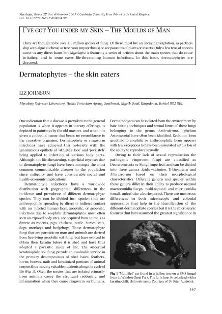
keratinophilic soil fungi provide an invaluable service in<br />
<strong>the</strong> primary decomposition of shed hairs, fea<strong>the</strong>rs,<br />
horns, hooves, nails and keratinized portions of animal<br />
corpses thus moving valuable nutrients along <strong>the</strong> cycle of<br />
life (Fig 1). Often <strong>the</strong> species that are isolated primarily<br />
from animals cause <strong>the</strong> strongest reddening and<br />
inflammation when <strong>the</strong>y cause ringworm on humans.<br />
<strong>Dermatophytes</strong> can be isolated from <strong>the</strong> environment by<br />
hair baiting techniques and sexual forms of <strong>the</strong>se fungi<br />
belonging to <strong>the</strong> genus Arthroderma, (phylum<br />
Ascomycota) have often been identified. Evolution from<br />
geophilic to zoophilic or anthropophilic forms appears<br />
with few exceptions to have been associated with a loss of<br />
<strong>the</strong> ability to reproduce sexually.<br />
Owing to <strong>the</strong>ir lack of sexual reproduction <strong>the</strong><br />
pathogenic ringworm fungi are classified as<br />
Deuteromycota or Fungi Imperfecti and can be divided<br />
into three genera Epidermophyton, Trichophyton and<br />
Microsporum based on <strong>the</strong>ir morphological<br />
characteristics. Different genera and species within<br />
those genera differ in <strong>the</strong>ir ability to produce asexual<br />
macroconidia (large, multi-septate) and microconidia<br />
(small, unicellular aleuriospores). There are numerous<br />
differences in both microscopic and colonial<br />
appearance that help in <strong>the</strong> identification of <strong>the</strong><br />
different dermatophyte species but it is <strong>the</strong> microscopic<br />
features that have assumed <strong>the</strong> greatest significance in<br />
Fig 1 ‘Mumified’ cat found in a hollow tree on a BMS fungal<br />
foray in Windsor Great Park. The fur is heavily colonized with a<br />
keratinophilic Arthroderma sp. Courtesy of Dr Peter Austwick.<br />
147
Mycologist, Volume 17, Part 4 November 2003<br />
Fig 2 Typical ringworm lesion caused by Trichophyton tonsurans.<br />
classical taxonomy.<br />
Athlete’s foot or tinea pedis <strong>the</strong> most widely suffered<br />
dermatophyte infection is considered a disease of<br />
affluence and a high standard of living. Most frequently<br />
caused by Trichophyton rubrum, it is common in<br />
countries where <strong>the</strong> majority of <strong>the</strong> population wears<br />
footwear and has ready access to communal sports or<br />
bathing facilities. Such conditions provide <strong>the</strong> ideal<br />
means of spread of shed infected <strong>skin</strong> squames which<br />
can remain contagious for prolonged periods in <strong>the</strong><br />
environment. The warm, occlusive atmosphere afforded<br />
by footwear allows dormant fungal propagules to invade<br />
<strong>the</strong> moist, softened tissue of <strong>the</strong> interdigital spaces.<br />
In contrast, scalp ringworm or tinea capitis was often<br />
seen as a sign of poor hygiene and a lower standard of<br />
living. From <strong>the</strong> early to <strong>the</strong> mid 1900’s <strong>the</strong> most<br />
prevalent cause of scalp ringworm was <strong>the</strong><br />
anthropophilic dermatophyte Microsporum audouinii<br />
Gruby which is highly infectious, this led to<br />
ostracization with <strong>the</strong> formation of special schools to<br />
segregate infected children. Even as late as <strong>the</strong> middle of<br />
<strong>the</strong> last century it was “considered by middle and upper<br />
class mo<strong>the</strong>rs to be a somewhat disreputable disease”<br />
(Ainsworth, 1952). The introduction of effective oral<br />
<strong>the</strong>rapy led to a drastic reduction. Today in <strong>the</strong> UK scalp<br />
ringworm (Fig 2) is more frequently caused by ano<strong>the</strong>r<br />
anthropophilic species Trichophyton tonsurans Molsten<br />
prevalent in <strong>the</strong> Afro-Caribbean population and also<br />
Microsporum canis Bodin acquired from cats and dogs.<br />
The incidence of tinea capitis due to Trichophyton<br />
tonsurans has increased more than 16 fold in <strong>the</strong> last 10<br />
years in <strong>the</strong> UK and a carrier state has been recognized<br />
in which an undetected index case may be responsible<br />
for direct or indirect transmission of infection to o<strong>the</strong>rs.<br />
Infections such as tinea cruris (groin infection<br />
commonly known as ‘jock itch’) and tinea corporis<br />
(body) are more often seen in adolescents or adults and<br />
are usually due to direct person to person or less<br />
commonly animal or soil to person spread.<br />
The term ‘tinea’ to denote ringworm infections arose<br />
from <strong>the</strong> observation that <strong>the</strong> lesions caused by<br />
dermatophyte fungi resemble <strong>the</strong> holes eaten in fabric by<br />
larvae of <strong>the</strong> clo<strong>the</strong>s moth Tineola biselliella and has been<br />
in use since <strong>the</strong> fourteenth century. The anglicized<br />
‘rynge-worme’ appears to date from <strong>the</strong> fifteenth century<br />
and was probably coined because dermatophyte lesions<br />
of smooth <strong>skin</strong> often heal in <strong>the</strong> centre as <strong>the</strong> fungus<br />
spreads out from a central inoculum point. This results in<br />
a circular lesion or ring with a raised, inflamed, flaky and<br />
red periphery and a clear centre (see Fig 1).<br />
Dr Sabouraud was one of <strong>the</strong> first to formally<br />
categorize this group of organisms and produced a<br />
remarkable tome in 1910 (Les Teignes, Libraires de<br />
l’Academie de Medicine) which describes culture of <strong>the</strong><br />
dermatophytes in detail along with extensive<br />
photogravure plates, drawings, clinical descriptions<br />
and discussion of treatment. Although <strong>the</strong> taxonomic<br />
classification has been modified since this time this<br />
rightly earned him a central position in twentienth<br />
century medical mycology and a lasting impact in <strong>the</strong><br />
form of Sabouraud’s Agar. This peptone-sugar medium<br />
developed by Sabouraud in <strong>the</strong> late 19th century was<br />
revolutionary in enabling fungi to be grown, studied<br />
and described under standardized conditions an<br />
important advance given <strong>the</strong> way in which most<br />
macroscopic and microscopic features are influenced<br />
by <strong>the</strong> nutritional value of <strong>the</strong> growth substrate.<br />
Although not part of <strong>the</strong> normal <strong>skin</strong> flora,<br />
dermatophytes have <strong>the</strong> ability to utilize keratin as a<br />
nutrient source and are thus well adapted to a role in<br />
cutaneous infection. It is interesting that<br />
<strong>Dermatophytes</strong> rarely if ever infect tissue below <strong>the</strong><br />
cornified layers. It can be argued in fact that <strong>the</strong>y really<br />
only colonize dead layers and are <strong>the</strong>refore not true<br />
pathogens. However <strong>the</strong>ir ability to cause<br />
inflammation, itching and acute discomfort is<br />
something that many people will give testament to!<br />
Initiation of infection relies on adherence of<br />
arthroconidia followed by rapid germination and<br />
hyphal penetration before epidermal cell proliferation<br />
results in desquamation of <strong>the</strong> infected cells.<br />
<strong>Dermatophytes</strong> produce a variety of proteinases<br />
including keratinases to facilitate rapid penetration of<br />
<strong>the</strong> stratum corneum. Growth in <strong>the</strong> <strong>skin</strong> is as<br />
branching septate mycelium sometimes with <strong>the</strong><br />
formation of arthrospores resulting from <strong>the</strong><br />
fragmentation of hyphae. Diagnosis is made by<br />
microscopic observation of fungal mycelium in <strong>skin</strong><br />
scales, hair (Fig 3) or nail specimens softened in 20%<br />
caustic potash (KOH) and squashed to produce a<br />
monolayer but this does not allow identification of <strong>the</strong><br />
148
Mycologist, Volume 17, Part 4 November 2003<br />
Fig 3 Calcofluor staining of mycelium of Trichophyton tonsurans<br />
in an infected hair.<br />
infecting species. Keratinized <strong>skin</strong> is a relatively poor<br />
nutritional medium and this may be one reason why<br />
<strong>the</strong> characteristic microconidia and macroconidia of<br />
dermatophytes are not formed in vivo. Thus in order to<br />
identify <strong>the</strong> infecting organism <strong>skin</strong>, nail or hair<br />
samples have to be cultured on a rich medium in order<br />
to encourage spore formation. The resulting production<br />
of spores in <strong>the</strong> laboratory may be <strong>the</strong> first time that <strong>the</strong><br />
organism has utilized this reproductive mechanism for<br />
many generations or hundreds of thousands of years.<br />
The observation that dematophyte infections occur<br />
significantly more commonly in males than females and<br />
that scalp ringworm is much less common after puberty<br />
has led to <strong>the</strong> discovery of steroid-mediated inhibition of<br />
growth. Infection with dermatophyte fungi elicits a cellmediated<br />
immune response directed primarily against<br />
<strong>the</strong> antigen trichophytin. This inflammatory response is<br />
particularly marked following human infection with<br />
zoophilic organisms reflecting <strong>the</strong>ir relative lack of<br />
adaptation to parasitic life on a human host. Factors<br />
that can predispose to chronic dermatophyte infection<br />
include collagen vascular disease, corticosteroid<br />
administration, diabetes mellitus, haematological<br />
malignancy, atopy and old age. An inherited tendency to<br />
develop chronic infection linked to an autosomal<br />
recessive trait has also been documented.<br />
Treatment of dermatophyte infections has had a<br />
somewhat chequered course. Early barbaric treatment of<br />
scalp ringworm included tar cap or pitch plaster epilation<br />
in which <strong>the</strong> hair was imbedded in a sticky cap which<br />
was allowed to set and <strong>the</strong>n torn from <strong>the</strong> head to remove<br />
<strong>the</strong> hair at <strong>the</strong> roots. A treatment that was considered far<br />
less barbaric but in truth was potentially much more<br />
damaging was introduced in <strong>the</strong> early 20th century by<br />
Sabouraud following <strong>the</strong> observation that doses of<br />
radiation caused hair loss. A ra<strong>the</strong>r sinister-looking<br />
apparatus was constructed and used to administer doses<br />
of radiation to targeted areas of <strong>the</strong> scalp.<br />
The first oral agent for dermatophyte infection<br />
followed <strong>the</strong> discovery by Professor Jimmy Gentles in<br />
1958 that griseofulvin, an antibiotic produced by <strong>the</strong><br />
mould Penicillium griseofulvum, had anti-dermatophyte<br />
activity. This remained <strong>the</strong> mainstay of oral <strong>the</strong>rapy for<br />
many years until <strong>the</strong> introduction of azole antifungals<br />
and <strong>the</strong> allylamine terbinafine and still retains a place<br />
in <strong>the</strong> treatment of scalp infections. There is a plethora<br />
of effective over-<strong>the</strong>-counter remedies for <strong>the</strong> topical<br />
treatment of localized <strong>skin</strong> infections ranging from<br />
zinc-based Whitfield’s ointment to a large number of<br />
imidazole creams. The total market for compounds,<br />
antibiotics and ointments that treat dermatophytes is as<br />
large or greater that that used to treat all life<br />
threatening fungal infections. Notable in recent years<br />
has been a sharp rise in <strong>the</strong> number of individuals<br />
seeking treatment for onychomycosis (nail infection)<br />
due to dermatophytes. Whe<strong>the</strong>r this indicates a true<br />
increasing incidence or is merely a reflection of <strong>the</strong><br />
perception that this is now a readily treatable condition<br />
thus encouraging infected individuals to seek <strong>the</strong>rapy is<br />
not clear. Despite <strong>the</strong> introduction of newer fungicidal<br />
agents such as terbinafine treatment of fungal nail<br />
infection is still an uncertain and lengthy procedure<br />
with treatment regimens of up to six months for<br />
toenails.<br />
Recent developments in <strong>the</strong> molecular analysis of<br />
dermatophyte fungi has led to some interesting<br />
epidemiological observations and will help to fur<strong>the</strong>r<br />
elucidate <strong>the</strong> relationships and population dynamics of<br />
this group of human parasites.<br />
References<br />
Ainsworth G. C. 1952. Medical Mycology: An Introduction to its<br />
Problems. Sir Isaac Pitman & Sons Ltd. London<br />
Padhye, A.A. & Weitzman, I. 1998. The <strong>Dermatophytes</strong>. In<br />
Topley and Wilson’s Microbiology and Microbial Infections<br />
9th Edition Vol 4 (Eds. L. Ajello and R.J. Hay) Edward<br />
Arnold, London.<br />
Sabouraud R, 1910. Les Teignes, Libraires de l’Academie de<br />
Medicine, Masson, Paris<br />
149