Species of Xerula from sub-Saharan Africa
Species of Xerula from sub-Saharan Africa
Species of Xerula from sub-Saharan Africa

You also want an ePaper? Increase the reach of your titles
YUMPU automatically turns print PDFs into web optimized ePapers that Google loves.
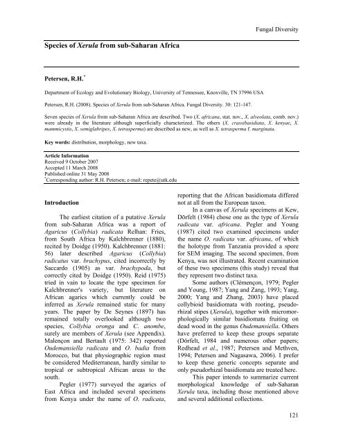
Fungal Diversity<br />
<strong>Species</strong> <strong>of</strong> <strong>Xerula</strong> <strong>from</strong> <strong>sub</strong>-<strong>Saharan</strong> <strong>Africa</strong><br />
Petersen, R.H. *<br />
Department <strong>of</strong> Ecology and Evolutionary Biology, University <strong>of</strong> Tennessee, Knoxville, TN 37996 USA<br />
Petersen, R.H. (2008). <strong>Species</strong> <strong>of</strong> <strong>Xerula</strong> <strong>from</strong> <strong>sub</strong>-<strong>Saharan</strong> <strong>Africa</strong>. Fungal Diversity. 30: 121-147.<br />
Seven species <strong>of</strong> <strong>Xerula</strong> <strong>from</strong> <strong>sub</strong>-<strong>Saharan</strong> <strong>Africa</strong> are described. Two (X. africana, stat. nov., X. alveolata, comb. nov.)<br />
were already in the literature although superficially characterized. The others (X. crassibasidiata, X. kenyae, X.<br />
mammicystis, X. semiglabripes, X. tetrasperma) are described as new, as well as X. tetrasperma f. marginata.<br />
Key words: distribution, morphology, new taxa.<br />
Article Information<br />
Received 9 October 2007<br />
Accepted 11 March 2008<br />
Published online 31 May 2008<br />
* Corresponding author: R.H. Petersen; e-mail: repete@utk.edu<br />
Introduction<br />
The earliest citation <strong>of</strong> a putative <strong>Xerula</strong><br />
<strong>from</strong> <strong>sub</strong>-<strong>Saharan</strong> <strong>Africa</strong> was a report <strong>of</strong><br />
Agaricus (Collybia) radicata Relhan: Fries,<br />
<strong>from</strong> South <strong>Africa</strong> by Kalchbrenner (1880),<br />
recited by Doidge (1950). Kalchbrenner (1881:<br />
56) later described Agaricus (Collybia)<br />
radicatus var. brachypus, cited incorrectly by<br />
Saccardo (1905) as var. brachypoda, but<br />
correctly cited by Doidge (1950). Reid (1975)<br />
tried in vain to locate the type specimen for<br />
Kalchbrenner's variety, but literature on<br />
<strong>Africa</strong>n agarics which currently could be<br />
inferred as <strong>Xerula</strong> remained static for many<br />
years. The paper by De Seynes (1897) has<br />
remained totally overlooked although two<br />
species, Collybia oronga and C. anombe,<br />
surely are members <strong>of</strong> <strong>Xerula</strong> (see Appendix).<br />
Malençon and Bertault (1975: 342) reported<br />
Oudemansiella radicata and O. badia <strong>from</strong><br />
Morocco, but that physiographic region must<br />
be considered Mediterranean, hardly similar to<br />
tropical or <strong>sub</strong>tropical <strong>Africa</strong>n areas to the<br />
south.<br />
Pegler (1977) surveyed the agarics <strong>of</strong><br />
East <strong>Africa</strong> and included several specimens<br />
<strong>from</strong> Kenya under the name <strong>of</strong> O. radicata,<br />
reporting that the <strong>Africa</strong>n basidiomata differed<br />
not at all <strong>from</strong> the European taxon.<br />
In a canvas <strong>of</strong> <strong>Xerula</strong> specimens at Kew,<br />
Dörfelt (1984) chose one as the type <strong>of</strong> <strong>Xerula</strong><br />
radicata var. africana. Pegler and Young<br />
(1987) cited two examined specimens under<br />
the name O. radicata var. africana, <strong>of</strong> which<br />
the holotype <strong>from</strong> Tanzania provided a spore<br />
for SEM imaging. The second specimen, <strong>from</strong><br />
Kenya, was not illustrated. Recent examination<br />
<strong>of</strong> these two specimens (this study) reveal that<br />
they represent two distinct taxa.<br />
Some authors (Clémençon, 1979; Pegler<br />
and Young, 1987; Yang and Zang, 1993; Yang,<br />
2000; Yang and Zhang, 2003) have placed<br />
collybioid basidiomata with rooting, pseudorhizal<br />
stipes (<strong>Xerula</strong>), together with micromorphologically<br />
similar basidiomata fruiting on<br />
dead wood in the genus Oudemansiella. Others<br />
have preferred to keep these groups separate<br />
(Dörfelt, 1984 and numerous other papers;<br />
Redhead et al., 1987; Petersen and Methven,<br />
1994; Petersen and Nagasawa, 2006). I prefer<br />
to keep these generic concepts separate and<br />
only pseudorhizal basidiomata are treated here.<br />
This paper intends to summarize current<br />
morphological knowledge <strong>of</strong> <strong>sub</strong>-<strong>Saharan</strong><br />
<strong>Xerula</strong> taxa, including those mentioned above<br />
and several additional collections.<br />
121
Materials and methods<br />
Specimens discussed here were borrowed<br />
<strong>from</strong> the herbaria <strong>of</strong> the Royal Botanic<br />
Gardens, Kew; the University <strong>of</strong> Helsinki,<br />
Finland; the Jardin Botanique National de<br />
Belgique; Royal Botanic Garden, Edinburgh;<br />
Plant Protection Research Institute, Pretoria,<br />
and New York Botanical Garden.<br />
PhC = phase contrast microscopy, under<br />
which some structures are refringent. Colors<br />
within quotation marks are <strong>from</strong> Ridgway<br />
(1912): colors cited alphanumerically are <strong>from</strong><br />
Kornerup and Wanscher (1976). Notes with<br />
specimens concerning fresh conditions were<br />
especially scanty, but where possible have been<br />
incorporated into species descriptions. E =<br />
spore length divided by spore width; E m =<br />
median E <strong>of</strong> at least ten spores; L m = median<br />
length <strong>of</strong> at least ten spores.<br />
Results<br />
Key to <strong>sub</strong><strong>Saharan</strong> <strong>Africa</strong>n <strong>Xerula</strong> taxa<br />
[Basidiospores 7 × 4 µm; pleurocystidia ten pin-shaped;<br />
pileus with significant, acute umbo ………… Collybia<br />
oronga, C. anombe; see Appendix]<br />
1. Basidia 2-spored; hyphae clampless ..........................6<br />
1. Basidia 4-spored; hyphae clamped ............................2<br />
2. Pileipellis constructed <strong>of</strong> only clavate to sphaeropedunculate<br />
pileocystidia (sect. Radicatae) ..............3<br />
2. Pileipellis constructed <strong>of</strong> clavate to sphaeropedunculate<br />
pileocystidia as well as extended, cylindrical<br />
pileal hairs (sect. Albotomentosae) ............................5<br />
3. Cheilocystidia mammilate; basidiospores 17.5-21 ×<br />
11-13 µm (E m = 1.58; L m = 19.20 µm), elongate-ovate<br />
to <strong>sub</strong>amygdaliform; pileus surface minutely farinose<br />
(30×); Nigeria................................ 5. X. mammicystis<br />
3. Cheilocystidia fusiform to clavate; basidiospores L m =<br />
Fungal Diversity<br />
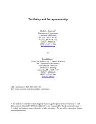
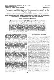
Figs 1-4. <strong>Xerula</strong> africana; holotype. 1. Basidioma (illustrative reconstruction). 2. Pileipellis elements. 3. Pleurocystidia.<br />
4. Basidia and basidiospores. Bars: 1 = 40 mm, 2-4 = 20 µm.<br />
123
clavate to <strong>sub</strong>sphaeropedunculate toward pileus<br />
margin, thin- to thick-walled (wall never more<br />
than 1 µm thick, always over pedicel and lower<br />
bulb), hyaline to olivebrown (especially in<br />
pedicel), without clamp connection, contents<br />
homogeneous, weakly pigmented in watery<br />
tan. Pleurocystidia (Fig. 3) 117-208 × 23-40<br />
µm, long-pedicellate, elongate-fusiform to<br />
fusiform-capitulate, the capitulum sometimes<br />
pronounced (-20 µm broad), sometimes a<br />
rounded extension <strong>of</strong> the pleurocystidial neck,<br />
hyaline, thick-walled (wall up to 2 µm thick)<br />
proximally, thin- to firm-walled over capitulum,<br />
without clamp connection; contents<br />
homogeneous, sometimes <strong>sub</strong>refringent (PhC)<br />
in capitulum. Basidia (Fig. 4) 57-72 × 15-20<br />
µm, clavate <strong>from</strong> somewhat pinched base,<br />
<strong>of</strong>ten somewhat bulbous apically, strictly 2-<br />
spored, without clamp connection; contents<br />
sludgy to multigranular. Basidiospores (Fig. 4)<br />
18-23 × 14-16 µm (E = 1.23-1.50; E m = 1.39;<br />
L m = 21.2 µm), ellipsoid, ovate to <strong>sub</strong>limoniform,<br />
thin-walled, delicately dimpled; contents<br />
multiguttulate, refringent (PhC). Lamellar<br />
margin sterile, a solid palisade <strong>of</strong> free cheilocystidia,<br />
extending significantly in KOH.<br />
Cheilocystidia (Fig.5) 38-108 × 8-22 µm, <strong>of</strong>ten<br />
clavate when small, sometimes developing a<br />
mammilate to digitate apical extension, to<br />
fusiform or clavate-fusiform, thin-walled,<br />
hyaline, without clamp connection; contents<br />
homogeneous. Apical caulocystidia (Fig. 6)<br />
occurring in a turf or as erumpent fascicles,<br />
similar to cheilocystidia, 40-110 × 13-23 µm,<br />
clavate to elongate-fusiform, thin-walled,<br />
hyaline, without clamp connection; contents<br />
homogeneous. Mid-stipe surface an appressed<br />
layer <strong>of</strong> coralloid (almost ramealis) hyphae 3-5<br />
µm diam, producing scattered (not in fascicles)<br />
caulocystidia; caulocystidia (Fig. 6) up to 25<br />
individuals in erumpent sori, 50- > 225 × 10-17<br />
µm, clavate when small, fusiform, elongatefusiform<br />
to cylindrical when larger, with<br />
narrow pedicel, without clamp connection,<br />
hyaline, thin-walled; contents homogeneous,<br />
not refringent.<br />
Commentary: Absence <strong>of</strong> pileal hairs<br />
places these specimens in sect. Radicatae.<br />
Spores are unusually large, which caused<br />
Redhead et al. (1987) to compare some North<br />
American collections to this taxon. Pegler and<br />
Young (1987), however, failed to recognize the<br />
124<br />
2-spored basidia in placing the taxon under O.<br />
radicata. Pegler (1977) cited several collections<br />
<strong>from</strong> Kenya as O. radicata, but none <strong>of</strong><br />
those collections was cited by Pegler and<br />
Young (1987), and are found here under X.<br />
semiglabripes and X. tetrasperma.<br />
Redhead et al. (1987) referred some<br />
North American collections to this name, but<br />
all were described (and confirmed in this<br />
study) as 4-spored. The character which<br />
separated them <strong>from</strong> other North American<br />
material was spore size, reported by Redhead et<br />
al. (1987) as 20-25 × 12.5-14.5 µm. No citation<br />
<strong>of</strong> type or authentic material <strong>of</strong> X. radicata var.<br />
africana was furnished in that study.<br />
Dörfelt's (1984) description <strong>of</strong> X.<br />
radicata var. africana was superficial, as<br />
follows (in its entirety, transl.): "The sample<br />
consists <strong>of</strong> one single fruitbody (Taf. XXIII,<br />
Fig. 10); cap diameter 5.8 cm, stipe length 13.5<br />
cm, stipe diameter about 3 mm, <strong>of</strong> the<br />
thickened base 10 mm, rhizomorphic appendix<br />
2.5 cm long, then torn; cap graybrown, stipe<br />
concolorous, somewhat lighter, lamellae <strong>of</strong><br />
exsiccate brownish white, without darker<br />
margin.<br />
“Spores measure 18.5-26 × 12.5-16 µm,<br />
average 21.5 × 13.5 µm; cystidia mostly<br />
somewhat capitate (Taf. XXIII, Figs 11, 12)<br />
and most narrower than in the typical variety,<br />
relatively abundant, 60-80 µm long, 12-25 µm<br />
diam (at widest point), 10-14 µm (neck), 12-18<br />
µm (capitulum); stipe macroscopically smooth,<br />
microscopically found to be a single compact<br />
hymenodermium.<br />
Discussion: The great variability <strong>of</strong><br />
<strong>Xerula</strong> radicata relative to many characters<br />
(form, color, structure <strong>of</strong> lamellar margin)<br />
leaves a circumscription with only a few fixed,<br />
relatively constant characters. Understanding<br />
<strong>of</strong> spore size points out that tropical collections<br />
have relatively large spores. Typical spore<br />
mass <strong>of</strong> tropical samples lies around 19.5 × 12<br />
µm, but there exists all transitions to<br />
compressed (also, only essentially shorter).<br />
Spores <strong>of</strong> middle-European samples <strong>of</strong>ten<br />
reach 15 × 11 µm typical dimensions. The<br />
fruitbody <strong>from</strong> Kilimanjaro has typical spores<br />
21.5 × 13.5 µm, and capitulate cystidia exhibit<br />
no transition (neither for <strong>Africa</strong>n collections!),<br />
so I accept only a single variety. Whether this<br />
is an endemic entity on Kilimanjaro or a
Fungal Diversity<br />
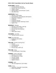
Figs 5-6. <strong>Xerula</strong> africana; holotype. 5. Cheilocystidia. 6. Upper, apical caulocystidia; lower; mid-stipe caulocystidia.<br />
Bars = 20 µm.<br />
tropical taxon with wider distribution is at<br />
present unclear." Not included, <strong>of</strong> course, were<br />
the 2-spored basidia and absence <strong>of</strong> clamp<br />
connections.<br />
Mid-stipe caulocystidia are welldeveloped<br />
but scattered almost individually<br />
rather than in erumpent fascicles as in X.<br />
furfuracea. Cheilocystidia and all caulocystidia<br />
(apical and mid-stipe) are similar, typically<br />
elongate-fusiform.<br />
This specimen resembles BPI 841566<br />
(viz. X. incognita var. bispora) in spore and<br />
general pleurocystidial shape. Even these<br />
characters fail, however. Spore dimensions in<br />
BPI 841566 are 17-20 × 12-14 µm (E m = 1.43;<br />
L m = 18.64 µm; i.e. somewhat shorter than<br />
those <strong>of</strong> X. africana); pleurocystidia are 83-155<br />
× 26-42 µm, broadly clavate with broadly<br />
rounded apex to <strong>sub</strong>capitate (i.e. wider and less<br />
capitulate than those <strong>of</strong> X. africana). Less<br />
obvious; pileicystidia <strong>of</strong> X. incognita var.<br />
bispora do not exhibit the "spur" common on<br />
the pedicel in X. africana.<br />
Specimens examined: PEOPLES REPUBLIC OF<br />
CONGO, Katanga Prov., Muhulu de la Luiswishi, 1210<br />
m, 19.XI.1971, leg D Thoen (as Oudemansiella aff.<br />
longipes), Thoen no. 4992, no. 70447-25 (BR). SOUTH<br />
AFRICA, Transvaal Prov., Pretoria, 5.IV.1921, coll AM<br />
Bottomley (as Collybia radicata), no 14519 (PREM).<br />
TANZANIA, Kilimanjaro Province, Mt. Kilimanjaro W<br />
slope, W. Kilimanjaro Forest Station, S 5°, E 37° 8′,<br />
10.II.1973, coll. L. Ryvarden (no. 10178) [K(M)<br />
124281; holotype].<br />
2. <strong>Xerula</strong> alveolata (Kalchbr.) R.H. Petersen,<br />
comb. nov. (Figs 7-11)<br />
MycoBank: 511152<br />
Basionym: Agaricus alveolatus Kalchbr.,<br />
Grevillea 9: 110 (1881).<br />
≡ Collybia alveolata (Kalchbr.) Sacc., Syll. Fung.<br />
5: 202 (1887).<br />
Lectotype (hic. design.): SOUTH AFRI-<br />
CA, Cape Prov., no date, coll MacOwan, s.n.<br />
[K(M) 144264]. [annot. DA Reid: “Clearly not<br />
type but could serve as lectotype.”]<br />
Basidiome (Fig. 7) collybioid, gracile,<br />
rooting. Pileus 33-57 mm broad, dark brown to<br />
dark nut brown (near "Saccardo's umber"),<br />
125
Figs 7-10. <strong>Xerula</strong> alveolata; Saarimäki 555 (H). 7. Basidioma (illustrative reconstruction). 8. Pileipellis elements. 9.<br />
Pleurocystidia. 10. Basidia, basidiospores and cheilocystidia. Bars: 7 = 40mm, 8-10 = 20 µm.<br />
126
Fungal Diversity<br />
mostly plane but with low umbo, smooth, matt,<br />
minutely laccate (30×), with <strong>sub</strong>tle blackish<br />
radiating streaks or reticulate pattern near<br />
margin, outward with scattered paler flecks;<br />
umbo darker, minutely roughened (30×), in<br />
part appearing frosted (i.e. hyaline hairs);<br />
margin entire, not striate, perhaps incurved,<br />
thin; flesh white, outward very thin. Lamellae<br />
adnate with significant decurrent tooth, white<br />
when fresh, near "light ochraceous buff" after<br />
drying (in one specimen developing deep rust<br />
color <strong>from</strong> necropigment), close to <strong>sub</strong>distant,<br />
somewhat ventricose, up to 10 mm deep, in<br />
three tiers; margin entire, smooth, sometimes<br />
with evidence <strong>of</strong> cheilocystidial palisade,<br />
delicately marginate in limited areas. Stipe 85-<br />
160 mm long to ground level, 2-5 mm broad<br />
upward, tapering slightly upward, swollen to 6-<br />
8 mm broad just above pseudorhiza, pale and<br />
somewhat flaired apically, neutral brown<br />
downward, longitudinally lined, obscurely<br />
furfuraceous, hollow; pseudorhiza beet-shaped,<br />
7-10 mm broad at widest point, then tapering<br />
gradually to at least 35 mm long, involving<br />
significant soil so as to obscure color, minute<br />
areas <strong>of</strong> thin pale tomentum between soil<br />
particles. Taste mild; odor weak.<br />
Pileipellis constructed <strong>of</strong> a single<br />
variable element. Pileocystidia (Fig. 8)<br />
pedicellate, thin-walled, hyaline to pigmented,<br />
not apparently clamped, <strong>of</strong> two types (with<br />
intermediates): 1) 27-52 × 13-24 µm, sphaeropedunculate,<br />
short-pedicellate, apparently<br />
arising <strong>from</strong> wide or inflated hyphae; contents<br />
homogeneous, pigmented olive brown in small<br />
individuals, <strong>sub</strong>hyaline in larger individuals;<br />
and 2) pileal hairs 50-198 × 6-14 µm (at widest<br />
point), pedicellate, clavate or extended into a<br />
<strong>sub</strong>cylindric or cylindric extension, commonly<br />
slightly inflated proximally (as though an<br />
extension <strong>of</strong> a clavate individual), thin-walled,<br />
hyaline, not apparently clamped. Pleurocystidia<br />
(Fig. 9) 78-122 × 19-30 µm, pedicellate,<br />
fusiform with wide, bluntly rounded<br />
extension to fusiform with prolonged neck and<br />
capitulum (<strong>sub</strong>lecythiform), not clamped,<br />
hyaline, thin-walled; contents homogeneous;<br />
capitulum not refringent. Basidia (Fig. 10) 47-<br />
75 × 12-19 µm, narrowly clavate with slightly<br />
pinched base, 2-spored, occasionally sclerified,<br />
without clamp connection; contents multiguttulate<br />
when young, with guttules coalescing to<br />
3-4 by maturity. Basidiospores (Fig. 10) <strong>from</strong><br />
hymenium 14.5-18 × 11-15 µm [E = (1.15-)<br />
1.23-1.44; E m = 1.23; L m = 16.2 µm], <strong>sub</strong>ovate,<br />
ellipsoid to <strong>sub</strong>tly <strong>sub</strong>limoniform, delicately<br />
dimpled, thin-walled, hyaline; contents<br />
opalescent to uniguttulate. Basidiospores <strong>from</strong><br />
stipe apex and/or pileus surface 16-21 × 13-18<br />
µm (E = 1.00-1.33; E m = 1.18; L m = 18.5 µm),<br />
<strong>sub</strong>globose, broadly ellipsoid, occasionally<br />
<strong>sub</strong>limoniform, thin- to thick-walled (wall<br />
never more than 1 µm thick), delicately<br />
dimpled. Lamellar margin sterile, extending<br />
significantly in KOH, a solid palisade <strong>of</strong><br />
cheilocystidia. Cheilocystidia (Fig. 10) (36-)<br />
56-146 × (8-)13-30 µm, pedicellate, clavate in<br />
smaller individuals, sometimes <strong>sub</strong>tly<br />
capitulate, fusiform to broadly cylindrical in<br />
larger individuals with slender pedicel, hyaline,<br />
firm-walled, without clamp connection;<br />
contents homogeneous. Stipe apex minutely<br />
scurfy with white tomentum (composed <strong>of</strong><br />
spores plus caulocystidia). Apical caulocystidia<br />
(Fig. 11) 50-165 × 15-25 µm, a turf <strong>of</strong> clavate<br />
to lobed individuals, producing broadly<br />
cylindrical individuals with narrow pedicel,<br />
hyaline, thin- to thick-walled (wall up to 1 µm<br />
thick); contents homogeneous. Stipe<br />
midsection apparently with a thin, appressed<br />
layer <strong>of</strong> surface hyphae 6-11 µm broad,<br />
perhaps involved in slime, producing a lawn <strong>of</strong><br />
side branches <strong>of</strong>ten gathered into erumpent<br />
fascicles; caulocystidia (Fig. 11) 49-223(-300)<br />
× 12-20 µm, clavate in smaller individuals,<br />
extended to fusiform, elongate-fusiform to<br />
cylindric in longer individuals, rarely furcate<br />
near apex, usually with slender pedicel, thin-,<br />
firm- or thick-walled (wall never more than 1<br />
µm thick); contents homogeneous, perhaps<br />
slightly pigmented toward tan.<br />
Commentary: These specimens are segregated<br />
<strong>from</strong> other similar basidiomata as<br />
follows: 1) pileipellis with extended pileocystidial<br />
hairs; 2) 2-spored basidia; 3) <strong>sub</strong>globose<br />
to broadly ellipsoid (rarely <strong>sub</strong>limoniform)<br />
spores; 4) pleurocystidia fusiform-capitate with<br />
extended, broadly rounded neck; and 5) welldeveloped<br />
midstipe caulocystidia. The taxon<br />
belongs in sect. Albotomentosae. Pileipellis<br />
"hairs" are extensions <strong>of</strong> clavate pileocystidia,<br />
but sphaeropedunculate individuals are more<br />
127
plentiful in some areas <strong>of</strong> pileipellis than in<br />
others. The areas with copious extended<br />
pileocystidia seem to be somewhat roughened<br />
(20×) or minutely furry. This texture does not<br />
assume a pattern, but is distributed over much<br />
<strong>of</strong> the umbo, extending only slightly into the<br />
pileus limb. Portions <strong>of</strong> the umbo and adjacent<br />
areas are very delicately frosted (20×) with<br />
these hyaline hairs.<br />
Two-spored basidia could be attributed to<br />
X. africana, together with the fusiform<strong>sub</strong>lecythiform<br />
pleurocystidia, but spores are<br />
rarely <strong>sub</strong>limoniform (most <strong>sub</strong>globose), and<br />
basidia are small with undersized sterigmata.<br />
Moreover, the pileipellis found in X. africana<br />
does not exhibit extended hairs. Spore<br />
dimensions, pileocystidial hairs and two-spored<br />
basidia could place the specimens in X. chiangmaiae<br />
var. raphanipes, but pleurocystidia <strong>of</strong><br />
that taxon are rotund-capitulate, not <strong>sub</strong>lecythiform,<br />
and its distribution in southeast to<br />
northern Indo-Asia would seem to exclude <strong>sub</strong>-<br />
<strong>Saharan</strong> <strong>Africa</strong>.<br />
A furfuraceous to scabrous stipe surface<br />
<strong>of</strong> well-developed caulocystidia is common to<br />
other <strong>Africa</strong>n taxa (q.v.), but two-spored X.<br />
africana does not exhibit this character. <strong>Xerula</strong><br />
kenyae produces utriform pleurocystidia and<br />
four-spored basidia. Singer (1964) considered<br />
A. alveolatus to be a synonym under Oudemansiella<br />
radicata, a judgement followed by<br />
Pegler (1977). In other publications, Pegler<br />
(1960) and Pegler and Young (1987) did not<br />
take up the name.<br />
Although only three specimens have been<br />
examined, it may be that this species occurs on<br />
the southeast coast <strong>of</strong> <strong>Africa</strong>, and should be<br />
sought in Madagascar.<br />
Handwritten notes in MacOwan’s hand<br />
pasted over the type basidiomata stipes: “I<br />
think this is what Kalchbr. has described as ‘A.<br />
alveolatus’ a grege ‘radicati & aff.’ But my<br />
sending was not numbered as I only then found<br />
one plant. For certainty it must await the<br />
publication <strong>of</strong> his work.” This indicates that the<br />
note was written BEFORE MacOwan received<br />
Kalchbrenner’s work, and may indicate that<br />
Kalchbrenner had given the species a provisional<br />
name when this specimen was collected.<br />
Although Reid annotated the collection<br />
as a candidate for lectotype, he designated it as<br />
a paratype in publication (Reid, 1975). I cannot<br />
128<br />
find any reference to the specimen by<br />
Kalchbrenner (1881) who merely cited the type<br />
as “Somers[et] East, MacOw., sine No.” This<br />
reference could be applied to the original<br />
MacOwan specimen (no longer known) or the<br />
present specimen. Because MacOwan implied<br />
that this specimen was sent to Kalchbrenner<br />
before the published proposal <strong>of</strong> the species,<br />
there is at least some chance that it was in the<br />
hands <strong>of</strong> Kalchbrenner at the time <strong>of</strong> compiling<br />
the publication (Kalchbrenner, 1881). Therefore,<br />
following Reid’s suggestion, the specimen<br />
is here designated as lectotype <strong>of</strong> Agaricus<br />
alveolatus.<br />
Specimen examined: SOUTH AFRICA, Natal<br />
Prov., Wintersklo<strong>of</strong>, vic Pietermaritzburg, grounds <strong>of</strong><br />
Cowan House, 29° 50′ S, 30° 05′ E, 23.I.1975, coll NG<br />
Sinnott & s.n. [K(M) 144264; lectotype]. TANZANIA,<br />
T3, Tanga Region, Pare District, South Pare Mts.,<br />
Mbaga Manka village, in small patch <strong>of</strong> natural montane<br />
forest, collection site 29, degree ref. system square 04 37<br />
BB, 1.XII.1990, coll. Tiina Saarimäki & al., no. 555 (H).<br />
3. <strong>Xerula</strong> crassibasidiata R.H. Petersen, sp.<br />
nov. (Figs 12-17)<br />
MycoBank: 511153<br />
Basidiomata collybioidea, gracilis, radicatia.<br />
Pileo 30-32 mm lato, brunneo, convexo, centro umbonato,<br />
viscido, innate atroburnnea radialiter striatulo;<br />
margine levi. Lamellis albis, adnatis, <strong>sub</strong>ventricosis,<br />
non-marginatis. Stipite 85-105 × 3-4 mm, apice albo,<br />
deorsum griseobrunneis; pseudorhiza dauciformis,<br />
longis.<br />
Pileocystidiis 27-150 × 7-15 µm, clavatis vel<br />
<strong>sub</strong>sphaeropedunculatis, firme tunicatis. Pleurocystidiis<br />
147-220 × 24-41 µm, pedicellatis, fusiformis-capitulatis,<br />
hyalinis, fibulatis. Basidiis 45-88 × 18-22 µm, clavatis,<br />
tetra-sporibus. Basidiosporis 15-20 × 11-15 µm (E m =<br />
1.30; L m = 17.4 µm), late ellipsoidis, hyalinis.<br />
Cheilocystidiis 41-150 × 7-31 µm, fusiformis, fusiformimammilatis,<br />
fibulatis. Caulocystidiis 45-300 × 10-23<br />
µm, cylindricis, fibulatis, hyalinis ad pallide fuscis.<br />
Holotype: BURUNDI, Prov. T.Muramvya,<br />
Teza, S 03° 13' E 29° 34', 2500 m,<br />
22.XII.1978, coll. J. Rameloo (as O. radicata,<br />
no. K 6238) (BR 032225,21).<br />
Basidiomata (Fig. 12) collybioid, gracile,<br />
rooting. Pileus ca 30-32 mm broad, plane with<br />
shallow umbo, slimy but appearing dry when<br />
dried; disc matt to minutely furry, dark brown<br />
("bister," 5D4) usually with a few dark brown<br />
radial streaks formed by darker colored<br />
pileicystidia (not raised in ridges), occasionally<br />
with raised, almost black, coarse radial ridges<br />
<strong>from</strong> umbo, becoming narrower over limb, and<br />
then delicately reticulate near and over margin,
Fungal Diversity<br />
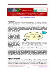
Figs 11-14. <strong>Xerula</strong> species. 11. X. alveolata; Saarimäki 555 (H). Left, apical caulocystidia; right, mid-stipe<br />
caulocystidia. 12-14. X. crassibasidiata. 12. Basidiomata (illustrative reconstruction). Left, holotype; right, Rameloo<br />
6580. 13. Elements <strong>of</strong> pileipellis (holotype). Upper, pileicystidia <strong>from</strong> disc; lower, pileicystidia <strong>from</strong> pileus margin. 14.<br />
Pleurocystidia (holotype). Bars: 11, 13, 14 = 20 µm, 12 = 40 mm.<br />
129
at 40x with slender, hyaline, flexuous, scattered<br />
hairs; outward brown (4D4), appearing smooth,<br />
with margin somewhat darker; margin entire,<br />
not striate or crenate, sometimes undulate;<br />
flesh white, loosely compact over stipe, very<br />
thin outward, extending beyond interlamellar<br />
hymenophore but not beyond lamellae.<br />
Lamellae white when fresh, becoming light<br />
ochraceous buff to ochraceous buff in time<br />
when dried, adnate with small decurrent tooth,<br />
<strong>sub</strong>ventricose, in four ranks (the outermost<br />
extremely small), close, entire, not marginate;<br />
interlamellar space with hymenophore, ribbed<br />
at margin. Stipe 85-105 mm long to ground<br />
level, 2 mm broad near apex, 3 mm toward<br />
ground line; apex white, slightly flaired,<br />
appearing glabrous, silky to minutely pustulate<br />
with white sori; stipe midsection hardly lined,<br />
downward pallid tan ground color with lattice<br />
layer brown and minutely hispid or with<br />
densely scattered, minute amorphous caulocystidial<br />
dots; pseudorhiza hardly swollen, carrotshaped,<br />
sometimes with fine white, tangled<br />
tomentum, apparently extended at least 65 mm,<br />
tapering slowly downward, appearing glabrous,<br />
<strong>of</strong>f-white or brown.<br />
Pileipellis constructed <strong>of</strong> a single<br />
variable element. Pileocystidia <strong>from</strong> disc (Fig.<br />
13) 27-150 × 7-15 µm (at widest point),<br />
varying <strong>from</strong> narrowly clavate to clavate, <strong>of</strong>ten<br />
extended into a cylindrical shaft with bluntly<br />
rounded apex, thin-walled (pedicel occasionally<br />
slightly thick-walled; wall never more<br />
than 0.7 µm thick); contents more or less<br />
homogeneous, hyaline to olive-brown, especiallly<br />
in pedicel. Pileocystidia <strong>from</strong> near<br />
pileus margin (Fig. 13) 20-48 × 9-27 µm,<br />
broadly clavate to sphaeropedunculate, perhaps<br />
obscurely clamped, thin-walled; contents<br />
homogeneous, usually olive-brown; pileal hairs<br />
60-112 × 11-16 µm (at widest point), pedicellate,<br />
extended pileocystidia, slightly inflated<br />
proximally, tapering to obtusely rounded tip,<br />
thin- to thick-walled (wall never more than 1<br />
µm thick) except for thin-walled apex, hyaline<br />
to weakly olivetan; contents more or less<br />
homogeneous. Pleurocystidia (Fig. 14) welldeveloped,<br />
sparse, arising deep in lamellar<br />
trama, 147-220 × 24-41 µm, short-pedicellate,<br />
fusiform with extended neck (7-11 µm diam)<br />
and distinct capitulum (11-15 µm diam),<br />
thickwalled over median portion (wall never<br />
130<br />
more than 2 µm thick), hyaline, clamped;<br />
contents homogeneous, <strong>sub</strong>refringent in upper<br />
neck and capitulum. Basidia (Fig. 15) 45-88 ×<br />
18-22 µm, clavate with hardly pinched base, 4-<br />
spored; contents multigranular when immature,<br />
coalescing to several-guttulate near maturity;<br />
several sclerotized basidia present. Basidiospores<br />
(Fig. 15) 15-20 × 11-15 µm (E = 1.13-<br />
1.48; E m = 1.30; L m = 17.4 µm), broadly<br />
ellipsoid to <strong>sub</strong>ovate, smooth or very delicately<br />
pock-marked, hyaline; contents opalescent<br />
when immature, uniguttulate when mature,<br />
refringent.<br />
Lamellar margin sterile, extending significantly<br />
in KOH, a solid beard <strong>of</strong> welldeveloped<br />
cheilocystidia. Cheilocystidia (Fig.<br />
16) 41-150 × 7-31 µm, digitate to narrowly<br />
clavate when young, extending to fusiform<br />
with various expressions <strong>of</strong> mammilate or<br />
<strong>sub</strong>capitulate apex, conspicuously clamped,<br />
hyaline; contents homogeneous. Caulocystidia<br />
at stipe apex (Fig. 17) appressed, sparse in<br />
pallid sori scattered over stipe apex, 45- > 300<br />
× 10-23 µm, hyaline, thin-to thick-walled (wall<br />
up to 2 µm thick), with rounded apex, clamped.<br />
Stipe midsection with extensive loose, lattice<br />
<strong>of</strong> wide hyphae (6-8 µm diam, thin-walled,<br />
clamped), producing superficial sori <strong>of</strong><br />
caulocystidia arising <strong>from</strong> a tangle <strong>of</strong> tortuous,<br />
thin-walled, pigmented hyphae producing<br />
small, sharply pointed olive-tan individuals and<br />
several (-15) larger individuals; contents <strong>of</strong><br />
small individuals homogeneous, olive-brown;<br />
caulocystidia (Fig. 17) 57-190 × 9-21 µm, with<br />
slender base, hyaline, thin- to thick-walled<br />
(wall up to 2 µm thick), inconspicuously<br />
clamped; contents homogeneous, <strong>of</strong>ten slightly<br />
refringent at very apex.<br />
Commentary: Pegler (1977) reported<br />
several collections <strong>of</strong> Oudemansiella radicata<br />
<strong>from</strong> Kenya, but later (Pegler and Young,<br />
1987) included additional collections <strong>from</strong> <strong>sub</strong>-<br />
<strong>Saharan</strong> <strong>Africa</strong>, including O. radicata var.<br />
africana. Examination <strong>of</strong> additional collections<br />
reveals several taxa, including X. crassibasidiata<br />
and X. kenyae (q.v.). Of these taxa, two<br />
(X. alveolata, X. crassibasidiata) belong in<br />
sect. Albotomentosae, for both produce<br />
extended pileicystidia as "hairs." The two taxa<br />
are separated by differences in basidia<br />
(extremely long and narrow in X. alveolata,<br />
long but broad in X. crassibasidiata) and
Fungal Diversity<br />
Figs 15-18. <strong>Xerula</strong> species. 15-17. X. crassibasidiata. 15. Basidia and basidiospores. Left, holotype; right, Rameloo<br />
6580. 16. Cheilocystidia. 17. Caulocystidia. Left, caulocystidia <strong>from</strong> stipe apex; right, caulocystidia <strong>from</strong> stipe<br />
midsection. 18. <strong>Xerula</strong> kenyae; holotype. Basidioma (illustrative reconstruction). Bars: 15-17 = 20 µm, 18 = 40 mm.<br />
131
spores (13-18 × 10-15 µm in X. alveolata). In<br />
sect. Radicatae (no pileisetae, no extended<br />
pileicystidia), <strong>Xerula</strong> africana (= O. radicata<br />
var. africana ss. Pegler and Young, 1987) is 2-<br />
spored, with its 4-spored analog as X. tetrasperma.<br />
The difference between pileocystidia<br />
<strong>from</strong> the disc and <strong>from</strong> the margin reflects the<br />
situation in several other species. It would<br />
appear that the disc, not as expanded as the<br />
limb, has less room for pileocystidial swelling<br />
than the outer pileus. But pileal hairs are<br />
present in all areas <strong>of</strong> the pileus surface,<br />
although somewhat shorter outward than over<br />
the disc. Furthermore, not more than 1:250<br />
pileipellis elements is a pileal hair. Therefore<br />
such structures are inconspicuous in sections or<br />
squashes and invisible at 10×. Intermediates<br />
are clavate, thick-walled and hyaline (not<br />
olive-tan like pileocystidia).<br />
A single basidiome (BM 466) <strong>from</strong> high<br />
altitude in Malawi (6000 ft.) differs <strong>from</strong><br />
typical X. crassibasidiata in its stout form<br />
(pileus 110 mm broad, stipe 40 × 6-8 mm,<br />
lamellae <strong>sub</strong>distant, ventricose, up to 10 mm<br />
deep, with distinct decurrent tooth) and<br />
olivaceous color (noted as “greenish” in notes<br />
on the fresh specimen). Otherwise, microscopic<br />
characters fit nicely.<br />
Specimens examined: BURUNDI, Prov.<br />
T.Muramvya, Teza, S 03° 13' E 29° 34', 2500 m,<br />
22.XII.1978, coll. J. Rameloo (as O. radicata, no. K<br />
6238) (BR 032225, 21); same location, 20.XII.1978, leg<br />
J. Rameloo (as O. radicata; no. 6146), K 1766 (BR no.<br />
032224,30); Prov. Bururi, Bururi, Forêt de Bururi, S 02°<br />
57' E 29° 37', 7.II.1979, 1950 m, leg. J. Rameloo (as O.<br />
radicata, no. 6580), (BR 032226,22). MALAWI, Nyita<br />
Nat. Park, surroundings <strong>of</strong> Chelinda Lodge, 6.XII.1981,<br />
leg K. Rameloo, no. 7688 (BR 032231,27); Zomba Mt.,<br />
27.XII.1981, coll B Morris, BM 466 [K(M) 144256].<br />
ZAMBIA, Chowo Forest, 7.XII.1981, leg J Rameloo, no<br />
7716 (BR no. 032233, 29); same location, 9.XII.1981,<br />
leg K. Rameloo, no. 7755 (BR 032234, 30); Manyanjere<br />
Forest, 16.XII.1981, leg J Rameloo, no 7938 (BR<br />
032237,33).<br />
4. <strong>Xerula</strong> kenyae R.H. Petersen, sp. nov.<br />
(Figs 18-23)<br />
MycoBank: 511154<br />
Basidiomata collybioidea, crasis, atro-brunneis.<br />
Pileo 44-57 mm lato, convexiumbonato, glabro, vel<br />
innate atrobrunneo radialiter striatulo. Lamellis albis,<br />
crassis, ventricosis, adnatis, non-marginatis. Stipe 100-<br />
150 × 4-8 mm, apice albo, deorsum brunneo; pseudorhiza<br />
inflata.<br />
Pileocystidiis 26-40 × 10-38 µm, <strong>sub</strong>sphaeropedunculatis,<br />
sine fibulis, crassi-tunicatis; trichomis<br />
132<br />
pilioris 107-136 × 9-13 µm, hyalinis, fibulatis. Pleurocystidiis<br />
72-136 × 22-36 µm, utriformis, tenuitunicatis.<br />
Basidiis 66-88 × 14-21 µm, clavatis, fibulatis, tetrasporibus.<br />
Basidiosporis 15-21 × 15-17 µm (E m = 1.22;<br />
L m = 17.8 µm). Late ellipsoidiis. Cheilocystidiis 41-156<br />
× 8-40 µm, clavatis ad cylindricis, firme tunicatis.<br />
Caulocystidiis 76-137 × 12-23 µm, cylindricis ad<br />
vermiformis, olivaceo-brunneis, fibulatis.<br />
Holotype: KENYA, Rift Valley Prov.,<br />
vic. Timboroa, Cengalo, IV.1970, coll. J.K.<br />
Dedan, I.A.S. Gibson no. 2172 [K(M) 124280].<br />
On litter under young Pinus radiata plantation.<br />
[annot. H. Dörfelt, 1982, as X. radicata].<br />
Basidiomata (Fig. 18) collybioid, stout,<br />
generally dark brown. Pileus 44-57 mm broad,<br />
abruptly and significantly umbonate, matt,<br />
without laccate surface; umbo and narrow<br />
radial streaks darker (near "sepia" or "Natal<br />
brown") and appearing minutely furry (30x),<br />
area surrounding umbo somewhat darker than<br />
"sayal brown," becoming neutral nut brown<br />
outward to margin; margin smooth to crenate<br />
with occasional narrow, radial, blackish ridges,<br />
entire, probably incurved when fresh, thin.<br />
Lamellae white when fresh, now "light ochraceous<br />
buff" (i.e. little or no necropigment),<br />
thick, close, ventricose (up to 8 mm deep), with<br />
short decurrent tooth, in 3-4 tiers, not<br />
marginate. Stipe 100-150 mm long to ground<br />
line, 4-6 mm diam near apex, 8 mm diam near<br />
ground line, <strong>of</strong>f-white apically and there<br />
closely lined, soon dark brown, hardly<br />
longitudinally lined or channeled, tapering<br />
gradually upward, swollen somewhat at ground<br />
line, distinctly but minutely furfuraceous or<br />
scabrous, probably lacerate when fresh, with<br />
extensive meandering patches <strong>of</strong> darker brown<br />
caulocystidia; flesh apparently woody, white to<br />
ivory color; pseudorhiza 10-13 mm broad,<br />
more or less beet- or carrot-shaped (tapering<br />
gradually), dark brown with pallid buff thatch<br />
over upper 5 mm. Spore print white.<br />
Habitat: Ethiopia: at edge <strong>of</strong> bamboo<br />
zone near top <strong>of</strong> mountain (3000 ft elev.). In<br />
drier area <strong>of</strong> montane forest, with Dombeya,<br />
Schefflera, Aningeria, etc. Kenya: on litter<br />
under young Pinus radiata plantation.<br />
Pileipellis near disc constructed <strong>of</strong> a<br />
single element. Pileocystidia 26-140 × 10-38<br />
µm, pedicellate, clavate to sphaeropedunculate,<br />
<strong>of</strong>ten with pedicel spur, thin- to firm-walled<br />
over bulb, <strong>of</strong>ten somewhat thick-walled (wall<br />
never more than 0.7 µm thick) over pedicel,
Fungal Diversity<br />
Figs 19-22. <strong>Xerula</strong> kenyae. 19. Pileipellis elements. Left, <strong>from</strong> disc; right, <strong>from</strong> pileus margin. Ash 3448. 20.<br />
Pleurocystidia; holotype. 21. Basidia and basidiospores; holotype. 22. Cheilocystidia; holotype. Bars= 20 µm.<br />
without clamp connections; contents homogeneous,<br />
<strong>sub</strong>hyaline. Pileipellis near pileus<br />
margin constructed <strong>of</strong> a single variable<br />
element. Pileocystidia (Fig. 19) 36-55 × 18-33<br />
µm, <strong>of</strong> two sorts: a) sphaeropedunculate, thinwalled,<br />
obscurely clamped; contents homogeneous,<br />
<strong>sub</strong>hyaline; and b) clavate, 47-83 × 12-<br />
17 µm, hardly pedicellate, without evidence <strong>of</strong><br />
133
clamp connections; contents homogeneous,<br />
<strong>sub</strong>hyaline to distinctly olive-brown; pileal<br />
hairs occasional, 107-136 × 9-13 µm, hardly<br />
inflated, clamped, tapering to narrowly<br />
rounded apex, thick-walled (wall never more<br />
than 1 µm thick); contents homogeneous to<br />
heterogeneous, hyaline. Pleurocystidia (Fig.<br />
20) sparsely scattered, not prominent, projecting<br />
<strong>from</strong> hymenium only with hemispherical<br />
dome, 72-136 × 22-36 µm, shortly pedicellate,<br />
utriform, thinwalled; contents hyaline and<br />
homogeneous proximally, deeply yellow<br />
refringent apically, and perhaps solidified (note<br />
the thin wall as a loose sheath in one example).<br />
Basidia (Fig. 21) 66-88 × 14-21 µm, clavate<br />
with slightly pinched base, clamped, hyaline,<br />
4-spored, usually geniculate, <strong>of</strong>ten sclerified in<br />
distal 2/3 (and there with refringent wall);<br />
contents with scattered granules or sludge but<br />
not congested. Basidiospores (Fig. 21) (13-)15-<br />
21 × (11-)15-17 µm (E = 1.08-1.31(-1.45); E m<br />
= 1.22; L m = 17.8 µm) broadly ellipsoid, never<br />
<strong>sub</strong>limoniform, hyaline, thin-walled, delicately<br />
dimpled, broadly rounded distally, rarely<br />
somewhat torulose adaxially; contents multiguttulate.<br />
Lamellar margin sterile, a solid<br />
palisade <strong>of</strong> cheilocystidia. Cheilocystidia (Fig.<br />
22) 41-186 × 8-40 µm, clavate when small,<br />
inflating and elongating to broadly clavate or<br />
fusiform, usually thin-walled but thick-walled<br />
(wall never more than 1.5 µm thick) over bulb<br />
in largest individuals. Stipe surface a scabrous<br />
lattice <strong>of</strong> caulocystidia. Caulocystidia <strong>from</strong><br />
stipe apex (Fig. 23) 76-137(-
Fungal Diversity<br />
X. furfuracea they are <strong>of</strong>ten rotund fusiformcapitulate);<br />
2) mid-stipe caulocystidia in X.<br />
furfuracea and X. chiangmaiae occur in<br />
scattered fascicles, while those <strong>of</strong> X. kenyae are<br />
so extensive as to give the stipe a scabrouslacerate<br />
appearance; and 3) basidiomatal color<br />
(especially pileus) in X. kenyae is significantly<br />
darker brown (sepia brown) than that in X.<br />
furfuracea ("clay color," "sayal brown") but<br />
comparable to that in X. chiangmaiae ("bister"<br />
to "Saccardo's umber"). The pileus in X.<br />
chiangmaiae is sometimes radially streaked<br />
just as in X. kenyae.<br />
A third specimen [MALAWI, Machemba<br />
Hill, 9.II.1980, coll B Morris, BM 98 (K[M]<br />
144259)] conforms to X. kenyae in<br />
macromorphology (i.e. dark brown pileus,<br />
support microscopic examination. It would<br />
represent an expanded geographic distribution<br />
in <strong>sub</strong>-<strong>Saharan</strong> east <strong>Africa</strong>.<br />
Specimens examined: ETHIOPIA, Kaffa Prov.<br />
(Katta <strong>of</strong> label), Mount Karkarha (Karkarta <strong>of</strong> label)<br />
(Mount Bamboo), c. 10 mi SSE <strong>of</strong> Mezan Tefari (Mezan<br />
Tetari <strong>of</strong> label), 35°25′ E, 6° 58′ N, 18.II.1976, coll J<br />
Ash, Ash 3448 [K(M) 144260]. KENYA, Rift Valley<br />
Prov., vic. Timboroa, Cengalo, IV.1970, coll. J.K.<br />
Dedan, I.A.S. Gibson no. 2172 [K(M) 124280].<br />
5. <strong>Xerula</strong> mammicystis R.H. Petersen, sp. nov.<br />
(Figs 24-29)<br />
MycoBank: 511155<br />
Basidiomata collybioidea, gracilis, radicata. Pileo<br />
25-30 mm lato, plano-convexo, vix umbonato, sicco,<br />
olivaceo-brunneo asolivaceo-nigro; margine crenato.<br />
Lamellis albis, adnatis, non-marginatis, <strong>sub</strong>ventricosis.<br />
Stipite 85-100 × 1-1.5 mm, apice albo, deorsum brunneo<br />
ad atrobrunneo, minuto piloso; pseudorhiza nonexpansis,<br />
longis.<br />
Pileocystidiis 24-82 × 14-30 µm, pedicellatis,<br />
<strong>sub</strong>sphaeropedunculatis, sine fibulis, pallide olivaceis.<br />
Pleurocystidiis 110-150 × 25-34 µm, fusiformis cum<br />
apex elongates, fibulatis, hyalinis. Basidiis 61-83 × 15-<br />
20 µm, clavatis, tetra-sporibus, fibulatis. Basidiosporis<br />
17.5-21 × 11-13 µm (E m = 1.58; L m = 19.2 µm),<br />
ellipsoidiis ad <strong>sub</strong>amygdaliformis. Cheilocystidiis 39-<br />
136 × 17-40 µm, pediucellatis, clavatis ad fusiformis,<br />
mammilatis, fibulatis, hyalinis. Caulocystidiis 85-178 ×<br />
15-19 µm, cylindricis ad clavatis, crassi-tunicatis.<br />
Holotype: NIGERIA, Cross River State,<br />
Obudu Ranch, 29.IV.1990, coll RA Nicholson<br />
(as O. radicata var. africana), Nicholson 400<br />
[K(M) 16682].<br />
Basidiomata (Fig. 24) gracile, collybioid,<br />
rooting. Pileus 25-30 mm broad, plano-convex<br />
with shallow, broad umbo, smooth to suedelike<br />
(dry, with no evidence <strong>of</strong> viscidity) over<br />
umbo, crenate over margin, matt (minutely<br />
farinose at 35×), deep olive-brown over disc,<br />
deep olive over limb, olive-black over margin;<br />
margin downturned. Lamellae white when<br />
fresh, now “light ochraceous buff,” adnate with<br />
decurrent tooth, not marginate, <strong>sub</strong>ventricose,<br />
up to 7 mm deep, in three ranks, minutely<br />
frosted with hyaline pleurocystidia (30×). Stipe<br />
85-100 mm to ground line, 1-1.5 mm broad<br />
through length, flaired slightly apically, white<br />
to <strong>of</strong>f-white apically, soon brown to dark<br />
brown, hardly lined, apically minutely roughened<br />
(30×) with white caulocystidia, downward<br />
minutely roughened (30×) with brown<br />
caulocystidia; pseudorhiza without expansion,<br />
rooting at least 40 mm, brown with sparse <strong>of</strong>fwhite<br />
tomentum.<br />
Habitat: unknown; 1600 m elev.<br />
Pileipellis near disc constructed <strong>of</strong> a single<br />
element. Pileocystidia (Fig. 25) 24-74 × 14-30<br />
µm, pedicellate or hardly so, sphaeropedunculate<br />
to strangulate, thinwalled, without clamp<br />
connection; contents homogeneous, weakly<br />
olive. Pileocystidia <strong>from</strong> pileus margin similar,<br />
33-62 × 14-23 µm, short- to long-pedicellate,<br />
sphaeropedunculate to strangulate, thin-walled,<br />
without clamp connection; contents homogeneous,<br />
weakly olive. Subpellis hyphae<br />
clamped. Pleurocystidia (Fig. 26) 110-150 ×<br />
25-34 µm, pedicellate, fusiform with extended<br />
neck (but not expanded into a capitulum), thinwalled,<br />
clamped; contents homogeneous,<br />
hyaline, sometimes refringent at apex. Basidia<br />
(Fig. 27) 61-83 × 15-20 µm, clavate, <strong>from</strong> wide<br />
base, 4-spored, <strong>of</strong>ten sclerified, obscurely<br />
clamped; contents multiguttulate and refringent<br />
at maturity. Basidiospores (Fig. 27) 17.5-21 ×<br />
11-13 µm (E = 1.42-1.74; E m = 1.58; L m = 19.2<br />
µm), ellipsoid, <strong>sub</strong>-elongate-ovate to slightly<br />
amygdaliform, delicately dimpled, appearing<br />
thick-walled (but probably not so); contents<br />
opalescent when immature, obscurely guttulate<br />
at maturity but hardly refringent. Lamellar<br />
trama composed <strong>of</strong> two hyphal widths: 1) 13-<br />
25 µm broad, restricted at septa, thick-walled<br />
(wall up to 1.0 µm thick), hyaline, obscurely<br />
clamped; and 2) 3.5-5.5 µm diam, thin-walled,<br />
prominently clamped, hyaline. Lamellar<br />
margin sterile, a solid beard <strong>of</strong> cheilocystidia,<br />
extended significantly in KOH. Cheilocystidia<br />
135
Figs 23-26. <strong>Xerula</strong> species. 23. X. kenyae. Caulocystidia. Left, <strong>from</strong> stipe apex; right, <strong>from</strong> stipe midsection. Ash 3448.<br />
24-26, <strong>Xerula</strong> mammicystis, holotype. 24. Basidiomata (illustrative reconstruction). 25. Pileocystidia. Left, <strong>from</strong> pileus<br />
disc; right, <strong>from</strong> pileus margin. 26. Pleurocystidia. Bars: 23, 25, 26 = 20 µm, 24 = 40 mm.<br />
136
Fungal Diversity<br />
Figs 27-29. <strong>Xerula</strong> mammicystis (<strong>from</strong> holotype). 27. Basidia and basidiospores. 28. Cheilocystidia. 29. Caulocystidia.<br />
Left, <strong>from</strong> stipe apex; right, <strong>from</strong> stipe midsection. Bars = 20 µm.<br />
(Fig. 28) welldeveloped, <strong>of</strong> two types: 1) 39-68<br />
× 17-23 µm, pedicellate, broadly clavate,<br />
thinwalled, clamped; contents homogeneous,<br />
hyaline in small individuals, distinctly pigmented<br />
olive-brown in largest; and 2) 122-136 ×<br />
38-40 µm, long-pedicellate, clamped, fusiform<br />
to broadly fusiform, usually mammilate, thickwalled<br />
(wall up to 3 µm thick over bulb);<br />
contents homogeneous, hyaline. Caulocystidia<br />
<strong>from</strong> stipe apex (Fig. 29) 52-125 × 20-36 µm,<br />
not pedicellate, broad at base (5-7.5 µm broad),<br />
obscurely clamped, thin-walled but with<br />
coagulated protoplasm appearing irregularly<br />
thickwalled, hyaline, in discrete sori. Caulocystidia<br />
<strong>from</strong> stipe midsection (Fig. 29) arising<br />
<strong>from</strong> heavily pigmented outer stipe surface<br />
layer, in indiscrete sori, 85-178 × 15-19 µm,<br />
slender proximally, cylindrical to rarely<br />
clavate, thick-walled (wall 1-2 µm thick),<br />
obscurely clamped; contents homogeneous,<br />
distinctly olive-brown.<br />
Commentary: With only two basidiomata<br />
<strong>of</strong> the type specimen to represent the species,<br />
little can be reported about infraspecific<br />
137
variation. Separation <strong>from</strong> other <strong>Africa</strong>n taxa<br />
includes: 1) large, ellipsoid to <strong>sub</strong>amygdaliform<br />
spores; 2) 4-spored basidia; 3) deep<br />
olive color <strong>of</strong> pileus; 4) minutely roughened to<br />
farinose texture <strong>of</strong> pileus; 5) mammilate, welldeveloped<br />
cheilocystidia; and 6) unexpanded<br />
pseudorhiza. In spite <strong>of</strong> pigmented cheilocystidia,<br />
lamellae are non-marginate. Caulocystidia<br />
<strong>from</strong> stipe apex are reminiscent <strong>of</strong><br />
cheilocystidia; well-developed, significantly<br />
inflated, hyaline and clamped.<br />
Specimen examined: NIGERIA, Cross River<br />
State, Obudu Ranch, 29.IV.1990, coll RA Nicholson (as<br />
O. radicata var. africana), Nicholson 400 [K(M) 16682].<br />
6. <strong>Xerula</strong> semiglabripes R.H. Petersen, sp.<br />
nov. (Figs 30-35)<br />
MycoBank: 511156<br />
Basidiomata collybioidea, gracilis, radicata. Pileo<br />
40-60 mm lato, convexo, vix umbonato, atrobrunneo,<br />
extra brunneo, levis; margine levi. Lamellis albis,<br />
<strong>sub</strong>ventricosis, adnatis, non-marginatis. Stipite 80-100 ×<br />
2 mm, apice pallidis, deorsum brunneis, glabris;<br />
pseudorhiza inflata, radicata.<br />
Pileocystidiis 24-42 × 10-26 µm, sphaeropedunculatis,<br />
fibulatis, olivaceo-brunneis. Pleurocystidiis 85-<br />
141 × 20-38 µm, pedicellatis, fusiformis ad fusiformis<strong>sub</strong>capitulatis,<br />
hyalinis, tenuitunicatis, fibulatis. Basidiis<br />
46-58 × 13-18 µm, clavatis ad urniformis, tetra-sporis,<br />
fibulatis. Basidiosporis 13-17.5 × 10.5-13 µm (E m =<br />
1.38; L m = 15.3 µm), ellipsoideis. Cheilocystidiis 33-100<br />
× 10-30 µm, pedicellatis, clavatis ad late fusiformis.<br />
Caulocystidiis 31-77× 11-16 µm, clavatis ad cylindricis,<br />
hyalinis, firme tunicatis, fibulatis.<br />
Holotype: KENYA, Central Prov.,<br />
Kiambu Dist., Muguga (EAAFRO),<br />
13.III.1968, coll DN Pegler (K47, as O.<br />
radicata var. africana) [K(M) 129460] [annot.<br />
H. Dörfelt, as X. radicata].<br />
Basidiome (Fig. 30) collybioid, gracile,<br />
rooting. Pileus 40-60 mm broad, shallowly<br />
convex with low, gradual umbo, dark brown<br />
over disc ("Saccardo's umber"), somewhat<br />
lighter over limb and margin (darker than<br />
"sayal brown"), smooth, not laccate; margin<br />
thin, inrolled. Lamellae adnate with little<br />
evidence <strong>of</strong> decurrent tooh, white when fresh,<br />
after drying light ochraceous buff, not<br />
marginate (but in dried specimens margin<br />
appearing hygrophanous, cartilaginous and<br />
somewhat darker than lamellar face), hardly<br />
ventricose, in three ranks. Stipe 80-100 mm<br />
long to ground line, 2 mm broad in midsection;<br />
apex pallid, flaired, minutely silky but not<br />
ornamented; mid-stipe brown, glabrous with no<br />
sign <strong>of</strong> caulocystidia (25×); pseudorhizal<br />
swelling seven mm broad, brown; pseudorhizal<br />
extension involved in clay soil, brown,<br />
glabrous (not with normal pallid tomentum).<br />
Fig. 30. <strong>Xerula</strong> semiglabripes; (<strong>from</strong> holotype).<br />
Basidioma (illustrative reconstruction). Bar = 40 mm.<br />
Pileipellis constructed <strong>of</strong> a single<br />
element. Pileocystidia (Fig. 31) <strong>from</strong> near<br />
pileus margin 24-42 × 10-26 µm, pedicellate<br />
(usually short), sphaeropedunculate, occasionally<br />
clavate, obscurely clamped, thin-walled;<br />
contents coagulated olive-tan, homogeneous.<br />
Pleurocystidia (Fig. 32) 85-141 × 20-38 µm,<br />
broadly jar-shaped, fusiform-capitulate to<br />
broadly fusiform-capitulate, hyaline, thinwalled,<br />
clamped; contents homogeneous,<br />
<strong>sub</strong>refringent in capitulum. Basidia (Fig. 33)<br />
46-58 × 13-18 µm, clavate to urniform-clavate<br />
<strong>from</strong> pinched base, 4-spored, obscurely<br />
clamped; sterigmata weak and small for this<br />
genus; contents axially sludgy. Basidiospores<br />
(Fig. 33) 13-17.5 × 10.5-13 µm (E = 1.17-1.59;<br />
E m = 1.38; L m = 15.3 µm), ellipsoid (not<br />
<strong>sub</strong>limoniform), smooth to delicately dimpled,<br />
flattened adaxially; contents uniguttulate when<br />
mature. Lamellar margin sterile, hardly<br />
extended in KOH, appearing as though repent<br />
(100×), a solid palisade <strong>of</strong> cheilocystidia.<br />
Cheilocystidia (Fig. 34) 33-100 × 10-30 µm,<br />
138
Fungal Diversity<br />
Figs 31-34. <strong>Xerula</strong> semiglabripes. 31. Pileipellis elements. Holotype. 32. Pleurocystidia. Holotype. 33. Basidia and<br />
basidiospores. Basidia and lower basidiospores, Pegler K112. Upper basidiospores, holotype. 34. Cheilocystidia, Pegler<br />
K112. Bars = 20 µm.<br />
clavate to broadly fusiform, <strong>of</strong>ten with<br />
suggestion <strong>of</strong> capitulum in smaller individuals,<br />
thin-walled, hyaline, inconspicuously clamped;<br />
contents homogeneous. Stipe apex s<strong>of</strong>t, silky,<br />
with no evidence <strong>of</strong> caulocystidia. Apical<br />
caulocystidia represented by an arachnoid layer<br />
<strong>of</strong> slender (1.5-3 µm diam), thin-walled,<br />
hyaline hyphae with rare inflated (-18 µm<br />
diam) termini curling outward (semi-erect).<br />
Stipe midsection with somewhat more complex<br />
arachnoid layer, commonly gathered in sori <strong>of</strong><br />
caulocystidial termini; caulocystidia (Fig. 35)<br />
31-77 × 11-16 µm (usually on the short side),<br />
pedicellate with slender base, clavate to<br />
<strong>sub</strong>cylindrical, hyaline, firm-walled (wall never<br />
more than 0.7 µm thick), inconspicuously<br />
clamped; contents homogeneous.<br />
139
Figs 35-38. <strong>Xerula</strong> species. 35. X. semiglabripes. Mid-stipe caulocystidia. Holotype. 36-38. <strong>Xerula</strong> tetrasperma; (<strong>from</strong><br />
holotype). 36. Pileocystidia. Upper, <strong>from</strong> pileus disc; lower, <strong>from</strong> pileus margin. 37. Pleurocystidia. 38. Basidia and<br />
basidiospores. Bars = 20 µm.<br />
Commentary: Ellipsoid, not <strong>sub</strong>limoniform,<br />
smooth or delicately dimpled spores<br />
dictate a place closer to X. radicata than to X.<br />
tetrasperma. From X. radicata, these specimens<br />
differ in pleurocystidial shape and welldeveloped<br />
caulocystidia in sori. Caulocystidial<br />
sori are at the limit <strong>of</strong> macroscopic visibility,<br />
whence the species epithet.<br />
140<br />
Of the specimens cited by Pegler (1977),<br />
two taxa are involved: X. tetrasperma<br />
(<strong>sub</strong>limoniform spores; extended, capitulate<br />
pleurocystidia); X. semiglabripes (ellipsoid<br />
spores, jar-shaped pleurocystidia). None <strong>of</strong> the<br />
specimens represents X. radicata (ellipsoid<br />
spores, repressed caulocystidia, utriform<br />
pleurocystidia). But with at least two different
Fungal Diversity<br />
taxa sheltered under Pegler's (1977) macroscopic<br />
description, it is difficult to tease apart<br />
that which applies to each taxon.<br />
Specimens examined: KENYA, Central Prov.,<br />
Kiambu Dist., Muguga (EAAFRO), 13.III.1968, coll DN<br />
Pegler (K47, as O. radicata var. africana) [K(M)<br />
129460] [annot. H. Dörfelt, 1982, as X. radicata];<br />
Central Prov., Nairobi Dist., Thika, Thiba River,<br />
16.III.1968, coll DN Pegler (K112, as O. radicata var.<br />
africana) [K(M) 129456] [annot. H. Dörfelt, 1982, as X.<br />
radicata].<br />
7. <strong>Xerula</strong> tetrasperma R.H. Petersen, sp. nov.<br />
(Figs 36-40)<br />
MycoBank: 511157<br />
Basidiomata collybioidea, gracilis, radicata. Pileo<br />
12-47 mm lato, plano ad conicoumbonato, brunneo<br />
(<strong>sub</strong>inde albo), innate radialiter striatulato. Lamellis<br />
albis, adnatis, <strong>sub</strong>ventricosis, non-marginatis. Stipite -<br />
140 × 2-5 mm, apice albis, deorsum brunneis,<br />
minutulifurfuraceis; pseudorhiza expansis, betiformis,<br />
atrobrunneis.<br />
Pileocystidiis 25-80 × 9-27 µm, pedicellatis,<br />
clavatis ad sphaeropedunculatis, fibulatis, hyalinis ad<br />
olivaceo-brunneis. Pleurocystidiis 106-200 × 20-30 µm,<br />
pedicellatis, fusiformicapitulatis cum apex extensis,<br />
hyalinis, fibulatis. Basidiis 52-71 × 14-24 µm, late<br />
clavatis, tetrasporis. Basidiosporis 15-21 × 10-15 µm<br />
(E m = 1.41; L m 17.9 µm), ovatis ad <strong>sub</strong>limoniformis.<br />
Cheilocystidiis 31-130 × 7-28 µm, pedicellatis, elongatodigitatis,<br />
tenuitunicatis, fibulatis. Caulocystidia 26-220 ×<br />
7-22 µm, digitatis ad late cylindricis, tenui- ad<br />
crassitunicatis, fibulatis.<br />
Holotype: TANZANIA, Southern<br />
Highlands Region, Iringa District, Mufindi,<br />
Lulando village, Lulando Forest Reserve,<br />
lower montane forest, Degree Ref. System<br />
Square: 08 35 DA, 15.XII.1990, leg. Tiina<br />
Saarimäki et al, no. 537 (H).<br />
Basidiomata collybioid, gracile, rooting,<br />
quite similar to those <strong>of</strong> X. kenyae. Pileus 12-<br />
47(-80) mm broad, dark neutral brown (in<br />
herbarium), occasionally white or <strong>of</strong>f-white,<br />
plane with a low, conical umbo, radially<br />
wrinkled, with (or without when white)<br />
delicate, radiating brown-black ridges or lines<br />
extending <strong>from</strong> umbo outward 3-4 mm,<br />
occasionally anastomosing, reappearing near<br />
margin and then widely lacy (i.e. anastomosing<br />
in large web-like pattern), otherwise smooth<br />
with evidence <strong>of</strong> laccate surface, suede-like;<br />
margin thin, sometimes wavy, <strong>sub</strong>tly striate<br />
over lamellae, concolorous with pileus limb;<br />
flesh white, hygrophanous. Lamellae white to<br />
<strong>of</strong>f-white when fresh, ochraceous buff after<br />
drying (i.e. no appreciable necropigment),<br />
adnate with significant decurrent tooth, usually<br />
seceding, somewhat ventricose, up to 6 mm<br />
deep, <strong>sub</strong>distant, nine/cm at margin, in three<br />
tiers, with a fourth tier merely an obscure<br />
raised ridge at the pileus margin; lamellar<br />
margin concolorous with lamellar face when<br />
fresh, after drying somewhat darker and<br />
appearing hygrophanous or occasionally<br />
abruptly delicately marginate to dark brown;<br />
interlamellar region ribbed, not solid hymenium.<br />
Stipe up to 140 mm long, 2-5 mm thick,<br />
flairing somewhat apically, abruptly swollen at<br />
ground line, pr<strong>of</strong>oundly hollow, white apically,<br />
downward pallid brown, minutely furfuraceous<br />
or scabrous with silky sheen; pseudorhiza up to<br />
10 mm thick at widest point, beet-shaped, at<br />
least 14 mm long, dark brown.<br />
Pileipellis involved in copious slime,<br />
constructed <strong>of</strong> a single element. Pileocystidia<br />
<strong>of</strong> umbo (Fig. 36) 25-80 × 9-27 µm, shortly to<br />
significantly pedicellate, usually clavate to<br />
occasionally sphaeropedunculate, occasionally<br />
lobed, firm-walled to appearing thick-walled<br />
(wall occluding pedicel lumen, perhaps by<br />
coagulation <strong>of</strong> protoplasm), perhaps obscurely<br />
clamped; contents homogeneous, commonly<br />
with or without two small amorphous dark<br />
bodies (?nuclei), hyaline to obviously<br />
pigmented olive tan; septum at pedicel base<br />
usually appearing thickened; pileicystidia <strong>from</strong><br />
pileus margin (Fig. 36) similar, 36-67 × 11-27<br />
µm, pedicellate, sphaeropedunculate (rarely<br />
clavate). Pleurocystidia (Fig. 37) sparsely to<br />
densely scattered, 106- > 200 × 20-30(-49) µm,<br />
pedicellate, fusiform-capitate with slender neck<br />
(8-13 µm) and minimal to accentuated,<br />
<strong>sub</strong>refringent capitulum (12-23 µm), conspicuously<br />
clamped, firm-walled; contents<br />
homogeneous, hyaline in bulb, <strong>sub</strong>refringent in<br />
upper neck and capitulum. Basidia (Fig. 38)<br />
52-71 × 14-24 µm, broadly clavate with<br />
somewhat pinched base, 4-spored, refringent<br />
(PhC); contents multigranular when immature,<br />
becoming granularguttulate, then developing a<br />
large guttule (or 1-3) at base (never grossly<br />
multiguttulate), <strong>of</strong>ten grossly, axially sludgy.<br />
Basidiospores (Fig. 38) 15-21 × 10-15 µm (E =<br />
(1.15-)1.21-1.54(-1.77); E m = 1.41; L m = 17.9<br />
µm) more or less ovate, <strong>sub</strong>tly <strong>sub</strong>limoniform<br />
or even torulose (with a distal, adaxial hump),<br />
141
Figs 39-40. <strong>Xerula</strong> tetrasperma, (<strong>from</strong> holotype). 39. Cheilocystidia. 40. Caulocystidia. Left, <strong>from</strong> stipe apex; right,<br />
<strong>from</strong> stipe midsection. Bars = 20 µm.<br />
delicately dimpled or pock-marked, refringent<br />
(PhC); contents opalescent with non-refringent<br />
guttules nearly filling lumen. Spores sometimes<br />
somewhat collapsed, with axial lines, which<br />
may raise the E m value somewhat. Lamellar<br />
margin sterile, significantly extended in KOH,<br />
a solid, irregular palisade <strong>of</strong> cheilocystidia.<br />
Cheilocystidia (Fig. 39) 31-130 × 7-28 µm,<br />
pedicellate, elongate-digitate when young,<br />
expanding to clavate and finally fusiform,<br />
sometimes <strong>sub</strong>capitate, thin- to firm-walled (in<br />
larger individuals firm-walled over midsection;<br />
wall never more than 1 µm thick, diaphanous<br />
over apex); conspicuously clamped, hyaline;<br />
contents homogeneous. Stipe apex minutely<br />
powdery (35×) with delicate, usually pyramidal,<br />
coherent fascicles <strong>of</strong> caulocystidia<br />
arising <strong>from</strong> a superficial layer <strong>of</strong> surface<br />
hyphae involved in slime. Apical caulocystidia<br />
(Fig. 40) 80-180 × 16-32 µm, clavate to<br />
fusiform, rarely with suggestion <strong>of</strong> capitulum,<br />
thin- to firm-walled, hyaline, obscurely<br />
clamped; contents homogeneous. Stipe<br />
midsection covered by a superficial layer <strong>of</strong><br />
pigmented hyphae, <strong>from</strong> which erupt densely<br />
scattered caulocystidia and patches <strong>of</strong> gnarled<br />
to tortuous hyphae involved in mucus, and<br />
<strong>from</strong> which are produced coherent fascicles <strong>of</strong><br />
well-developed caulocystidia. Mid-stipe<br />
caulocystidia (Fig. 40) 26->220 × 7-22 µm,<br />
digitate to clavate when small, broadly<br />
142<br />
cylindrical with bluntly rounded apex when<br />
larger, thin- to thick-walled (wall never more<br />
than 1.5 µm thick), conspicuously clamped;<br />
contents homogeneous, <strong>of</strong> smaller, less welldeveloped<br />
individuals pigmented brownish, <strong>of</strong><br />
larger individuals hyaline.<br />
Commentary: These specimens exhibit<br />
the following characters similar to those <strong>of</strong> X.<br />
africana: 1) pileocystidia not extended into<br />
hairs, and <strong>of</strong> similar size and shape; 2) similar<br />
pleurocystidia which are somewhat unique in<br />
the genus; 3) basidia <strong>of</strong> similar dimensions and<br />
contents; 4) <strong>sub</strong>limoniform (to ellipsoid),<br />
dimpled spores. Conversely, the following<br />
characters are shared by X. kenyae: 1) blackish<br />
radial lines over pileus; 2) furfuraceous to<br />
scabrous stipe beset with well-developed<br />
caulocystidia; and 3) 4-spored basidia. Basidiomata<br />
macroscopically more closely resemble<br />
those <strong>of</strong> X. kenyae.<br />
Three characters appear to separate X.<br />
tetrasperma <strong>from</strong> X. africana: 1) 4-spored<br />
basidia vs 2-spored; 2) spore dimensions<br />
slightly smaller; 3) radiating black streaks on<br />
pileus; and 4) scabrous stipe surface. The latter<br />
character is ameliorated by well-developed<br />
caulocystidia in X. africana, but not in<br />
discernable, discrete fascicles. All in all, X<br />
tetrasperma appears to be a four-spored form<br />
<strong>of</strong> X. africana.
Fungal Diversity<br />
Basidiospores are not only <strong>sub</strong>limoniform<br />
but dimpled as well, quite like those <strong>of</strong> X.<br />
megalospora <strong>from</strong> eastern North America.<br />
From SEM images <strong>of</strong> basidiospores <strong>of</strong> X.<br />
megalospora and other species <strong>of</strong> <strong>Xerula</strong>, this<br />
dimpling seems common to the outer wall <strong>of</strong><br />
spores in this generic complex (Petersen,<br />
2007).<br />
The Goossens specimen was accompanied<br />
by a copy <strong>of</strong> outline drawings <strong>of</strong> two<br />
small fruitbodies. The two basidiomata <strong>of</strong> the<br />
Allard specimen are in poor condition, covered<br />
with mold. The characters observed, however,<br />
seem to indicate the four-spored version <strong>of</strong> X.<br />
africana.<br />
In K(M)144257, two basidiomata are<br />
included in a single packet, both collected in<br />
the same place on the same date. Similar in<br />
stature and size, one was noted as white, the<br />
other having no information in this regard and<br />
assumed to have exhibited a brown pileus (as it<br />
is now). Such “albino” forms are relatively<br />
common in some species (i.e. X. megalospora<br />
<strong>from</strong> eastern North America, X. radicata <strong>from</strong><br />
Scandinavia, X. orientalis var. margaritella<br />
<strong>from</strong> Japan). Another specimen (cited under X.<br />
crassibasidiata) was also collected with<br />
identical data, attesting to the variation to be<br />
expected even in a single day’s gatherings.<br />
Specimens examined: DEMOCRATIC<br />
REPUBLIC OF CONGO, Djongo-Akuba, XII.1925, coll<br />
Mme Goossens-Fontana, Goossens no. 508, no.<br />
32228,24 (BR); Vicariat apostolique du Kwano, Région<br />
des Bambata, II.1910, coll RP Allard, leg Hyaç.<br />
Vanderyst (as Collybia radicata), s.n. (BR 032227, 23).<br />
KENYA, Central Prov., Nairobi Dist., Nairobi, City<br />
Park, 10.III.1968, coll DN Pegler (K1; as O. radicata<br />
var. africana), [K(M) 129458]; same location,<br />
12.III.1968, coll DN Pegler (K18, as O. radicata var.<br />
africana), [K(M) 129459]; same location, coll DN<br />
Pegler (K 371; as O. radicata var. africana), [K(M)<br />
129457]; same location, 2.IV.1968, coll DN Pegler (K<br />
372; as O. radicata var. africana), [K(M) 129455];<br />
Nairobi, IV.1986, coll R Gatumbi & W Karia (as O.<br />
radicata var. africana), NAL 3854 [K(M) 144261].<br />
MALAWI, Nyika Nat. Park, surroundings <strong>of</strong> Chelinda<br />
Lodge, 4.XII.1981,leg J Rameloo, no. 7658 (BR; SEM<br />
images 24044-24049); Zomba Mt., 27.XII.1981, coll B<br />
Morris, BM 477B [K(M) 144257]. SOUTH AFRICA,<br />
Natal Prov., Zululand, vic Sibayi, 29.III.1965, coll J<br />
Vahlmeyer (as Oudemansiella radicata), Vahlmeyer 726<br />
(PREM 43114); Cape Prov., Somerset East, Boschberg<br />
Mts., 1845, coll P MacOwan (as Collybia radicata),<br />
MacOwan 1245 (PREM 22041). TANZANIA, Southern<br />
Highlands Region, Iringa District, Mufindi, Lulando<br />
village, Lulando Forest Reserve, lower montane forest,<br />
alt. c. 2000 m, Degree Ref. System Square: 08 35 DA,<br />
15.XII.1990, leg. T. Saarimäki et al., no. 537(H;<br />
holotype). ZAMBIA, Chowo Forest, 14.XII.1981, leg J<br />
Rameloo, no 7899 (BR 032235,31); Manyanjare Forest,<br />
15.XII.1981, leg J. Rameloo, no. 7903 (BR 032236,32).<br />
7A. <strong>Xerula</strong> tetrasperma forma marginata<br />
R.H. Petersen, f. nov.<br />
MycoBank: 511158<br />
Basidiomata ad X. tetrasperma, vel lamellis<br />
marginatis; margine atrobrunneis. Basidiosporis 15.5-21<br />
× 11-16 µm (E m = 1.42; L m = 18.8 µm).<br />
Holotype: ZAMBIA, Chowo Forest,<br />
7.XII.1981, leg J Rameloo, no. 7715 (BR<br />
032232,28).<br />
Basidiomata unusually large for this<br />
species, collybioid, rooting. Pileus 90 mm<br />
diam, shallowly convex with low umbo, dark<br />
brown, smooth, not laccate. Lamellae adnate,<br />
ventricose, white when fresh becoming<br />
ochraceous buff after drying, in three ranks;<br />
margin delicately, abruptly brown-black. Stipe<br />
200 mm to ground line, <strong>of</strong>f-white, minutely<br />
silky and flairing apically, downward sooty<br />
grayish tan, appearing smooth. Spore print <strong>of</strong>fwhite<br />
(2A2).<br />
Pileipellis over pileus margin constructed<br />
<strong>of</strong> a single element; pileocystidia 26-55 × 8-27<br />
µm, pedicellate (usually shortly so), clavate to<br />
sphaeropedunculate, thinwalled, conspicuously<br />
clamped; contents hyaline and homogeneous in<br />
clavate individuals, blotchy deep olive-brown<br />
in sphaeropedunculate individuals. Pleurocystidia<br />
occasional, prominent, 111-184 × 22-30<br />
µm, lecythiform to matchstick-shaped with<br />
somewhat inflated proximal portion, hyaline,<br />
thin-walled, conspicuously clamped; contents<br />
homogeneous below, dull <strong>sub</strong>refringent over<br />
neck, <strong>sub</strong>refringent in capitulum. Basidia 52-<br />
72 × 14-22 µm, clavate <strong>from</strong> pinched base,<br />
four-spored, refringent; contents multiguttulate<br />
when immature, coalescing into several large<br />
guttules filling the basidium by maturity.<br />
Basidiospores 15.5-21 × 11-16 µm (E = 1.27-<br />
1.58; E m = 1.42; L m = 18.8 µm), ovate to<br />
<strong>sub</strong>limoniform, delicately dimpled, hyaline,<br />
refringent: contents opalescent. Lamellar<br />
margin sterile, greatly extending in KOH, a<br />
solid palisade <strong>of</strong> cheilocystidia. Cheilocystidia<br />
36-100 × 9-20 µm, clavate to (occasionally)<br />
broadly fusiform, thin-walled (except rarely in<br />
143
small <strong>sub</strong>apical areas <strong>of</strong> firm wall), conspicuously<br />
clamped; contents homogeneous,<br />
hyaline proximally, pallid olive-tan near apex<br />
(individuals). Apical and mid-stipe caulocystidia<br />
typical. Sori over stipe apex widely<br />
scattered, merely pin-points. Sori over midstipe<br />
scattered, rarely pyramidal, usually<br />
repent, hardly visible (35×).<br />
Commentary: Although spores are<br />
slightly larger than typical, all other microscopic<br />
characters agree with X. tetrasperma.<br />
Two macroscopic characters are atypical,<br />
however: 1) large size <strong>of</strong> basidiome; and 2)<br />
abruptly marginate lamellae. Although cheilocystidia<br />
are intracellularly pigmented, they are<br />
otherwise uninteresting. The marginate form <strong>of</strong><br />
X. radicata seems also to form somewhat<br />
larger spores than the typical form.<br />
Specimen examined: ZAMBIA, Chowo Forest,<br />
7.XII.1981, leg J Rameloo, no. 7715 (BR 032232,28).<br />
Discussion<br />
A taxo-nomenclatural problem occurs<br />
over the correct rank <strong>of</strong> <strong>Xerula</strong> africana and X.<br />
alveolata. Redhead et al (1987) described X.<br />
radicata var. bispora based on an exsiccate<br />
specimen <strong>from</strong> Sweden. In that specimen,<br />
basidia were two-spored and hyphae (i.e.<br />
lamellar trama, <strong>sub</strong>hymenium) were without<br />
clamp connections. Later, Petersen and<br />
Methven (1994) showed that the two-spored<br />
version <strong>of</strong> X. radicata was asexual. The<br />
unclamped hyphae were uninucleate and<br />
basidia did not undergo meiosis. Furthermore,<br />
single-basidiospore isolates <strong>from</strong> two-spored<br />
basidiomata did not yield a patterned self-cross<br />
(i.e. only a single mating type was found).<br />
Thus, technically, although all features <strong>of</strong><br />
normal basidiomata were present, these twospored<br />
basidiomata were essentially haploid<br />
clones.<br />
The ICBN accepts as its premise the<br />
application <strong>of</strong> the Linnaean system <strong>of</strong><br />
nomenclature, which is based exclusively on<br />
sexual reproductive structures. As a result,<br />
asexual stages have been segregated into a<br />
separate nomenclatural system based on "formtaxa"<br />
(i.e. form-species, form-genus, etc.).<br />
Under this dogma, the two-spored form <strong>of</strong> X.<br />
radicata should have been described as a<br />
"form-variety." This understandable error<br />
144<br />
(Redhead et al., 1987, did not know <strong>of</strong> the<br />
asexual nature <strong>of</strong> their "form bispora") has<br />
gone uncorrected, and will not be so here.<br />
This opens the question <strong>of</strong> the rank <strong>of</strong> X.<br />
africana and X. alveolata, both also twospored.<br />
In both taxa, all examined tissues<br />
appear to be without clamp connections. Thus,<br />
it could be strongly suspected that these<br />
basidiomata are asexual, and since both taxa<br />
are known only <strong>from</strong> dried specimens, more<br />
data await analyses <strong>of</strong> fresh collections with<br />
nuclear stains. For now, the taxa are proposed<br />
at species rank, with the understanding that<br />
future research may change the situation.<br />
All species described here are characterized<br />
by relatively large spores, as opposed to<br />
the several species <strong>from</strong> Europe, the Orient and<br />
North America with smaller, sometimes<br />
<strong>sub</strong>globose spores. Whether consistently large<br />
spores presents an evolutionary trend remains<br />
unknown, but the phenomenon was noted first<br />
by Dörfelt (1984). Likewise, the commonplace<br />
occurrence <strong>of</strong> dark radial streaks and/or ridges<br />
on an otherwise monotonous brown pileus<br />
seems to occur chiefly in <strong>Africa</strong> and Asia.<br />
Pegler and Young (1987) produced<br />
scanning electron micrographs <strong>of</strong> <strong>Xerula</strong><br />
spores (as Oudemansiella) and showed that<br />
although the spore surface appeared smooth<br />
under light microscopy, spore walls were<br />
variously sculptured at higher magnification.<br />
Petersen and Hughes (2004) and Petersen<br />
(2007) showed that the sculptured wall was<br />
beneath the outer, smoother wall. Redhead et<br />
al. (1987) described the spores <strong>of</strong> X. megalospora<br />
as "finely roughened," a feature best seen<br />
under phase contrast microscopy just above the<br />
median plane <strong>of</strong> the spore. As experience with<br />
<strong>Xerula</strong> spores grows, however, this fine<br />
"dimpling" can be discerned on numerous<br />
spores, as described above. Whether an inner<br />
wall <strong>of</strong> any or all <strong>of</strong> these spores would be<br />
found significantly sculptured remains to be<br />
seen, although the report by Petersen (2007)<br />
would indicate so.<br />
Appendix<br />
Two specific epithets proposed by De<br />
Seynes (1897) must qualify as belonging to<br />
<strong>Xerula</strong>, but no type specimens (or other<br />
material) remain, and the protologue is
Fungal Diversity<br />
insufficient to link the names to taxa heret<strong>of</strong>ore<br />
known or proposed here. No described species<br />
<strong>of</strong> <strong>Xerula</strong> <strong>from</strong> <strong>Africa</strong> produces spores as small<br />
as 7 × 4 µm. The names await neotypification.<br />
A third De Seynes species has been discussed<br />
as a possible <strong>Xerula</strong> and thus warrants some<br />
explanation. These species follow:<br />
<strong>Xerula</strong> oronga (De Seynes) R.H. Petersen,<br />
comb. nov.<br />
Basionym: Collybia oronga De Seynes, 1897.<br />
Recherches pour server à l'histoire naturelle et à la flore<br />
des champignons de Congo Français. Paris. pp. 4-5, pl II<br />
Figs. 1-12.<br />
Pileus 70-80 mm broad, plano-depressed<br />
with proiminent acute umbo, umbrinous grayisabelline<br />
(illustration shows reddish brown);<br />
margin down-turned, undulate, perhaps<br />
striatulate. Lamellae broad, thick, <strong>sub</strong>distant,<br />
adnexed to almost free with small decurrent<br />
tooth, rounded forward and back, gray-white;<br />
lamellar margin eroded or serrate. Stipe 90-100<br />
mm long, elongated, straight upwards, strongly<br />
lined, perceptibly attenuate, <strong>sub</strong>fibrillose,<br />
stuffed, pallid umber with whitish base;<br />
pseudorhiza hardly inflated, very short.<br />
Pilepellis including <strong>sub</strong>sphaeropedunculate<br />
pileicystidia. Pleurocystidia ten pinshaped<br />
("<strong>sub</strong>sporiferes" <strong>of</strong> Fayod). Basidia<br />
ovoid with attenuate base, with short sterigmata,<br />
?2-spored. Basidiospores 7 × 4 µm,<br />
hyaline, smooth, ovoid. Lamellar trama<br />
perhaps sarcodimitic, including inflated, barrelshaped<br />
cells and slender, equal hyphae.<br />
Cheilocystidia prominent, broadly ellipsoid to<br />
<strong>sub</strong>spherical, resembling pileicystidia.<br />
Edible, on soil, Talagouga, February,<br />
March.<br />
Commentary: It is impossible to determine<br />
several characters diagnostic in the key<br />
above. For example, while pileicystidia are<br />
illustrated by De Seynes (Pl. II, Fig. 8), the<br />
presence <strong>of</strong> pileal hairs cannot be determined.<br />
Of three basidia illustrated, two are clearly 2-<br />
sterigmate, while the third basidium appears to<br />
exhibit a small, bent-over third sterigma.<br />
Although perhaps insignificant, no illustrated<br />
hyphae show clamp<br />
connections.<br />
<strong>Xerula</strong> anombe (De Seynes) R.H. Petersen,<br />
comb. nov.<br />
Basionym: Collybia anombe De Seynes, 1897.<br />
Recherches pour server à l'histoire naturelle et à la flore<br />
des champignons de Congo Français. Paris. p. 5, Pl. II.<br />
Figs. 13-17.<br />
Pileus 20-30 mm broad, umbonate<strong>sub</strong>depressed<br />
with prominent acute center,<br />
mouse-gray to umber, fleshy membranous;<br />
margin clearly striate. Lamellae wide, thick,<br />
<strong>sub</strong>distant, adnate, rounded forward and back,<br />
with crenate margin, in three ranks, white<br />
becoming sordid. Stipe 25-65 mm long, straight<br />
or with curved base, gray becoming white,<br />
stuffed.<br />
Hymenium and spores as in C. oronga.<br />
Edible, on soil, Talagouga, February,<br />
March.<br />
Commentary: According to De Seynes<br />
(1897; 5-6, introduction) these two species<br />
have a good fresh odor <strong>of</strong> mushrooms; are<br />
edible and are much appreciated by the<br />
indigenous people.<br />
De Seynes (transl.): “The general form <strong>of</strong><br />
the basidiome <strong>of</strong> C. oronga bears some<br />
resemblance to that <strong>of</strong> C. radicata Relh., but<br />
the consistency and disposition <strong>of</strong> lamellae<br />
resemble C. butyracea Bull, but most <strong>of</strong> the<br />
other characters remove these two species <strong>from</strong><br />
their place in sect. Levipedes Fr.<br />
“In a section <strong>of</strong> lamellae one encounters<br />
almost isodiametric epidermal ‘hyphocysts’<br />
[pileicystidia]. Distributed between the<br />
hymenial elements which extend very little are<br />
pleurocystidia <strong>of</strong> a particular form figured<br />
under Naucoria melinoides Bull. (Flore Mycol.<br />
de Montpellier, etc., pl. III, fig. 12). It is the<br />
type which M. Fayod called <strong>sub</strong>sporiferal<br />
cystidia, a constriction separating the summit<br />
<strong>of</strong> the elongated cystidia <strong>from</strong> a spherical<br />
‘spore form.’ The cheilocystidia (‘hyphocysts’)<br />
<strong>of</strong> lamellar margin are large and are similar in<br />
form and dimension to the cells <strong>of</strong> the exterior<br />
covering <strong>of</strong> the pileus with which they merge at<br />
the pileus margin.“<br />
Clitocybe verruculosa De Seynes, 1897.<br />
Recherches pour server à l'histoire naturelle et<br />
à la flore des champignons de Congo Français.<br />
Paris. Pp. 7-8, Pl. III. Figs. 8-10.<br />
145
Commentary: Singer (1953) described<br />
<strong>Xerula</strong> verruculosa Singer for an organism<br />
collected in semitropical Argentina. He<br />
compared it to De Seynes’ fungus, which he<br />
opined might be the same. Both organisms<br />
were further compared to <strong>Xerula</strong> chrysopepla<br />
(Berk. & M.A. Curtis) Singer [= Cyptotrama<br />
asprata (Berk.) Redhead & Ginns; ≡ <strong>Xerula</strong><br />
asprata (Berk.) Aberdeen]. De Seynes’<br />
description, illustrations <strong>of</strong> a small basidioma<br />
with bright red, ornamented pileus, large<br />
pleurocystidia and polycystoderm warts on the<br />
pileipellis all point in this direction, but until<br />
authentic or topotype material can be<br />
examined, a transfer <strong>of</strong> C. verruculosa to<br />
Cyptotrama is insecure.<br />
Agaricus radicatus var. brachypus, Kalchbr.,<br />
Grevillea 10: 52 (1881).<br />
≡ Collybia radicata var. brachypus (Kalchbr.)<br />
Sacc., 1887. Syll. Fung. 5: 201. (as “brachypoda”).<br />
Holotype: SOUTH AFRICA, Cape<br />
District, Somerset East, no. 1424.<br />
Pileus gibbous, brown. Stipe striate,<br />
usually gracile; base swollen and with short,<br />
acute pseudorhiza. Terrestrial. Between Ag.<br />
radicatus and Agaricus (Collybia) butyraceus.<br />
Ambiguous.<br />
Commentary: Although cited by Doidge<br />
(1950), no specimen is known to have<br />
survived. Reid (1975) could not find a<br />
specimen at BPI, B, K, PC, S, SAM or UPS. A<br />
specimen at PREM has a handwritten<br />
annotation “1245 type,” but there is no<br />
evidence <strong>of</strong> misnumbering and the handwriting<br />
is not reliably that <strong>of</strong> MacOwan or<br />
Kalchbrenner. Strangely, however, Reid’s<br />
search did not include PREM. With a dearth <strong>of</strong><br />
material <strong>from</strong> South <strong>Africa</strong>, no correlation can<br />
presently be made for this variety to a modern<br />
specimen, but the search for a neotype should<br />
continue.<br />
It is coincidental that De Seynes (1895)<br />
should have picked out the same two taxa (Ag.<br />
radicatus and Collybia butyracea) for<br />
comparison <strong>of</strong> his Collybia anombe.<br />
Acknowledgements<br />
It is a pleasure to acknowledge the fine cooperation<br />
by Dr. André Fraiture (Brussels Herbarium, Meise)<br />
who not only made available numerous <strong>Africa</strong>n<br />
specimens, but also translations <strong>of</strong> the Dutch and French<br />
146<br />
labels. Equally generous were Dr. Tuomo Niemelä and<br />
Dr. Marja Harkonen (University <strong>of</strong> Helsinki Herbarium)<br />
who searched through their recent <strong>Africa</strong>n collections<br />
and provided pivotal specimens. The staff <strong>of</strong> the<br />
mycology herbarium at Kew, especially Dr. Begoña<br />
Aguirre-Hudson, provided space and advice during some<br />
days there. The University <strong>of</strong> Tennessee provided <strong>of</strong>fice<br />
and laboratory space for this study. As usual, Dr. Karen<br />
Hughes greatly aided the technological aspects <strong>of</strong><br />
manuscript and illustration manipulation.<br />
References<br />
Clémençon, H. (1979). Taxonomic structure <strong>of</strong> the genus<br />
Oudemansiella (Agaricales). Sydowia 32: 74-80.<br />
Doidge, E. (1950). The South <strong>Africa</strong>n fungi and lichens<br />
to the end <strong>of</strong> 1945. Bothalia 5: 1094 pp. + maps.<br />
Dörfelt, H. (1984). Taxonomische Studien in der<br />
Gattung <strong>Xerula</strong> R. Mre. (IX). Feddes Repertorium<br />
95: 189-200.<br />
Kalchbrenner, K. (1880). Fungi MacOwaniana.<br />
Grevillea 9: 107-116, 131-137.<br />
Kalchbrenner, K. (1881). Fungi MacOwaniana.<br />
Grevillea 10: 52-59.<br />
Kornerup, A. and Wanscher, J.H. (1976). Methuen<br />
handbook <strong>of</strong> colour. 2nd ed. Methuen, London.<br />
Malençon, G. and Bertault, R. (1975). Flore des<br />
champignons superieurs du Maroc. Vol. 2. Rabat.<br />
Pegler, D.N. (1960). Tropical <strong>Africa</strong>n Agaricales.<br />
Persoonia 4: 73-124.<br />
Pegler, D.N. (1977). A preliminary agaric flora <strong>of</strong> east<br />
<strong>Africa</strong>. Kew Bull., addit. Ser. VI. Her Majesty’s<br />
Stationery Office, London.<br />
Pegler, D.N. and Young, T.W.K. (1987). Classification<br />
<strong>of</strong> Oudemansiella (Basidiomycota, Tricholomataceae),<br />
with special reference to spore structure.<br />
Transactions <strong>of</strong> the British Mycological Society<br />
87: 583-602.<br />
Petersen, R.H. (2007). Scanning electron microscope<br />
images <strong>of</strong> basidiospores <strong>of</strong> <strong>Xerula</strong> (Physalacriaceae,<br />
Agaricales). Mycoscience 49: 19-34.<br />
Petersen, R.H. and Hughes, K.W. (2004). Une nouvelle<br />
espèce de <strong>Xerula</strong>: <strong>Xerula</strong> limonispora. Bulletin<br />
trimestrel de la Societé Mycologique de France<br />
120: 37-49.<br />
Petersen, R.H. and Methven, A.S. (1994). Mating<br />
systems in the <strong>Xerula</strong>ceae: <strong>Xerula</strong>. Canadain<br />
Journal <strong>of</strong> Botany 72: 1151-1163.<br />
Petersen, R.H. and Nagasawa, E. (2006). The genus<br />
<strong>Xerula</strong> in temperate east Asia. Report <strong>of</strong> the<br />
Tottori Mycological Institute 43: 1-49.<br />
Redhead, S.A, Ginns, J. and Shoemaker, R.A. (1987).<br />
The <strong>Xerula</strong> (Collybia, Oudemansiella) radicata<br />
complex in Canada. Mycotaxon 30: 357-405.<br />
Reid, D.A. (1975). Type studies <strong>of</strong> the larger basidiomycetes<br />
described <strong>from</strong> southern <strong>Africa</strong>.<br />
Contributions <strong>from</strong> the Bolus Herbarium 7: 1-<br />
255.<br />
Ridgway, R. (1912). Color standards and color nomenclature.<br />
Washington, D.C. Publ by the author.<br />
Saccardo, P.A. (1905). Sylloge Fungorum 17: 1-991.<br />
Padua.
Fungal Diversity<br />
Seynes, J. De. (1897). Recherches pour server à l'histoire<br />
naturelle et à la flore des champignons de Congo<br />
Français. I. Paris, Masson et Cie., 29 pp + 3<br />
colored plates (and associated legends).<br />
Singer, R. (1953). Quelques agarics nouveaux de<br />
l’Argentine. Revue de Mycologie 18: 3-23.<br />
Singer, R. (1964). Oudemansiellinae, Macrocystidiinae,<br />
Pseudohiatulinae in South America. Darwinia 13:<br />
145190.<br />
Yang, Z.L. (2000). Further notes on the genus<br />
Oudemansiella <strong>from</strong> southwestern China.<br />
Mycotaxon 74: 357-366.<br />
Yang, Z.L. and Zang, M. (1993). Classification <strong>of</strong> the<br />
genus Oudemansiella Speg. in southwest China<br />
(in Chinese). Acta Mycologia Sinica 12: 16-27.<br />
Yang, Z.L. and Zhang L.F. (2003). Type studies on<br />
Clitocybe macrospora and <strong>Xerula</strong> furfuracea var.<br />
bispora. Mycotaxon 88: 447-454.<br />
147
148