Management of Parathyroid Adenoma
Management of Parathyroid Adenoma
Management of Parathyroid Adenoma

Create successful ePaper yourself
Turn your PDF publications into a flip-book with our unique Google optimized e-Paper software.
www.downstatesurgery.org<br />
<strong>Management</strong> <strong>of</strong> <strong>Parathyroid</strong><br />
<strong>Adenoma</strong>:<br />
A Case <strong>of</strong> the Missing <strong>Parathyroid</strong><br />
M&M 8/23/2012<br />
Long Island College Hospital<br />
Verena Liu, MD
www.downstatesurgery.org<br />
Case Report<br />
• 61 F referred for primary hyperparathyroidism with hypercalcemia<br />
and elevated PTH, otherwise asymptomatic<br />
• PMH- osteopenia, seasonal allergies<br />
• PSH- none<br />
• Meds- vitamin D, singulair PRN<br />
• All- Sulfa<br />
• PE-<br />
NAD<br />
Trachea midline, no neck masses palpable<br />
Clear to auscultation b/l<br />
Abdomen s<strong>of</strong>t, nontender<br />
Extremities warm, normal motor and sensory function
www.downstatesurgery.org<br />
Case Report<br />
• Labs: Ca2+ - 11.1 mg/dL (normal: 8.5 to 10.2 mg/dL)<br />
PTH - 127 pg/mL (normal: 10 to 72 pg/mL )<br />
• Sestamibi scan: small parathyroid adenoma next to inferior pole<br />
<strong>of</strong> right thyroid
www.downstatesurgery.org<br />
Case Report<br />
• Operation: Bilateral paratracheal exploration with right<br />
thyroid lobectomy and parathyroidectomy with<br />
intraoperative PTH monitoring<br />
– After extensive right cervical exploration including posterior to<br />
the trachea and the thyroid, as well as along the carotid sheath<br />
and in the tracheoesophageal groove, no parathyroid adenoma<br />
was identified<br />
– Tissue resembling a parathyroid was removed from the right<br />
lower paratracheal area, but was found to be a lymph node and<br />
normal parathyroid on frozen section. PTH did not drop after<br />
removal.<br />
– The superior right parathyroid was identified and protected
www.downstatesurgery.org<br />
Case Report<br />
– Left cervical exploration also did not yield any parathyroid<br />
adenoma, a normal appearing superior and inferior gland were<br />
identified<br />
– Right thyroid lobectomy was performed<br />
– Ectopic parathyroid adenoma was then identified in a<br />
retroesophageal superior mediastinal position and removed<br />
– PTH dropped from 143 after anesthesia induction to 7 eight<br />
minutes after removal <strong>of</strong> the specimen<br />
– Incision was closed, pathology confirmed parathyroid adenoma<br />
2x0.8x0.8 cm (270 g)
Hyperparathyroidism: Pathophysiology<br />
Hypersecretion <strong>of</strong> PTH, leading to<br />
Hypercalcemia:<br />
• Classic pentad:<br />
– kidney stones, painful bones,<br />
abdominal groans, psychic moans,<br />
and fatigue overtones<br />
• Nonspecific symptoms:<br />
– Bone pain, osteopenia/<br />
osteoporosis<br />
– Constipation<br />
– Pruritus<br />
– Decreased appetite, nausea,<br />
heartburn<br />
– Urinary frequency, incontinence<br />
– Muscle weakness, joint pain<br />
– Fatigue, lethargy, depression<br />
• Truly asymptomatic patients<br />
are rare < 5%<br />
www.downstatesurgery.org
www.downstatesurgery.org<br />
Hyperparathyroidism: Classification<br />
Primary Hyperparathyroidism:<br />
• <strong>Adenoma</strong> 80%<br />
• Double <strong>Adenoma</strong> 5-10%<br />
• Four-Gland Hyperplasia 5-<br />
10%<br />
• <strong>Parathyroid</strong> carcinoma 1%<br />
Secondary and tertiary<br />
Hyperparathyroidism:<br />
• Chronic hypocalcemia, which<br />
stimulates PTH secretion and<br />
parathyroid hyperplasia due to<br />
renal failure and other<br />
malabsorptive or metabolic<br />
disorders<br />
• Autonomous hyperfunction can<br />
develop after long-standing<br />
secondary HPT/transplantation
www.downstatesurgery.org<br />
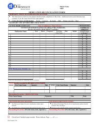
Primary Hyperparathyroidism: Diagnosis<br />
Patient Laboratory Values<br />
Serum calcium = 11.0 mg/dL<br />
Intact PTH = 92 pg/mL<br />
Serum calcium = 11.2 mg/dL<br />
Intact PTH = 49 pg/mL<br />
Serum calcium = 9.9 mg/dL<br />
Intact PTH = 81 pg/mL<br />
Diagnosis <strong>of</strong> Primary<br />
Hyperparathyroidism?<br />
Yes, classic disease<br />
Yes, mild disease<br />
Probably mild disease, but<br />
rule out vitamin D deficiency<br />
Normal serum calcium = 8.5 to 10.2 mg/dL; normal intact PTH = 10 to 72 pg/mL.
www.downstatesurgery.org<br />
Primary Hyperparathyroidism: Indication for<br />
<strong>Parathyroid</strong>ectomy<br />
Criteria for surgical referral and parathryroidectomy:<br />
• All symptomatic patients<br />
• All asymptomatic patients with any <strong>of</strong> the following:<br />
– Serum calcium concentration >1 mg/dL (>0.25 mM/liter) above<br />
the upper limits <strong>of</strong> normal<br />
– Bone density at the lumbar spine, hip, or distal end <strong>of</strong> the<br />
radius that is >2 SD below peak bone mass (T-score
www.downstatesurgery.org<br />
Primary Hyperparathyroidism: Benefits <strong>of</strong><br />
Surgical <strong>Management</strong><br />
Benefits from surgical management <strong>of</strong> primary HPT:<br />
• Renal function and bone density improvement<br />
• Resolution <strong>of</strong> neuropsychiatric symptoms<br />
• Quality <strong>of</strong> life better<br />
• Prolongs survival (10% reduction if untreated)<br />
• Reduction in cardiovascular incidents<br />
• Low complication rates<br />
• Cost <strong>of</strong> parathyroidectomy at 5 years is less than the cost <strong>of</strong><br />
surveillance
www.downstatesurgery.org<br />
<strong>Parathyroid</strong>: Embryology<br />
• Lower parathyroids are<br />
derived from the 3rd branchial<br />
pouch and migrate with the<br />
thymus<br />
• Upper parathyroids are derived<br />
from the 4th branchial pouch<br />
and lie in close proximity to the<br />
ultimobranchial bodies<br />
A- 8- to 10-mm embryo<br />
B- 13- to 14-mm embryo
www.downstatesurgery.org<br />
<strong>Parathyroid</strong> Anatomy<br />
Anatomical Locations:<br />
• Superior Glands:<br />
posteromedial aspect <strong>of</strong> the<br />
thyroid, above junction <strong>of</strong><br />
inferior thyroid artery and<br />
recurrent laryngeal nerve<br />
• Inferior Glands:<br />
posteriolateral aspect <strong>of</strong> the<br />
lower thyroid pole, below the<br />
inferior thyroid artery
www.downstatesurgery.org<br />
<strong>Parathyroid</strong> Anatomy<br />
Ectopic Locations (5-15%):<br />
• Thyrothymic ligament<br />
• Tracheoesophageal groove<br />
• Retroesophageal space<br />
• Retropharyngeal/high cervical<br />
• Carotid sheath<br />
• Intrathyroid<br />
• Ant/post superior mediastinum<br />
• Retropharyngeal<br />
• Intrathymic<br />
• Aorto-pulmonary window
www.downstatesurgery.org<br />
Preoperative Imaging<br />
IMAGING MODALITY SENSITIVITY SPECIFICITY COST SAFETY<br />
Noninvasive<br />
Sestamibi Moderate Moderate Moderate Safe<br />
Sestamibi SPECT High High Moderate Safe<br />
Ultrasound Moderate Moderate Low Safe<br />
4D-CT High High High Radiation<br />
MRI Low Moderate Moderate Safe<br />
Invasive<br />
Angiography Moderate Moderate Very high Hematoma, CVA, nephropathy*<br />
Venous localization High High Very high Hematoma, nephropathy*<br />
Ultrasound, biopsy High High Moderate Hematoma, infection
www.downstatesurgery.org<br />
Preoperative Imaging: Sestamibi Scan<br />
• Intravenous injection <strong>of</strong> 25mCi<br />
<strong>of</strong> 99mTechnetium<br />
• AP and oblique views <strong>of</strong> thorax<br />
and neck with gamma camera<br />
immediately after injection and<br />
at 1h and 4h or SPECT (single<br />
photon emission computed<br />
tomography)<br />
• Limitations with coexistence <strong>of</strong><br />
thyroid pathology or other<br />
metabolically active tissue can<br />
be overcome with double-tracer<br />
subtraction technique
www.downstatesurgery.org<br />
Preoperative Imaging: Ultrasound<br />
• Effective, noninvasive and<br />
inexpensive<br />
• Limitations are operator<br />
dependent, restriction to lesions<br />
in the neck<br />
• Often combined with sestamibi
www.downstatesurgery.org<br />
Preoperative Imaging: CT<br />
• Higher sensitivity than<br />
ultrasound, but involves<br />
radiation<br />
• 4D-CT is derived from 3D CT<br />
scanning, with added<br />
dimension from changes in<br />
perfusion <strong>of</strong> contrast over time,<br />
which allows to characterize<br />
hyperfunctioning parathyroid<br />
glands
www.downstatesurgery.org<br />
Preoperative Imaging: Venous Sampling<br />
• Selective arteriography in conjunction<br />
with venous sampling for PTH<br />
• Requires catheterization <strong>of</strong> multiple<br />
veins in the neck and mediastinum,<br />
from which blood samples are<br />
obtained with rapid PTH<br />
measurement in angio suite<br />
• <strong>Parathyroid</strong> adenomas have<br />
increased vascularity, demonstrating<br />
a characteristic blush on arteriography<br />
• Indicated for patients requiring reexploration<br />
with negative or<br />
discordant imaging studies
www.downstatesurgery.org<br />
<strong>Parathyroid</strong>ectomy<br />
Bilateral Neck exploration<br />
• 4 gland exploration, removal <strong>of</strong><br />
enlarged parathyroid<br />
• Intraoperative, histopathologic<br />
frozen section examination <strong>of</strong><br />
excised parathyroid tissue<br />
• Complication rate 1-3%<br />
• Cure rate 95-99%<br />
Focused <strong>Parathyroid</strong>ectomy<br />
(Minimally-Invasive <strong>Parathyroid</strong>ectomy)<br />
• Preoperative localization<br />
• Unilateral exploration<br />
• Intraoperative PTH monitoring<br />
• Local/ regional anesthesia<br />
• Ambulatory surgery<br />
• Complication rate 1.2%<br />
• Cure rate 93-99%<br />
Classic approach, now used when:<br />
– 4 gland hyperplasia is suspected<br />
– Family history <strong>of</strong> MEN1, MEN2A,<br />
PHPT<br />
– Concomitant thyroid disorder<br />
– <strong>Parathyroid</strong> localization studies are<br />
negative<br />
– ioPTH does not fall after unilateral<br />
exploration<br />
Widely used when positive<br />
localization available, patients with sporadic<br />
PHPT
www.downstatesurgery.org<br />
Bilateral Exploration vs Focused<br />
<strong>Parathyroid</strong>ectomy<br />
• Most studies show equal results in terms <strong>of</strong> complications, operative<br />
failure and cure rate, but large prospective trials with long-term<br />
follow-up are lacking<br />
• RCT with 5-year follow-up comparing unilateral vs. bilateral neck<br />
exploration did not note any difference in the rates <strong>of</strong> recurrent or<br />
persistent disease in the two groups <strong>of</strong> patients<br />
Westerdahl et al: Unilateral vs Bilateral Neck Exploration for Primary Hyperparathyroidism. Ann Surg. 2007;246(6):976-81
www.downstatesurgery.org<br />
Focused <strong>Parathyroid</strong>ectomy<br />
• 2-4 cm transverse incision just<br />
below cricoid cartilage,division<br />
<strong>of</strong> platysma +/- flaps, strap<br />
muscles divided in midline<br />
• Identification and mobilization<br />
<strong>of</strong> thyroid pole according to<br />
preop localization<br />
• Dissection <strong>of</strong> parathyroid<br />
adenoma, control <strong>of</strong> blood<br />
supply<br />
• ioPTH monitoring to confirm<br />
removal <strong>of</strong> hyperfunctioning<br />
adenoma<br />
• Closure in layers
www.downstatesurgery.org<br />
Focused <strong>Parathyroid</strong>ectomy: Other<br />
Techniques<br />
Video-Assisted <strong>Parathyroid</strong>ectomy<br />
and Endoscopic <strong>Parathyroid</strong>ectomy<br />
• Creation <strong>of</strong> working space in the neck<br />
with CO2 insufflation (5-8 mmHg)<br />
• Variable port placement<br />
• Operative space created between<br />
platysma and strap muscles<br />
• Increased operative time and expenses<br />
• Small amount <strong>of</strong> blood <strong>of</strong>ten diminishes<br />
view<br />
• Metabolic derangements due to<br />
absorption <strong>of</strong> CO2<br />
• Greatest use for thoracoscopic resection<br />
<strong>of</strong> mediastinal parathyroid adenomas
www.downstatesurgery.org<br />
Intraoperative Monitoring: PTH<br />
Rapid PTH assay:<br />
• PTH level is sent before<br />
induction <strong>of</strong> anesthesia, at<br />
resection <strong>of</strong> adenoma and 5<br />
and 10 minutes after resection<br />
<strong>of</strong> adenoma<br />
• 50% reduction at 10 minutes<br />
compared to original level<br />
confirms removal <strong>of</strong> parathyroid<br />
adenoma
Intraoperative Monitoring: Gamma Probe<br />
Radioguided parathyroidectomy:<br />
www.downstatesurgery.org<br />
• Injection <strong>of</strong> sestamibi 1-4h prior<br />
to procedure<br />
• Background count by scanning<br />
the thyroid isthmus with the γ-<br />
probe<br />
• Resected parathyroid tissue is<br />
placed on the probe for an ex<br />
vivo count<br />
• Ratio <strong>of</strong> ex vivo to in vivo<br />
background counts is > 20%<br />
confirms hyperfunctioning<br />
parathyroid tissue<br />
• Not widely used because yields<br />
little additional information over<br />
that obtained by adequate<br />
preoperative localization and<br />
the intraoperative PTH assay
www.downstatesurgery.org<br />
Steps to Find a Missing <strong>Parathyroid</strong><br />
Retrospective analysis <strong>of</strong> 115<br />
patients (2003-2005):<br />
Operative strategy:<br />
• Systematic perithyroid<br />
exploration, PTH monitoring<br />
• Extended cervical exploration:<br />
bilateral exploration along thyrothymic<br />
ligament, esophagotracheal sulcus,<br />
carotid sheath,<br />
retropharyngeal/esophageal region,<br />
cranial ventral and dorsal mediastinum<br />
• Hemithyroidectomy:<br />
on the side with higher suspicion/preop<br />
localization<br />
Herden et al: Intrathyroid adenomas in primary hyperparathyroidism: are they frequent enough to guide surgical strategy?<br />
Surg Innov. 18(4):373-8. 2011
www.downstatesurgery.org<br />
Steps to Find a Missing <strong>Parathyroid</strong><br />
Herden et al: Intrathyroid adenomas in primary hyperparathyroidism: are they frequent enough to guide surgical strategy?<br />
Surg Innov. 18(4):373-8. 2011
www.downstatesurgery.org<br />
Steps to Find a Missing <strong>Parathyroid</strong><br />
Herden et al: Intrathyroid adenomas in primary hyperparathyroidism: are they frequent enough to guide surgical<br />
strategy? Surg Innov. 18(4):373-8. 2011
www.downstatesurgery.org<br />
Steps to find a missing <strong>Parathyroid</strong><br />
Herden et al: Intrathyroid adenomas in primary hyperparathyroidism: are they frequent enough to guide surgical<br />
strategy? Surg Innov. 18(4):373-8. 2011
www.downstatesurgery.org<br />
Other Steps to Find a Missing <strong>Parathyroid</strong><br />
• Perform bilateral internal jugular venous sampling for PTH<br />
• Perform a cervical thymectomy<br />
• Open the carotid sheath<br />
• Search for an undescended gland, occasionally found in<br />
undescended thymic tissue<br />
• Perform intraoperative ultrasound <strong>of</strong> the thyroid gland<br />
• Sternotomy is not recommended during initial exploration<br />
• If the gland cannot be found, terminate the operation, leaving normal<br />
parathyroid gland intact
www.downstatesurgery.org<br />
Summary<br />
• Patients with symptomatic and asymptomatic primary<br />
hyperparathyroidism benefit from parathyroidectomy<br />
• After biochemical diagnosis <strong>of</strong> HPT, technique <strong>of</strong> choice for PHPT<br />
due to parathyroid adenoma is focused parathyroidectomy with<br />
preoperative localization (sestamibi scan most widely used) and<br />
intraoperative PTH monitoring<br />
• Drop <strong>of</strong> ioPTH to under 50% <strong>of</strong> preop value 10 minutes after<br />
resection confirms resection <strong>of</strong> hyperfunctioning parathyroid<br />
adenoma<br />
• If adenoma cannot be found and/or ioPTH does not drop<br />
appropriately, bilateral extended cervical exploration and if needed<br />
hemithyroidectomy on the side <strong>of</strong> localization should ensue (10-15%<br />
<strong>of</strong> parathyroid adenomas are ectopic, 5-10% double adenoma)<br />
• If adenoma still cannot be found after extended cervical exploration<br />
and hemithyroidectomy, the procedure should be aborted and<br />
further localization studies performed
www.downstatesurgery.org<br />
References<br />
• Townsend: Sabiston Textbook <strong>of</strong> Surgery, 19th ed.: Chapter 39 – The <strong>Parathyroid</strong><br />
Glands<br />
• Cameron: Current Surgical Therapy, 10th ed.: Primary Hyperparathyroidism<br />
• Townsend: Atlas <strong>of</strong> General Surgical Techniques, 1st ed.: Chapter 3 –<br />
<strong>Parathyroid</strong>ectomy<br />
• Schwartz's Principles <strong>of</strong> Surgery, 9e: Chapter 38. Thyroid, <strong>Parathyroid</strong>, and Adrenal<br />
• Westerdahl et al: Unilateral vs Bilateral Neck Exploration for Primary<br />
Hyperparathyroidism. Ann Surg. 2007;246(6):976-81<br />
• Herden et al: Intrathyroid adenomas in primary hyperparathyroidism: are they<br />
frequent enough to guide surgical strategy? Surg Innov. 18(4):373-8. 2011<br />
• Bilezikian JP, Potts JT Jr, Fuleihan Gel H, et al. Summary statement from a workshop<br />
on asymptomatic primary hyperparathyroidism: a perspective for the 21st century. J<br />
Clin Endocrinol Metab 2002;87(12):5353-5361.<br />
• Yeh et al: Surgery for Primary Hyperparathyroidism: Are the Consensus Guidelines<br />
being followed ? Ann Surg 255(6):1179-83, 2012<br />
• From Wang C-A. A clinical and pathological study <strong>of</strong> 112 cases. Ann Surg<br />
1977;186:140-145.)
www.downstatesurgery.org<br />
Questions<br />
A 47 year old female develops symptoms <strong>of</strong> hypercalcemia and further<br />
workup demonstrates her to have primary hyperparathyroidism. What is<br />
the most likely etiology <strong>of</strong> her disease?<br />
A- single adenoma<br />
B- double adenoma<br />
C- hyperplasia<br />
D- carcinoma
www.downstatesurgery.org<br />
Questions<br />
A 45 year old female without history <strong>of</strong> previous hospitalization or<br />
significant medical history presents to her primary care physician with<br />
complaints <strong>of</strong> headache, lethargy and constipation. EKG shows<br />
shortended QT interval and a widened T wave. Which <strong>of</strong> the following<br />
is the most likely etiology <strong>of</strong> her disease?<br />
A- vitamin D toxicity<br />
B- malignancy<br />
C- primary hyperparathyroidism<br />
D- sarcoidosis<br />
E- secondary hyperparathyroidism
www.downstatesurgery.org<br />
Questions<br />
A 55 year old woman with primary hyperparathyroidism is noted to<br />
have an asymptomatic kidney stone on an abdominal radiograph. What<br />
is the next step in management?<br />
A- cystoscopy<br />
B- serum oxalate measurement<br />
C- parathyroid localization studies<br />
D- bilateral neck exploration<br />
E- observation
www.downstatesurgery.org<br />
Questions<br />
Surgical exploration for a patient with primary hyperparathyroidism<br />
reveals all 4 glands to be enlarged. What is the most appropriate way<br />
to manage this?<br />
A- closure with localization study<br />
B- incisional biopsy <strong>of</strong> all glands<br />
C- subtotal parathyroidectomy<br />
D- excision <strong>of</strong> the largest enlarged gland<br />
E- selective venous PTH sampling