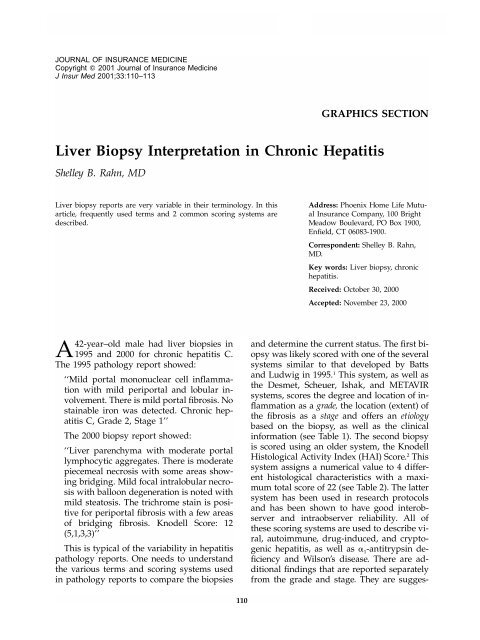
Liver Biopsy Interpretation in Chronic Hepatitis
Liver Biopsy Interpretation in Chronic Hepatitis
Liver Biopsy Interpretation in Chronic Hepatitis

Create successful ePaper yourself
Turn your PDF publications into a flip-book with our unique Google optimized e-Paper software.
JOURNAL OF INSURANCE MEDICINE<br />
Copyright 2001 Journal of Insurance Medic<strong>in</strong>e<br />
J Insur Med 2001;33:110–113<br />
GRAPHICS SECTION<br />
<strong>Liver</strong> <strong>Biopsy</strong> <strong>Interpretation</strong> <strong>in</strong> <strong>Chronic</strong> <strong>Hepatitis</strong><br />
Shelley B. Rahn, MD<br />
<strong>Liver</strong> biopsy reports are very variable <strong>in</strong> their term<strong>in</strong>ology. In this<br />
article, frequently used terms and 2 common scor<strong>in</strong>g systems are<br />
described.<br />
Address: Phoenix Home Life Mutual<br />
Insurance Company, 100 Bright<br />
Meadow Boulevard, PO Box 1900,<br />
Enfield, CT 06083-1900.<br />
Correspondent: Shelley B. Rahn,<br />
MD.<br />
Key words: <strong>Liver</strong> biopsy, chronic<br />
hepatitis.<br />
Received: October 30, 2000<br />
Accepted: November 23, 2000<br />
A42-year–old male had liver biopsies <strong>in</strong><br />
1995 and 2000 for chronic hepatitis C.<br />
The 1995 pathology report showed:<br />
‘‘Mild portal mononuclear cell <strong>in</strong>flammation<br />
with mild periportal and lobular <strong>in</strong>volvement.<br />
There is mild portal fibrosis. No<br />
sta<strong>in</strong>able iron was detected. <strong>Chronic</strong> hepatitis<br />
C, Grade 2, Stage 1’’<br />
The 2000 biopsy report showed:<br />
‘‘<strong>Liver</strong> parenchyma with moderate portal<br />
lymphocytic aggregates. There is moderate<br />
piecemeal necrosis with some areas show<strong>in</strong>g<br />
bridg<strong>in</strong>g. Mild focal <strong>in</strong>tralobular necrosis<br />
with balloon degeneration is noted with<br />
mild steatosis. The trichrome sta<strong>in</strong> is positive<br />
for periportal fibrosis with a few areas<br />
of bridg<strong>in</strong>g fibrosis. Knodell Score: 12<br />
(5,1,3,3)’’<br />
This is typical of the variability <strong>in</strong> hepatitis<br />
pathology reports. One needs to understand<br />
the various terms and scor<strong>in</strong>g systems used<br />
<strong>in</strong> pathology reports to compare the biopsies<br />
and determ<strong>in</strong>e the current status. The first biopsy<br />
was likely scored with one of the several<br />
systems similar to that developed by Batts<br />
and Ludwig <strong>in</strong> 1995. 1 This system, as well as<br />
the Desmet, Scheuer, Ishak, and METAVIR<br />
systems, scores the degree and location of <strong>in</strong>flammation<br />
as a grade, the location (extent) of<br />
the fibrosis as a stage and offers an etiology<br />
based on the biopsy, as well as the cl<strong>in</strong>ical<br />
<strong>in</strong>formation (see Table 1). The second biopsy<br />
is scored us<strong>in</strong>g an older system, the Knodell<br />
Histological Activity Index (HAI) Score. 2 This<br />
system assigns a numerical value to 4 different<br />
histological characteristics with a maximum<br />
total score of 22 (see Table 2). The latter<br />
system has been used <strong>in</strong> research protocols<br />
and has been shown to have good <strong>in</strong>terobserver<br />
and <strong>in</strong>traobserver reliability. All of<br />
these scor<strong>in</strong>g systems are used to describe viral,<br />
autoimmune, drug-<strong>in</strong>duced, and cryptogenic<br />
hepatitis, as well as 1 -antitryps<strong>in</strong> deficiency<br />
and Wilson’s disease. There are additional<br />
f<strong>in</strong>d<strong>in</strong>gs that are reported separately<br />
from the grade and stage. They are sugges-<br />
110
RAHN—LIVER BIOPSY INTERPRETATION<br />
Table 1. Batts and Ludwig Stag<strong>in</strong>g and Grad<strong>in</strong>g of <strong>Chronic</strong> <strong>Hepatitis</strong> 1<br />
Stage<br />
Score Description Criteria<br />
0 No fibrosis Normal connective tissue<br />
1 Portal fibrosis Fibrous portal expansion<br />
2 Periportal fibrosis Periportal or rare portal-portal septa<br />
3 Septal fibrosis Fibrous septa with architectural distortion;<br />
no obvious cirrhosis<br />
4 Cirrhosis Cirrhosis<br />
Grade<br />
Lobular<br />
Score Description Piecemeal Necrosis Inflammation/Necrosis<br />
0 Portal <strong>in</strong>flammation only (no lobular or None<br />
None<br />
piecemeal necrosis)<br />
1 M<strong>in</strong>imal M<strong>in</strong>imal, patchy M<strong>in</strong>imal/few areas of<br />
patchy necrosis<br />
2 Mild Mild; <strong>in</strong>volv<strong>in</strong>g some<br />
or all portal tracts<br />
Mild/mild hepatocellular<br />
damage<br />
3 Moderate Moderate; <strong>in</strong>volv<strong>in</strong>g all<br />
portal tracts<br />
Moderate/noticeable<br />
hepatocellular<br />
4 Severe Severe; with bridg<strong>in</strong>g<br />
necrosis<br />
damage<br />
Severe/prom<strong>in</strong>ent, diffuse<br />
hepatocellular<br />
damage<br />
Schematic representation of liver histology with various pathologic changes. Area with<strong>in</strong> the dotted l<strong>in</strong>es demonstrates cirrhosis.<br />
Portal-to-portal and portal-to-central fibrosis results <strong>in</strong> nodule formation.<br />
111
JOURNAL OF INSURANCE MEDICINE<br />
Periportal<br />
Bridg<strong>in</strong>g Necrosis<br />
Table 2. Knodell Histological Activity Index 2<br />
Score<br />
Intralobular<br />
Degeneration<br />
and Focal<br />
Necrosis Score Portal Inflammation Score Fibrosis Score<br />
None 0 None 0 No portal <strong>in</strong>flammation<br />
Mild piecemeal necrosis 1 Mild (acidophilic 1 Mild (spr<strong>in</strong>kl<strong>in</strong>g of<br />
bodies, balloon<strong>in</strong>g<br />
<strong>in</strong>flammatory<br />
degeneration cells <strong>in</strong> less than<br />
and/or scattered one third of portal<br />
foci of hepatocellular<br />
tracts)<br />
necrosis<br />
<strong>in</strong> one third of<br />
lobules or nodules)<br />
Moderate piecemeal necrosis<br />
(<strong>in</strong>volves 50% of the circumference<br />
of most portal<br />
tracts)<br />
Marked piecemeal necrosis (<strong>in</strong>volves<br />
50% of circumference<br />
of most portal tracts)<br />
Moderate piecemeal necrosis 5<br />
plus bridg<strong>in</strong>g necrosis<br />
Marked piecemeal necrosis plus 6<br />
bridg<strong>in</strong>g necrosis<br />
Multilobular necrosis 10<br />
3 Moderate (<strong>in</strong>volvement<br />
of one<br />
third to two<br />
thirds of lobules<br />
or nodules)<br />
4 Marked (<strong>in</strong>volvement<br />
of greater<br />
than two thirds<br />
of lobules or<br />
nodules)<br />
4 Moderate (<strong>in</strong>creased<br />
<strong>in</strong>flammatory<br />
cells <strong>in</strong><br />
one third to two<br />
thirds of portal<br />
tracts)<br />
4 Marked (dense<br />
pack<strong>in</strong>g of <strong>in</strong>flammatory<br />
cells<br />
<strong>in</strong> greater than<br />
two thirds of<br />
portal tracts)<br />
0 No fibrosis 0<br />
1 Fibrous portal<br />
expansion<br />
1<br />
3 Bridg<strong>in</strong>g Fibrosis<br />
(portal-portal<br />
or portalcentral<br />
l<strong>in</strong>kage)<br />
3<br />
4 Cirrhosis 4<br />
tive, though not diagnostic, of the specific etiologies<br />
<strong>in</strong>dicated. These <strong>in</strong>clude steatosis<br />
(hepatitis C), Mallory bodies (alcohol, Wilson’s<br />
disease), copper deposits (Wilson’s disease),<br />
ground glass hepatocytes (hepatitis B),<br />
and hepatocellular carc<strong>in</strong>oma.<br />
The terms used <strong>in</strong> liver biopsies are def<strong>in</strong>ed<br />
below and illustrated <strong>in</strong> the Figure:<br />
Inflammation and Necrosis<br />
Portal <strong>in</strong>flammation: lymphocytes conf<strong>in</strong>ed<br />
to the portal tract (triad).<br />
Piecemeal (periportal) necrosis: the extension<br />
of lymphocytes or monocytes from the portal<br />
tract <strong>in</strong>to the lobule (cross<strong>in</strong>g the limit<strong>in</strong>g<br />
plate) with the destruction of the periportal<br />
hepatocytes.<br />
Interface hepatitis: same as piecemeal necrosis<br />
but <strong>in</strong>troduced to reflect that apoptosis<br />
rather than necrosis is the predom<strong>in</strong>ant process<br />
at the limit<strong>in</strong>g plate.<br />
Bridg<strong>in</strong>g necrosis: the extension of <strong>in</strong>flammation<br />
and necrosis from one portal tract to<br />
another.<br />
Lobular <strong>in</strong>flammation: presence of <strong>in</strong>creased<br />
number of lymphocytes <strong>in</strong> the lobule.<br />
Lobular necrosis: hepatocyte damage separate<br />
from the portal tract.<br />
Fibrosis and Cirrhosis<br />
Portal fibrosis: expansion of the portal tract by<br />
fibrosis without extension outside the tract.<br />
Periportal fibrosis: extension of fibrosis outside<br />
the portal tract.<br />
Bridg<strong>in</strong>g fibrosis: extension of fibrosis from<br />
one portal tract to another.<br />
112
RAHN—LIVER BIOPSY INTERPRETATION<br />
Cirrhosis: diffuse fibrosis with nodule formation.<br />
Nodules are groups of cells r<strong>in</strong>ged by<br />
fibrosis as a result of portal-to-portal and<br />
portal-to-central ve<strong>in</strong> fibrosis. They have no<br />
organized circulation or biliary dra<strong>in</strong>age.<br />
The terms, chronic persistent and chronic<br />
active hepatitis, are obsolete. These were <strong>in</strong>consistently<br />
used to describe the morphology<br />
and disease etiology. Importantly, prognosis<br />
cannot be predicted by these terms.<br />
The first biopsy is reported as Grade 2<br />
based on the f<strong>in</strong>d<strong>in</strong>g of mild periportal necrosis<br />
and mild lobular activity, and Stage 1<br />
based on the presence of fibrosis limited to<br />
the portal tract. The diagnosis of chronic hepatitis<br />
C is made us<strong>in</strong>g both the histologic appearance<br />
and the cl<strong>in</strong>ical <strong>in</strong>formation. The<br />
Knodell score for this biopsy would be<br />
1,1,1,1.<br />
The second biopsy is scored accord<strong>in</strong>g to<br />
the Knodell system: 5 for moderate piecemeal<br />
necrosis with bridg<strong>in</strong>g necrosis; 1 for mild <strong>in</strong>tralobular<br />
degeneration and necrosis; 3 for<br />
moderate portal <strong>in</strong>flammation; and the last 3<br />
for bridg<strong>in</strong>g fibrosis. This would be scored by<br />
the Batts-type scale as Grade 4, Stage 2. When<br />
there is a disparity between the criteria, the<br />
higher score is used. Us<strong>in</strong>g either system,<br />
there clearly has been progression of disease<br />
from 1995 to 2000.<br />
REFERENCES<br />
1. Knodell RG, Ishak KG, Black WC, et al. Formulation<br />
and application of a numerical scor<strong>in</strong>g system for<br />
assess<strong>in</strong>g histological activity <strong>in</strong> asymptomatic<br />
chronic active hepatitis. Hepatology. 1981;1:431–435.<br />
2. Batts KP, Ludwig J. <strong>Chronic</strong> hepatitis an update on<br />
term<strong>in</strong>ology and report<strong>in</strong>g. Am J Surg Pathol. 1995;<br />
19:1409–1417.<br />
113