GABA & Behaviour II - Godzilla - Kennedy Krieger Institute
GABA & Behaviour II - Godzilla - Kennedy Krieger Institute
GABA & Behaviour II - Godzilla - Kennedy Krieger Institute

Create successful ePaper yourself
Turn your PDF publications into a flip-book with our unique Google optimized e-Paper software.
B r i e f com m u n i c at i o n s<br />
© 2010 Nature America, Inc. All rights reserved.<br />
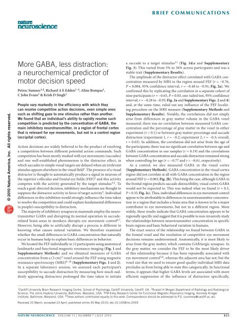
More <strong>GABA</strong>, less distraction:<br />
a neurochemical predictor of<br />
motor decision speed<br />
Petroc Sumner 1,4 , Richard A E Edden 1–4 , Aline Bompas 1 ,<br />
C John Evans 1 & Krish D Singh 1<br />
People vary markedly in the efficiency with which they<br />
can resolve competitive action decisions, even simple ones<br />
such as shifting gaze to one stimulus rather than another.<br />
We found that an individual’s ability to rapidly resolve such<br />
competition is predicted by the concentration of <strong>GABA</strong>, the<br />
main inhibitory neurotransmitter, in a region of frontal cortex<br />
that is relevant for eye movements, but not in a control region<br />
(occipital cortex).<br />
Action decisions are widely believed to be the product of resolving<br />
a competition between different potential action commands. Such<br />
competition has been mostly studied with eye movements (saccades)<br />
and one well-established phenomenon is the distractor effect, in<br />
which saccades to simple visual targets are delayed when an irrelevant<br />
stimulus appears elsewhere in the visual field 1 . The presence of a visual<br />
distractor is thought to automatically produce a signal in neurons of<br />
the superior colliculus and frontal eye fields (FEF) 2 and this activity<br />
competes with the activity generated by the target stimulus 3,4 . To<br />
reach a goal-directed decision, inhibitory mechanisms are thought to<br />
suppress the distractor activity in favor of target activity 5 . Individual<br />
differences in this inhibition would strongly influence the time taken<br />
to resolve the competition and could explain fundamental differences<br />
in people’s susceptibility to distraction.<br />
The majority of inhibitory synapses in mammals employ the neurotransmitter<br />
<strong>GABA</strong> and disrupting its normal operation in saccaderelated<br />
brain areas in monkeys disrupts eye movement control 6,7 .<br />
However, being able to artificially disrupt a process is different to<br />
knowing what causes natural variation. We therefore examined<br />
whether the small differences in <strong>GABA</strong> concentration that naturally<br />
occur in humans help to explain basic differences in behavior.<br />
We located the FEF individually in 12 participants using anatomical<br />
landmarks and functional magnetic resonance imaging (Fig. 1 and<br />
Supplementary Methods) and we obtained measures of <strong>GABA</strong><br />
concentration from a (3 cm) 3 voxel around the FEF using magnetic<br />
resonance spectroscopy (MRS) 8–10 (Supplementary Figs. 1 and 2).<br />
In a separate laboratory session, we assessed each participant’s<br />
susceptibility to saccade distraction by measuring how much suddenly<br />
appearing distractors prolonged the time taken to initiate<br />
a saccade to a target stimulus 11 (Fig. 1d,e and Supplementary<br />
Fig. 3). This varied from 5% to 36% across participants and was a<br />
stable trait (Supplementary Results).<br />
The amplitude of the distractor effect correlated with <strong>GABA</strong> concentration<br />
measured by MRS in the region around FEF (r = −0.76,<br />
P = 0.004, 95% confidence interval, r = −0.48 to −0.91; Fig. 2a). We<br />
confirmed this by replicating the correlation in a separate cohort of<br />
nine participants (r = −0.65, P = 0.03, one-tailed test, 95% confidence<br />
interval, r = −0.28 to −0.95; Fig. 2a and Supplementary Figs. 2 and 4)<br />
and, at the same time, ruled out any influence of the FEF localizing<br />
procedure on the MRS measure (Supplementary Methods and<br />
Supplementary Results). Notably, the correlations did not simply<br />
arise from differences in gray matter volume in the <strong>GABA</strong> voxel<br />
measured; there was no correlation between measured <strong>GABA</strong> concentration<br />
and the percentage of gray matter in the voxel in either<br />
experiment (r < 0.1) or between gray matter percentage and saccade<br />
distraction (experiment 1, r = −0.2; experiment 2, r = −0.3; overall,<br />
r = 0.03). In addition, the correlation did not arise from the age of<br />
the participants; there was no significant correlation between age and<br />
<strong>GABA</strong> concentration in our samples (r = 0.19) and the correlations<br />
between <strong>GABA</strong> concentration and saccade distraction remained strong<br />
when controlling for age (r = −0.77 and r = −0.61, respectively).<br />
As a control, we also measured <strong>GABA</strong> in the visual cortex<br />
(Supplementary Methods). <strong>GABA</strong> concentration in the visual cortex<br />
region did not correlate at all with <strong>GABA</strong> concentration in the region<br />
around FEF (r = 0.003; Fig. 2b). This being the case, although <strong>GABA</strong> in<br />
the frontal region predicts saccade distractibility, visual cortex <strong>GABA</strong><br />
would not be expected to. This was indeed what we found (r = 0.3,<br />
P = 0.35; Fig. 2c). Thus, individual differences in eye movement control<br />
appear to be attributable to differences in neurotransmitter concentration<br />
in a region that includes a brain area that is known to be a major<br />
contributor to eye movements, but not in a different region. More<br />
widely, these results indicate that <strong>GABA</strong> concentration appears to be<br />
regionally specific and suggest that it is possible to non-invasively study<br />
the relationships between neurotransmitter concentration in specific<br />
brain regions and basic behavioral variation in humans.<br />
The exact source of the relationship we found between <strong>GABA</strong> in<br />
the frontal voxel and the resolution of competitive eye movement<br />
decisions remains undetermined. Anatomically, it is most likely to<br />
arise from the gray matter, which contains <strong>GABA</strong>ergic synapses. In<br />
the gray matter, we consider the FEF to be the most likely driver<br />
of this relationship because it has been repeatedly associated with<br />
eye movement control 3,6 , whereas the adjacent area has not, but the<br />
voxel size that we used to ensure good-quality individual MRS data<br />
precludes us from being able to state this categorically. In functional<br />
terms, it appears that higher <strong>GABA</strong> levels are associated with more<br />
efficient suppression of the influence of distractors specifically,<br />
1 Cardiff University Brain Research Imaging Centre, School of Psychology, Cardiff University, Cardiff, UK. 2 Russell H Morgan Department of Radiology and Radiological<br />
Science, The Johns Hopkins University, Baltimore, Maryland, USA. 3 FM Kirby Research Center for Functional Magnetic Resonance Imaging, <strong>Kennedy</strong> <strong>Krieger</strong><br />
<strong>Institute</strong>, Baltimore, Maryland, USA. 4 These authors contributed equally to this work. Correspondence should be addressed to P.S. (sumnerp@cardiff.ac.uk).<br />
Received 22 March; accepted 23 April; published online 30 May 2010; doi:10.1038/nn.2559<br />
nature neuroscience VOLUME 13 | NUMBER 7 | JULY 2010 825
i e f com m u n i c at i o n s<br />
© 2010 Nature America, Inc. All rights reserved.<br />
Figure 1 Methodology (example data from one<br />
individual). (a,b) The bilateral activation of<br />
FEF revealed by functional magnetic resonance<br />
imaging (red/yellow, see scale bar for t value)<br />
was used to locate the MRS voxel (green, 3 ×<br />
3 × 3 cm 3 ), which was also aligned with the<br />
brain surface and remained anterior to the<br />
central sulcus. (c) Edited magnetic resonance<br />
spectra allowed the quantification of <strong>GABA</strong><br />
concentration 8,10 (glutamine/glutamate (Glx) and<br />
N-acetyl-aspartate (NAA) peaks are also marked).<br />
(d) In the saccade distractor procedure, targets<br />
(black) occurred either alone or accompanied<br />
by distractors (light gray), which could appear<br />
at various delays before (as illustrated) or after<br />
target onset. The characteristic rise and fall in<br />
the distractor effect as distractor onset time<br />
varies relative to target onset can be seen in e.<br />
The peak distractor effect for this individual<br />
(yellow star and dotted line) was extracted<br />
by fitting a Gaussian curve to the data 11 (see<br />
Supplementary Methods and Supplementary<br />
Figures 1–4 for all participants).<br />
rather than more general inhibition or caution, which would also be<br />
expected to influence overall response time and error rate, neither<br />
of which correlated with <strong>GABA</strong> (r = 0.08 and r = −0.06, respectively;<br />
Supplementary Fig. 5).<br />
In physiological terms, naturally occurring individual differences in<br />
<strong>GABA</strong> may reflect differences in the number of <strong>GABA</strong> interneurons<br />
in certain regions, the number of synapses per neuron or simply differences<br />
in <strong>GABA</strong> concentration per synapse. Thus, we should not<br />
expect natural variation to mimic the effects of pharmacological<br />
agents, which tend to manipulate the efficacy of <strong>GABA</strong> at the synapse<br />
rather than changing the overall concentration, synapse density<br />
or cell numbers. We expect future research to clarify the relationship<br />
between natural variation and induced modulation of <strong>GABA</strong>,<br />
but we already have a hint that they are different: injections of<br />
the <strong>GABA</strong> antagonist bicuculline in monkey FEF produce greater<br />
a<br />
Distractor effect (%)<br />
40<br />
30<br />
Distractor effect (%)<br />
40<br />
30<br />
20<br />
10<br />
0<br />
1.0<br />
1.2<br />
1.2 1.4 1.6 1.8<br />
1.4 1.6 1.8<br />
0 0<br />
1.2 1.3 1.4 1.5 1.6 1.7 1.8 1.0 1.4 1.8<br />
20<br />
Frontal region <strong>GABA</strong><br />
Frontal region <strong>GABA</strong><br />
conc. (i.u.) c<br />
40<br />
conc. (i.u.)<br />
10<br />
20<br />
Frontal region <strong>GABA</strong> conc. (i.u.)<br />
Occipital <strong>GABA</strong><br />
conc. (i.u.)<br />
Figure 2 <strong>GABA</strong> in the frontal region correlates with saccade distraction.<br />
(a) Higher <strong>GABA</strong> concentration in the region around human FEF predicted<br />
smaller distractor effects across individuals. This result was replicated in<br />
a second cohort (inset). (b,c) There was no correlation between the frontal<br />
<strong>GABA</strong> concentration and the control, occipital <strong>GABA</strong> concentration, and<br />
<strong>GABA</strong> concentration in visual cortex did not correlate with the distractor<br />
effect. <strong>GABA</strong> concentration measurements are relative, not absolute, and<br />
thus stated in institutional units (i.u.)<br />
b<br />
Occ. <strong>GABA</strong> conc. (i.u.)<br />
Distractor effect (%)<br />
2.0<br />
1.8<br />
1.6<br />
1.4<br />
1.2<br />
a<br />
c<br />
6<br />
5<br />
Glx<br />
4<br />
b<br />
FEF<br />
<strong>GABA</strong><br />
3<br />
2<br />
NAA<br />
MRS voxel<br />
1 0<br />
ppm<br />
10<br />
0<br />
d<br />
e<br />
Distractor effect (%)<br />
40<br />
20<br />
0<br />
–20<br />
0 ms<br />
660 ms<br />
Distractor<br />
700 ms<br />
Target<br />
710 ms<br />
–40 –20 0 20 40 60<br />
Distractor timing (ms)<br />
1,000 ms<br />
Time<br />
variance in saccade latency 7 , but in our data, although some<br />
participants were more variable in their response times than others,<br />
this did not correlate with natural differences in <strong>GABA</strong> concentration<br />
(r = 0.12) Studying naturally occurring <strong>GABA</strong> differences in restricted<br />
brain regions therefore promises to offer different insights from studying<br />
the consequences of artificially manipulating <strong>GABA</strong> signaling in<br />
humans or animals.<br />
The reasons why humans differ from each other, even in basic mechanisms<br />
of sensory-motor behavior, is crucial to our understanding of<br />
normal brain function, but it also has important implications for many<br />
clinical disorders where there appears to be a spectrum from normal<br />
to pathological without any clear boundary in between. <strong>GABA</strong>mediated<br />
inhibition (interacting with other neurotransmitters, such<br />
as dopamine) has been implicated in many such disorders, including<br />
schizophrenia, epilepsy, anxiety, depression, obsessive compulsive<br />
disorder (OCD), attention deficit hyperactivity disorder (ADHD),<br />
autism and Tourette’s syndrome 7,12,13 . Furthermore, in many of these<br />
disorders there appear to be deficits or differences in basic motor or<br />
oculomotor behavior 14 . In schizophrenia, deficits of eye movement<br />
competition and inhibition have been reliably measured over two<br />
decades 15 , which, on the basis of our current findings, we predict may<br />
correlate with regionally specific <strong>GABA</strong> concentration.<br />
In sum, we found a link between individual variability in action<br />
control and neurotransmitter levels in a specific brain region.<br />
Moreover, our finding that <strong>GABA</strong> concentration may be regionally<br />
specific in the brain—an individual with low <strong>GABA</strong> in one brain area<br />
does not necessarily have low <strong>GABA</strong> in another area—is of crucial<br />
importance for the associations between <strong>GABA</strong> transmission and<br />
clinical disorders.<br />
Note: Supplementary information is available on the Nature Neuroscience website.<br />
Acknowledgments<br />
We thank F. Boy, S. Muthukumaraswamy, B. Taud, M. Husain, C. Chambers,<br />
T. Freeman, R. Honey and L. Wilkinson. R.A.E.E. thanks G. Barker and D. Shungu<br />
for pulse programming advice. This work was supported by the ESRC, the<br />
Wellcome Trust, the Wales <strong>Institute</strong> of Cognitive Neuroscience and the Schools of<br />
Psychology, Biosciences and Chemistry at Cardiff University. Cardiff University<br />
Brain Research Imaging Centre was established with support from the UK<br />
Department of Trade and Industry, Cardiff University and the Welsh Assembly<br />
government. R.A.E.E. held an RCUK fellowship.<br />
826 VOLUME 13 | NUMBER 7 | JULY 2010 nature neuroscience
i e f com m u n i c at i o n s<br />
AUTHOR CONTRIBUTIONS<br />
P.S., R.A.E.E. and K.D.S. conceived and planned the experimental study. R.A.E.E.,<br />
P.S. and C.J.E. carried out and analyzed the magnetic resonance imaging and<br />
P.S. and A.B. performed and analyzed the behavioral experiments. All authors<br />
contributed to interpretation and presentation.<br />
COMPETING FINANCIAL INTERESTS<br />
The authors declare no competing financial interests.<br />
Published online at http://www.nature.com/natureneuroscience/.<br />
Reprints and permissions information is available online at http://www.nature.com/<br />
reprintsandpermissions/.<br />
1. Walker, R., Mannan, S., Maurer, D., Pambakian, A.L.M. & Kennard, C. Proc. R.<br />
Soc. Lond. B Biol. Sci. 267, 431–438 (2000).<br />
2. Olivier, E., Dorris, M.C. & Munoz, D.P. Behav. Brain Sci. 22, 694–695 (1999).<br />
3. Schlag, J., Dassonville, P. & Schlag-Rey, M. J. Neurophysiol. 79, 64–72 (1998).<br />
4. Godijn, R. & Theeuwes, J. J. Exp. Psychol. Hum. Percept. Perform. 28, 1039–1054<br />
(2002).<br />
5. McSorley, E., Haggard, P. & Walker, R. J. Neurophysiol. 96, 1420–1424 (2006).<br />
6. Schiller, P.H. & Tehovnik, E.J. Eur. J. Neurosci. 18, 3127–3133 (2003).<br />
7. Pouget, P., Wattiez, N., Rivaud-Pechoux, S. & Gaymard, B. PLoS One 4, e5208<br />
(2009).<br />
8. Mescher, M., Merkle, H., Kirsch, J., Garwood, M. & Gruetter, R. NMR Biomed. 11,<br />
266–272 (1998).<br />
9. Muthukumaraswamy, S.D., Edden, R.A.E., Jones, D.K., Swettenham, J.B. & Singh, K.D.<br />
Proc. Natl. Acad. Sci. USA 106, 8356–8361 (2009).<br />
10. Evans, C.J., McGonigle, D.J. & Edden, R.A. J. Magn. Reson. Imaging 31, 204–209<br />
(2010).<br />
11. Bompas, A. & Sumner, P. J. Vision 9, 1–14 (2009).<br />
12. Lewis, D.A., Hashimoto, T. & Volk, D.W. Nat. Rev. Neurosci. 6, 312–324 (2005).<br />
13. Kalueff, A.V. & Nutt, D.J. Depress. Anxiety 24, 495–517 (2007).<br />
14. Rommelse, N.N., Van der Stigchel, S. & Sergeant, J.A. Brain Cogn. 68, 391–414<br />
(2008).<br />
15. Thaker, G.K., Nguyen, J.A. & Tamminga, C.A. Biol. Psychiatry 25, 49–59<br />
(1989).<br />
© 2010 Nature America, Inc. All rights reserved.<br />
nature neuroscience VOLUME 13 | NUMBER 7 | JULY 2010 827