Congenital Cytomegalovirus Conference - Congenital CMV ...
Congenital Cytomegalovirus Conference - Congenital CMV ...
Congenital Cytomegalovirus Conference - Congenital CMV ...
Create successful ePaper yourself
Turn your PDF publications into a flip-book with our unique Google optimized e-Paper software.
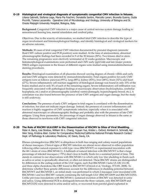
O-19 Histological and virological diagnosis of symptomatic congenital <strong>CMV</strong> infection in fetuses.<br />
Liliana Gabrielli, Stefania Lega, Maria Pia Foschini, Donatella Santini, Marcello Lanari, Brunella Guerra, Giulia<br />
Piccirilli, Tiziana Lazzarotto. Operative Unit of Microbiology and Virology, University of Bologna and St.<br />
Orsola Malpighi University Hospital, Bologna, Italy.<br />
Background: <strong>Congenital</strong> <strong>CMV</strong> infection is a major cause of central nervous system damage leading to<br />
sensorineural hearing loss, mental retardation and cerebral palsy.<br />
Objectives. Due to the scarcity of information, we studied fetal <strong>CMV</strong> infection to describe the type of<br />
organ involvement and histomorphological findings, and identify histological and virological markers for<br />
an adverse outcome.<br />
Methods: 30 cases of fetal congenital <strong>CMV</strong> infection documented by prenatal diagnosis (amniotic<br />
fluid <strong>CMV</strong> culture positive and PCR positive) were studied. At the time of amniocentesis, abnormal<br />
ultrasonographic findings had been recorded in 9 of the 30 fetuses (30%). Two fetuses died in utero.<br />
The remaining pregnancies were electively terminated at 22 weeks gestation. Macroscopic and<br />
histomorphological examinations were performed and <strong>CMV</strong> early (ppUL44) and late (major protein<br />
55KD) antigen expression in the tissues of different organs were studied using immunohistochemical<br />
staining procedures.<br />
Results: Histological examination of all placentas showed varying degrees of chronic villitis and early<br />
and late <strong>CMV</strong> antigens were detected by immunohistochemistry. Fetal organs positive for early <strong>CMV</strong><br />
antigens were as follows: pancreas (95%), lung (88%), kidney (83%), liver (68%), brain (52%) and heart<br />
(27%). Severe inflammatory cell reaction suggestive of histological damage was found in only 33% of the<br />
brains studied and in only 4% of each of the remaining organs. Severe inflammatory cell infiltration was<br />
frequently associated with pathological findings at macroscopic observation (hydrocephalus, cerebellar<br />
hypoplasia, etc.) and/or at ultrasonography (cerebral ventriculomegaly, hyperechogenic bowel, etc.). A<br />
correlation was also found between the presence of late <strong>CMV</strong> antigens and organ damage, but the study<br />
is still underway.<br />
Conclusions: The presence of early <strong>CMV</strong> antigens in fetal organs is correlated with the dissemination<br />
of infection, but does not indicate organ damage. Instead, the presence of a severe inflammatory cell<br />
reaction is highly suggestive of <strong>CMV</strong> symptomatic infection, especially when it is associated with<br />
abnormal macroscopic or ultrasonographic findings and probably also with the presence of late <strong>CMV</strong><br />
antigens. Using these parameters, the percentage of organ damage observed in fetuses is the same as<br />
those observed in newborns with <strong>CMV</strong> congenital infection.<br />
O-20 The Role of Rh<strong>CMV</strong> ULb1ORF in the Dissemination of Rh<strong>CMV</strong> to Sites of Viral Shedding.<br />
Peter A. Barry, Lisa Strelow, William W.-L. Chang, Yujuan Yue, Kristie L. Oxford, Kimberli A. Schmidt, Kai-<br />
Wen Yang, Kristina Abel. Center for Comparative Medicine/California National Primate Research Center/<br />
Dept. of Pathology & Laboratory Medicine, UC Davis, Davis, CA.<br />
Rhesus cytomegalovirus (Rh<strong>CMV</strong>) is ubiquitous in both free-ranging and colony-reared populations<br />
of rhesus macaques. Clinical signs of Rh<strong>CMV</strong> infection are almost never observed in either population<br />
following either natural exposure to wild-type virus (Rh<strong>CMV</strong>WT) or experimental inoculation with<br />
the 68-1 strain of virus (Rh<strong>CMV</strong>68-1). A hallmark of natural infection with Rh<strong>CMV</strong>WT is that a large<br />
percentage of infected macaques (~75%) shed virus in bodily fluids for a sustained period of time. This<br />
stands in contrast to our observations with Rh<strong>CMV</strong>68-1 in which only low titer shedding in fluids such<br />
as saliva or urine, is sporadically observed, or often not detected. These Rh<strong>CMV</strong> strains are distinguished<br />
by differences in the coding content of the ULb’ region, such that Rh<strong>CMV</strong>68-1 lacks UL128, UL130,<br />
and three alpha chemokine-like ORF found in Rh<strong>CMV</strong>WT (Oxford et al, Virology, 373:181, 2008). To<br />
determine whether these genetic differences account for the differences in shedding potential between<br />
Rh<strong>CMV</strong>WT and Rh<strong>CMV</strong>68-1, an initial study was performed in which 2 macaques were inoculated with<br />
Rh<strong>CMV</strong>68-1 and two Rh<strong>CMV</strong> variants containing the full-length ULb’ (Rh<strong>CMV</strong>22659 and Rh<strong>CMV</strong>21252).<br />
Unlike previous inoculations with Rh<strong>CMV</strong>68-1, sustained excretion of Rh<strong>CMV</strong> was detected in urine<br />
and saliva. Using differential PCR to distinguish between the three variants, only Rh<strong>CMV</strong>22659 and<br />
Rh<strong>CMV</strong>21252 were detected in urine and/or saliva, or recovered from parotid gland explanted in<br />
39


