64 Slice CT
64 Slice CT
64 Slice CT

You also want an ePaper? Increase the reach of your titles
YUMPU automatically turns print PDFs into web optimized ePapers that Google loves.
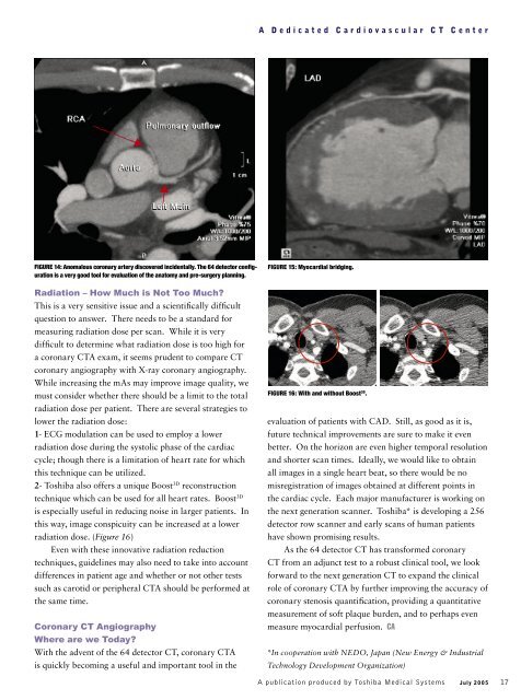
A Dedicated Cardiovascular <strong>CT</strong> Center<br />
FIGURE 14: Anomalous coronary artery discovered incidentally. The <strong>64</strong> detector confi g-<br />
uration is a very good tool for evaluation of the anatomy and pre-surgery planning.<br />
Radiation – How Much is Not Too Much?<br />
This is a very sensitive issue and a scientifi cally diffi cult<br />
question to answer. There needs to be a standard for<br />
measuring radiation dose per scan. While it is very<br />
diffi c ult to determine what radiation dose is too high for<br />
a coronary <strong>CT</strong>A exam, it seems prudent to compare <strong>CT</strong><br />
coronary angiography with X-ray coronary angiography.<br />
While increasing the mAs may improve image quality, we<br />
must consider whether there should be a limit to the total<br />
radiation dose per patient. There are several strategies to<br />
lower the radiation dose:<br />
1- ECG modulation can be used to employ a lower<br />
radiation dose during the systolic phase of the cardiac<br />
cycle; though there is a limitation of heart rate for which<br />
this technique can be utilized.<br />
2- Toshiba also offers a unique Boost 3D reconstruction<br />
technique which can be used for all heart rates. Boost 3D<br />
is especially useful in reducing noise in larger patients. In<br />
this way, image conspicuity can be increased at a lower<br />
radiation dose. (Figure 16)<br />
Even with these innovative radiation reduction<br />
techniques, guidelines may also need to take into account<br />
differences in patient age and whether or not other tests<br />
such as carotid or peripheral <strong>CT</strong>A should be performed at<br />
the same time.<br />
Coronary <strong>CT</strong> Angiography<br />
Where are we Today?<br />
With the advent of the <strong>64</strong> detector <strong>CT</strong>, coronary <strong>CT</strong>A<br />
is quickly becoming a useful and important tool in the<br />
FIGURE 15: Myocardial bridging.<br />
FIGURE 16: With and without Boost 3D .<br />
evaluation of patients with CAD. Still, as good as it is,<br />
future technical improvements are sure to make it even<br />
better. On the horizon are even higher temporal resolution<br />
and shorter scan times. Ideally, we would like to obtain<br />
all images in a single heart beat, so there would be no<br />
misregistration of images obtained at different points in<br />
the cardiac cycle. Each major manufacturer is working on<br />
the next generation scanner. Toshiba* is developing a 256<br />
detector row scanner and early scans of human patients<br />
have shown promising results.<br />
As the <strong>64</strong> detector <strong>CT</strong> has transformed coronary<br />
<strong>CT</strong> from an adjunct test to a robust clinical tool, we look<br />
forward to the next generation <strong>CT</strong> to expand the clinical<br />
role of coronary <strong>CT</strong>A by further improving the accuracy of<br />
coronary stenosis quantifi c ation, providing a quantitative<br />
measurement of soft plaque burden, and to perhaps even<br />
measure myocardial perfusion. CA<br />
*In cooperation with NEDO, Japan (New Energy & Industrial<br />
Technology Development Organization)<br />
A publication produced by Toshiba Medical Systems<br />
July 2005 17