64 Slice CT
64 Slice CT
64 Slice CT

Create successful ePaper yourself
Turn your PDF publications into a flip-book with our unique Google optimized e-Paper software.
Patient Focused Imaging<br />
A<br />
A<br />
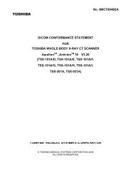
FIGURE 6: Images of a 300 lb patient with 0.5 mm slices, 120 kV, 140 mAs without (a)<br />
and with (b) the Boost 3D algorithm.<br />
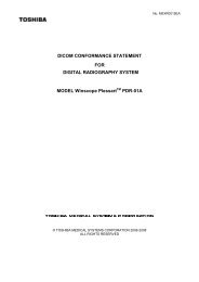
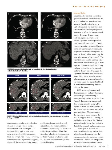
150mAs 80mAs 80mAs (w/QDS)<br />
A B C<br />
Std. Dev: 32.7 Std. Dev: 47.8 Std. Dev: 31.6<br />
FIGURE 7: Effect of QDS. Noise levels with (a) standard technique, (b) low dose technique, and (c) low dose<br />
technique with QDS.<br />
demonstrates cardiac and abdominal<br />
images of a large patient using a<br />
relatively low scan technique. The<br />
images exhibit typical structured<br />
noise and streak artifacts resulting<br />
from the low photon count. However,<br />
when the Boost 3D algorithm is applied,<br />
Figure 6b shows the resulting image<br />
B<br />
B<br />
quality: the image noise is greatly<br />
reduced and the streak artifacts<br />
disappear. By reducing the noise and<br />
mitigating the effects of low dose<br />
scanning, adaptive techniques such<br />
as Boost 3D are an invaluable asset<br />
in Toshiba’s commitment to patient<br />
focused imaging.<br />
Image Data<br />
Once the detectors and acquisition<br />
systems have been optimized and the<br />
streaks and excess noise have been<br />
removed from localized areas of<br />
high attenuation, we must turn our<br />
attention to minimizing the general<br />
noise that is left in the reconstructed<br />
image. To tackle this problem,<br />
Toshiba engineers developed a<br />
unique algorithm called Quantum<br />
Denoising Software (QDS). QDS is<br />
an adaptive noise reduction fi lter that<br />
works on reconstructed image data<br />
by preferentially smoothing areas of<br />
uniform density while preserving the<br />
edge information of the image 14 . The<br />
algorithm uses locally sampled edge<br />
information within the image to blend<br />
together variable strength smoothing<br />
and sharpening fi lters. In areas of<br />
uniform density with few edges, the<br />
algorithm smoothes and reduces the<br />
noise. Near tissue boundaries and<br />
other complex structures where there<br />
are many edges, the algorithm blends<br />
in more of the sharpening fi lter to<br />
enhance the image.<br />
QDS works in both two and<br />
three dimensions and can drastically<br />
reduce image noise allowing a<br />
corresponding savings in patient dose.<br />
Figure 7 illustrates the substantial<br />
dose savings possible using QDS.<br />
Figure 7a shows the relative noise in<br />
the liver of a patient using a standard<br />
scan technique. 7b demonstrates<br />
the increase in image noise as the<br />
mAs is dropped by 47%. Finally, 7c<br />
highlights the ability of the QDS to<br />
reduce the noise in the liver to below<br />
that of the original, higher dose image.<br />
Techniques of this sort are<br />
most useful in reducing patient dose<br />
when they are integrated into the<br />
scanner’s automatic exposure control<br />
mechanism. Toshiba’s SURE Exposure<br />
software adjusts the mAs technique<br />
A publication produced by Toshiba Medical Systems<br />
July 2005 7